Jolanta Rysiakiewicz, Maciej Bagłaj
Treatment of Hydrocephalus in Children with Open
Meningomyelocele – a 10-year Experience
Leczenie wodogłowia u dzieci z otwartą przepukliną oponowo-rdzeniową
– 10-letnie doświadczenia
Department of Pediatric Surgery and Urology, Wroclaw Medical University, Poland
Abstract
Objectives. Analysis of authors own experience in the management of hydrocephalus in patients with meningomy-elocele (MMC) in order to identify potential risk factors of ventricular shunt complications.
Material and Methods. A retrospective analysis of the medical files of all newborns affected by open MMC and referred for surgical management to the University Department of Pediatric Surgery and Urology from 1997 to 2006. The data regarding prenatal history, gestational age, location of spina bifida, surgical management of the spinal defect and its results and mode of ventricular shunt insertion and its complications were studied. Chi-square tests were used to determine the statistical differences between the categorical data.
Results. Eighty-six babies with meningomyelocele and hydrocephalus constituted the study group. All had a ven-triculo-peritoneal (VP) shunt with medium-pressure valve implanted. One stage repair of the meningomyelocele and VP shunt was performed in 25 children, while in the other 50 babies, two-stage operative management was conducted. Eleven patients had delayed presentation of hydrocephalus and had a VP shunt inserted between 3 and 18 weeks of life. Complication of the VP shunt were noted in 37 children (40%). There were a total of 60 episodes of complications. Early and late complications occurred in 12 and 31 patients respectively. VP shunt infections were recorded in 12 children (13.9%), while mechanical failures were noted in 33 patients (38.3%). Detailed analysis of gestational age, prenatal diagnosis, location of MMC, mode of operative management and complications of dorsal wound healing did not reveal any statistical significance, although evident trends for a higher risk of shunt com-plications were found among those patients with a complicated postoperative course after MMC repair, and with a positive prenatal diagnosis. The one stage operative strategy was not associated with a higher risk of complica-tions when compared to sequential management of hydrocephalus.
Conclusions. One-stage insertion of a VP shunt and repair of MMC is a very good alternative to classical sequential treatment of hydrocephalus and should be recommended in babies with evident hydrocephalus at birth. A delayed insertion of the VP shunt in a baby with MMC should be considered only after the dorsal wound has healed cor-rectly. A precise identification of the risk factors of VP shunt complications in the population of patients with MMC requires prospective multi-institutional clinical studies (Adv Clin Exp Med 2011, 20, 5, 543–551).
Key words: meningomyelocele, hydrocephalus, ventriculo-peritoneal shunt, complications.
Streszczenie
Cel pracy. Analiza własnych doświadczeń w leczeniu wodogłowia u dzieci z otwartą przepukliną oponowo- -rdzeniową z podjęciem próby określenia czynników ryzyka powikłań drenującego układu zastawkowego.
Materiał i metody. Przeprowadzono retrospektywną analizę dokumentacji medycznej wszystkich noworodków leczonych z powodu otwartej przepukliny oponowo-rdzeniowej w Klinice Chirurgii i Urologii Dziecięcej AM we Wrocławiu w latach 1997–2006. Badaniom poddano dane dotyczące wywiadu położniczego, wieku ciążowego, topografii wady rozszczepowej rdzenia, leczenia chirurgicznego przepukliny, sposobu i trybu czasowego wszcze-pienia układu zastawkowego oraz potencjalnych powikłań. Uzyskane wyniki poddano ocenie statystycznej z zasto-sowaniem testu c2.
Wyniki. Grupa badawcza objęła łącznie 86 noworodków z otwartą przepukliną rdzeniową i towarzyszącym wodo-głowiem. U wszystkich dzieci wszczepiono drenujący układ komorowo-otrzewnowy z zastawką średniociśnieniową. Jednoczasowo zabieg implantacji układ zastawkowy i zabieg zamknięcia przepukliny rdzeniowej przeprowadzono u 25 dzieci. U 50 podjęto leczenie etapowe w ramach jednej hospitalizacji. U 11 dzieci z późnymi objawami kli-nicznymi wodogłowia zestaw drenujący wszczepiono między 3. a 18. tygodniem życia. Powikłania układu
komoro-Adv Clin Exp Med 2011, 20, 5, 543–551 ISSN 1230-025X
EDITORIAl
Meningomyelocele (MMC) is a congenital anomaly of the central nervous system leading to serious sequelae related to various systems and or-gans of the affected patients. In most clinical series, hydrocephalus is reported to occur in 75–90% of babies with open spina bifida [1, 2]. Up-to-date, early implantation of a shunting system remains the gold standard in this subset of children. It must, however, be emphasized that treatment of hydro-cephalus is only part of a complex multidirectional medical and surgical management of MMC. Tak-ing into account the true nature of the spinal defect, the potential failure or complications of ventricu-lar drainage may not only induce a pathological le-sion of the brain, but may seriously affect the other aspects of the mental and physical development of a child with a dysraphic defect as well [3]. MMC--associated hydrocephalus has seldom been the subject of separate clinical studies in the literature. Most authors, presenting their experience, discuss the various diagnostic and therapeutic aspects of all types of hydrocephalus collectively [4–6]. Tak-ing the above mentioned facts into account, the authors have undertaken a retrospective analysis of their own experience in the management of hy-drocephalus in a consecutive series of patients af-fected by open MMC. They specifically aimed to analyze the complications of ventricular drainage and attempted to identify their risk factors.
Material and Methods
The medical files of all newborns affected by open MMC and referred for surgical management between 1997 and 2006 were identified from the hospital database and subjected to a detailed analy-sis. The children with dysraphic defects other than open spina bifida were excluded from this study. The Department of Pediatric Surgery and Urology in Wroclaw is a referral center for all babies born in lower Silesia and requiring surgical management.
During the study period, all babies with open spina bifida, regardless of birth place, were referred to the surgical center on day 1 or 2 of life. Apart from a thorough clinical review, including a neurologi-cal and orthopedic examination, the patients had a babygram, cranial and abdominal ultrasound studies (US) and a cardiac echogram performed on admission. A computed tomography of the head was not routinely performed in the preoperative pe-riod. Diagnosis of hydrocephalus was based on the results of the prenatal ultrasound studies, if avail-able, clinical examination of the head and cranial US. All patients were subjected to operative man-agement of the MMC during the first 24–48 hours of life. In patients with evident hydrocephalus noted on the clinical and/or US examination at admission, simultaneous insertion of a ventricular shunt and repair of the spinal dysraphic defect was considered. The final decision was arbitrarily taken in each case by the attending surgeon. In the case of such one-stage procedures, repair of the MMC was always preceded by implantation of ventricu-lar drainage. During the postoperative period, head and chest circumference were measured once dai-ly. Routinely, a cranial US was carried out on the 5th and 10th postoperative day in babies subjected
to MMC repair only. Those showing a progressive dilatation of the ventricular system were qualified for operative insertion of shunting drainage unless complications in the healing of the dorsal wound occurred or inflammatory markers were still posi-tive. The patients without progressive enlargement of the ventricular system or without ventriculom-egaly were discharged from the hospital as soon the back wound had healed. A routine check-up pro-gram including cranial US was offered to them and the parents were instructed to measure chest and head circumference every other day. In the case of progressive ventriculomegaly noted on follow-up, those children were readmitted to the surgical unit and had ventricular drainage inserted. Apart from the regular follow-up reviews, the parents of all the
wo-otrzewnowego odnotowano u 37 dzieci (40%). Łączna liczba epizodów powikłań wyniosła 60. Wczesne i późne powikłania odnotowano odpowiednio u 12 i 31 dzieci. Powikłania infekcyjne wystąpiły u 12 dzieci (13,9%). U 33 dzieci (38,3%) stwierdzono niedrożność mechaniczną elementów zestawu drenującego. Wnikliwa analiza nie wykaza-ła istotnej zależności między częstością powikwykaza-łań a wiekiem ciążowym, rozpoznaniem prenatalnym wady, umiejsco-wieniem przepukliny rdzeniowej, sposobem leczenia operacyjnego wady rdzenia i powikłaniem gojenia rany grzbietu o znaczeniu statystycznym, zauważalny jednak wyraźnie był większy odsetek niepowodzeń u pacjentów z powikłanym przebiegiem gojenia rany grzbietu oraz dodatnim rozpoznaniem prenatalnym. Jednoetapowa strategia leczenia ope-racyjnego wady rdzenia i wodogłowia nie jest obarczona większym odsetkiem powikłań w porównaniu z klasycznym etapowym postępowaniem operacyjnym.
Wnioski. Jednoetapowe leczenie przepukliny rdzeniowej i wodogłowia wydaje się korzystną strategią postępowania operacyjnego i powinno być rekomendowane u noworodków mających cechy kliniczne wodogłowia po urodzeniu. Odroczone wszczepienie układu zastawkowego może być wdrożone wyłącznie po uzyskaniu pełnego zagojenia rany grzbietu po operacji przepukliny. Wnikliwa identyfikacja czynników ryzyka powikłań układu drenującego wymaga prospektywnych wieloośrodkowych badań klinicznych (Adv Clin Exp Med 2011, 20, 5, 543–551).
children were advised to return in case of the oc-currence of worrisome signs and symptoms poten-tially related to ventricular drainage.
During the study period, all children had a ventriculo-peritoneal (VP) drainage system with medium-pressure valve inserted. Regarding complications of the ventricular drainage, two principal categories were differentiated, namely mechanical and infectious. Early complications occurred within 30 days of the initial insertion of the VP shunt. Those occurring later than 1 month postoperatively were labeled as late complications. The results of the treatment of hydrocephalus were analyzed separately for three groups of chil-dren with regard to the mode of operative man-agement. The first group consisted of babies who underwent simultaneous repair of the MMC and shunt insertion. Those who had delayed insertion of the ventricular shunt during the first hospital-ization formed the second group. The third group included the patients with late presentation of hy-drocephalus and they had a shunt inserted during re-admission to the surgical department. In the ex-amined subgroups, complications of the VP shunt were analyzed with regard to clinical factors such as location of the spinal defect, final result of back closure, mode of insertion of ventricular drainage, antenatal diagnosis of ventriculomegaly and ges-tational age.
In order to comply with the principal aim of the study, only babies in whom a VP shunt was in-serted and at least a 1-month follow-up was avail-able, were included in the study group. Therefore, the patients presenting with hydrocephalus but not subjected to VP insertion or those dying with-in 30 days of with-initial operative treatment of MMC were excluded from further analysis.
The results were analyzed using a statistics program (Statistica 8.0 for Windows). Chi-square tests were used to determine the differences be-tween the categorical data. A p-value of < 0.05 was taken as significant.
Results
During the study period, 110 babies with open spina bifida were referred for surgical manage-ment. Hydrocephalus was noted on admission in 84 babies (76.4%). Seventy-eight patients present-ed with typical clinical features and evident ven-triculomegaly was confirmed on a US cranial scan. In 6 others, diagnosis was made on a US examina-tion only.
One hundred and seven (97.3%) babies were subjected to the operative management within the first 48 hours of life. Three patients with severe
cardio-respiratory insufficiency were disqualified from early surgical procedure and they died on day 2, 10 and 12 of life, respectively, not being oper-ated on at all. All three had hydrocephalus noted on admission.
There were 8 deaths (7.4%) within 30 days of initial surgical procedure. Two of these patients were subjected to simultaneous repair of the MMC and VP shunt. A further 3 babies with evident hydrocephalus died before the delayed insertion of ventricular drainage could be performed. One child succumbed due to respiratory insufficiency after delayed surgical treatment for massive hydro-cephalus. Two early deaths occurred in the group of babies without ventriculomegaly.
Fifty-one patients had sequential operative treatment of hydrocephalus during the first hospi-tal stay. Age at the time of delayed VP shunt inser-tion ranged from 6 to 21 days. Twenty-four chil-dren were discharged from the surgical unit after the MMC repair, not presenting with ventriculom-egaly. On a further follow-up, hydrocephalus was eventually diagnosed in 12 of them. The parents of one child from this group refused operative man-agement of hydrocephalus and this patient was lost from follow-up. Eleven babies required re-admis-sion. Their age at the time of the VP shunt ranged from 3 to 16 weeks (mean 8.2 weeks).
In summary, 86 children with open spina bifida constituted the study group. All of them had hydro-cephalus requiring insertion of a VP shunt. Twenty-five of them (29%) underwent one stage treatment, a further 50 (58.1%) had delayed treatment of hy-drocephalus during the first hospitalization and 11 (12.8%) required re-admission due to late presen-tation of hydrocephalus. They were regularly re-viewed at the out-patient clinic of the department. Their follow-up ranged from 3 month to 13 years.
Complication of VP Shunt
Early complications were recorded in 12 pa-tients (13.9%). Four babies had infectious com-plications, while a further 9 children developed mechanical failure of the VP shunt. One child had both mechanical and infectious complication in the early postoperative period. Two babies with in-fectious complications were included in this study group although in both, CNS infection occurred as a sequel of a complicated course of dorsal wound healing with its partial dehiscence. In both chil-dren, the initial treatment of hydrocephalus con-sisted of insertion of external drainage followed by formal insertion of a VP shunt as soon as sterile CSF had been confirmed on bacteriological studies. One of these patients developed a second episode of ventriculitis 3 months after VP shunt insertion. Among those with early mechanical complications, occlusion of the ventricular catheter was noted in 8. In 3 children only (33.3%), the postoperative
course was uneventful. Six patients developed fur-ther complications and all of these events occurred within the first year of life. In four of them, infec-tious complications were reported, while in two, there were mechanical complications noted. One girl developed both types of complications.
There were 47 episodes (78.3%) of late VP shunt complications occurring in 31 patients (36%). In 10 children, a single episode of infectious complication was noted and all occurred within the first year of life. One patient had a history of an early infectious complication, while in 3 children, previous early mechanical complication had been reported with intervals between those events being one, 2.5 and 11 months respectively. In one patient, VP shunt infection occurred as a complication of emergency laparotomy due to malrotation with ileus. A fur-ther 4 children developed VP shunt infection fol-lowing a previous late mechanical complication. In
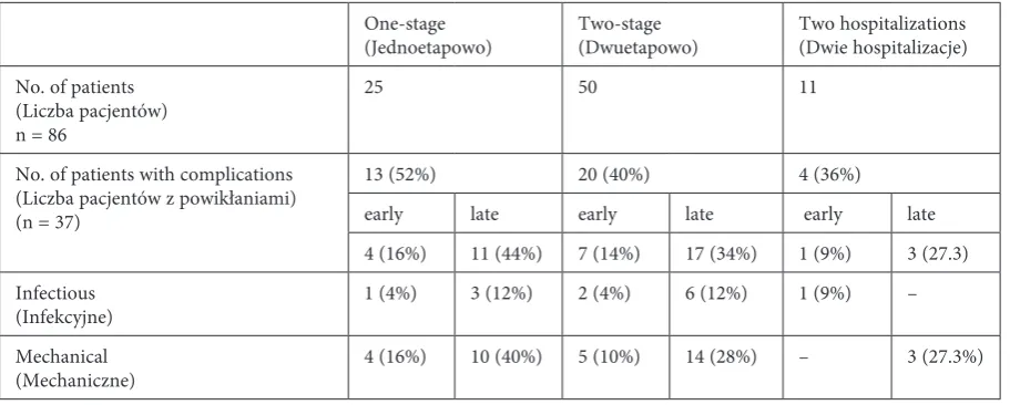
Table 1 Frequency and type of complications with regard to mode of VP shunt insertion
Tabela 1. Częstość i rodzaj powikłań w zależności od trybu czasowego wszczepienia drenującego układu zastawkowego One-stage
(Jednoetapowo) Two-stage(Dwuetapowo) Two hospitalizations(Dwie hospitalizacje) No. of patients
(liczba pacjentów) n = 86
25 50 11
No. of patients with complications (liczba pacjentów z powikłaniami) (n = 37)
13 (52%) 20 (40%) 4 (36%)
early late early late early late 4 (16%) 11 (44%) 7 (14%) 17 (34%) 1 (9%) 3 (27.3) Infectious
(Infekcyjne) 1 (4%) 3 (12%) 2 (4%) 6 (12%) 1 (9%) – Mechanical
(Mechaniczne) 4 (16%) 10 (40%) 5 (10%) 14 (28%) – 3 (27.3%)
Table 2. Frequency of episodes of complications in analyzed groups of patients with regard to mode of VP shunt insertion
Tabela 2. Rozkład epizodów powikłań w analizowanych grupach pacjentów w zależności od trybu czasowego wszczepienia drenującego układu zastawkowego
One-stage
(Jednoetapowo) Two-stage(Dwuetapowo) Two hospitalizations(Dwie hospitalizacje) Patients
(Pacjenci) n = 86
25 50 11
No. of episodes of complications (liczba epizodów powikłań) n = 60
24 (40%) 32 (53.3%) 4 (6.7%)
early late early late early late
5 (8.3%) 19 (31.6%) 7 (11.6%) 25 (41.6%) 1 (1.6%) 3 (5%) Infectious
(Infekcyjne) 1 (1.6%) 3 (5%) 2 (3.3%) 6 (10%) 1 (1.6%) -Mechanical
two of them, it occurred 2 weeks after shunt revi-sion, while in two other children it was noted after 2.5 and 4 months respectively. Two children from this group had further late complications. In both, mechanical failure of the VP shunt occurred at the age of 4 years.
There were 37 episodes of late mechanical com-plications noted in 27 patients. Occlusion of the ventricular catheter was the cause of the failure in 28 cases (75.7%). Eighteen such episodes (64.3%) were reported within the first 6 months of life. Between 6 and 12 months of age, there were only 5 episodes (17.8%). Similar incidence was found in children older than 1 year. Mechanical obstruction of the peritoneal catheter was noted in 6 patients. One patient with a cardiac catheter required two re-visions due its occlusion. All of these complications occurred in older children, aged between 3 and 10 years. Six children developed two episodes of late
mechanical VP failure, while 2 further children had 3 such episodes. Twenty-seven late mechani-cal complications (72.9%) were recorded within the first year of life. Among those occurring in children older than 1 year, 50% of failures were caused by oc-clusion of the peritoneal/cardiac catheter.
Six children developed both late infectious and mechanical complications. Two children had VP shunt infection within the first year of life, while mechanical failure occurred late, after 4 years. Two patients had infectious complications in the early postoperative period after previous shunt revision due to mechanical obstruction. One girl had mechanical complications at the age of 3 and 5 weeks and subsequently developed VP infection 4 months later. The last patient had infection of the VP shunt at the age of 7 months. Four months ear-lier, he had had mechanical complications because of ventricular catheter obstruction.
Table 3. Complications of VP shunt with regard to dorsal wound healing
Tabela 3. Powikłania układu zastawkowego w zależności od przebiegu gojenia rany grzbietu
VP shunt complication (Powikłania układu zastawkowego)
Study group
(Grupa objęta badaniami) Babies with uneventful healing of dorsal wound (Dzieci z niepowikłanym przebiegiem gojenia rany)
Babies with complication of dorsal wound healing (Dzieci z powikłanym przebiegiem gojenia rany) No. of patients
(liczba pacjentów) 86 64 22
Early complications (Wczesne powikłania) infectious
mechanical
12 (13.9%)
4 (4.7%) 9 (10.5%)
6 (9.3%)
2 (3.1%) 5 (5.8%)
6 (27.2%)
2 (9 %) 4 (18%) late complications
(Późne powikłania) infectious mechanical
31 (36%)
10 (11.6%) 27 (36%)
21 (32.8%)
5 (5.8%) 20 (31.3%)
10 (45.4%)
5 (22.7%) 7 (31.8%)
Table 4. Complications of VP shunt with regard to gestational age
Tabela 4. Powikłania układu zastawkowego w zależności od wieku ciążowego noworodków
Complication
(Powikłanie) All babies(Badana grupa) Babies born prematurelyWcześniaki (< 38 Hbd)
Babies born at term (Dzieci urodzone o czasie)
No. of babies
(liczba noworodków) 86 21 65
Infectious (Infekcyjne) 13 (15.1%) 5 (23.8%) 8 (12.3%) Mechanical (Mechaniczne) 33 (38.4%) 12 (57.4%) 21 (32.3%) Early (Wczesne)
infectious mechanical
12 (13.9%) 4 (4.6%) 9 (10.5%)
2 (9.5%) 1 (4.8%) 2 (9.6%)
10 (15.3%) 3 (4.6%) 7 (10.7%) late (Późne)
infectious mechanical
31 (36%) 10 (11.6%) 27 (31.4%)
11 (52.3%) 3 (14.3%) 10 (47.6%)
In the study group of 86 children, infectious complications were recorded in 12 of them (13.9%). Mechanical causes of VP shunt failure were noted in 33 children (38.3%). Infectious complications exclusively were noted in 4 patients (4.7%). A fur-ther 25 children (29.1%) were affected by mechani-cal complications only. Eight children (9.3%) had both types of complications, including 5 patients in whom infectious complications occurred within one month of previous VP shunt revision due to mechanical failure.
All infectious complications occurred within the first year of life. All complications noted in chil-dren older than one were of a mechanical nature. Thirty-seven episodes (63.3%) of complications in the study group occurred within the first six months of life. A further 7 episodes (11.7%) were reported in infants aged between 6 and 12 months.
In the whole group, three late deaths oc-curred (3.4%) at the ages of 55 days, 2 months and 12 months. All of these patients had been previ-ously treated because of infectious complications. Considering the occurrence of complications with regard to the mode of VP shunt insertion, there were no statistically significant differences between the analyzed groups noted.
Sixty-four babies of all 86 had an uneventful postoperative course after the initial repair of the MMC. In 22 patients (25.6%), complicated healing of the dorsal wound was reported. The incidence of this complication with regard to operative strat-egy (one-stage, two-stage, two hospitalizations) was 16%, 32% and 18.8% respectively.
Table 3 and Table 4 illustrate the incidence of VP shunt complications with regard to the course of dorsal wound healing and gestational age re-spectively. Statistical analysis did not reveal sig-nificant differences between the examined groups when early and late, and mechanical and infec-tious complications were analyzed collectively (p > > 0.05). There were, however, differences between groups of babies in the frequency of early infec-tious and early mechanical complications when dorsal wound healing was an examined parameter. Unfortunately, a small number of babies in these subgroups does not render an objective statistical analysis possible.
Among 86 children, in 38 of them (44.1%), prenatal US examination had revealed a congeni-tal anomaly of the central nervous system. Hydro-cephalus was noted in 36 of these children (42.9%). It was detected along with MMC in 17 cases. Both mechanical and infectious complications occurred twice as frequently in the group of babies with pre-natally noted hydrocephalus than in those with a negative prenatal diagnosis, but statistical analysis did not reveal significant differences (p = 0.8944)
Discussion
The natural history of MMC-associated hy-drocephalus can be variable. It may be detected prenatally or may present with evident clinical or sonographic features at birth. In a considerable number of babies, however, ventriculomegaly is of a minor degree on a first postnatal clinical ex-amination but shows a progressive nature after back closure (1, 7). In our series of 110 babies, hy-drocephalus was noted immediately after birth in 84 babies (76.4%). Twelve patients (10.9%) were last presenters with hydrocephalus diagnosed in early infancy. Therefore it may be stated that the association of MMC and early active hydrocepha-lus is a rule. In the reverse situation, when the af-fected child did not present with progressive dila-tation of the ventricular system as happened with 12 (10.9%) of described patients only, this is rather the exception to the rule and not a clinical variant of the defect. The incidence of hydrocephalus with 87.3% presenting early in the neonatal period or early infancy in present report corroborates well with data from the literature. Caldarelli et al., re-porting their experience with 170 children with spina bifida, undertook surgical treatment of hy-drocephalus within the first 48 hours in 33 (19.4%) of them, during the first week in 34 (20%) and later during the first month in a further 70 (41.2%). On-ly 19.4% of their patients had a VP shunt inserted beyond the first month of life [8]. In the series of 264 patients with open spina bifida presented by Wakhlu et al., all had a VP shunt in the neonatal period or no later than by the 8th month of life [9]. Considering the early presentation of MMC--associated hydrocephalus, all children not show-ing ventriculomegaly early after back repair should remain under very close medical surveillance with regular US examinations during the first months of life. In late infancy, the intervals between follow-up reviews can be extended.
function-al abnormfunction-alities and defects related to dysraphic anomalies [2, 10].
The authors have undertaken a review of their own experience in an attempt to identify potential risk factors for early and late complications. Fol-low-ups for the consecutive group of 86 patients, excluding three cases of late death, ranged from 3 to 13 years which is a unique attribute of this study and gives an objective insight into long-term results. Only a few authors have discussed these aspects in their reports. Others focused on com-plications occurring early or up to 1 year after VP shunt insertion [8, 11]. Among the 86 patients, in 40% of them, complications of the VP shunt were reported. This indicates clearly that this is a seri-ous clinical problem which should be considered when planning medical and surgical follow-ups for these children. Other authors present similar experience. Caldarelli et al. reported that compli-cations of the VP shunt within the first 12 months occurred in 45.9% of them [8]. Tulli et al. noted that 64% of their 189 children required shunt re-vision during the first postoperative year [11]. In the group of 31 children with MMC with one-stage or sequential insertion of a VP shunt, Gamache et al. reported a very high incidence of early compli-cations of 74% [7]. liptak et al. presented a series of 67 children. Within the first 6 postoperative months, they noted complications of the VP shunt in 28% of them. When the follow-up period was extended to 1 year, the rate of complications rose to 37%. It was 50% when the period of late analysis exceeded 4 years [12]. A report from a pediatric neurosurgical center in luckow (India) needs to be emphasized. This is the largest series of patients with MMC-associated hydrocephalus reported to date in the literature and consists of 264 children. In the group of babies subjected to simultaneous repair of MMC and hydrocephalus, they noted complications in 11% of them. Among those in whom VP shunt insertion was delayed, only 9% had complications. The follow-up in their study ranged up to 2.5 years [9].
Early complications of the VP shunt require special attention. First, in most instances, they should be attributed to the surgeon, who either did not follow the generally recommended principles of shunt insertion in a baby with hydrocephalus or made a technical error [13]. Secondly, early com-plications, especially infectious ones, pose a high risk of impairment of further mental development. In present series, there were 4 early infectious com-plications. One patient was born prematurely. Two babies had complications in dorsal wound heal-ing and infection of the CNS had occurred before a permanent shunt could be inserted. Therefore these episodes were not true VP shunt
complica-tions and resulted from secondary ventriculitis. Excluding these two babies, the incidence of early infectious complications falls to 2.3%. There was no absolute difference in early shunt infection be-tween the babies with one-stage repair and those with delayed insertion of drainage. The same holds true in the case of early mechanical complications. It indicates clearly that simultaneous repair of the MMC and VP shunt is not a procedure of higher risk of postoperative failure and therefore should be strongly considered for babies with clinically evident hydrocephalus on admission to the surgi-cal department.
Early mechanical complication of the VP shunt carries a high risk of subsequent failure. Only 3 of 9 children did not require any further shunt revi-sion. Three patients developed secondary infec-tious complications in the period ranging from 2 weeks to 2.5 months postoperatively.
Analyzing collectively the group of 14 patients with infectious complications, either early or late, it must be stressed that in at least 8 of them, a poten-tial etiological factor could be precisely identified as previous shunt revision due to mechanical ob-struction with a short interval between procedures, complication of the dorsal wound with subsequent ventriculitis and finally emergency laparotomy due to ileus. They all may be considered as iatrogenic, potentially-avoidable factors.
late mechanical complications occurred with much higher frequency in the one-stage group than in those with delayed a VP shunt. Such a correla-tion is difficult to explain. It may be hypothesized that patients subjected to simultaneous insertion of a VP shunt and MMC repair presented initially with a much larger ventricular system. Successful decompression with a decrease of ventricular pres-sure and concomitant growth of cerebral tissue within the first postoperative weeks might predis-pose the patient to displacement of the ventricular catheter and its eventual obstruction. Such a the-ory seems to find its support in clinical observa-tions. Episodes of mechanical complication due to ventricular catheter obstruction occurred predom-inantly within first 6 months of life and in very few instances in children older than 1 year of age. The authors have not analyzed the relation between complications and rate of ventricular dilatation in the examined patients. This aspect undoubtedly deserves further investigation and should be the subject of a prospective multi-institutional study. Caldarelli et al. noted a higher rate of VP shunt complication among patients with significant ven-triculomegaly than in those with moderate or mild dilatation of the ventricular system [8, 14].
prena-tally-noted hydrocephalus. Early mechanical fail-ure might again be attributed to the larger volume of the ventricular system at the time of VP shunt insertion. But this hypothesis can’t explain the higher frequency of infectious episodes. Vinchon et al. made similar observations, including prenatal diagnosis of hydrocephalus into risk factors of VP shunt complications. They, however, conducted their studies on patients with various types of hy-drocephalus and at much wider range of age [15]. The choice of the optimal mode of treatment of hydrocephalus in a patient with open spina bi-fida remains controversial. Since the late 1980’s, simultaneous insertion of the VP shunt and MMC repair has been gaining popularity. It is associated with longer operative time and this factor might potentially be responsible for a higher risk of VP shunt infections. This is an argument quoted by opponents of this surgical strategy [16]. Clinical practice seems to contradict this statement. Chub-ballah et al. and Chadduck et al. did not report VP shunt infections in the early postoperative period, but it must be stressed that these authors presented small groups of children subjected to one-stage re-pair [17, 18]. Other authors noted infectious com-plications but still their frequency after one-stage repair, ranging from 4.5% to 29.4%, did not exceed those after sequential operative treatment of spina bifida and hydrocephalus [19, 20]. Present authors results strongly confirm that the one-stage strategy is a valuable option for babies with MMC.
Only a few authors have undertaken clini-cal studies aimed at identifying specific risk fac-tors for VP shunt complication in children with MMC. Surprisingly, opinions between them vary significantly. Gamache et al. claim that the risk of infectious complication is the highest when the procedure is performed after day 3 of life [7]. Con-versely, Ammirati et al. recommend strongly that the shunt should be inserted no sooner than the third week of life as in their experience, the inci-dence of shunt complication was the lowest among patients operated on between week 9 and 52 [10]. Tulli et al. were not able to identify a specific risk factor of shunt failure despite a very complex sta-tistical analysis conducted in their study [11]. Cal-darelli et al. demonstrated that, in children with high MMC operated on within the first week of life with significant dilatation of the ventricular system, on whom one-stage repair was carried out with a VP shunt preceding MMC repair, and in those with abnormal cerebral fluid, the risk of in-fectious complication is high [8]. In present study, the authors have not been able to identify a risk factor for VP shunt complications of statistical significance. The small number of patients in the analyzed subgroups must therefore be regarded
as insufficient for objective investigation. This may explain, to some extent, the differences be-tween various authors reporting their experience. In present material, the location of the dysraphic defect was not relevant with regard to complica-tion of the VP shunt. In all the patients subjected to one-stage repair, the VP shunt was inserted as the first procedure in each case. Therefore a very high rate of complications of 83.3% in the babies operated on with this strategy by Caldarelli et al. is difficult to explain [8]. The authors can’t support the observations by other authors that the age of the patient at the time of VP shunt insertion may be a factor affecting further postoperative course.
In the present study, one trend, although again of not statistically significant, must be emphasized. Analysis of the study groups revealed that a com-plicated course of dorsal wound healing must be regarded as a predisposing factor for further shunt complication. Apart from late mechanical failures, the rate of complications in babies with complicated healing of the dorsal wound was 3 times higher than in those with an uneventful postoperative course af-ter back closure. The authors strongly recommend that repair of the MMC must be performed by the most experienced pediatric surgeons familiar with various operative techniques, especially in cases with extensive defects. Considering the operative strategy, the authors noted that one-stage repair was associated with half as many postoperative compli-cations than the two-stage modality. In the former group, the babies’ wound dehiscence was usually su-perficial and amenable to conservative treatment.
Analyzing other factors, the authors surpris-ingly noted that prematurity was not associated with a higher risk of early complications. Similar observations were made by Hubballah et al., al-though they analyzed a small group of babies sub-jected to the one-stage operation [18]. Kulkarni et al. included prematurity among risk factors of infectious complications. These authors, however, studied 299 patients with hydrocephalus, of whom only 23.1% were affected by MMC and therefore a comparative analysis is rather difficult [13].
References
[1] Akar Z: Myelomeningocoele. Sem Neurol 1995, 43, 113–118.
[2] Lemire R: Neural tube defects . JAMA 1988, 259, 76–82.
[3] Bell WO, Arbit E, Fraser R: One-stage meningomyocele closure and ventriculoperitoneal shunt placement. Surg Neurol 1987, 27, 233–236.
[4] Hirsch J–F: Consensus : long term outcome in hydrocephalus. Child’s Nerv Syst 1994, 10, 64–69.
[5] Lazaroff J, Peacock W, Holly L, Ver Halen J, Wong A, Olmstead C: Multiple shunt failures: an analysis of risk factors. Child’s Nerv Syst 1998, 14, 271–275.
[6] Rocco C, Marches E, Velardi F: A survey of the first complication of newly implanted CSF shunt device for the treatment of nontumoral hydrocephalus. Child’s Nerv Syst 1994, 10, 321–327.
[7] Gamache F Jr: Treatment of hydrocephalus in patients with meningomyelocele or encephalocele: a recent series. Child’s Nerv Syst 1995, 11, 487–488.
[8] Caldarelli M, Di Rocco C, La Marca F: Shunt complications in the first postoperative year in children with menin-gomyelocele. Child’s Nerv Syst 1996, 12, 748–754.
[9] Wakhlu A, Wakhlu G, Saxena S, Tandon RK: Single-stage treatment of spina bifida with hydrocephalus based on a prediction rule derived from preoperative cranial ultrasound. Pediatr Neurosurg 2009, 45, 271–275.
[10] Ammirati M, Raimondi AJ: Cerebrospinal fluid shunt infections in children. Child’s Nerv Syst 1987, 3, 106–109.
[11] Tuli S, Drake J, Lamberti–Pasculli M: long-term outcome of hydrocephalus management in myelomeningoceles. Childs Nerv Syst 2003, 19, 286–291.
[12] Liptak G: Screening for ventricular shunt function in children with hydrocephalus secondary to meningomyelo-cele. Pediatr Neurosurg 2001, 34, 281–285.
[13] Kulkarni A, Drake J, Lamberti-Pasculli M: Cerebrospinal shunt infection, a prospective study of risk factor. 2001, 94, 195–201.
[14] Sato O, Yamaguchi: Hydrocephalus: full of contentious points and a lot of questions to be solved. Crit Rev Neurosurg 1998, 8, 131–140.
[15] Vinchon M, Dhellemmes P: Cerebrospinal fluid shunt infection: risk factors and long-term follow-up. Child’s Nerv Syst 2006, 22, 692–697.
[16] Oktem I, Menku A Ozdemir: When should ventriculoperitoneal shunt placement be performed in cases with myelomeningocele and hydrocephalus? Turk Neurosurg 2008, 18, 387–391.
[17] Chadduck WM, Redding D: Experience with simultaneous ventriculo-peritoneal shunt placement and myelom-eningocele repair. J Pediatric Surg 1988, 23, 913–916
[18] Hubballah M, Hoffman H: Early repair of myelomeningocele and simultaneous insertion of ventriculoperitonal shunt: technique and results. Neurosurgery 1987, 20, 1987, 21–23.
[19] Epstein N, Rosenthal A, Zito J, Osipoff M: Shunt placement and myelomeningocele repair: simultaneous vs sequential shunting. Review of 12 cases. Child’s Nerv Syst 1985, 1, 145–147.
[20] Machado H: Simultaneous repair of myelomeniningocele and shunt insertion. Child’s Nerv Syst 2004, 20, 107–109.
Address for correspondence:
Maciej Bagłaj
Department of Pediatric Surgery Wroclaw Medical University M. Skłodowskiej-Curie 52 50-367 Wrocław
Poland
Conflict of interest: None declared