C A S E R E P O R T
Open Access
Epidural spinal cord compression as initial
clinical presentation of an acute myeloid
leukaemia: case report and literature review
Dominique N
’
Dri Oka
1*, Alpha Boubacar Bah
2, André Valentin Tokpa
1and Louis Derou
1Abstract
Epidural localization of myeloid leukaemia is rarely reported. Spinal cord compression as an initial presentation of acute myeloid leukaemia is extremely rare. This is a report of a 17-year-old black boy who presented to emergency department with neurological symptoms of spinal cord compression. Imaging modalities showed multiple soft tissue masses in the epidural space. After surgical treatment, histopathological examination of the epidural mass showed myeloid leukaemia cells infiltration. Literature review on Medline and“scholar Google”database was done. The characteristics and management of extra-medullary leukaemia are discussed. Granulocytic sarcoma, myeloid sarcoma or chloroma with acute myeloid leukaemia should be considered as part of epidural spinal cord compression. Therefore surgery is indicated on an emergent basis.
Keywords:Spinal cord compression, Initial epidural localization, Acute myeloid leukaemia
Background
Acute myeloid leukaemia corresponds to malignant mono-clonal proliferation of medullary blastic myeloid cells with an interrupted differentiation [1]. Clinically acute myeloid leukaemia will result in adenopathy and hepatosplenic syn-drome associated with bone marrow failure synsyn-drome. Extra nodal involvement is rare and mostly made of fre-quently observed forms of lymphoblastic leukemia in chil-dren. These attacks represent 2 to 8 % of acute myeloid leukaemia. Epidural localization is unusual and is a rare cause of spinal cord compression. Spinal cord compression as an initial presentation of acute myeloid leukaemia is ex-tremely rare. In this study, we did not consider Spinal cord compression as the initial presentation of acute biphenoty-pic leukaemia [2]. The authors reported a case of acute myeloid leukaemia revealed by thoracic spinal cord com-pression. Literature review follows the case report.
Clinical case
A 17-year –old A black patient with no known signifi-cant medical history, complained of a 3-week history of
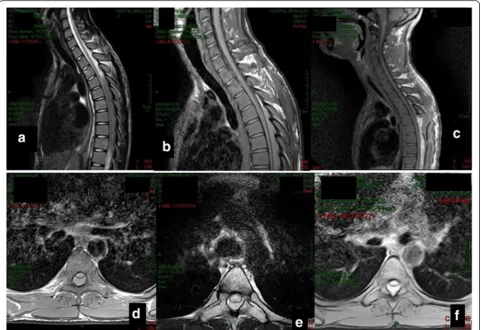
inter-scapular back pain progressively exacerbating with insomnia associated with paraesthesia, hypoesthesia of the lower limbs and trunk. The rapidly worsening deficit led to emergent Neurosurgery consultation. Neurological examination found an almost paraplegic patient with in-creased deep tendon reflexes, a bilateral Babinski, a dis-tended bladder, hypoesthesia and dysesthesia with at T10 sensitive level. Further clinical examination found no evidence of lymphadenopathy or hepatosplenome-galy. The diagnosis of spinal cord compression was made and a spinal CT scan showed an extensive epidural mass at T4-T9. MRI confirmed the presence of a thor-acic epidural mass posterior to the spinal cord between T4 and T9 (Fig. 1a, b, c, d, e, f ). The MR imaging showed hypointense lesion on T1 weighted sequence, iso-intense on T2 weighted sequence and enhanced on T1 with gadolinium. There was a distinct line of dura demar-cation between the tumor mass and the spinal cord.
Full blood count revealed leucocytosis with a import-antly increased white blood cell count 200,000/mm3, with 77 % blasts, haemoglobin: 10.7 g/dL-1; platelet count: 102 × 109 L-1. The myelogram diagnosis was acute myelogenous leukaemia. Histologically the tumor is composed of immature cells. There are some imma-ture eosinophile and neutrophile. Cytogenetic evaluation * Correspondence:ndriokad@gmail.com
1Department of Neurosurgery, Teaching Hospital of Yopougon in Abidjan
Ivory Coast, 21 BP 632 Abidjan, Côte d’Ivoire
Full list of author information is available at the end of the article
© 2016 N’Dri Oka et al.Open AccessThis article is distributed under the terms of the Creative Commons Attribution 4.0 International License (http://creativecommons.org/licenses/by/4.0/), which permits unrestricted use, distribution, and reproduction in any medium, provided you give appropriate credit to the original author(s) and the source, provide a link to the Creative Commons license, and indicate if changes were made. The Creative Commons Public Domain Dedication waiver (http://creativecommons.org/publicdomain/zero/1.0/) applies to the data made available in this article, unless otherwise stated.
could not be done. Additional blood test did not find bleeding disorders or ion anomaly. Given the severity of the neurological picture and predicted rapid deteri-oration, a laminectomy from T4 to T10 was per-formed in emergency and found that the posterior epidural space was occupied by greyish, haemorrhagic
tumour tissue, completely separated from the dura and pushes forwards. A macroscopically complete tumour resection was performed. Haemostasis was difficult. Histopathological examination of the tumour mass was favourable to leukemic blast cells. It was type 2 leukemia (Fig. 2). Advanced treatment of acute Fig. 1Spine MRI showing spinal cord compression at T4-T9 level by posterior epidural mass.aSagittal view showing a T4T9 posterior mass which is isointense on T2 weighted sequence.bSagittal view T1 sequence showing a hypointense T4T9 posterior lésion well demarcated from the dura.
cSagittal view showing a contrast enhancement of the T4-T9 lesion.dAxial view: The lesion is hypointense on T1 weighted sequence.eAxial view: The lesion is isointense on T2 sequence at the level of T5.fAxial view showing the enhancing lesion at the level of T5 wth a good demarcation from the dura
Fig. 2HE X 100: Tumor proliferation with blasts exhibiting cytonuclear atypia (a). Immunohistochemistry X 200: Nuclear expression of antibodies anti myeloperoxidase (MPO) by the majority of undifferentiated blasts (b)
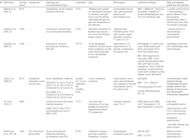
Table 1Literature review from 2005 to 2015. characterization of acute myeloid leukemia associated to spinal cord compression
Nb References Sex/age (years)
Cytogenetic Histology and immunohistochemistry
Localization signs MR Imaging Laboratory findings Delay and diagnosis treatement and outcome
1 Meltzer et al. 2005 [3]
M/10 - -Myeloblastic Acute myeloid leukemia-Chloroma
T2-T6 -Midback pain-weight loss-decreased sensation to vibration and light touch over the left leg -decreased sensation to pain and temperature in the right leg
-paravertebral lesions with spinal-posterior epidural and prevertebral mass
WBC 12,800/mm3, HB 9.4 g/
dl, platelet 74,800/mm3 .-lactate dehydrogenase was 570 IU/l,-immature cells
-3 weeks-Laminectomie + Chemotherapy + spinal radiation-Total Neurological improvement after 6 month-well more than 18 months of therapy
2 Ghalaut et al. 2005. [4]
23/M - Granulocytic sarcoma due to acute myeloid leukemia
T4-T6 -Backache-numbness of bilateral legs-sensory loss up to mid thoracic region anemia
-Epidural mass extending from T4-T6 with marrow signal alteration involving T1 and T5 to T7
- - surgical excision + chemotherapy -neurological improvement
3 Amalraj et al. 2009 [5]
14/M - Granulocytic sarcoma promyelocytic leukemia AML M3
T3-T10 -Acute paraplegia -weakness of both lower limbs-numbness of both lower limbs-back pain for the last-bladder symptoms
hypointense in T1 hyperintense in T2 dorsally compressing the spinal cord
Hemoglobin 13 gm%, total count 8,400, polymorph 60 %, and lymph 40 %, with high erythrocyte.
-paraplegia improved with radiotherapy and chemotherapy
RBC mild hypochromic microcytic, WBC count normal predominantly blast cells with high nuclear cytoplasmic ratio, moderate cytoplasm, hyperchromatic nuclei with multiple nucleoli, platelet count decreased.
4 Olcay et al. 2009 [6]
M/12 Cytogenetic evaluation could not be done.
acute myeloblastic leukemia Lumbar (L3) -Conus medullaris-left maxillary mass -Conus medullaris syndrome
Cauda equine nerve roots were thickened and clumped in the left anterolateral side of the spinal canal.
Hb:8 g/dL -Chemotherapy + triple intratecal therapy-Radiotherapy-Symptom partially resolved died because of pneumonia and disseminated intravascular coagulation CD45:95.15 %, CD15: 35.56 %,
CD33:77.57 %, CD117:79.3 %, CD34:43.38 %, CD13:29.32 %.
WBC : 7.4 × 10⁹/mm3
Thrombocyte:60.109/mm3
Lactate dehydrogenase: 691 U/l
HLA-DR:86, 4 %,
CDw65:23.9 %, CD19:49.54 % CD20:21.65 %, CD14:3.14 %.
5 Yin et al. 2010 [7]
28/M - myeloid sarcoma with acute myeloid leukemia
T12-L1 -low back pain-numbness of his legs- bladder-incontinence-hypoesthesia below T12 paraparesis
-Posterior epidural mass T12-L1
White blood cell 3,900/ mm3- hemoglobin :11.0 g/dL-hematocrit of 38.2 %.
High-dose methylprednisolone
-Decompression with T12-L1 laminectomy and tumor resection. CD68, CD45, CD43, CD117
and lysozyme but not of MPO, CD20
Paraparesis Improvement
- anti-AML therapy, but died of sepsis
7 Bittencourt et al. 2011 [8]
53/M t(15;17)(q22q21) abnormality
-Acute promyelocytic leukemia presenting as extradural mass-PCR
T6-T8 -Progressive fatigue, pain legs weakness Paraparesis- Gingival
Posterolateral extradural mass from
Hb:10,6 g/dl-1 delais4 months/ chemotherapy (daunorubicin) + WBC: 7,5.109/L-1
Table 1Literature review from 2005 to 2015. characterization of acute myeloid leukemia associated to spinal cord compression(Continued)
for the PML-RARαgene was positive
hemorrhage, hepatomegaly
T6-T8 with medullar compression
radiotherapy/Evolution: no neurologic improvement + died of sepsis Platelet:12.109/l-1
Fibrinogen:1.4 g/L-1
INR: 1.86 (N:0,8-1,2)
Uric acid and lactate dehydrogenase were both elevated
8 Kyaw et al. 2012 [9]
26/M - -Myeloid sarcoma: acute promyelocytic leukemia-CD33, CD117, CD64, CD34 and cytoplasmic MPO were presented Reverse transcriptase-polymerase chain reaction showed BCR1-type PML-RARαfusion copies
T2-T4 and T12–L2
Progressive back pain and bilateral leg weakness: -paraparesis Loss of pain and sensory perception
Multiple hyperintense T12,L1,L2, L4 and L5 vertebral bodies and sacrum
Hb:10,7 g/dL-1
-Radiotherapy-Chemotherapy: retinoic acid + ida rubicin-Neurological improvement -Good remission WBC:2,8.109/L-1
Platelet : 102.109/L-1
Intraspinal extradural masses L4 and L5 vertebral bodies and sacrum
Intraspinal extradural masses located fromT2 toT4 and T12 to L2.
9 Ben et al. 2013 [10]
21/M karyotype: 46, XY, t (8, 21) (q22; q22) / 46, XY
chloromaCD3, CD20 = --positive for myeloperoxidaseacute myeloblastic leukaemia (AML) of the French-American-British M2 subtype
T4/ T7, T1-T2
Progressive paraplegia. urinary retention
intermediate in T1, hyperintense in T2
hemoglobin 8 g/dL-1, white
blood cell count 3100/mm3, platelet count 44000/mm3
and 3 % neutrophils, 44 % lymphocytes and 44 % .
laminectomy + tumour was totally removed
-rubidomycine (45 mg/m2 daily for 3 days) and cytosine arabinoside (200 mg/m2continuous
infusion for 7 days). -improvement paraplegia- complete remission
10 Krishnamurthy et al. 2014 [11]
16 /M - Granulocytic sarcoma associated with leukaemia,-acute myeloblastic leukaemia (AML) of the French-American-British M2 subtype.-subleukemic leukemia
Low backaches follow proptosis, spinal cord causing significant compression of the spinal cord.
Multiple midline fusiform extra-dural masses which are iso- intense to cord on T1W images seen extending from C3-C7, D3-D5, D11-D12 & S1-S2 are seen posteriorly
Hb : 7.2 g/dl, 3 RBC 2.5 million/mm3,TC : 11200 cells/mm3, with a differential
ount of N 41 %, L-28 %, E-01 %, M-30 %, 3ESR 120 mm/hr, Platelet count: 51,000/mm3, PT/INR–
-dimorphic anemia, thrombocytopenia and myeloblasts
-radiotherapy and chemotherapy
11 Present case. 17/M -Cytogenetic evaluation could not be done.
-Myeloid leukemia-type 2 leukemia
T4T9 Acute paraplegia hypointense lesion on T1 weighted sequence isointense T2 weighted sequence enhanced on T1 with gadolinium. (epidural mass (hyperintense)
blood count revealed leucocytosis with a major white blood cell count 200,000 / mm3, with 77 %
blasts, hemoglobin: 10.9 g dl; platelet count: 106 × 109L-1
-Chemotherapy-Died one week after diagnosis
Literature review from 2005 to 2015. characterization of acute myeloid leukemia associated to spinal cord compression N b: Number
myeloid leukaemia was indicated. But the patient died a week after the diagnosis of spinal cord compression because of sepsis.
Discussion
The spinal cord compression revealing acute myeloid leu-kaemia is unusual. Nine cases have been reported from 2005 to 2015 [3–11] (Table 1). It often appears like a solid tumour known as granulocytic sarcoma or myeloid, or also chloroma as seen in our patient [12]. It corresponds to the migration outside of the bone marrow of myeloid cells that proliferate on their own [1]. The first case was described in 1811 by Burns in 1893 and Dock who reported its associ-ation with leukaemia [13, 14]. It is reported in 3.1 to 9.1 % of patients with acute myeloid leukaemia [15]. We found ten cases from 2005 to 2015 which were reported in litera-ture. In those cases epidural space was occupied by granu-locyt sarcoma [3-9 11, 15]. Surgically excision of the tumour appeared to be the first treatment option in these cases. Granulocytic sarcoma is frequently diagnosed simul-taneously with or after the start of an acute myeloid leukae-mia or may be the initial sign of a relapse in a patient in remission [3–9, 11, 15]. In non-leukemic patients, myeloid granulocytic sarcoma usually precedes acute myeloid leu-kaemia [16]. In 87 to 88 % of patients without hematologic abnormalities at diagnosis, acute myeloid leukaemia de-velops in the 10.5 to 11 following months [17]. Myeloid sarcoma may occur in all tissues but they are frequently lo-calized on the skin, bone, soft tissues of the head and neck (especially on the orbit) and adenopathies [18]. Spinal localization and especially epidural is rare. Thoracic localization is the most frequent spinal localization followed by lumbar and sacral localizations. Clinical signs are in most cases made of spinal cord compression. Some-times they involve isolated rachialgia [3–9, 11, 15]. MRI is the best choice for neuroimaging examination to show epi-dural tumour lesions. It is also the best neuroimaging examination [3–9, 11, 15] to show the epidural tumour le-sions without being specific. Granulocytic sarcoma is iso-intense in T1 and T2 with in general a contrast enhance-ment [3–9, 11, 15]. The therapeutic strategy of this myeloid sarcoma is based on chemotherapy, radiation therapy, sur-gical decompression and bone marrow transplantation and any combination of these methods [3–9, 11, 15]. 1n cases of granulocytic sarcoma in particular by acute myeloid leu-kaemia with neurological signs, priority should be given to chemotherapy and/or radiation therapy rather than sur-gery. In our patient the diagnosis of acute myeloid leukae-mia was made from full blood count after surgery. Surgery was only justified by the acuteness and quickly worsening neurological disorders. Despite some cases of more or less complete neurological recovery with a variable remission, most patients present with neurological sequellae and die from haematological complications dominated by sepsis.
Conclusion
This case associated with the literature review has shown that granulocytic sarcoma with acute myeloid leukaemia should be considered as part of epidural spinal cord compression .it might be the initial sign of epidural spinal cord compression . Therefore surgery is indicated in emergency. Chemotherapy and radiotherapy are secondly introduced according to the diagnosis.
Competing interests
The authors declare that they have no competing interests.
Authors’contributions
DNO has performed the surgery, evaluated the patient, and drafted and revised the manuscript. ABB assisted the surgeon at the operation room, grammatically reviewed the manuscript and participated to the discussion. AVT evaluated the patient, carried out the literature review. LD evaluated the patient, carried out the literature review. All authors read and approved the final manuscript.
Acknowledgements
We thank Sir Djo BI doctor Alban Mbende Professor koffi Gustave in haematology department in yopougon and Dr Doukouré Brahim Histopatology department for their help.
Author details
1Department of Neurosurgery, Teaching Hospital of Yopougon in Abidjan
Ivory Coast, 21 BP 632 Abidjan, Côte d’Ivoire.2Department of Neurosurgery,
Teaching Hospital of Conakry, Conakry, Guinea.
Received: 11 September 2015 Accepted: 20 November 2015
References
1. Pileri SA, Ascani S, Cox MC, Campidelli C, Bacci F, Paccioli M, et al. Myeloid sarcoma. Clinico-pathologic, phenotypic and cytogenetic analysis of 92 adult patients. Leukemia. 2007;21:340–50.
2. Dimou J, Jithoo R, Tsui A, Morokoff AP. Spinal cord compression as the initial presentation of acute biphenotypic leukaemia. J Clin Neurosci. 2009; 16(12):1696–8.
3. Meltzer JA, Jubinsky PT. Acute myeloid leukemia presenting as spinal cord compression. Pediatr Emerg Care. 2005;21(10):670–2.
4. Ghalaut PS, Jindal S, Jeevan O. Granulocytic sarcoma as initial presentation of acute myeloid leukaemia. J Assoc Physicians India. 2005;53:828–30. 5. Amalraj P, Syamlal S. Unusual case of paraplegia. Ann Indian Acad Neurol.
2009;12(3):188–90.
6. Olcay L, AribaşBK, Gökçe M. A patient with acute myeloblastic leukemia who presented with conus medullaris syndrome and review of the literature. J Pediatr Hematol Oncol. 2009;31(6):440–7.
7. Yin Q, Zhou YY, Chen D, Li WL. Different outcome of myeloid sarcoma with spinal cord compression preceding acute myeloid leukemia: report of two cases and review of literature. Chin J Cancer Res. 2010;22(2):156–62. 8. Bittencourt H, Teixeira Junior AL, Glória AB, Ribeiro AF, Fagundes EM. Acute
promyelocytic leukemia presenting as an extradural mass. Rev Bras Hematol Hemoter. 2011;33(6):478–80.
9. Kyaw TZ, Maniam JAS, Bee PC, Cin EFM, Nadarajan VS, Shanmugam H, et al. Myeloid sarcoma: an unusual presentation of acute promyelocytic leukemia causing spinal cord compression. Turk J Hematol. 2012;29:278–82. 10. Ben FW, Zahra K, Ben HI, Hadhri R, Khelif A. Paraplegia due to chloroma as
the initial presenting feature of acute myeloid leukaemia. Pan Arab J Neurosurg. 2013;17(1):4.
11. Krishnamurthy VG, Narendra SS, Bhagwat KA, Shashikala P, Prashanth S, Khanpur R. Granulocyte sarcoma presenting as acute paraplegia and Proptosis. J Pub Health Med Res. 2014;2(2):52–4.
12. Frohna BJ, Quint DJ. Granulocytic sarcoma (chloroma) causing spinal cord compression. Neuroradiology. 1993;35:509–11.
13. Dock G. Chloroma and its relation to leukemia. Am J Med Sci. 1893;106:152–85. 14. Burns EW. Chloroma and paraplegia: report of a case. N Z Med J.
1963;62:422–5.
15. Eser B, Cetin M, Kontas O, Er AUO, Coskun HS, Altinbas MR. Facial nerve paralysis and paraplegia as presenting symptoms of acute myeloid leukaemia. Jpn J Clin Oncol. 2001;31(2):86–8.
16. Serefhanoglu S, Goker H, Aksu S, Buyukasik Y, Sayinalp N, Haznedaroglu IC, et al. Spinal myeloid sarcoma in two non-leukemic patients. Intern Med.
2010;49(22):2493–7.
17. Neiman RS, Barcos M, Berard C, Bonner H, Mann R, Rydell RE, et al. Granulocytic sarcoma: a clinicopathologic study of 61 biopsied cases. Cancer. 1981;48:1426–37.
18. Balleari E, Panarello S, Capello E, Grosso M, Passalia C, Pitto P, et al. Granulocytic sarcoma : an unusual cause of spinal compression. Int J Clin Oncol. 2007;12:234–7.
• We accept pre-submission inquiries
• Our selector tool helps you to find the most relevant journal
• We provide round the clock customer support
• Convenient online submission
• Thorough peer review
• Inclusion in PubMed and all major indexing services
• Maximum visibility for your research
Submit your manuscript at www.biomedcentral.com/submit