RESEARCH
Effect of
IKZF1
deletions on signal
transduction pathways in Philadelphia
chromosome negative pediatric B-cell precursor
acute lymphoblastic leukemia (BCP-ALL)
Naomi E van der Sligte
1, Frank J G Scherpen
1, Arja ter Elst
1, Victor Guryev
2, Frank N van Leeuwen
3and Eveline S J M de Bont
1*Abstract
Background: IKZF1 deletions are an unfavorable prognostic factor in children with Philadelphia chromosome posi-tive (Ph+) as well as negative (Ph−) acute lymphoblastic leukemia (ALL). Although IKZF1 deletions occur in 10–15%
of Ph− ALL cases, effects of IKZF1 deletions on signaling pathways in this group have not been extensively studied.
Therefore, in this study we aimed to study the effect of IKZF1 deletions on active signal transduction pathways. Methods: Multiplex ligation-dependent probe amplification (MLPA) was used to determine IKZF1 deletions and other copy number alterations in 109 pediatric B-Cell Precursor ALL (BCP-ALL) patients. Kinase activity profiling of 45 primary Ph− BCP-ALL patients (31 IKZF1 wild type patients and 14 patients harboring an IKZF1 alteration) and western
blot analysis of 14 pediatric BCP-ALL samples was performed to determine active signal transduction pathways. Results: Unsupervised hierarchical cluster analysis of kinome profiles of 45 pediatric Ph− ALL cases showed no
clustering based on IKZF1 status. Comparing the phosphorylation intensities of peptides associated with signaling pathways known to be involved in BCP-ALL maintenance, we did not observe differences between the two groups. Western blot analysis of 14 pediatric BCP-ALL samples showed large variations in phosphorylation levels between the different ALL samples, independent of IKZF1 status.
Conclusions: Based on these results we conclude that, although IKZF1 deletions appear to be an important clinical prognostic factor, we were unable to identify a unique IKZF1 dependent protein expression signature in pediatric Ph−
ALL and consequently no specific targets for future therapy of Ph−IKZF1 deleted BCP-ALL could be identified.
Keywords: Acute lymphoblastic leukemia, IKZF1, Signaling, Kinome profiling
© 2015 van der Sligte et al. This article is distributed under the terms of the Creative Commons Attribution 4.0 International License (http://creativecommons.org/licenses/by/4.0/), which permits unrestricted use, distribution, and reproduction in any medium, provided you give appropriate credit to the original author(s) and the source, provide a link to the Creative Commons license, and indicate if changes were made. The Creative Commons Public Domain Dedication waiver (http://creativecommons. org/publicdomain/zero/1.0/) applies to the data made available in this article, unless otherwise stated.
Background
Overall survival rates for children with Acute Lympho-blastic Leukemia (ALL), the most common type of can-cer in children, are approaching 90% [1]. Historically, risk stratification of newly diagnosed children was based on age and white blood cell count (WBC), but nowadays
includes extensive cytogenetic and molecular analyses. In the past 5 years, genome wide approaches, studying DNA copy number alterations in ALL, have identified novel molecular markers that can be used for further risk stratification, including IKZF1 deletions as a predic-tor of poor outcome. IKZF1 deletions can be identified in approximately 70% of the children with Philadelphia chromosome positive (Ph+) ALL and in 10–15% of the
children with Philadelphia chromosome negative (Ph−)
ALL and are associated with an increased relapse risk and decreased overall survival in both groups [2–4]. More recent studies indicate that the genomic context
Open Access
*Correspondence: E.S.J.M.de.Bont@umcg.nl
1 Division of Pediatric Oncology/Hematology, Department of Pediatrics, Beatrix Children’s Hospital, University Medical Center Groningen, University of Groningen, PO Box 30.001, 9700 RB Groningen, The Netherlands
in which IKZF1 deletions occur is more important for prognosis as for example CRLF2 and JAK2 mutations are more common in IKZF1 deleted BCP-ALL [5–7]. In pediatric B-cell progenitor ALL (BCP-ALL), 80% of the IKZF1 deletions are found in a Philadelphia chromosome negative background.
IKZF1, which encodes the transcription factor Ikaros, is essential for normal lymphoid development, whereas for erythroid and myeloid lineage differentiation IKZF1 is less critical [8]. Mice deficient for IKZF1 show a com-plete arrest in B-lymphocyte development while mice heterozygous for a dominant-negative mutation of IKZF1 develop T cell leukemia and lymphoma with a 100% penetrance [9, 10]. During normal development, Ikaros restricts the G1-S transition of the cell cycle when it binds
to the DNA, by regulating transcription of cell cycle reg-ulator genes e.g. a positive effect on cell cycle inhibitors CDKN1A (p21Cip1) and CDKN1B (p27Kip1) [11]. Phos-phorylation of Ikaros by casein kinase II (CK2) temporar-ily reduces Ikaros binding to DNA and thereby facilitates progression through the S phase of the cell cycle [11]. Furthermore, Ikaros can be phosphorylated by spleen tyrosine kinase (SYK) and bruton’s tyrosine kinase (BTK) [12, 13]. These phosphorylation events are essential for nuclear localization, regulation of DNA binding activity, and an optimal transcriptional function of Ikaros [12, 13].
IKZF1 deletions observed in BCP-ALL are typically mono-allelic, either resulting in a loss of function or the expression of a dominant-negative isoform [14]. The dominant-negative isoforms lack the DNA binding N-terminal zinc fingers, preventing DNA binding after dimerization with Ikaros [15]. As a result, the control of Ikaros on the G1-S transition is abolished leading to
hyperproliferation and the development of leukemia [11]. Although the cure rates for children with BCP-ALL have improved substantially, the outcome after ALL relapse remains poor. Since IKZF1 deletions increase the risk of relapse, new therapeutic options aiming to improve cure rates for this specific subtype of ALL are needed. We have previously shown that insight into active signal transduction pathways allows identifica-tion of interesting targets for future therapy [16–19]. At the level of signal transduction, Iacobucci et al. showed on western blot analysis a higher STAT5 phosphoryla-tion in IKZF1 deleted compared to IKZF1 wild type adult BCP-ALL patients with unknown cytogenetic back-ground [20]. However, this observation might also be associated with BCR-ABL1 activity as in adult BCP-ALL patients IKZF1 deletions are more common in Ph+ ALL
[21]. Additionally, Ikaros-reconstitution in two IKZF1 deleted Philadelphia positive ALL patients resulted in an upregulation of the B-cell receptor (BCR) signaling path-way and a concomitant cell cycle arrest; showing that in
Ph+ ALL pre-B cell receptor signaling suppresses
prolif-eration through an Ikaros-mediated cell cycle arrest [22]. Although IKZF1 deletions in children are most com-monly found in a Philadelphia negative background, the effect of IKZF1 deletions on signaling pathways in Phila-delphia negative ALL have not been extensively studied. Therefore, in this study we aimed to study the effect of IKZF1 deletions on active signal transduction path-ways in Philadelphia negative pediatric BCP-ALL using kinome profiling.
Methods Patients
Primary blood and bone marrow samples from newly diagnosed ALL patients were collected after getting writ-ten informed consent in accordance with the regulations and protocols of the medical ethics committee of the University Medical Center Groningen. Overall, we col-lected material of 109 Philadelphia negative BCP-ALL patients. Mononuclear cells were isolated by Lympho-prep (Nycomed, Zürich, Switzerland) density gradients and cryopreserved in liquid nitrogen until use. The cry-opreserved leukemia cells were thawed rapidly at 37°C and diluted in a 6 ml volume of newborn calf serum, as described earlier [23].
DNA isolation
Genomic DNA was extracted from mononuclear cells using the QIAamp DNA easy kit (Qiagen, Hilden, Ger-many) according to the manufacturer’s instructions. All isolated DNA was quantified by NanoDrop spectropho-tometry (NanoDrop, Wilmington, DE, USA).
Multiplex ligation‑dependent probe amplification (MLPA)
Data analysis: Data were analyzed using Gene Mapper v.4.0 software (Applied Biosystems). Normalization of the data was carried out by dividing the peak area of each probe by the average peak area of the control probes. This normalized peak pattern was divided by the average peak pattern of all the samples in the same experiment. The resulting values were 1 for every wild-type peak, 0.5 for heterozygous deletions and 1.5 for heterozygous duplications.
PepChip
Kinase activity profiles of 45 primary Ph− BCP-ALL
patients were determined using the PepChip™ Kinom-ics microarray system (Pepscan, Lelystad, the Nether-lands) and performed as described previously [18, 19]. The microarray contain 1,024 peptides in triplicate (1,008 unique target peptides and 16 peptides used for produc-tion) derived from known phosphorylation sites from human protein sequences that can be phosphorylated by the kinases in the sample lysate. Per sample, 0.5 × 10⁶
cells were lysed in 100 μl of M-PER Mammalian Protein
Extraction Buffer containing 1 μl Phosphatase Inhibitor
and 1 μl Protease Inhibitor (Pierce, Rockford, IL, United
States). Peptide array incubation mix was produced by adding 10 μl of filter-cleared activation mix onto 90 μl
cell lysate. Peptide array incubation mix was loaded on the chip and incubated for 2 h at 37°C in a closed humid box at saturated humidity. Subsequently, the peptide array was washed and blow dried with compressed air or N2 and the chips were exposed to a phospho-storage
screen for 24–96 h. The amount of bound 33P-labelled
ATP to the peptides specifies the amount of peptide phosphorylation and was analyzed with array software (ScanAlyze, Eisen Lab, University of California at Berkely, Berkely, CA, United States).
Data analysis: Data were analyzed as described previ-ously [18, 19]. In short, background was subtracted and the spot intensities were quantile normalized. A Pearson’s correlation coefficient was determined over the triplicates (Excel 2003, Microsoft Office, Redmond, WA, United States). Slides with a correlation <0.6 over the triplicates were excluded from further analysis. The correlation over the triplicates was <0.6 in none of the samples. Cluster, statistical and heatmap analysis were performed using Qlucore Omics Explorer 3.0 (Qlucor AB, Lund, Sweden). The file containing the processed raw data can be found in the additional information (Additional file 1: Table S1).
Western blot analysis
Primary ALL cells were solved in laemmli sample buffer (Bio-Rad laboratories, Veenendaal, the Neth-erlands). Proteins were separated by sodium dode-cyl sulphate–polyacrylamide gel electrophoresis, and
transported to nitrocellulose membranes. Membranes were blocked in 7.5% skimmed milk and incubated overnight with primary antibodies for phospho-Src_ Y416, phospho-Syk_Y323, phospho-ERK1/2_T202/ Y204, phospho-CREB_S133, phospho-p38_T180/ Y182, phospho-Akt_S473, phospho-mTOR_S2448,
phospho-GSK3α/β_S21/S9, phospho-MDM2_S166,
Chk2_T68, RB1_S807/811, phospho-STAT3_Y705, phospho-STAT5_Y694 (Cell Signaling, Danvers, MA, USA), or phospho-p27_T187 (Abgent, San Diego, CA, United States) and for 1 h with HRP conju-gated secondary antibodies (Dako, Glosturp, Denmark). Protein bands were visualized using the ChemiDoc MP imaging system (Bio-Rad, Hercules, CA, United States) and ImageLab software (version 5.0, Bio-Rad Laborato-ries). Loading control was visualized using β-actin (Santa
Cruz Biotechnology, Dallas, TX, United States).
Results
Generation of kinase activity profiles in IKZF1 deleted versus IKZF1 wild type Philadelphia negative pediatric BCP‑ALL
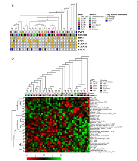
MLPA analysis revealed that among the 109 Philadel-phia negative pediatric BCP-ALL patients tested, 17 (15.6%) patients harbor an IKZF1 deletion whereas one patient (0.9%) showed a gain of IKZF1. We generated kinase activity profiles of 31 IKZF1 wild type patients and 14 patients with an IKZF1 alteration (13 patients with an IKZF1 deletion and 1 patient with a gain of IKZF1). No material was available for kinome profiling of the other three patients with an IKZF1 deletion. Among the 14 patients harboring an IKZF1 alteration, deletions in exons 1 through 8 (4 patients, 28.6%), 4 through 7 (3 patients, 21.4%), and 4 through 8 (3 patients, 21.4%) were most frequent (Fig. 1). Patients’ characteristics are shown in Fig. 1. Patients harboring an IKZF1 alteration appeared to be older compared to IKZF1 wild type patients (8.4 versus 5.7 years, respectively, P = 0.038). Furthermore, MLPA analysis showed more CSF2RA alterations in the group with IKZF1 alterations compared to the IKZF1 wild type group (P = 0.003).
Kinase activity profiles were generated to reveal poten-tial differences in signaling between IKZF1 deleted and non-deleted Ph− pediatric BCP-ALL cases. Compared to
(Fig. 2a). The clustering in peptide activity was not corre-lated to patients’ characteristics as age, gender, and white blood cell count (WBC) at diagnosis (Additional file 2: Figure S1). To evaluate whether IKZF1 deletions display a unique kinase signature we studied differences in pep-tide phosphorylation intensities by t-test analysis. Thirty-eight peptides were differentially phosphorylated between IKZF1 deleted (N = 13) and IKZF1 wild type (N = 31)
pediatric Ph− BCP-ALL patients: phosphorylation of 14
peptides was higher in the IKZF1 deleted group and 24 peptides showed reduced phosphorylation intensities (P ≤ 0.05, Fig. 2b, Additional file 3: Table S2). In Fig. 2b,
normalized peptide intensities are shown with each vari-able normalized to mean 0 and variance 1. Although we observed thirty-eight peptides to be differentially
previously defined as activated peptides in pediatric ALL including cAMP responsive element binding protein 1 (CREB1), peptides derived from protein kinases related to the PI3K/Akt-signaling pathway including peptides related to phosphatidylinositol 3 kinases and ribosomal S6 kinases, and peptides derived from regulators of the cell cycle including checkpoint kinase 2 and retinoblastoma 1 [19].
Signal transduction pathway activation in response to IKZF1 status
To focus more closely on active signal transduction pathways, we determined peptide phosphorylation
of proteins involved in important signaling pathways for BCP-ALL cell proliferation and survival (e.g. the BCR signaling pathway, the MAPK, PI3K/Akt/mTOR, JAK/STAT5 signaling pathways), adhesion pathways, and regulators of the cell cycle (including p21Cip1 and p27Kip1, Fig. 3). Activity of these signaling path-ways could be observed in both IKZF1 wild type and IKZF1 deleted pediatric Ph− ALL patients.
Compar-ing phosphorylation intensities of peptides associ-ated with these main signaling pathways showed no differences in pathway activation between IKZF1 wild type and IKZF1 deleted Ph− ALL (Additional file 6:
Table S4).
Phosphorylation levels of key signaling proteins in IKZF1
deleted and wild type BCP‑ALL
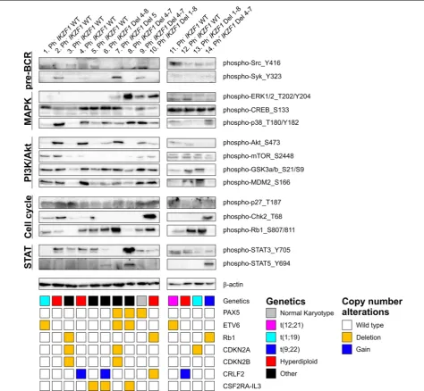
Previously, upregulation of the B-cell receptor (BCR) signaling pathway was reported in adult Ph+ ALL in
response to Ikaros-reconstitution, as well as an increased STAT5 phosphorylation in IKZF1 deleted versus wild type adult ALL cases with unknown cytogenetic back-ground [20, 22]. Although various peptides involved in the BCR signaling pathway and peptides derived from STAT5 were present on the array, we did not observe these differences in our Ph− cases. Therefore, in addition
to the kinome profiles, we performed western blot analy-sis of 14 pediatric BCP-ALL samples; seven Philadel-phia negative IKZF1 wild type patients, six Philadelphia negative IKZF1 deleted patients, and one Philadelphia positive IKZF1 deleted patient (Fig. 4). The western blot results showed a large variation in phosphorylation lev-els between the different ALL samples. STAT5_Y694 phosphorylation could be detected in the Philadelphia positive patient (patient no 14) and in two Philadelphia negative patients with an IKZF1 deletion (patients no 6 and 8), but was not detectable in the other IKZF1 deleted Philadelphia negative patients (patients no 7, 9, 10, and 13, Fig. 4). The western blot results showed that protein phosphorylation of Src_Y416, Syk_Y323, ERK1/2_T202/ Y204, CREB_S133, p38_T180/Y182, Akt_S473, mTOR_
S2448, GSK3α/β_S21/S9, MDM2_S166, p27_T187,
Chk2_T68, Rb1_S807/811, and STAT3_Y705 was clearly independent of IKZF1 status. Neither did we observe a clear relation between the western blot results and known copy number alterations of PAX5, ETV6, Rb1, CDKN2A, CDKN2B, or CRLF2 (Fig. 4). In conclusion, our kinome and western blot results suggest that IKZF1 deletions do not predict a unique protein expression sig-nature in pediatric Ph− ALL.
Discussion
IKZF1 deletions are found in approximately 70% of
the children with Philadelphia chromosome positive (Ph+) ALL and in 10–15% of the children with
Philadel-phia chromosome negative (Ph−) ALL. In both groups,
IKZF1 deletions are associated with an increased risk on relapse and decreased overall survival [2–4]. In Ph−
ALL, the effect of IKZF1 deletions on outcome is most pronounced in children with an intermediate treatment response based on the minimal residual disease at days 42 and 84 [24]. Although multiple studies have established IKZF1 as a prognostic factor in pediatric BCP-ALL, the effect of IKZF1 deletions on signaling pathways in Phila-delphia negative B-cell precursor ALL is poorly under-stood. In previous studies, we have shown that kinome profiling can be used successfully to describe active signal transduction pathways in pediatric malignancies [16–19].
In this study we used kinome profiling to elucidate the effect of IKZF1 deletions on active signal transduction pathways.
Unexpectedly, unsupervised hierarchical cluster analy-sis revealed no clustering between IKZF1 deleted and wild type patients. Furthermore, peptide phosphoryla-tion intensities between IKZF1 deleted and wild type Ph−
BCP-ALL patients were very comparable as shown by the phosphorylation intensities of the thirty-eight differen-tially phosphorylated peptides and by the list of top 100 most highly phosphorylated peptides. While focusing on important signaling pathways involved in cell prolifera-tion and survival of BCP-ALL cells we showed activity of all these pathways, however, no differences between the IKZF1 deleted versus the IKZF1 wild type group could be observed. Moreover, western blots of several key proteins involved in ALL signaling showed a variety of phospho-rylation events, clearly unrelated to IKZF1 status.
Although no clear differences in peptide phospho-rylation intensities could be observed, kinome profiles showed a remarkable high phosphorylation of peptides derived from Cytohesin-1_S394 and Cytohesin-2_S392. Cytohesins have been described as ErbB receptor acti-vators [25]. It has been described that Cytohesin over-expression enhances epidermal growth factor receptor (EGFR) signaling in human cancers including lung cancer and colorectal cancer [25, 26]. Although kinase domain mutations of ErbB receptors are uncommon in acute leu-kemias, in vitro inhibition of ErbB2 reduces cell prolifera-tion especially when combined with BCR-ABL tyrosine kinase inhibitors in Ph+ ALL, suggesting a role for ErbB
signaling pathway activation in leukemia [27, 28]. There-fore, it will be interesting to further explore the role of Cytohesin in BCP-ALL.
Although we did not identify a unique kinome signa-ture as a result of IKZF1 status, effects of IKZF1 deletions on gene expression were described previously. Iacobucci et al. showed that IKZF1 deletions display a unique gene expression signature in a cohort of adult B-ALL patients, including patients with a Philadelphia translocation and B-ALL patients negative for known molecular rearrange-ments [20]. The gene expression signature was charac-terized by the downregulation of genes regulating B-cell lineage development and DNA repair upon DNA damage response genes and upregulation of cell cycle/apoptosis genes, JAK/STAT signaling and stem cell self-renewal [20]. More recently, besides an upregulation of genes associated with B-cell proliferation, an upregulation of genes involved in cell adhesion and communication was also observed in pediatric Ph− ALL [29].
Philadelphia-positive ALL was identified several years ago (Ph-like ALL) [2, 30]. Importantly, 68% of the patients with Ph-like ALL showed deletions in IKZF1 [31]. Within the Ph-like ALL subtype, 5-year event-free survival rates in patients with IKZF1 alterations were inferior compared to Ph-like IKZF1 wild type ALL patients [31]. Recently, Roberts et al. defined the genomic landscape of Ph-like ALL in a large cohort of children and adolescents to elu-cidate kinase-activating genetic alterations which might include potential leads for targeted therapy [31]. Genomic
heterogeneous subgroup when evaluated at the level of active signal transduction pathways. The identification of potential targets for tyrosine kinase inhibitors therefore appears to be dictated by upstream genomic alterations that activate kinase signaling or cytokine receptor path-ways rather than IKZF1 status per se.
Conclusions
The aim of this study was to elucidate the effect of IKZF1 deletions on active signal transduction pathways using kinome profiling and western blot analysis in children with Ph− BCP-ALL. Although IKZF1 deletions are an
important clinical prognostic factor we were unable to identify a unique IKZF1 associated protein expression signature in pediatric Ph− BCP-ALL and consequently no
specific targets for future therapy of Ph−IKZF1 deleted
BCP-ALL were identified.
Abbreviations
ALL: acute lymphoblastic leukemia; BCP-ALL: B-cell progenitor acute lymphoblastic leukemia; BCR: B-cell receptor; BTK: Bruton’s tyrosine kinase; Additional files
Additional file 1: Table S1. Data file containing processed raw data values of the PepChip.
Additional file 2: Figure S1. Supplementary unsupervised hierarchical clustering. Kinase activity profiles of 45 pediatric BCP-ALL patients were generated. Unsupervised hierarchical clustering of 1,008 unique target peptides using Qlucore Omics Explorer 3.0 showed no distinct clustering based on the patients’ characteristics age, gender and white blood cell count (WBC).
Additional file 3: Table S2. List of 38 differentially phosphorylated peptides. Thirty-eight peptides were differentially phosphorylated between IKZF1 deleted (N= 13) and IKZF1 wild type (N= 31) Philadelphia negative pediatric BCP-ALL patients as determined by t-test analysis. The phosphorylation of 14 peptides was higher in the IKZF1 deleted group and 24 peptides showed reduced phosphorylation intensities. Shown are the normalized peptide phosphorylation intensities as well as P values. Additional file 4: Figure S2. Absolute phosphorylation intensities of 38 differentially phosphorylated peptides. Supervised hierarchical clustering of 44 pediatric BCP-ALL cases; 13 IKZF1 deleted and 31 IKZF1 wild type Philadelphia chromosome negative patients based on 38 peptides identi-fied by t-test. Each row represents a peptide, each column represents a single ALL sample. Absolute phosphorylation intensities are shown by the color saturation, red and green spots display high and low phosphoryla-tion intensities, respectively.
Additional file 5: Table S3. Top 100 most highly phosphorylated peptides. Shown is the list of top 100 highest phosphorylated peptides in IKZF1 deleted and IKZF1 wild type Philadelphia negative pediatric BCP-ALL patients. Highlighted peptides are overlapping peptides between IKZF1 deleted and IKZF1 wild type.
Additional file 6: Table S4. List of proteins involved in important signaling pathways for BCP-ALL. Shown are proteins involved in important signaling pathways for BCP-ALL cell proliferation and survival (e.g. the BCR signaling pathway, the MAPK, PI3 K/Akt/mTOR, JAK/STAT5 signaling pathways), adhesion pathways, and regulators of the cell cycle (including p21Cip1 and p27Kip1). The mean normalized phosphorylation intensities of multiple peptides derived from indicated proteins as well as P-values are shown for IKZF1 deleted (N= 13) and IKZF1 wild type (N= 31) pediat-ric patients.
CK2: casein kinase II; MLPA: multiplex ligation-dependent probe amplification; Ph−: Philadelphia chromosome negative; Ph+: Philadelphia chromosome posi-tive; SYK: spleen tyrosine kinase.
Authors’ contributions
NEvdS performed research, collected data, analyzed data and wrote the paper. FJGS performed research and collected data. AtE supervised pepchip data analysis. VG performed quantile normalization and supervised pepchip data analysis. FNvL supervised and edited the paper. ESJMdB designed research, analyzed data, supervised and edited the paper. All authors read and approved the final manuscript.
Author details
1 Division of Pediatric Oncology/Hematology, Department of Pediatrics, Beatrix Children’s Hospital, University Medical Center Groningen, University of Groningen, PO Box 30.001, 9700 RB Groningen, The Netherlands. 2 European Research Institute for the Biology of Ageing, University Medical Center Groningen, University of Groningen, Groningen, The Netherlands. 3 Labora-tory of Pediatric Oncology, Department of Pediatrics, Radboud Institute for Molecular Life Sciences, Radboud University Medical Center, Nijmegen, The Netherlands.
Acknowledgements
We thank the Junior Scientific Masterclass, University of Groningen, Gronin-gen, The Netherlands for financial support.
Compliance with ethical guidelines
Competing interests
The authors declare that they have no competing interests.
Received: 24 July 2015 Accepted: 27 July 2015
References
1. Inaba H, Greaves M, Mullighan CG. Acute lymphoblastic leukaemia. Lancet. 2013;381:1943–55.
2. Mullighan CG, Su X, Zhang J, Radtke I, Phillips LA, Miller CB, et al. Deletion of IKZF1 and prognosis in acute lymphoblastic leukemia. N Engl J Med. 2009;360:470–80.
3. Kuiper RP, Waanders E, van der Velden VH, van Reijmersdal SV, Venka-tachalam R, Scheijen B, et al. IKZF1 deletions predict relapse in uniformly treated pediatric precursor B-ALL. Leukemia. 2010;24:1258–64. 4. van der Veer A, Waanders E, Pieters R, Willemse ME, Van Reijmersdal SV,
Russell LJ, et al. Independent prognostic value of BCR-ABL1-like signature and IKZF1 deletion, but not high CRLF2 expression, in children with B-cell precursor ALL. Blood. 2013;122:2622–9.
5. Mullighan CG, Zhang J, Harvey RC, Collins-Underwood JR, Schulman BA, Phillips LA, et al. JAK mutations in high-risk childhood acute lymphoblas-tic leukemia. Proc Natl Acad Sci USA. 2009;106:9414–8.
6. Olsson L, Albitar F, Castor A, Behrendtz M, Biloglav A, Paulsson K, et al. Cooperative genetic changes in pediatric B-cell precursor acute lympho-blastic leukemia with deletions or mutations of IKZF1. Genes Chromosom Cancer. 2015;54:315–25.
7. Olsson L, Johansson B. Ikaros and leukaemia. Br J Haematol. 2015;169:479–91.
8. Georgopoulos K, Bigby M, Wang JH, Molnar A, Wu P, Winandy S, et al. The Ikaros gene is required for the development of all lymphoid lineages. Cell. 1994;79:143–56.
9. Wang JH, Nichogiannopoulou A, Wu L, Sun L, Sharpe AH, Bigby M, et al. Selective defects in the development of the fetal and adult lymphoid system in mice with an Ikaros null mutation. Immunity. 1996;5:537–49. 10. Winandy S, Wu P, Georgopoulos K. A dominant mutation in the Ikaros gene leads to rapid development of leukemia and lymphoma. Cell. 1995;83:289–99.
12. Uckun FM, Ma H, Zhang J, Ozer Z, Dovat S, Mao C, et al. Serine phospho-rylation by SYK is critical for nuclear localization and transcription factor function of Ikaros. Proc Natl Acad Sci USA. 2012;109:18072–7. 13. Ma H, Qazi S, Ozer Z, Zhang J, Ishkhanian R, Uckun FM. Regulatory
phosphorylation of Ikaros by Bruton’s tyrosine kinase. PLoS One. 2013;8:e71302.
14. Dupuis A, Gaub MP, Legrain M, Drenou B, Mauvieux L, Lutz P, et al. Biclonal and biallelic deletions occur in 20% of B-ALL cases with IKZF1 mutations. Leukemia. 2013;27:503–7.
15. Sun L, Liu A, Georgopoulos K. Zinc finger-mediated protein interactions modulate Ikaros activity, a molecular control of lymphocyte develop-ment. EMBO J. 1996;15:5358–69.
16. Sikkema AH, Diks SH, den Dunnen WF, ter Elst A, Scherpen FJ, Hoving EW, et al. Kinome profiling in pediatric brain tumors as a new approach for target discovery. Cancer Res. 2009;69:5987–95.
17. Ter Elst A, Diks SH, Kampen KR, Hoogerbrugge PM, Ruijtenbeek R, Boender PJ, et al. Identification of new possible targets for leukemia treat-ment by kinase activity profiling. Leuk Lymphoma. 2011;52:122–30. 18. Kampen KR, Ter Elst A, Mahmud H, Scherpen FJ, Diks SH, Peppelenbosch
MP, et al. Insights in dynamic kinome reprogramming as a consequence of MEK inhibition in MLL-rearranged AML. Leukemia. 2014;28:589–99. 19. van der Sligte NE, Scherpen FJ, Meeuwsen-de Boer TG, Lourens HJ, Ter Elst
A, Diks SH, et al. Kinase activity profiling reveals active signal transduction pathways in pediatric acute lymphoblastic leukemia: a new approach for target discovery. Proteomics. 2015;15:1245–54.
20. Iacobucci I, Iraci N, Messina M, Lonetti A, Chiaretti S, Valli E, et al. IKAROS deletions dictate a unique gene expression signature in patients with adult B-cell acute lymphoblastic leukemia. PLoS One. 2012;7:e40934. 21. Safavi S, Hansson M, Karlsson K, Biloglav A, Johansson B, Paulsson K.
Novel gene targets detected by genomic profiling in a consecutive series of 126 adults with acute lymphoblastic leukemia. Haematologica. 2015;100:55–61.
22. Trageser D, Iacobucci I, Nahar R, Duy C, von Levetzow G, Klemm L, et al. Pre-B cell receptor-mediated cell cycle arrest in Philadelphia chromo-some-positive acute lymphoblastic leukemia requires IKAROS function. J Exp Med. 2009;206:1739–53.
23. Dokter WH, Tuyt L, Sierdsema SJ, Esselink MT, Vellenga E. The spontane-ous expression of interleukin-1 beta and interleukin-6 is associated with spontaneous expression of AP-1 and NF-kappa B transcription factor in acute myeloblastic leukemia cells. Leukemia. 1995;9:425–32.
24. Waanders E, van der Velden VH, van der Schoot CE, van Leeuwen FN, van Reijmersdal SV, de Haas V, et al. Integrated use of minimal residual disease classification and IKZF1 alteration status accurately predicts 79% of relapses in pediatric acute lymphoblastic leukemia. Leukemia. 2011;25:254–8.
25. Bill A, Schmitz A, Albertoni B, Song JN, Heukamp LC, Walrafen D, et al. Cytohesins are cytoplasmic ErbB receptor activators. Cell. 2010;143:201–11.
26. Pan T, Sun J, Hu J, Hu Y, Zhou J, Chen Z, et al. Cytohesins/ARNO: the func-tion in colorectal cancer cells. PLoS One. 2014;9:e90997.
27. Irwin ME, Nelson LD, Santiago-O’Farrill JM, Knouse PD, Miller CP, Palla SL, et al. Small molecule ErbB inhibitors decrease proliferative signaling and promote apoptosis in philadelphia chromosome-positive acute lympho-blastic leukemia. PLoS One. 2013;8:e70608.
28. Lee JW, Soung YH, Kim SY, Nam SW, Park WS, Lee JY, et al. Kinase domain mutation of ERBB family genes is uncommon in acute leukemias. Leuk Res. 2006;30:241–2.
29. Vitanza NA, Zaky W, Blum R, Meyer JA, Wang J, Bhatla T, et al. Ikaros dele-tions in BCR-ABL-negative childhood acute lymphoblastic leukemia are associated with a distinct gene expression signature but do not result in intrinsic chemoresistance. Pediatr Blood Cancer. 2014;61:1779–85. 30. Den Boer ML, van Slegtenhorst M, De Menezes RX, Cheok MH,
Buijs-Glad-dines JG, Peters ST, et al. A subtype of childhood acute lymphoblastic leukaemia with poor treatment outcome: a genome-wide classification study. Lancet Oncol. 2009;10:125–34.
31. Roberts KG, Li Y, Payne-Turner D, Harvey RC, Yang YL, Pei D, et al. Targeta-ble kinase-activating lesions in Ph-like acute lymphoblastic leukemia. N Engl J Med. 2014;371:1005–15.
Submit your next manuscript to BioMed Central and take full advantage of:
• Convenient online submission
• Thorough peer review
• No space constraints or color figure charges
• Immediate publication on acceptance
• Inclusion in PubMed, CAS, Scopus and Google Scholar
• Research which is freely available for redistribution