Volume 14, Issue 2, Spring 2015
Editorial in Charge
Hossein Pakdaman, M.D.
Professor of Neurology, Shahid Beheshti
University of Medical Sciences,
Tehran, Iran
Editor-in-Chief
Shahriar Nafissi, M.D.
Associate Professor of Neurology,
Neurology Department, Tehran University of
Medical Sciences, Tehran, Iran
Deputy Editor
Farzad Fatehi, M.D.
Assistant Professor of Neurology, Neurology Department, Tehran University of
Medical Sciences, Tehran, Iran
Section Editors
Headache: Mansooreh Togha, M.D.,
Tehran
University of Medical Sciences
, Tehran, Iran
Multiple
Sclerosis:
Mohammad
Ali
Sahraian, M.D., Neurology Department, Tehran
University of Medical Sciences
, Tehran, Iran
Stroke: Afshin Borhanin Haghighi, M.D.,
Shiraz University of Medical Sciences, Shiraz,
Iran
Movement Disorders: Mohammad Rohani,
M.D., Iran University of Medical Sciences,
Tehran, Iran
Associate Editors
Shahin
Akhondzadeh,
Pharm.D.,
Ph.D,
Tehran University of Medical Sciences,
Tehran, Iran
Majid Ghafarpour, M.D., Tehran University of
Medical Sciences, Tehran, Iran
Massoud Nabavi, M.D., Shahed University of
Medical Sciences, Tehran, Iran
Scientific Assistant Editor
Ali Amini-Harandi, M.D., Shahid Beheshti University of Medical Sciences, Tehran, Iran
Editorial Board
Shahram Attarian, M.D., Centre de Référence
des Maladies Neuromusculaires et de la SLA,
France
Mahmoud R. Azarpazhooh, M.D., Mashhad
University of Medical Sciences, Mashhad, Iran
Keivan Basiri, M.D., Isfahan University of
Medical Sciences, Isfahan, Iran
Ahmad R. Dehpour, Pharm.D., Ph.D., Tehran
University of Medical Sciences, Tehran, Iran
Masoud Etemadifar, M.D., Isfahan University
of Medical Sciences, Isfahan, Iran
Kavian
Ghandehari,
M.D.,
Mashhad
University of Medical Sciences, Mashhad, Iran
Kurosh Gharagozli, M.D., Shahid Beheshti
University of Medical Sciences, Tehran, Iran
Mohammad H. Harirchian, M.D., Tehran
University of Medical Sciences, Tehran, Iran
Payam Kabiri, M.D., Ph.D., Tehran University
of Medical Sciences, Tehran, Iran
Hossein Kalani, M.D., Shahid Beheshti
University of Medical Sciences, Tehran, Iran
Jamshid Lotfi, M.D., Tehran University of
Medical Sciences, Tehran, Iran
Alireza Minagar, M.D., Louisiana State
University Health Sciences Center, USA
Ali Moghtaderi, M.D., Zahedan University of
Medical Sciences, Zahedan, Iran
Mahmood Motamedi, M.D., Tehran University
of Medical Sciences, Tehran, Iran
Alireza Nikseresht, M.D., Shiraz University of
Medical Sciences, Shiraz, Iran
Abdolmohamad M. Rostami, M.D., Thomas
Jefferson University Hospitals, USA
Mohammad
Saadatnia,
M.D.,
Isfahan
University of Medical Sciences, Isfahan, Iran
Mohammad K. Salajegheh, M.D., Brigham and
Women's Hospital and Harvard Medical School,
USA
Gholam A. Shahidi, M.D., Tehran University
of Medical Sciences, Tehran, Iran
Vahid
Shaygannejad,
M.D.,
Isfahan
University of Medical Sciences, Isfahan, Iran
Akbar Soltanzadeh, M.D., Tehran University
of Medical Sciences, Tehran, Iran
Amir A. Zamani, M.D., Brigham and Women's
Hospital and Harvard Medical School, USA
Babak Zamani, M.D., Tehran University of
Medical Sciences, Tehran, Iran
Secretary: Samaneh Bahraminejad, BSc
Email: [email protected]
http://ijnl.tums.ac.ir
Copy Edit, Layout Edit, Proof Reading and Design: Farzanegan Radandish Co. Postal Code: 81465-1798, Isfahan, Iran; Telefax: +98 311 6686302
www.farzaneganco.ir; Email: [email protected]
Indexed in
• PubMed,• PubMed Central,
• Academic Keys,
• Cite Factor (Directory Indexing of International Research Journals),
• Directory of Open Access Journals (DOAJ),
• Directory of Research Journal Indexing (DRJI),
• Ebsco,
• Electronic Journals Library,
• InfoBase Index,
• Islamic World Science Citation Center (ISC),
• LocatorPlus,
• Scientific Information Database (SID),
• Ulrichsweb Global Serials Directory,
• Universal Impact Factor,
• WorldCat
Iranian Journal of Neurology © 2015
INFORMATION FOR AUTHORS
Aim and Scope
The Iranian Journal of Neurology is dedicated to the
Iranian Neurological Association. The journal is a
peer-reviewed journal published quarterly and publishes
neurological experiences in basic or clinical fields in
English Language. The Iranian Journal of Neurology aims
to publish manuscripts of a high scientific quality
representing original clinical, diagnostic or experimental
works or observations in neurological sciences. Papers in
English are welcomed, particularly those which bring
novel information and researches in clinical or basic fields
from the neurological disorders. All received manuscripts
coving the scope of the journal will be evaluated by
properly competent referees.
Submission
Cover Letter:
Submissions should be accompanied by a cover letter
including a declaration by the first author on behalf of the
others to the effect that
(1) The paper has not been published to date (except
for abstracts of conference materials).
(2) The paper has not been accepted for publication
elsewhere.
(3) All persons listed as the authors have read it and
approved it for publication. The cover letters should be
submitted in section "Comments for the Editor".
Articles must be written in accurate scientific English
appropriate for publication. The articles are subject to
review and editing; however, the authors are responsible
for the correctness the manuscript's English language.
The articles must be submitted only online:
ijnl.tums.ac.ir
Policies
The Editorial Board reserves the right to reject a paper
without seeking reviewers’ opinion provied the content or
the form of the paper does not meet minimum acceptance
criteria or if the subject of the paper is beyond the aims
and scope of the journal.
Everyone listed as the author of a paper is responsible
for the reliability and completeness of data presented in
the paper.
Do not submit papers that copy fully or partially
previously published papers.
Indicate that this submission is ready to be considered
by this journal by checking off the following:
•
The submission has not been previously published,
nor is it before another journal for consideration (or an
explanation has been provided in Comments to the Editor).
•
The submission file is in Microsoft Word document
file format.
•
Where available, URLs for the references have
been provided.
•
The text is double-spaced; uses an Arial 12-point
font; and all illustrations, figures, and tables are placed
within the text at the appropriate points, rather than at the
end.
•
The text adheres to the stylistic and bibliographic
requirements outlined in the Author Guidelines, which is
found in About the Journal.
If the Editorial Board is not notified in advance and the
paper is found to have been copied during editorial work,
the paper shall be rejected.
We expect that all studies reported in the journal
conform to the requirements of the Declaration of Helsinki
(1989). Information on the consent of a relevant ethics
committee to perform the trial and the informed consent of
the patients to participate in the trial should be given in the
Material and methods section of each paper in which
diagnostic or therapeutic intervention does not follow from
the standard procedure. Authors of case reports must not
disclose personal data of patients described.
Manuscripts
The journal publishes:
•
Original Article
•
Review Article
•
Case Report
•
Short Communication
•
Clinical Notes
•
Editorial
•
Letters to Editor
•
Neurological Images
•
Neurological Videos
•
Iranian Neurological Events
•
Clinical Quiz
Details
Original and review papers: The maximum length of
original and review papers (including tables and figures
materials) is 3000 words.
Case reports: Should not be longer than 1200 words,
while letters to the Editor, reports and critical reviews
should not exceed 800 words.
Short communications: The maximum word number of
short communications should be below 1200 words with
maximum one table or figure and 10 references. The
manuscript should be structured including introduction,
materials and methods, results, discussion, and conclusion
with a structured abstracts as original articles.
Iranian Journal of Neurology © 2015
Neurological images or videos: Interesting cases as
neurological images or videos are welcome. They should
be maximally 400 words with legends without abstract and
unstructured. The videos should be uploaded as
supplementary files.
Letter to the Editor: May concern short scientific reports
and comments. The maximum number of words should be
below 800 words with maximum 5 references, no abstract,
no table or figure, and unstructured.
Clinical notes: Refer to important interesting observations
which are imperative for reminders in clinical practice.
The maximum number is 1000 words with maximum 5
references, 1 table and 1 figure with no abstract.
Iranian neurological events: Include the brief description
of major regional events (congresses or seminar)
implemented in Iran.
Structure of Articles
•
Manuscripts should be submitted in 12 points, Arial
font, with double line spacing and sufficient margins of
2.5 cm.
•
The text should not be formatted.
•
Each section of the paper should begin on a new page
The manuscript must include:
•
Page 1: Title Page
•
Page 2: Abstract and Key Words
•
Page 3 and subsequent pages: manuscript body
including Introduction, Materials and Methods, Results,
Discussion, Conclusion, References, Tables, Figures
1. Title page:
Title page should contain paper title, full names of
authors, authors’ place of work, full name and address of
the corresponding author (including e-mail address and
telephone number), given in that order.
2. Abstract page:
•
The length of the abstract should be at least 200
and not more than 250 words for original papers and not
more than 150 words for review papers and case reports.
Abstracts of original papers should be structured to
include the background, methods, results and conclusion.
•
Below the abstract authors should provide between
three and six keywords conforming to Medical Subject
Headings (Index Medicus).
3. Page three and subsequent pages of the original paper
and short communication should include the text arranged
in the following order (for other mansucript type, see
above):
1.
Introduction: The introduction should be as
concise as possible and introduce the context of the paper
to the reader; the paper should clearly state the research
hypothesis and the objective of the study.
2.
Materials and Methods: Description of the
studied population or material should be detailed and
include all information necessary to assess the reliability
of results obtained in the study and/or allow the
experiment to be repeated by other researchers; the section
related to statistical analysis should have information on
applied statistical tests and programs.
3.
Results: Present results directly related to the topic
of the paper only; tables and/or figures are recommended.
4.
Discussion
5.
Conclusions: These should be brief, follow directly
from results presented above and correspond to the aim of
the paper outlined in the introduction.
6.
Acknowledgements: Should comprise information
on sources of funding (grant numbers); acknowledgements
should concern those who made a significant contribution
to the paper, but who did not meet the criteria to be listed
as authors.
7.
References: References should be listed in the
order quoted in the paper. Please cite source and major
papers that offer interested readers an opportunity to
obtain more detailed information. Avoid citing review
papers and conference reports, if they are not the only
materials on a given topic.
References
In the paper references should be given in superscripts
with no space between the comma and the consecutive
number.
Authors are advised to carefully verify citation details.
Give names of first six authors; if there are more
authors, add “et al.“. Use Index Medicus abbreviations for
journal titles. Then mention the volume and the issue of
the journal.
The recommended style for journal references is as
follows:
[Reference number][Authors]. [Article title]. [Journal
Name] [Year of publication]; [volume](issue): [Pages
range].
For Journal Example:
1. Janghorbani M, Amini M, Willett WC, Mehdi Gouya
M, Delavari A, Alikhani S, et al. First nationwide survey
of prevalence of overweight, underweight, and abdominal
obesity in Iranian adults. Obesity (Silver Spring) 2007;
15(11): 2797-808.
For Books Example:
2. Ropper AH, Brown RJ. Adams and Victors principles
of neurology. 8
thed. New York, NY: McGraw Hill
Professional; 2005. p. 271.
Iranian Journal of Neurology © 2015
Photographs sent electronically should be of the resolution
of 300 dpi and in the .tif or .jpg format. Figures and
photographs are placed in the paper in the form delivered,
so they must be prepared carefully. Please indicate where
they should be placed in the text.
Abbreviations should be always clarified when used for
the first time in the text (including the abstract).
Abbreviations should not be used in paper titles, unless in
exceptional circumstances.
Review process: All papers submitted for publication in
the journal are assessed by two independent reviewers
with the mutual anonymity rule as to the names of
reviewers and authors observed.
Plagiarism policy: According to the plagiarism policy of
Iranian Journal of Neurology, plagiarism is defined as a
paper which replicates another publication with as a
minimum 25% resemblance and devoid of citation.
In any time the evidence of plagiarism is detected, the
manuscript will be withdrawn and the author will be
sanctioned from publishing papers permanently.
Iranian Journal of Neurology © 2015
Table of Contents
Original Article(s)
Effects of hydroalcoholic extract of Coriandrum sativum on oxidative damage in
pentylenetetrazole-induced seizures in rats
Reza Karami, Mahmoud Hosseini, Toktam Mohammadpour, Ahmad Ghorbani, Hamid Reza
Sadeghnia, Hassan Rakhshandeh, Farzaneh Vafaee, Mahdi Esmaeilizadeh………..59-66
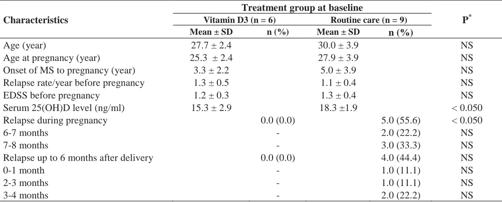
Efficacy of high-dose vitamin D3 supplementation in vitamin D deficient pregnant
women with multiple sclerosis: Preliminary findings of a randomized-controlled trial
Masoud Etemadifar, Mohsen Janghorbani ………67-73
Effects of pyridoxine supplementation on severity, frequency and duration of migraine
attacks in migraine patients with aura: A double-blind randomized clinical trial study
in Iran
Omid Sadeghi, Morteza Nasiri, Zahra Maghsoudi, Naseh Pahlavani, Masoud Rezaie,
Gholamreza Askari………...74-80
Comparison of serum vitamin D level in multiple sclerosis patients, their siblings, and
healthy controls
Ghazaleh Eskandari, Mahsa Ghajarzadeh, Mir Saeed Yekaninejad, Mohammad Ali Sahraian,
Razieh Gorji, Faezeh Rajaei, Abbas Norouzi-Javidan, Alireza Faridar, Amirreza Azimi.81-85
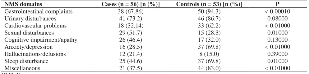
Comparison of frequencies of non motor symptoms in Indian Parkinson’s disease
patients on medical management versus deep brain stimulation: A case-control study
Kandadai Rukmini Mridula, Rupam Borgohain, Shaik Afshan Jabeen, Gaddamanugu
Padmaja, VCS Srinivasarao Bandaru, Praveen Ankathi, Meena A Kanikannan, Mohammed
Shujath Ali Khan……….86-93
Stroke specific quality of life questionnaire: Test of reliability and validity of the
Persian version
Mojtaba Mahmoodi, Anahid Safari, Mehrdad Vossoughi, Fatemeh Golbon-Haghighi,
Maliheh Kamali-Sarvestani, Haleh Ghaem, Afshin Borhani-Haghighi ……….94-100
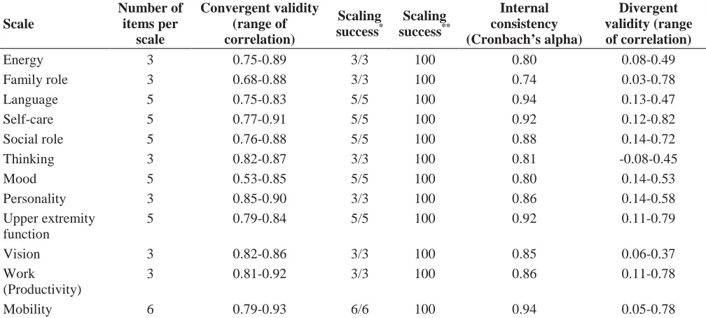
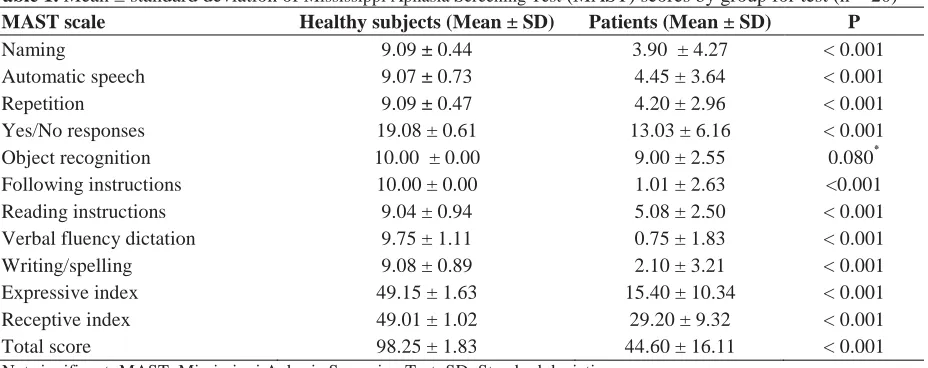
Development, cross-cultural adaptation, and validation of the Persian Mississippi
Aphasia Screening Test in patients with post-stroke aphasia
Ahmad
Reza
Khatoonabadi,
Noureddin
Nakhostin-Ansari,
Amin
Piran,
Hamid Tahmasian ………..101-107
Neurological Images
Unilateral cortical hyperintensity in diffusion-weighted MRI; New criteria for early
sporadic Creutzfeldt-Jakob disease
Nasim Tabrizi, Mahmoud Abedini……….108-109
Cyclic headaches in
β
-thalassemia intermedia case presenting as moyamoya syndrome
süha akpınar, Güliz Yılmaz, Emre Çelebio
ğ
lu………...110-112
Letter(s) to Editor
Intracranial hypertension and cerebellar symptoms due to Lhermitte-Duclos disease
Farhad Anssarzadegan, Atoosa Gharib, Shirin Behbahani, Meysam Ebrahimi-Abyaneh...113-115
Coexistence of Ehlers-Danlos syndrome and multiple sclerosis
Iranian Journal of Neurology © 2015 Corresponding Author: Mahmoud Hosseini
Original Paper
Iran J Neurol 2015; 14(2): 59-66
Effects of hydroalcoholic extract of
Coriandrum sativum on oxidative
damage in
pentylenetetrazole-induced seizures in rats
Reza Karami
1, Mahmoud Hosseini
2, Toktam Mohammadpour
3, Ahmad Ghorbani
4, Hamid Reza Sadeghnia
4,
Hassan Rakhshandeh
4, Farzaneh Vafaee
3, Mahdi Esmaeilizadeh
51
Student Research Committee, School of Medicine, Mashhad University of Medical Sciences, Mashhad, Iran
2
Neurocognitive Research Center AND Department of Physiology, School of Medicine, Mashhad University of Medical Sciences, Mashhad, Iran
3
Neurogenic Inflammation Research Center AND Department of Physiology, School of Medicine, Mashhad University of Medical Sciences, Mashhad, Iran
4 Pharmacological Research Center of Medicinal Plants AND Department of Pharmacology, School of Medicine, Mashhad University of
Medical Sciences, Mashhad, Iran
5
Esfarayen Faculty of Medical Sciences, Esfarayen, Iran
Keywords
Coriandrum sativum, Pentylenetetrazole, Seizures, Rat,
Oxidative Stress, Brain
Abstract
Background: An important role for oxidative stress,
as a consequence of epileptic seizures, has been
suggested. Coriandrum sativum has been shown that
have antioxidant effects. Central nervous system
depressant effects of C. sativum have also been
reported. In this study, the effects of hydroalcoholic
extract of aerial parts of the plants on brain tissues
oxidative damages following seizures induced by
pentylenetetrazole (PTZ) was investigated in rats.
Methods: The rats were divided into five groups and
treated: (1) Control (saline), (2) PTZ (90 mg/kg, i.p.),
(3-5) three doses (100, 500 and 1000 mg/kg of C.
sativum extract (CSE) before PTZ. Latencies to the first
minimal clonic seizures (MCS) and the first generalized
tonic-clonic seizures (GTCS) were recorded. The
cortical and hippocampal tissues were then removed
for biochemical measurements.
Results:
The extract significantly increased the MCS
and GTCS latencies (P < 0.01, P < 0.001) following
PTZ-induced seizures. The malondialdehyde (MDA)
levels in both cortical and hippocampal tissues of PTZ
group were significantly higher than those of the control
animals (P < 0.001). Pretreatment with the extract
prevented elevation of the MDA levels (P < 0.010–P <
0.001). Following PTZ administration, a significant
reduction in total thiol groups was observed in both
cortical and hippocampal tissues (P < 0.050).
Pre-treatment with the 500 mg/kg of the extract caused a
significant prevention of decreased in total thiol
concentration in the cortical tissues (P < 0.010)
.
Conclusion:
The present study showed that the
hydroalcoholic extract of the aerial parts of C. sativum
possess significant antioxidant and anticonvulsant
activities
.
Introduction
Epilepsy is a common neurological disease, which
affects approximately 1% of the population.
1It is
characterized by abnormal episodic bursts of electrical
activity in neurons, which is followed by a significant
impact on the behavior of the affected patients.
2An
important role for oxidative stress both as a
consequence and as a cause of epileptic seizures has
been suggested.
3It has been reported that production
Iranian Journal
of Neurology
of free radicals increases during seizures, which is
lead to oxidative damage to lipids, DNA and
susceptible proteins.
4Due to high levels of membrane lipid constituents,
the central nervous system (CNS) is very susceptible
to oxidative injury.
5In addition, oxidative damage
plays a significant role in the pathogenesis of various
CNS disorders and neurobehavioral impairments.
5The functional impairments of CNS, which occur
during seizures have also been suggested to be at least
in part, related to the brain tissues oxidative
damages.
6Furthermore, the anticonvulsant activities
of several agents with antioxidant effects such as
melatonin, vineatrol, trans-resveratrol and alpha
lipoic acid have been documented.
7,8There are also
some reports that reactive oxygen species (ROS) may
underlie the convulsant and neurotoxic effects of
pentylenetetrazole (PTZ).
9The results of human and
animal studies imply that epilepsy and seizures are
lead to the brain tissues oxidative damages, especially
in the cortical and hippocampal regions, which are
accompanied with cognitive, learning and memory
deficits.
4,6,8,10Medicinal plants are good sources to find new
therapeutic agents for human diseases. Coriandrum
sativum, an annual herb belonging to the Apiaceae
family, has been reported to have a wide range of
biological
activities
including
sedative-hypnotic,
antidiabetic,
hypolipidemic, and
hepatoprotective
effects.
11-15Experimental studies have also revealed a
strong antioxidant activity for C. sativum that is superior
to the well-known antioxidant agents like ascorbic
acid.
15-20In our previous work, we found that the
hydroalcoholic extract of aerial parts of this plant bearing
some compounds with the hypnotic effects.
21Regarding
the antioxidant and CNS depressant effects of C.
sativum, we aimed to evaluate the possible protective
effects of aerial parts of the plant on PTZ-seizures and
the brain tissues oxidative damages in rats.
Materials and Methods
PTZ was purchased from Sigma-Aldrich Company
(St. Louis, USA). Other chemical compounds such as
thiobarbituric acid (TBA), trichloroacetic acid (TCA),
hydrochloric acid (HCL), ethylenediaminetetraacetic
acid (EDTA) and 2, 2'-dinitro-5, 5'-dithiodibenzoic
acid (DTNB) were bought from Merck Company.
In this study, 40 virgin male Wistar rats, 250 ± 20 g
in weight were used. The animals were maintained at
the animal house under controlled conditions
including 12 h light and dark cycle, 22-24 °C
temperature
and
appropriate
humidity
with
laboratory chow and water provided ad libitum.
The animals were randomly divided into five
groups and treated (n = 8 in each group) as follows:
(1) Control (saline), (2) PTZ, (3) C. sativum extract
(CSE) 100 mg/kg (CSE 100) + PTZ, (4) CSE 500 mg/kg
(CSE 500) + PTZ and (5) CSE 1000 mg/kg (CSE 1000)
+ PTZ. The doses were chosen regarding our previous
study.
21The number of animals was also based on our
previous studies.
9,21-24The animals in groups 2-5 were treated
intraperitoneally (i.p.) with saline or the extract 30 min
before i.p. injection of a single dose (90 mg/kg) of
PTZ. In our previous works, we showed that PTZ in
this dose induces generalized tonic-clonic seizures
(GTCS) in rats.
9,22,24,25The time interval between
injection of the extract and PTZ was chosen regarding
our previous work in which injection of the extract 30
min before injection of pentobarbital increased the
sleeping time.
21The cortical and hippocampal regions were then
removed for biochemical measurements. In the
control group, saline was injected instead of both PTZ
and CSE and the brain tissues were removed without
inducing the seizures. All efforts were made to
maintain the animals in good general health, in
accordance with the European Communities Council
Directive (2010/63/UE). All behavioral tests were
conducted between 10:00 and 14:00 O'clock. Animal
handling and all related procedures were confirmed
by the Mashhad University of Medical Sciences, Iran,
Ethical Committee.
The aerial parts (leaves, stems, twigs) of C. sativum
were collected from Neyshabur, Iran. The identity of
the plant was confirmed and for future reference a
voucher specimen (10068) was deposited at the
herbarium of School of Pharmacy (Mashhad University
of Medical Sciences). To prepare the hydroalcoholic
extract, the plant materials (50 g) were dried and
extracted with 300 ml ethanol-water (70/30, v/v) using
a Soxhalet apparatus. The extract reduced to dryness
with a rotary vacuum evaporator (Stuart RE300, UK).
26In order to observe ictal behavior, PTZ was
injected and the animals were placed in a Plexiglas
arena (30 cm × 30 cm × 30 cm) on the day of the
experiment. The animals were observed during 60
min after PTZ (90 mg/ kg) administration.
9,22,24,25,27The behavioral responses of the animals to PTZ
administration were evaluated using these criteria:
latency to the first minimal clonic seizure (MCS),
incidence of MCS, latency to the first GTCS, incidence
of GTCS, protection percentage against GTCS and
protection percentage against mortality.
23-25minimum pain, suffering, and distress. The method
was performed as set out in that Annex IV of the
guidelines from Directive EU/2010/63 of the
European Parliament.
For total thiol (SH) content measurement, the
cortical and hippocampal regions were dissected on
an ice-cold surface and homogenized in iced-cold
phosphate-buffered saline to give 10% homogeny.
Total SH groups were measured using DTNB as the
reagent. This reagent reacts with the thiol groups to
produce a yellow colored complex, which has a peak
absorbance at 412 nm. Briefly, 1 ml Tris-EDTA buffer
(pH = 8.6) was added to 50 µl of the brain
homogenates, and the sample absorbance was read at
412 nm against Tris-EDTA buffer alone (A1). Then, 20
µl DTNB reagents (10 mm in methanol) were added to
the mixture and after 15 min (stored in laboratory
temperature), the sample absorbance was read again
(A2). The absorbance of DTNB reagent was also read
as a blank (B).
Total thiol concentration (mm) was
calculated from the following equation.
9,28-30Total thiol concentration (mM) = (A2-A1-B) ×
1.07/0.05 × 13.6
Malondialdehyde (MDA) levels, as an index of
lipid peroxidation, were also measured. MDA reacts
with TBA as a TBA reactive substance to produce a
red colored complex, which has a peak absorbance at
535 nm. The TBA/TCA/HCL reagent was added to
the homogenates, and the solution was heated in a
water bath for 40 min. After cooling, the whole
solutions were centrifuged within 1000 g for 10 min.
The absorbance was measured at 535 nm.
9,28-30The
MDA concentration was calculated as follows:
C (M) = Absorbance/(1.56 × 10
5)
All data were expressed as mean ± standard error
of the mean and analyzed by using ANOVA, followed
by Tukey’s post-hoc comparison test. P < 0.0500 were
considered to be statistically significant.
Results
Effect of C. sativum on PTZ-induced seizures
All the animals in different groups (except the control
group, which did not receive PTZ) showed MCS and
GTCS following administration of a high dose of PTZ.
Data analysis using one-way ANOVA showed that
there was a significant difference between the groups in
MCS latencies (F3,28 =
19.65, P < 0.0001). MCS latencies
in the extract pre-treated groups were significantly
higher than that of PTZ group. When compared with
PTZ group (61.66 ± 4.76 s), 100, 500 and 1000 mg/kg of
the extract significantly (P < 0.0100 to P < 0.0010)
increased the MCS latencies to 77.3 ± 2.07, 93.5 ± 4.35
and 339.3 ± 58.96 s, respectively (Figure 1).
Data analysis using one-way ANOVA also showed
that there was a significant difference between the
groups in GTCS latencies (F3,28 =
24.13, P < 0.0001). The
GTCS latencies in the animals, which had received 100,
500 and 1000 mg/kg of CSE before PTZ, were 183 ±
4.69, 341.88 ± 44.16, and 710.3 ± 98.84 c, respectively. All
3 doses of the extract significantly increased the GTCS
latencies (P < 0.0100 to P < 0.0010) compared with PTZ
group (114 ± 1.8 c) (Figure 2). There were no significant
differences
in
mortality
rate
following
PTZ
administration between groups.
Figure 1. Latencies to minimal clonic seizures (MCS) onsets
in pentylenetetrazole (PTZ), C. sativum extract (CSE) 100
mg/kg (CSE 100)-PTZ, CSE 500 mg/kg (CSE 500)-PTZ,
CSE 1000 mg/kg (CSE 1000)-PTZ groups. The animals were
treated with saline or CSE (100, 500 or 1000 mg/kg) before a
single injection (90 mg/kg) of PTZ;
**P < 0.010;
***
P < 0.001 as compared to PTZ group
Figure 2. Latencies to generalized tonic-clonic seizures
Effect of C. sativum on brain tissues oxidative damage
Data analysis using one-way ANOVA showed that there
was a significant difference between the groups in MDA
concentrations of cortical tissues (F4,35 =
6.93, P < 0.001).
The MDA levels in cortical regions of PTZ group were
significantly higher than those of control animals
(P < 0.001) (Figure 3). As shown in figure 3, pretreatment
with both 100 and 1000 mg/kg of the extract resulted in
a significant reduction in the free radical-mediated lipid
peroxidation as indicated by a decrease in the MDA
levels (P < 0.001 and P < 0.010, respectively).
Data analysis using one-way ANOVA also showed
that there was a significant difference between the
groups in total thiol contents of cortical tissues
(F4,35 =
6.78, P < 0.001). Following PTZ administration, a
significant reduction in total SH groups in cortical
samples was observed (P < 0.050, Figure 4).
Pretreatment with 500 mg/kg of the extract prevented
of decreased total thiol concentration in cortical tissues,
as compared with PTZ group (P < 0.010).
Figure 3. Comparison of the malondialdehyde (MDA) levels
in cortical tissues of control, pentylenetetrazole (PTZ), C.
sativum extract (CSE) 100 mg/kg (CSE 100)-PTZ, CSE 500
mg/kg (CSE 500)-PTZ, CSE 1000 mg/kg (CSE 1000)-PTZ
groups. The animals were treated with saline or CSE (100,
500 or 1000 mg/kg) before a single injection (90 mg/kg) of
PTZ; The animals in control group received saline instead of
PTZ;
***P < 0.001 as compared to control group;
++
P < 0.010;
+++P < 0.001 as compared to PTZ group
Data analysis using one-way ANOVA showed that
there was a significant difference between the groups in
MDA
concentrations
of
hippocampal
tissues
(F4,35 =
24.53, P < 0.0001). The MDA levels in the
hippocampal regions of PTZ group were significantly
higher than those of control animals (P < 0.001)
(Figure 5). The results also showed that all three doses of
CSE prevented the elevation of MDA concentration in
hippocampal tissues (P < 0.001 for all, Figure 5).
Data analysis using one-way ANOVA also showed
that there was a significant difference between the
groups in total thiol contents of hippocampal tissues
(F4,35 =
3.19, P < 0.050). Following PTZ administration, a
significant reduction in total SH groups in the
hippocampal samples was observed (P < 0.050, Figure 6).
There were no significant differences between CSE
treated rats and PTZ group when total thiol content in
hippocampal tissues was compared (Figure 6).
Figure 4. Comparison of the total SH groups in cortical
tissues of control, pentylenetetrazole (PTZ), C. sativum
extract (CSE) 100 mg/kg (CSE 100)-PTZ, CSE 500 mg/kg
(CSE 500)-PTZ, CSE 1000 mg/kg (CSE 1000)-PTZ groups.
The animals were treated with saline or CSE (100, 500 or
1000 mg/kg) before a single injection (90 mg/kg) of PTZ.
The animals in the control group received saline instead of
PTZ;
*P < 0.0500 as compared to Control group;
++
P < 0.0100 as compared to PTZ group
Figure 5. Comparison of the malondialdehyde (MDA) levels
Figure 6. Comparison of the total SH groups in hippocampal
tissues of control, pentylenetetrazole (PTZ), C. sativum
extract (CSE) 100 mg/kg (CSE 100)-PTZ, CSE 500 mg/kg
(CSE 500)-PTZ, CSE 1000 mg/kg (CSE 1000)-PTZ groups.
The animals were treated with saline or CSE (100, 500 or
1000 mg/kg) before a single injection (90 mg/kg) of PTZ;
The animals in control group received saline instead of PTZ;
*
P < 0.050 as compared to control group
.
Discussion
Oxidative stress is a basis etiology for many
neurological
and
neurodegenerative
disorders.
Previous studies demonstrated that oxidative stress
plays an important role in the pathogenesis of
epileptic seizures
.
3,4,31Elevation of free radical levels
during seizures
7,31has been well documented
therefore, it is suggested that oxidative stress has an
important role in the brain damages due to
epilepsy.
9,32Furthermore, the brain tissues oxidative
damage contributes to the complications of seizures
and epilepsy including cognitive, learning and
memory impairments.
2,10In the present study the
possible protective effects of C. sativum aerial parts on
PTZ-induced seizures and the brain tissues oxidative
damages was investigated. PTZ, is a selective inhibitor
of the chloride channel which is coupled to the
gamma-aminobutyric acid receptor.
33It is a
well-known chemoconvulsant which is frequently used for
evaluation of antiepileptic drugs.
34,35A high dose of
PTZ induces a continued seizure activity which
progress from mild myoclonic jerks to face and
forelimbs clonus without loss of righting reflex (which
is known as MCS), to clonic seizures of limbs with loss
of righting reflex which is followed by full tonic
extension of both forelimbs and hindlimbs (GTCS).
36PTZ has been repeatedly used in 90-100 mg/ kg to
induce MCS and GTCS seizures.
9,22-25,27,28We also
previously showed that PTZ-induced seizures are
accompanied with brain tissues oxidative damage.
9The contribution of ROS to the neurotoxic effects of
earlier, CSE prevented PTZ-induced thiol depletion
only in the cortex and not hippocampus. One possible
explanation for this observation might be due to brain
regional-dependent glutathione (GSH) metabolism, as
a major source of thiol group.
51It has been observed
that brain GSH concentrations varied in the range of
0.2-10 mM.
52It was reported that GSH levels are
highest in cortex, followed by hippocampus and brain
stem.
53,54Consistent with this finding, some studies
reported a strong antioxidant activity for C.
sativum.
15-20Furthermore, we showed that this
activity of C. sativum was accompanied by an
anticonvulsant effect as it increased both MCS and
GTCS latencies. In keeping with these observations,
the anticonvulsant activity of several agents with
antioxidant effect such as melatonin, vineatrol,
trans-resveratrol and alpha lipoic acid have also been
shown.
7,8Using pentobarbital-induced hypnosis
animal model, we previously found that the injection
of 50, 100, 200 and 400 mg/kg of hydroalcoholic
extract of C. sativum before pentobarbital increased
the sleep time of the rats.
12Even a higher dose of the
extract has also been previously used in the rats
without observation of any toxic effect.
55In the
current study, therefore we used comparable doses of
CSE 30 min before PTZ injection to test its
anticonvulsive effect. Previously, Hosseinzadeh and
Madanifard reported an anticonvulsant effect for the
aqueous and ethanolic extracts of C. sativum seeds.
56The same effect was found by Emamghoreishi and
Heidari-Hamedani for aqueous and hydroalcoholic
extracts and essential oil of the seeds.
57Regarding the
results of present study the beneficial effects of aerial
parts of the plant on seizures and its complications
such as brain tissues oxidative is suggested however,
further studies using other animal models are needed
to be done in the future. The electrophysiological
studies are also suggested for future.
However, no pharmacological studies have been
yet evaluated the anticonvulsant activity of aerial
parts of this plant. These parts of C. sativum are
widely consumed as a vegetable all over the world.
With the present study, we showed that aerial parts of
C. sativum are effective in protection against seizures
and oxidative stress induced by PTZ.
In the present study, the chemical compound(s)
responsible for these effects of C. sativum were not
identified and needs to be more investigated in future
studies. The presence of the flavonoids such as
quercitin has been reported in CSE.
58On the other
hand, it has been shown that the flavonoids have
considerable anticonvulsant effects.
59,60Regarding
sedative and CNS depressant effects of falvonoids
such as quercetin, these effects could be attributed to
the affinity of the compounds for the central
benzodiazepine receptors.
61-65The beneficial effect of
linalool in PTZ as well glutamate-related seizure
models has been suggested.
66,67It might be suggested
the beneficial effects of the extract which was
observed in the present study, might be at least in
part, due to linalool which is one the main
constituents of coriander and has considerable
antioxidant effects.
68Conclusion
The present data demonstrate that the hydroalcoholic
extract of C. sativum aerial parts possesses
anticonvulsant activity. This activity is accompanied
by an antioxidant effect in brain tissues. Isolation of
the active compound(s) from the extract needs to be
done in the future.
Conflict of Interests
The authors declare no conflict of interest in this study.
Acknowledgments
The authors would like to thank the Vice Presidency of
Research, Mashhad University of Medical Sciences, for
its financial supports.
References
1. Sander JW. The epidemiology of epilepsy revisited. Curr Opin Neurol 2003; 16(2): 165-70.
2. Meador KJ. Cognitive outcomes and predictive factors in epilepsy. Neurology 2002; 58(8 Suppl 5): S21-S26.
3. Patel M. Mitochondrial dysfunction and oxidative stress: cause and consequence of epileptic seizures. Free Radic Biol Med 2004; 37(12): 1951-62.
4. Kudin AP, Kudina TA, Seyfried J, Vielhaber
S, Beck H, Elger CE, et al. Seizure-dependent modulation of mitochondrial oxidative phosphorylation in rat hippocampus. Eur J Neurosci 2002; 15(7): 1105-14.
5. Sharma DR, Sunkaria A, Bal A, Bhutia YD, Vijayaraghavan R, Flora SJ, et al. Neurobehavioral impairments, generation of oxidative stress and release of pro-apoptotic factors after chronic exposure to sulphur mustard in mouse brain. Toxicol Appl Pharmacol 2009; 240(2): 208-18.
6. Mehla J, Reeta KH, Gupta P, Gupta YK. Protective effect of curcumin against seizures and cognitive impairment in a pentylenetetrazole-kindled epileptic rat model. Life Sci 2010; 87(19-22): 596-603. 7. Gupta YK, Briyal S. Protective effect of
vineatrol against kainic acid induced seizures, oxidative stress and on the expression of heat shock proteins in rats. Eur Neuropsychopharmacol 2006; 16(2): 85-91. 8. Sharma M, Briyal S, Gupta YK. Effect of
alpha lipoic acid, melatonin and trans
How to cite this article: Karami R, Hosseini M,
resveratrol on intracerebroventricular streptozotocin induced spatial memory deficit in rats. Indian J Physiol Pharmacol 2005; 49(4): 395-402.
9. Hosseini M, Harandizadeh F, Niazamand S, Soukhtanloo M, Mahmoudabady M. Antioxidant effect of Achillea wilhelmsii extract on pentylenetetrazole (seizure model)-induced oxidative brain damage in Wistar rats. Indian J Physiol Pharmacol 2013; 57(4): 418-24.
10. Rosche J, Uhlmann C, Froscher W. Cognitive deficits and psychiatric disorders in patients with new-onset epilepsy. Fortschr Neurol Psychiatr 2010; 78(1): 18-26. 11. Nematy M, Kamgar M, Mohajeri SM,
Tabatabaei Zadeh SA, Jomezadeh MR, Akbarieh HO, et al. The effect of hydroalcoholic extract of Coriandrum sativum on rat appetite. Avicenna J Phytomed 2013; 3(1): 91-7.
12. Rakhshandeh H, Sadeghnia HR, Ghorbani A. Sleep-prolonging effect of Coriandrum sativum hydro-alcoholic extract in mice. Nat Prod Res 2012; 26(22): 2095-8.
13. Eidi M, Eidi A, Saeidi A, Molanaei S, Sadeghipour A, Bahar M, et al. Effect of coriander seed (Coriandrum sativum L.) ethanol extract on insulin release from pancreatic beta cells in streptozotocin-induced diabetic rats. Phytother Res 2009; 23(3): 404-6.
14. Dhanapakiam P, Joseph JM, Ramaswamy VK, Moorthi M, Kumar AS. The cholesterol lowering property of coriander seeds (Coriandrum sativum): mechanism of action. J Environ Biol 2008; 29(1): 53-6.
15. Samojlik I, Lakic N, Mimica-Dukic N, Dakovic-Svajcer K, Bozin B. Antioxidant and hepatoprotective potential of essential oils of coriander (Coriandrum sativum L.) and caraway (Carum carvi L.) (Apiaceae). J Agric Food Chem 2010; 58(15): 8848-53. 16. de Almeida Melo E, Bion FM, Filho JM,
Guerra NB. In vivo antioxidant effect of aqueous and etheric coriander (Coriandrum sativum L.) extracts. European Journal of Lipid Science and Technology 2003; 105(9): 483-7.
17. Harsha SN, Anilakumar KR. In vitro free radical scavenging and DNA damage protective property of Coriandrum sativum L. leaves extract. J Food Sci Technol 2014; 51(8): 1533-9.
18. Tang EL, Rajarajeswaran J, Fung SY, Kanthimathi MS. Antioxidant activity of Coriandrum sativum and protection against DNA damage and cancer cell migration. BMC Complement Altern Med 2013; 13: 347.
19. Sultana S, Ripa FA, Hamid K. Comparative antioxidant activity study of some commonly used spices in Bangladesh. Pak J Biol Sci 2010; 13(7): 340-3.
20. Wangensteen H, Samuelsen AB, Malterud KE. Antioxidant activity in extracts from coriander. Food Chemistry 2004; 88(2): 293-7.
21. Rakhshandah H, Hosseini M. Potentiation of pentobarbital hypnosis by Rosa damascena in mice. Indian J Exp Biol 2006; 44(11): 910-2. 22. Ebrahimzadeh Bideskan AR, Hosseini M,
Mohammadpour T, Karami R, Khodamoradi M, Nemati KH, et al. Effects of soy extract on pentylenetetrazol-induced seizures in ovariectomized rats. Zhong Xi Yi Jie He Xue
Bao 2011; 9(6): 611-8.
23. Hosseini M, Ghasemzadeh RM, Sadeghnia HR, Rakhshandeh H. Effects of different extracts of Rosa damascena on pentylenetetrazol-induced seizures in mice. Zhong Xi Yi Jie He Xue Bao 2011; 9(10): 1118-24.
24. Hosseini M, Sadeghnia HR, Salehabadi S, Alavi H, Gorji A. The effect of L-arginine and L-NAME on pentylenetetrazole induced seizures in ovariectomized rats, an in vivo study. Seizure 2009; 18(10): 695-8. 25. Hosseini M, Harandizadeh F, Niazmand S,
Soukhtanloo M, Faizpour A, Ghasemabady M. The role for nitric oxide on the effects of hydroalcoholic extract of Achillea wilhelmsii on seizure. Avicenna J Phytomed 2014; 4(4): 251-9.
26. Kamkar AM, Nazariborun A, Hosseini M. Analgesic effect of the aqueous and ethanolic extracts of clove. Avicenna J Phytomed 2013; 3(2): 186-92.
27. Hosseini M, Pkan P, Rakhshandeh H, Aghaie A, Sadeghnia HR, Ghasemzadeh Rahbardar M. The Effect of Hydro-Alcoholic Extract of Citrus Flower on Pentylenetetrazole and Maximal Electroshock-Induced Seizures in Mice. World Applied Sciences Journal 2011; 15(8): 1104-9.
28. Hosseini M, Pourganji M, Khodabandehloo F, Soukhtanloo M, Zabihi H. Protective Effect of L-Arginine against Oxidative Damage as a Possible Mechanism of its Beneficial Properties on Spatial Learning in Ovariectomized Rats. Basic & Clinical Neuroscience 2012; 3(5): 36-44.
29. Vafaee F, Hosseini M, Sadeghinia HR, Hadjzadeh MA, Soukhtanloo M, Rahimi M. The effects of soy extract on spatial learning and memory damage induced by global ischemia in ovariectomised rats. Malays J Med Sci 2014; 21(3): 19-30.
30. Pourganji M, Hosseini M, Soukhtanloo M, Zabihi H, Hadjzadeh MA. Protective role of endogenous ovarian hormones against learning and memory impairments and brain tissues oxidative damage induced by lipopolysaccharide. Iran Red Crescent Med J 2014; 16(3): e13954.
31. Costello DJ, Delanty N. Oxidative injury in epilepsy: potential for antioxidant therapy? Expert Rev Neurother 2004; 4(3): 541-53. 32. Zhen J, Qu Z, Fang H, Fu L, Wu Y, Wang H,
et al. Effects of grape seed proanthocyanidin extract on pentylenetetrazole-induced kindling and associated cognitive impairment in rats. Int J Mol Med 2014; 34(2): 391-8. 33. Sejima H, Ito M, Kishi K, Noda A, Serikawa
T. Regional excitatory and inhibitory amino acid concentrations in Noda epileptic rat (NER) brain. Brain Dev 1999; 21(6): 382-5. 34. Porter RJ, Cereghino JJ, Gladding GD,
Hessie BJ, Kupferberg HJ, Scoville B, et al. Antiepileptic Drug Development Program. Cleve Clin Q 1984; 51(2): 293-305. 35. Hosseinzadeh H, Sadeghnia HR. Protective
effect of safranal on pentylenetetrazol-induced seizures in the rat: involvement of GABAergic and opioids systems. Phytomedicine 2007; 14(4): 256-62. 36. Loscher W, Honack D, Fassbender CP,
Nolting B. The role of technical, biological and pharmacological factors in the laboratory evaluation of anticonvulsant drugs. III. Pentylenetetrazole seizure models. Epilepsy Res 1991; 8(3): 171-89.
37. Xie T, Wang WP, Mao ZF, Qu ZZ, Luan SQ, Jia LJ, et al. Effects of epigallocatechin-3-gallate on pentylenetetrazole-induced kindling, cognitive impairment and oxidative stress in rats. Neurosci Lett 2012; 516(2): 237-41.
38. Liu SH, Chang CD, Chen PH, Su JR, Chen CC, Chaung HC. Docosahexaenoic acid and phosphatidylserine supplementations improve antioxidant activities and cognitive functions of the developing brain on pentylenetetrazol-induced seizure model. Brain Res 2012; 1451: 19-26.
39. Sudha K, Rao AV, Rao A. Oxidative stress and antioxidants in epilepsy. Clin Chim Acta 2001; 303(1-2): 19-24.
40. Rodrigues AD, Scheffel TB, Scola G, Santos MT, Fank B, de Freitas SC, et al. Neuroprotective and anticonvulsant effects of organic and conventional purple grape juices on seizures in Wistar rats induced by pentylenetetrazole. Neurochem Int 2012; 60(8): 799-805.
41. Reilly C, Agnew R, Neville BG. Depression and anxiety in childhood epilepsy: a review. Seizure 2011; 20(8): 589-97.
42. Maldonado A, Ramos W, Perez J, Huaman LA, Gutierrez EL. Convulsive status epilepticus: clinico-epidemiologic characteristics and risk factors in Peru. Neurologia 2010; 25(8): 478-84.
43. Liang LP, Beaudoin ME, Fritz MJ, Fulton R, Patel M. Kainate-induced seizures, oxidative stress and neuronal loss in aging rats. Neuroscience 2007; 147(4): 1114-8. 44. Golechha M, Bhatia J, Arya DS.
Hydroalcoholic extract of Emblica officinalis Gaertn. affords protection against PTZ-induced seizures, oxidative stress and cognitive impairment in rats. Indian J Exp Biol 2010; 48(5): 474-8.
45. Velaga MK, Yallapragada PR, Williams D, Rajanna S, Bettaiya R. Hydroalcoholic seed extract of Coriandrum sativum (Coriander) alleviates lead-induced oxidative stress in different regions of rat brain. Biol Trace Elem Res 2014; 159(1-3): 351-63.
46. Cioanca O, Hritcu L, Mihasan M, Trifan A, Hancianu M. Inhalation of coriander volatile oil increased anxiolytic-antidepressant-like behaviors and decreased oxidative status in beta-amyloid (1-42) rat model of Alzheimer's disease. Physiol Behav 2014; 131: 68-74. 47. Cioanca O, Hritcu L, Mihasan M, Hancianu
M. Cognitive-enhancing and antioxidant activities of inhaled coriander volatile oil in amyloid beta(1-42) rat model of Alzheimer's disease. Physiol Behav 2013; 120: 193-202. 48. Shila S, Kokilavani V, Subathra M,
Panneerselvam C. Brain regional responses in antioxidant system to alpha-lipoic acid in arsenic intoxicated rat. Toxicology 2005; 210(1): 25-36.
49. Devi SA, Kiran TR. Regional responses in antioxidant system to exercise training and dietary vitamin E in aging rat brain. Neurobiol Aging 2004; 25(4): 501-8. 50. Soszynski M, Bartosz G. Decrease in
accessible thiols as an index of oxidative damage to membrane proteins. Free Radic Biol Med 1997; 23(3): 463-9.
Experimental Models. Oxid Med Cell Longev 2014; 2014: 759293.
52. Anderson ME. Glutathione: an overview of biosynthesis and modulation. Chem Biol Interact 1998; 111-112: 1-14.
53. Chen TS, Richie JP, Lang CA. The effect of aging on glutathione and cysteine levels in different regions of the mouse brain. Proc Soc Exp Biol Med 1989; 190(4): 399-402. 54. Abbott LC, Nejad HH, Bottje WG, Hassan
AS. Glutathione levels in specific brain regions of genetically epileptic (tg/tg) mice. Brain Res Bull 1990; 25(4): 629-31. 55. Lal AA, Kumar T, Murthy PB, Pillai KS.
Hypolipidemic effect of Coriandrum sativum L. in triton-induced hyperlipidemic rats. Indian J Exp Biol 2004; 42(9): 909-12. 56. Hosseinzadeh H, Madanifard M.
Anticonvulsant effects of coriandrum sativum l. Seed extracts in mice. Archives of Iranian Medicine 2000; 3(4): 81-4.
57. Emamghoreishi M, Heidari-Hamedani G. Anticonvulsant effect of extract and essential oil of Coriandrum sativum seed in concious mice. Iran J Pharm Res 2004; 3(1): 71. 58. Kunzemann J, Herrmann K. Isolation and
identification of flavon(ol)-O-glycosides in caraway (Carum carvi L.), fennel (Foeniculum vulgare Mill.), anise
(Pimpinella anisum L.), and coriander (Coriandrum sativum L.), and of flavon-C-glycosides in anise. I. Phenolics of spices (author's transl). Z Lebensm Unters Forsch 1977; 164(3): 194-200.
59. Nassiri-Asl M, Mortazavi SR, Samiee-Rad F, Zangivand AA, Safdari F, Saroukhani S, et al. The effects of rutin on the development of pentylenetetrazole kindling and memory retrieval in rats. Epilepsy Behav 2010; 18(1-2): 50-3.
60. Sefil F, Kahraman I, Dokuyucu R, Gokce H, Ozturk A, Tutuk O, et al. Ameliorating effect of quercetin on acute pentylenetetrazole induced seizures in rats. Int J Clin Exp Med 2014; 7(9): 2471-7.
61. Medina JH, Viola H, Wolfman C, Marder M, Wasowski C, Calvo D, et al. Overview--flavonoids: a new family of benzodiazepine receptor ligands. Neurochem Res 1997; 22(4): 419-25.
62. Griebel G, Perrault G, Tan S, Schoemaker H, Sanger DJ. Pharmacological studies on synthetic flavonoids: comparison with diazepam. Neuropharmacology 1999; 38(7): 965-77.
63. Paladini AC, Marder M, Viola H, Wolfman C, Wasowski C, Medina JH. Flavonoids and the central nervous system: from forgotten
factors to potent anxiolytic compounds. J Pharm Pharmacol 1999; 51(5): 519-26. 64. Youdim KA, Shukitt-Hale B, Joseph JA.
Flavonoids and the brain: interactions at the blood-brain barrier and their physiological effects on the central nervous system. Free Radic Biol Med 2004; 37(11): 1683-93. 65. Kang TH, Jeong SJ, Kim NY, Higuchi R,
Kim YC. Sedative activity of two flavonol glycosides isolated from the flowers of Albizzia julibrissin Durazz. J Ethnopharmacol 2000; 71(1-2): 321-3. 66. Elisabetsky E, Brum LF, Souza DO.
Anticonvulsant properties of linalool in glutamate-related seizure models. Phytomedicine 1999; 6(2): 107-13.
67. de Sousa DP, Nobrega FF, Santos CC, de Almeida RN. Anticonvulsant activity of the linalool enantiomers and racemate: investigation of chiral influence. Nat Prod Commun 2010; 5(12): 1847-51.
Iranian Journal of Neurology © 2015 Corresponding Author: Masoud Janghorbani
Original Paper
Iran J Neurol 2015; 14(2): 67-73
Efficacy of high-dose vitamin D3
supplementation in vitamin D
deficient pregnant women with
multiple sclerosis: Preliminary
findings of a randomized-controlled
trial
Masoud Etemadifar
1, Mohsen Janghorbani
21
Department of Neurology, School of Medicine, Isfahan University of Medical Sciences, Isfahan, Iran
2
Department of Epidemiology and Biostatistics, School of Medicine, Isfahan University of Medical Sciences, Isfahan, Iran


![Table 4. Frequency of each non motor symptom in cases and controls Individual symptoms [n (%)] Cases (n = 56) Controls (n = 53)](https://thumb-us.123doks.com/thumbv2/123dok_us/8752285.1748908/37.595.72.532.78.539/table-frequency-symptom-controls-individual-symptoms-cases-controls.webp)