138
Original article
Antioxidant and antibacterial activities of methanol, ethanol and
aqueous extracts of leaf and root of Gentiana kurroo Royle
Karishma Joshi, Nave en Chandra Pant, Vandana A. Kumar* and Atul Kumar* Dolphin (P.G.) Institute of Biomedical and Natural Sciences, Dehradun-248007, Uttarakhand, India
*College of Basic Sciences and Humanities, G.B.Pant University of Agriculture and Technology, Pantnagar-263145, Uttarakhand, India Received July 3, 2017: Revised August 24, 2017: Accepted August 27, 2017: Published online December 30, 2017
Abstract
Phytochemical content, antioxidant and antimicrobial activity in various extracts, i.e., methanol, ethanol and water (aqueous), prepared from leaves and roots of Gentiana kurroo Royle were evalua ted. Highest phenol, fla vonoid, anthocya nin and gallotanin content was obser ved in methanolic root extract, i.e., 77.85 ± 3.91, 44.761 ± 0.63, 6.253 ± 0.14 and 3.956 ± 0.09 mg/g extract, respectively. Aqueous extract showed least phytochemical content among all the extracts. Highest antioxidant activity was also observed in methanol root and leaf extracts. Highest DPPH and superoxide radical scavenging activity (IC50; µg/ml), i.e., 316.290 ± 1.72 and 142.759 ± 0.258, respectively was observed in methanolic root extract. The extract also showed highest metal ion chelating ability (EC
50), i.e., 1162.525 ± 81.293, mg/ml. There was a positive correlation between phytochemical and antioxidant activities in different extracts. The extracts showed significant (p0.05) cytotoxic activity against pathogens like S. aureus, B. cereus, E. coli and S. typhimurium. In general, highest cytotoxicity was observed in root extracts against the respective bacteria. The result of the present study clearly reveals the presence of significant phytochemical content and antioxidant activity in roots and leaves of G. kurroo Royle. The plant also showed antibacterial properties and, therefore, tends to be a potential candidate for treatment of oxidative stress and other pathogenic diseases.
Key words: Gentiana kurroo Royle, medicinal plant, polyphenols, antioxidants, MTT
Copyright @ 2017 Ukaaz Publications. All rights reserved. Email: ukaaz@yahoo.com; Website: www.ukaazpublications.com Author for correspondence: Dr. Naveen Chandra Pant
Assistant Professor, Department of Agriculture, Dolphin (P.G.) Institute of Biomedical and Natural Sciences, Dehradun-248007, Uttarakhand, India
E-mail: naveen_cpant@rediffmail.com Tel.: +91-9639653975
1. Introduction
Plant based bioactive compounds have been known to exert pronounced effect on human health, apart from having little or no side effects. In recent years, there has been a shift towards medicines of plant origin, due to changing consumer preferences towards herbal medication. In India, ayurvedic system of medicine has been practiced since centuries. 80% of total population in developing countries like India use herbal drugs for the treatment of various diseases (Pathak and Das, 2013). As research on pharmacological properties of plant based bioactive compounds have gained momentum, the components responsible for medicinal properties are now well known in most of the plants. Polyphenols, one of the largest groups of secondary metabolites have been known to have antioxidant and antimicrobial properties. Most antioxidants isolated from higher plants are phenolic compounds (phenolic acids, flavonoids, flavonols, and tannins, etc.), which have diverse biological activities, such as anti-in flammatory, an ticarcin ogenic and antiatherosclerotic effects, as a result of their antioxidant activity (Kaisoon et al., 2011; Krishnaiah et al., 2011). Naturally occurring
an tioxidan ts have significant advantage over the synthetic antioxidants because of the adverse effect of synthetic antioxidants on human health as they are very commonly used as food preservatives, cosmetics and also in several medicines (Kahl and Kappus, 1993). Other than antioxidants, significance of polyphenols is also emerged to prevent the cancers, cardiovascular diseases, diabetes, osteoporosis and neurodegenerative diseases (Pandey and Rizvi, 2009).
Gentiana kurroo Royle (family Gentianaceae) is a critically endangered small perennial herb, found in northwestern and western Himalayan biomes (Behera and Raina, 2012). The plant is well known for its profound medicinal values. The roots and leaves of this plant are enriched with polyphenols and several other secondary metabolites, but roots possess immen se medicinal an d pharmaceu tical importance. T he roots of G. kurroo h ave immunomodulatory, antiarthritic, anti-inflammatory and analgesic activities (Maurya et al., 2012; Mubashir et al., 2014a; Mubashir et al., 2014b; Wani et al., 2011). The root stock and other parts of the plant are used as expectorant, stomachic, blood purifier and carminative (Kirtikar and Basu, 1935). The leaf powder of the plant is also used for treating ulcers and fungal infections (Uniyal and Shiva, 2005). The leaf and root of the plant has been known to have antioxidant properties (Baba and Malik, 2014). Thus, the present study have been undertaken to estimate the antioxidant and cytotoxic activities of roots and leaves of G. kurroo, so as to have a precise understanding of the role of polyphenolics in antioxidant and cytotoxic activities exerted by the plant.
Annals of Phytomedicine 6(2): 138-148, 2017
2. Materials and Methods
2.1 Sample preparation
Plants of G. kurroo were collected from Garhwal region at the altitude of 2000-2500 m above sea level of Uttarakhand, India. The leaves and roots were cleaned to remove the damaged and diseased parts. These leaves and roots were shade dried and then crushed in fine powder. The powder was kept inside the airtight container in room temperature for further use. Dry root and leaf powder was extracted with methanol and ethanol for 24 h in orbital shaker and then filtered through Whatman filter paper No.1(thrice). Extract was concentrated by using rotary evaporator under vacuum in 50oC. The concentrated residues of extracts were dissolved in
methanol, ethanol and water. The extracts were stored in 4oC. 2.2 Chemicals
Butylated Hydroxy Anisole (BHA), Butylated Hydroxy Toluene
(BHT) were purchased from Sigma (Sigma-Aldrich). All other chemicals were purchased from Hi-media chemicals.
2.3 Test microorganisms and growth conditions
Test microorganisms were provided by Department of Veterinary Public Health and Epidemiology in College of Veterinary and Animal Sciences, G.B.P.U.A. and T, Pantnagar, Uttarakhand, India. Four animal pathogenic microbial strains, i.e.,Staphylococcus aureus
strain-3160, Bacillus cereus strain-1272, E. coli strain-3 and
Salmonella typhimurium strain-247were tested for evaluating the cytotoxic activity of each extract. All bacterial strains were grown in nutrient agar plates at 37°C. Stock cultures were maintained at 4°C.
2.4 Phytochemical estimation
2.4.1 Total phenolic content
Total phenolic content of G. kurroo leaf and roots in methanol, ethanol and aqueous extract was determined by Folin-Ciocalteau reagent based method, using gallic acid as standard (Swain and Hills, 1959). Each extract (0.2 ml) was mixed with 0.5 ml of FCR
reagent, followed by addition of 2 ml of saturated Na2CO3. Final
volume was made up to 5 ml with distilled water. Mixture was incubated in room temperature for 1 h and absorbance was recorded at 650 nm, using the UV-Vis spectrophotometer.
2.4.2 Total flavonoid content
Total flavonoid content was estimated by the method given by Kim et al. (2003). Each extract (0.2 ml) was mixed with 0.3 ml of 5% NaNO2 and 0.3 ml of 10% AlCl3. After 5 min, 2 ml of 1MNaOH
was added to reaction mixture. Final volume was made up to 5 ml with distilled water. Absorbance was measured at 510 nm. Total flavonoids were estimated with the help of calibration curve of quercetin as standard.
2.4.3 Total flavonol content
Flavonol content was estimated by procedure developed by Yermakov et al. (1987). Extract sample (0.2 ml) was mixed with 2 ml of (20 mg/ml) AlCl3 and 6 ml of (50 mg/ml) CH3COONa. After
incubation for 2.5 h, absorbance was taken at 440 nm. Calibration curve of quercetin was prepared and amount of flavonols was calculated as mg quercetin equivalent/gram extract.
2.4.5 Proanthocyanidin content
Proanthocyanidins were estimated by vanillin-HCl method (Sun et al., 1998). The 0.5 ml (1mg/ml) of plant extract was mixed with 3.0 ml of 4% of vanillin-methanol solution, followed by addition of 1.5 ml of HCl. The vanillin reagent was prepared by adding 4% HCl in 0.5% vanillin in methanol. This mixture was incubated in water bath at 37oC for 15 min and absorbance was measured at 500 nm
against blank. Proanthocyanidins were estimated in mg catechin equivalent/gram extract by calibration curve of catechin.
2.4.6 Anthocyanin content
Total anthocyanins were determined by pH differential method (Cheng and Breen, 1991). Two dilutions of the same sample were prepared, the first one in potassium chloride buffer (0.025 M, pH
1.0) and the second one in sodium acetate buffer (0.4 M, pH 4.5),
pH being adjusted with 0.2 N HCl. After equilibration at room temperature for 15 min, the absorbance of two dilutions was read at 510 nm and 700 nm against a blank (distilled water). All measurements were made between 15 min and 1 h of sample preparation for preventin g increased observed readin gs. Anthocyanins were expressed as cynadin 3-glycoside equivalents. Absorbance of the sample was calculated as follows:
AAbs = (A510 – A700) pH1.0 – (A510 – A700) pH4.5
2.4.7 Gallotannin content
Gallotannins were estimated by the method given by Haslam (1965). Each extract (0.5 ml) was mixed with 1.5 ml of saturated potassium iodate (KIO3) solution. Mixture was incubated at 15oC until
maximum absorbance was achieved. The concentration of red colored intermediate was read in spectrophotometer at 550 nm. Gallotannin content was determined as mg methyl gallate equivalents/gram extract.
2.5 Antioxidant activity 2.5.1 Total antioxidant activity
Spectroscopic method was used for calculating the total antioxidant activity of methanol, ethanol and aqueous extract of G. kurroo leaf and root (Prieto et al., 1999). The 0.1 ml reagent solution (0.6 M H2SO4, 128 mM, Na3PO4 and 4 mM ammonium molybdate) was
added to 0.1 ml of each extract in the eppendorfs. Bluish green color appeared after incubating the samples for 90 min at 95oC.
Absorbance was read at 695 nm against blank after cooling at room temperature. Total antioxidant activity was calculated as mg ascorbic acid equivalents/gram extract.
2.5.2 DPPH assay
DPPH free radical scavenging activity of all the extracts was calculated by the method given by Braca et al. (2001). Extract solutions with
varying concentration (200, 400, 600, 800, 1000 µg/ml) were mixed
with 3 ml of DPPH solution (0.4 M in methanol). Mixture was shaking vigorously and then incubated in dark for 30 min and absorbance was read at 517 nm ascorbic acid, BHT and BHA were taken as positive control. Percentage DPPH scavenging by the samples was calculated by the following equation:
% Scavenging =
100
Control
Sample
1
Abs
Abs
2.5.3 Superoxide anion scavenging assay
The superoxide anion scavenging ability of G. kurroo leaf and root extract was determined by the assay developed by Liu with slight modification (Liu et al., 1997). The superoxide radicals were generated in 3 ml of Tris-HCl buffer (16 mM, pH 8.0) containing 1 ml of NBT (50 µM),1 ml NADH (78 µM) and different plant
extracts (200-800 µg/ml). The reaction was started by adding 1 ml
of PMS solution (10 µM) to the mixture. The reaction mixtures were incubated at 25oC for 5 min. The absorbance was read at 560 nm, using a spectrophotometer against blank samples using L-ascorbic acid as standard. Decreasing absorbance of the reaction mixture indicated increased superoxide anion scavenging activity. The percentage inhibition of superoxide anion was calculated using the following formula:
% Scavenging =
100
Control
Sample
1
Abs
Abs
2.5.4 Metal ion chelation activity
The metal ion chelation activity was determined by the protocol established by Decker and Welch (1990), modified by Aparadh et al. (2012). Extracts of various concentrations (0.2, 0.4, 0.6, 0.8 mg/ ml) were taken and made up to equal volume with distilled water.
Then 80 µl of 2 mM FeCl2 was added to the test tube. Reaction was
started after adding 160 µl of 5mM ferrozine. After 10 min absorbance was read at 562 nm. Distilled water instead of ferrozine was used as a blank while distilled water instead of sample was used as control. EDTA was used as positive control. Metal ion chelation activity was calculated as per following equation:
% Chelation = 100 Control
Sample 1
Abs
Abs
2.5.5 Total Reducing activity
Reducing power of extracts was evaluated by the method given by Oyaizu (1986). Aliquot of leaf and root extracts of various concentration (0.2, 0.4, 0.6 and 0.8 mg/ml) were mixed with 2.5 ml of 0.2 M phosphate buffer (pH 6.6) and than 2.5 ml of 1% K3FeCN6
was added to the reaction mixture. After incubation at 50oC for 20
min, 2.5 ml of 10% TCA was added for stopping the reaction. The reaction mixtures were centrifuged for 5 min at 4000 rpm and after that 1.5 ml of supernatant was mixed with 1.5 ml of distilled water and 0.1 ml of 0.1% FeCl3. This mixture was incubated for 10 min at
room temperature and absorbance was read at 700 nm. Ascorbic acid, BHA and BHT were used as positive control. Increasing absorbance was indicator of high reducing power.
2.6 Determina tion of minimum inhib itory concentration (MIC) via MTT assay
Cytotoxicity was determined by tetrazolium-based colorimetric assay (MTT) reported by Levitt and Diamond (1985) with slight modifications. In this assay reduction of tetrazoliumsalt MTT (3-[4, 5-dimethylthiazol-2-yl]-2, 5-diphenyltetrazolium bromide) to blue formazan product by mitochondrial enzymes was measured spectrophotometrically. Extracts were serially diluted and volume
was made up to 100 µl with methanol in each well 96-well
microplate. Last well was used as methanol blank without any
extract in it. The bacterial cell suspension (100 µl) was plated at a
density of approximate 1.5 ×10⁸ cells per well and the incubation was done for 24 h, followed by adding 20 µl (5 mg/ml) of MTT dye to each well. Excess MTT was discarded after 3 h incubation at 37°C and the intracellular purple insoluble formazan was solubilized
by adding 100 µl/well lysis buffer. Following shaking on orbital
shaker for the optical density (OD) was measured at 540 nm. The cytotoxicity was calculated after comparing with control (antibiotic ampicillin). All tests and analyses were run in triplicate and mean values were recorded.
3. Results
3.1 Phytochemical content
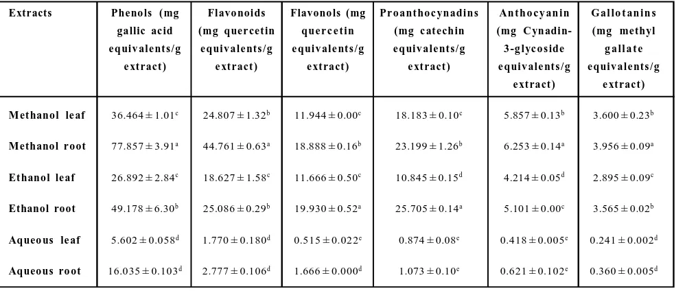
Table 1: Phytochemical content in various extracts of leaf and root of G. kurroo Royle
Extracts Pheno ls ( mg Flavo noids Flavono ls (mg Pr o ant ho c ynadins An t h o c y an in G al lo t an in s gallic ac id (mg quer cet in que r c e t in (mg c at echin (mg C ynadin- (mg methyl e quiv ale nt s /g e quiv ale nt s /g e quiv ale nt s /g e quiv ale nt s /g 3 -g lyc o s ide g a ll a t e
e xt ract ) e xt ract ) e xt ract ) e xt ract ) e quiv ale nt s /g e quiv ale nt s /g e xt ract ) e xt ract )
Met hanol le af 36.464 ± 1.01c 24.807 ± 1.32b 11.944 ± 0.00c 18.183 ± 0.10c 5.857 ± 0.13b 3.600 ± 0.23b
Methanol r oot 77.857 ± 3.91a 44.761 ± 0.63a 18.888 ± 0.16b 23.199 ± 1.26b 6.253 ± 0.14a 3.956 ± 0.09a
Et hanol le af 26.892 ± 2.84c 18.627 ± 1.58c 11.666 ± 0.50c 10.845 ± 0.15d 4.214 ± 0.05d 2.895 ± 0.09c
Ethanol root 49.178 ± 6.30b 25.086 ± 0.29b 19.930 ± 0.52a 25.705 ± 0.14a 5.101 ± 0.00c 3.565 ± 0.02b
Aqueo us le af 5.602 ± 0.058d 1.770 ± 0.180d 0.515 ± 0.022e 0.874 ± 0.08e 0.418 ± 0.005e 0.241 ± 0.002d
Aqueous ro ot 16.035 ± 0.103d 2.777 ± 0.106d 1.666 ± 0.000d 1.073 ± 0.10e 0.621 ± 0.102e 0.360 ± 0.005d
3.1.1 Total phenolics
Phenolics are the group of compounds primarily responsible for the antioxidant activities of the medicinal plants. The content of total phenolics is expressed in gallic acid equivalents (mg gallic acid/g extract). Phenolic content in various extracts are given in Table 1. Leaves of G. kurroo have lesser phenolics comparative to roots in each solvent. Highest phenolic content was observed in the methanolic extract of roots (77.857 ± 3.91 mg GAE g-1extract), followed by
ethanolic root extract (49.178 ± 6.30 mg GAE g-1extract). 3.1.2. Total flavonoids
Total flavonoid was also higher in roots of G. kurroo than in leaves. Flavonoid content in methanolic extract of roots had (44.761 ± 0.63 mg quercetin g-1extract) while ethanolic extract of roots contained
(25.086 ± 0.29 mg quercetin g-1extract). Methanol leaf extract had
higher flavonoids comparative to ethanol extraction (Table 1). Aqueous extract of both leaf and root parts have least flavonoid content.
3.1.3 Total flavonols
The concentration of flavonols was expressed in as mg quercetin equivalentsg-1extract. Ethanol was slightly better for the extraction
of flavonols. The high amount of flavonols was extracted in ethanolic extract (19.930 ± 0.52 mg quercetin g-1extract) than in methanolic
extract (18.888 ± 0.16 mg quercetin g-1extract) (Table 1). Flavonol
content in leaves was at par in both solvents while aqueous extract of leaf and root also have similar flavonol content.
3.1.4 Total proanthocyanidins
Proanthocyanidins are the class of polyphenols which are also responsible for antioxidant activities like metal ion chelation and free radical scavenging. Root extract of both methanol and ethanol have higher proanthocyanidins in comparison to leaf extracts. The ethanolic extract of roots has the highest antioxidant content (25.705 ± 0.14 mg catechin g-1extract), followed by methanolic root
extract. In case of leaf extracts, methanolic leaf extract has higher proanthocynanidins than ethanolic leaf extract. There was no significant difference (p0.05) between proanthocyanidin content in aqueous leaf and root extracts (Table 1).
3.1.5 Total anthocyanins
Anthocyanins are very strong antioxidants. They can prevent oxidation of low density lipoproteins in the blood vessels. Methanolic root and leaf extract of G. kurroo have the highest amount of anthocyanins (6.253 ± 0.14 mg, 5.857 ± 0.13 mg cyanide 3 glucoside g-1extract, respectively) compared to ethanolic root and
leaf extracts, respectively (Table 1).
3.1.6 Gallotannin content
Gallotannin content was higher in methanolic extract of roots and leaves (3.956 ± 0.09 mg, 3.600 ± 0.23 mg MGE g-1extract). This was
Figure 1: Antioxidant activities of various extracts of G. kurroo Royle. (A) Total antioxidant activity, (B) DPPH radical scavenging activity, (C) Superoxide radical scavenging activity, (D) Metal ion chelation activity, (*Note: The values with same superscript are not significantly (p0.05) different according to Duncan Post Hoc analysis).
3.2 Antioxidant activities
3.2.1 Total antioxidant activity
Antioxidant activity of ascorbic acid is used as a positive control from which the potential antioxidant activity of plant extracts is compared. Higher total antioxidant activity was observed in different root extracts, i.e., 274.520 ± 2.113 (methanol), 269.600 ± 1.508 (ethanol), 75.214 ± 0.783 (aqueous) mg ascorbic acid equivalents g -1 extract). The extracts, viz., methanol, ethanol and aqueous differed
significantly (p0.05) with reference to (w.r.t) total antioxidant activity. Aqueous root and leaf extracts showed lowest antioxidant activity among all the extracts. (Both root and leaf aqueous extracts had significantly lowest antioxidant activity (Figure 1A).
3.2.2 DPPH radical scavenging activity
DPPH radical scavenging activity in different extracts increased with the concentration of the extract. The radical scavenging was expressed as IC50 values of the extracts. The IC50 values of the extracts differed significantly (p0.05) w.r.t DPPH radical scavenging activity. Lowest IC50 was observed in methanolic root (316.290 ±
1.720 µg/ml) and ethanolic root (339.430 ± 2.489 µg/ml) extracts.
In general, lower concentration of all the root extracts was able to scavenge DPPH free radical (Figure 1B). Aqueous root and leaf extracts were able to scavenge 50% of DPPH free radical at much higher concentration. Methanol was proved better among all solvents for extraction in terms of DPPH radical scavenging activity than ethanol and water. When plant parts were compared, roots were found to possess higher DPPH scavenging than leaves in each solvent (Figure 1B). Methanolic root extract had better scavenging activity among all extracts. IC50 values of every extract were significantly different at p0.05. Aqueous extract had least DPPH radical scavenging activity.
3.2.3 Superoxide radical scavenging activity
Superoxide radical scavenging activity in different extracts increased with the concentration of the extract. Ascorbic acid (positive control) was used as a standard compound and showed the lowest IC50. The radical scavenging was expressed as IC50 values of the extracts. There was no significant (p0.05) difference between root extracts
of methanol (142.759 ± 0.258 µg/ml) and ethanol (145.404 ± 0.068 µg/ml) w.r.t superoxide radical scavenging activity. Scavenging
activities of both methanol and ethanol root extracts were not significantly different from each other and at par to the positive control (p0.05). Ethanolic leaf extract showed lower IC50 value
(159.310 ± 3.522 µg/ml) and was significantly different (p0.05) than methanolic and aqueous leaf extracts (Figure 1C). The IC50
values for superoxide radical scavenging activity in each extract had the following order: Ascorbic acid>methanol, root>ethanol, root>ethanol, leaf>methanol, leaf>aqueous, root>aqueous leaf. The
IC50 value for superoxide radical scavenging activity in each extract had the following order: Ascorbic acid>methanol, root>ethanol, root>ethanol, leaf>methanol, leaf>aqueous, root>aqueous leaf.
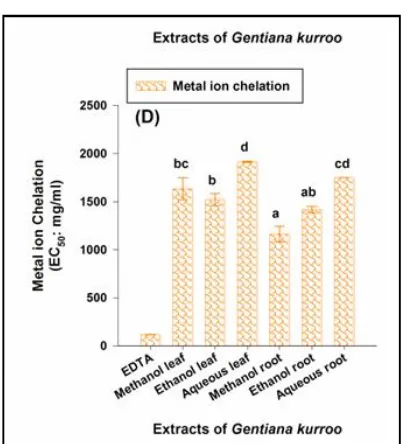
3.2.4 Metal ion chelation activity
The iron (II) chelating activity of the extracts was expressed as
EC50 value of the extracts signifying the extent of metal ion oxidation capacity of respective extract. EDTA was used as the positive control and had highest metal chelating ability. The extracts differed significantly (p0.05) w.r.t metal ion chelation activity. Highest metal ion chelating activity was observed in methanolic root extract (1162.525 ± 81.293 mg/ml), followed by ethanolic leaf extract (1521.710 ± 63.410 mg/ml). In general, root extracts had significant higher chelating activity than corresponding leaf extracts in all the solvents under investigation. The decreasing order of chelation activity was: EDTA>methanol, root>ethan ol, root>ethan ol, leaf>methanol, leaf>aqueous, root>aqueous leaf (Figure 1D). Root extracts possessed high chelation capacity in comparison to leaf extracts in each solvent. EDTA was used as standard antioxidant molecule and it had highest chelating capacity. The decreasing order of ch elation activity was: EDTA>meth an ol, root>eth an ol, root>ethanol, leaf>methanol, leaf>aqueous, root>aqueous leaf (Figure 1D).
3.2.5 Total reducing activity
Total reducing power of the extracts under investigation was used as the measure of antioxidant capacity of the respective extract. The total reducing power of root and leaf extracts was measured as a function of their concentration. The reducing power of all the extracts increased with increase in concentration of the extracts which well correlates with higher polyphenol content of the respective extracts. Thus, polyphenols present in leaf and roots of
G. kurroo might act as efficient electron donors which could reduce the oxidized intermediates of lipid peroxidation (Figure 2A).
3.3 Antimocrobial activity
3.3.1 Against gram positive bacteria (Staphylococcus aureus and
Bacillus cereus)
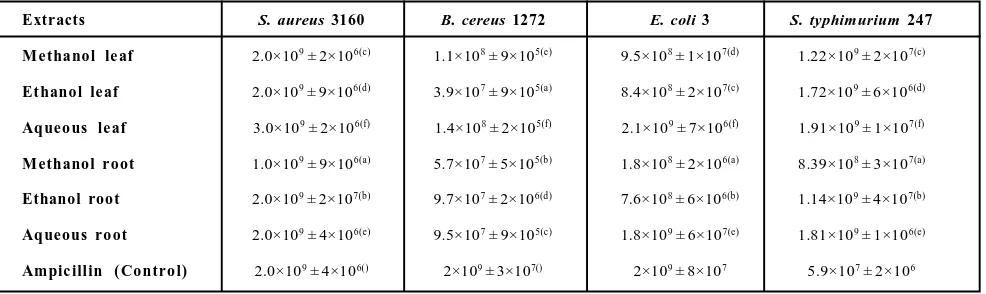
Antimicrobial activities of the extracts against gram positive bacteria,
positive control and was found to be effective in inhibiting the growth of both bacteria. Ethanolic root and leaf extracts showed significantly lower MIC values (15.62 µg) w.r.t ampicillin (31.25 µg) against S. aureus. All other extracts, viz., methanolic and aqueous leaf and root differed significantly w.r.t MIC values against the bacterium. Among all the extracts, aqueous extracts (leaf and root) showed least cytotoxicity. All the extracts were able to inhibit bacterial growth, with methanolic and ethanolic extracts showing more cytotoxicity. The increasing order of antimicrobial activity of different extracts of G. kurroo against S. aureus was as follows: ethanol leaf = ethanol root<ampicillin=methanol root<methanol leaf<aqueous leaf = aqueous roots (Table 2).
Methanolic leaf extract showed the lowest MIC (7.81 µg) which was significantly lower than positive control (15.62 µg) against B. cereus. Aqueous extracts (leaf and root) were also able to inhibit bacterial growth significantly. The increasing order of antimicrobial activity of various extracts of G. kurroo leaves and roots against
B. cereu s was in followin g order: eth an ol root<aq ueou s leaf<aq ueou s roots<methanol root<ampicillin = ethanol leaf<methanol leaf. The antimicrobial activity of various extracts in terms of resulting colony forming units (CFUs), followed similar trends (Table 3). Methanol leaf extract showed best cytotoxicity as there was least number of CFUs left in well containing respective minimum inhibitory concentration (1.1×10⁸). The antimicrobial
activity shown by the extracts of G. kurroo under investigation may be ascertained to the various bioactives present in different plant parts.
3.3.2 Against gram negative bacteria (E.coli-3 and Salmonella
typhimurium-247)
MIC of the extracts against the gram negative bacteria, viz.,
E. coli-3 and Salmonella typhimurium-247 was estimated after incubating bacterial cultures with serially diluted extracts. All the extracts were able to inhibit bacterial growth (Table 2). Against
E. coli-3, methanolic root extract showed significantly lower MIC value (15.625 µg) which was 2 fold lower than that of positive
control (ampicillin). The CFU count of 1.8×10⁸ (Table 3) was also
lowest w.r.t positive control. MIC of methanol leaf extract (31.25
µg) was at par with ampicillin. There was no significant difference
between ethanolic and aqueous leaf extracts (125 µg). Ethanolic
and aqueous root extracts also showed similar MIC values (62.5
µg). The increasing effectiveness in terms of antimicrobial activity
of each leaf and root of G. kurroo against E. coli-3 was in following order: aq ueous leaf=eth an ol leaf<aqu eous roots=ethanol root<ampicillin=methanol leaf<methanol root.
The antimicrobial activity of the extracts against Salmonella typhimurium strain-247 differed significantly w.r.t positive control (ampicillin). Ethanolic root extract showed the lowest MIC value
(7.812 µg) which was 8 fold lower than that of positive control (62.5 µg). Methanolic and ethanolic extracts (root and leaf) showed
significantly lower MIC values w.r.t ampicillin. MIC of different extracts of G. kurroo against S. typhimurium strain-247 in decreasing order was: ethanol root>methanol root>methanol leaf > ampicillin = ethanol leaf>aqueous leaf=aqueous roots. Aqueous extracts (leaf and root) were also able to inhibit bacterial growth but with half efficacy than that of control. Lower CFU count was also observed in methanol root and ethanol root extract (Table 3).
Table 2:Minimum inhibitory concentration of various extracts of root and leaf parts of G. Kurroo against pathogenic bacterial stra ins 62.5 31.25 15.62 31.25 Ampicillin (Control) 125 62.5 125 250 Aqueous root 7.812 62.5 500 15.62 Ethanol root 15.625 15.625 31.25 31.25 Methanol root 125 125 250 250 Aqueous leaf 62.5 125 15.62 15.52 Ethanol leaf 31.25 31.25 7.81 62.5 Methanol leaf S. typhimurium 247 (µg) E. coli
3 (µg) B. cereus 1272 (µg) S. aureus
3160 (µg) Extracts 62.5 31.25 15.62 31.25 Ampicillin (Control) 125 62.5 125 250 Aqueous root 7.812 62.5 500 15.62 Ethanol root 15.625 15.625 31.25 31.25 Methanol root 125 125 250 250 Aqueous leaf 62.5 125 15.62 15.52 Ethanol leaf 31.25 31.25 7.81 62.5 Methanol leaf S. typhimurium 247 (µg) E. coli
3 (µg) B. cereus 1272 (µg) S. aureus
3160 (µg) Extracts
Table 3:Colony forming unit (CFU) count of various bacterial strains as observed against minimum inhibitory concentration (MIC) of the extracts of G. Kurroo Royle
Extracts S. aureus 3160 B. cereus 1272 E. coli 3 S. typhimurium 247
Me thanol le af 2.0×10⁹ ± 2×10⁶(c) 1.1×10⁸ ± 9×10⁵(e) 9.5×10⁸ ± 1×10⁷(d) 1.22×10⁹ ± 2×10⁷(c)
Et hanol le af 2.0×10⁹ ± 9×10⁶(d) 3.9×10⁷ ± 9×10⁵(a) 8.4×10⁸ ± 2×10⁷(c) 1.72×10⁹ ± 6×10⁶(d)
Aqueo us le af 3.0×10⁹ ± 2×10⁶(f) 1.4×10⁸ ± 2×10⁵(f) 2.1×10⁹ ± 7×10⁶(f) 1.91×10⁹ ± 1×10⁷(f)
Methanol r oot 1.0×10⁹ ± 9×10⁶(a) 5.7×10⁷ ± 5×10⁵(b) 1.8×10⁸ ± 2×10⁶(a) 8.39×10⁸ ± 3×10⁷(a)
Ethanol root 2.0×10⁹ ± 2×10⁷(b) 9.7×10⁷ ± 2×10⁶(d) 7.6×10⁸ ± 6×10⁶(b) 1.14×10⁹ ± 4×10⁷(b)
Aqueous ro ot 2.0×10⁹ ± 4×10⁶(e) 9.5×10⁷ ± 9×10⁵(c) 1.8×10⁹ ± 6×10⁷(e) 1.81×10⁹ ± 1×10⁶(e)
Ampic illin ( Contro l) 2.0×10⁹ ± 4×10⁶() 2×10⁹ ± 3×10⁷() 2×10⁹ ± 8×10⁷ 5.9×10⁷ ± 2×10⁶
4. Discussion
The phytochemical analysis in roots and leaves of G. kurroo
revealed the presence of significant amount of bioactive compounds,
viz., phenols, flavonoids, flavonols, anthocyanidins, proantho-cyanidins and gallotanins. Presence of substantial amounts of these compounds apart from other bioactives makes G. Kurroo a potential source for the development of therapeutic agents imparting myriad pharmacological effects, i.e., free radical scavenging and antimicrobial properties.
In the present investigation, significant difference (p0.05) was observed in the amounts of bioactive compounds and antioxidant activities in six different (leaf and root) extracts prepared in three different solvents. Total phenol content (mg/g GAE g-1 extract)
within methanolic root extract was significantly higher than methanolic leaf extract as well as ethanolic and aqueous leaf and root extracts. Apart from aqueous (root and leaf) extracts, all other extracts differed significantly w.r.t their phenol content. The analysis revealed presence of higher phenol content in roots than in leaves of G. kurroo. Similar observations have been reported by Baba and Malik (2014). High phenol content in roots of G. kurroo is one of the reasons for its immense medicinal importance. Crude extracts of medicinal herbs rich in polyphenols are now having great importance in the pharmaceutical industry. Polarity of various solvents used for extraction as well as nature of plant species used was found to have significant effect on extraction of bioactive compounds (Ghasemzadeh et al., 2011). Similar results were observed in H. radicata where higher amount of phenolics were present in methanolic extracts (leaf and root) in comparision to aqueous extracts (Senguttuvan et al., 2014).
Flavonoids are known to exert high antioxidant activities and have ability of scavenge free radicals which damage cell membranes and biological molecules. Both leaf and root of G. kurroo were rich in flavonoids expressed as mg quercetin equivalents g-1 extract.
Methanolic root extract had significantly high content of flavonoids than other five extracts. No significant difference (p0.05) was observed in aqueous leaf and root extracts. Higher flavonoid content in methanolic root extract was also reported by Baba and Malik (2014). In similar studies on Azadirachta indica (Meliaceae),
Hemid esmus ind icus (Asclepiadaceae), Man ilkara zap ota
(Sapotaceae), Psorelea corylifolia (Fabaceae), Rubia cordifolia
(Ru biaceae), Tin ospo ra co rd ifolia (Men ispermaceae) an d Piccrorhiza kurroo (Scrophulariaceae) methanol proved better solvent for extraction of flavonoids than water (Kaneria et al.,
2012; Rajkumar et al., 2011). Flavonols, a subclass of flavonoids have substantial radical scavenging properties (Miliauskas et al., 2004). In general the extracts differed significantly (p0.05) w.r.t their flavonol content. Higher flavonol content was observed in methanolic extracts (leaf and root). Flavonols are known to play an important role in large number of biochemical signaling pathways and therefore, physiological and pathological processes, could be affected by flavonols. They could potentially interact with many of th e molecu lar targets, known to be in volved in th e pathophysiology of ischemic heart disease and stroke (Vizcaino and Duarte, 2010). They might act by multiple mechanisms operating both in the long term prevention and in the acute phase of cardiovascular events. A number of pharmacological effects of G. kurroo may well be attributed to the presence of significant amount of flavonols in leaf and root of the plant. Anthocyanins one of the
most prominent flavonoids present in plants are not only known for their antioxidant activities but are also used as food colorant (Naz et al., 2007; Mak et al., 2013). They help in reduction of oxidative (cell) damage, i.e., effects of various herbal formulations in hemodialysis patients have been especially attributed to the presen ce of h igh an th ocyan in /polyph en ol con ten t in these formulations. In general, all the extracts differed significantly w.r.t their anthocyanin content. Higher anthocyanin content was observed in methanolic extracts (leaf and root). Whereas no significant difference (p0.05) was observed in aqueous extracts (leaf and roots). Methanol proved to be the best solvent for extraction of anthocyanin than other solvents, i.e., ethanol and water (aqueous). Proanthocyanidins are bioflavanoids and are well known for their potent antioxidant and free radical scavenging properties. In general, higher proanthocyanidin content (i.e., ethanol and methanol root extracts) was observed in roots of G. kurroo. No significant difference in proanthocyanidin content was observed in aqueous extracts (leaf and root). Involvement of proanthocyanidins free radical scavenging and their potent antioxidant properties are well established (Amo et al., 2011). Similar results were observed in ethanolic and aqueous extracts of C. umbellatum and P. officinalis
(Neagu et al., 2016). Higher proanthocyanidin content in roots of
G. kurroo might be one of the decisive factors for its potent medicinal and pharmacological properties. Gallotannins are h ydrolysable tan nin polymer which also comes un der th e polyphenolic antioxidants. In general, higher gallotannin content was observed in root extracts. No significant difference (p0.05) in gallotannin content was observed in aqueous extracts (leaf and root). Gallotannins possess antimicrobial and anticancer activities (Hagerman et al., 1999; Van Molle et al., 2000; Feldman et al., 2001). Recent studies have shown that gallotannins not only exhibited cytotoxic activity, but also their activity was associated with conventional anticancer drugs, demonstrating once again the efficiency of natural products as potential sources of adjuvants that could be used in antitumor therapy. The cytotoxic effects of the extracts of G. kurroo may well be attributed to the presence of significant amount of gallotannins in the respective extracts. In the present study, methanolic root and leaf extracts revealed presence of significantly (according to Duncan post hoc analysis) high amount of various bioactive compounds than other extracts,
Figure 2:Total reducing activity and correlation analysis. (A) Total reducing activity of various extracts of G. kurroo Royle. (B), (C) , ( D) Corr ela tion of tota l phenol content with various antioxidant activities and phytochemicals. (*Note: The values with same superscr ipt ar e not significantly (p0.05) different according to Duncan Post Hoc analysis).
Total antioxidant activity of different extracts (leaf and root) of G. kurroo was expressed as ascorbic acid equivalents g-1 extracts. All
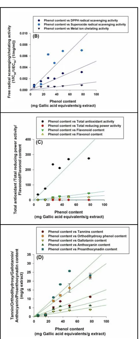
the extracts differed significantly (p0.05) w.r.t total antioxidant activity. High est total an tioxidan t activity was observed in methanolic root extract. Higher total antioxidant activity was observed in root extracts of various solvents. The total antioxidant activity in different extracts showed significant (p0.01) positive correlation with corresponding phytochemicals, i.e., 0.837, 0.886, 0.970, 0.925, 0.945 and 0.968 with phenols, flavonoids, flavonols, proanthocynadins, anthocyanins and gallotannins, respectively. The presence of high polyphenol content in roots could be a possible reason for higher antioxidant activity in roots. Methanolic extracts (leaf and root) had higher total antioxidant activity than ethanol and water. Thus, methanol was the best solvent for extraction of polyphenols leadin g to higher antioxidant activities of the corresponding extracts. DPPH radical scavenging activity was expressed as IC50 values of the extracts. The IC50 values of the extracts differed significantly (p0.05) w.r.t DPPH radical scavenging activity. Root extracts (methanol and ethanol) showed significantly higher DPPH radical scavenging activity than other extracts. In general, among different plant parts, root extracts showed higher
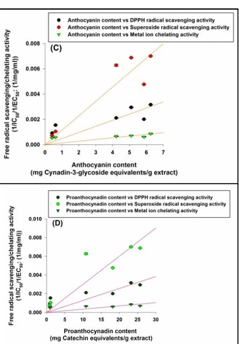
Figure 3:Correlation analysis of va rious phytochemicals with antioxidant activities, (A) Correlation between flavonoids content with antioxidant activities, (B) Correlation between flavonol content with a ntioxidant a ctivities, ( C) Correlation between anthocyanin content with antioxidant activities, (D) Correlation between proanthocyanin content with antioxidant activities.
DPPH radical scavenging activity (IC50) of different extracts showed significant (p0.01) negative correlation with corresponding phytochemicals, i.e., – 0.811, – 0.802, – 0.878, – 0.830, – 0.839 and – 0.857 with phenols, flavonoids, flavonols, proanthocynadins, anthocyanins and gallotannins, respectively. Thus, clearly showing that higher polyphenol content was responsible for lower IC50 values (i.e., concentration of the extract that was able to scavenge 50% of
DPPH radical) of the respective extracts. Methanol proved to be a better solvent for extraction in terms of DPPH radical scavenging activity than ethanol and water. Similar observations, i.e., higher
DPPH scavenging activity in methanolic root extracts than leaf extracts in G. kurroo have also been reported by Baba and Shahidi (2014). Similar results were also observed in G. lutea methanol, ethanol and aqueous root extract (Nastasijevic et al., 2016). Superoxide radical scavenging activity expressed as IC50 values of the extracts. The IC50 values of the extracts (except root extracts prepared in meth an ol an d ethanol, respectively) differed significantly (p0.05) w.r.t superoxide radical scavenging activity.. Superoxide radical scavenging activity (IC50) of different extracts showed significant (p0.01) negative correlation with corresponding phytochemicals, i.e., – 0.755, – 0.825, – 0.905, – 0.865, – 0.937
and – 0.954 with phenols, flavonoids, flavonols, proanthocynadins, anthocyanins and gallotannins, respectively. Thus, clearly showing that higher polyphenol content was responsible for lower IC50 values (i.e., concentration of the extract that was able to scavenge 50% of superoxide radical) of the extracts under investigation. Root extracts (methanol and ethanol) showed significantly higher superoxide radical scavenging activity than other extracts. Superoxide radical scavenging was significant for protection against oxidative damage (Fu and Mao, 2008). Superoxide radical scavenging activity of various Indian medicinal plants was higher in methanolic extracts than aqueous extracts (Kaneria et al., 2012; Rakholiya et al., 2011; Wu et al., 2011). Metal ion chelation was expressed as EC50 values of the extracts. The EC50 values of the extracts differed significantly (p0.05) w.r.t metal ion chelating activity. Metal ion chelation
(EC50) of different extracts showed significant (p0.01) negative
correlation with corresponding phytochemicals, i.e., – 0.918, – 0.907, – 0.882, – 0.815, – 0.802 and – 0.808 with phenols, flavonoids, flavon ols, proanth ocynadins, an thocyanin s and gallotannins. respectively. Thus, clearly showing that higher polyphenol content was responsible for lower EC50 values (i.e., concentration of the extract that was able to chelate 50% of Fe (II)
ions) of the extracts, leading to higher chelation of Fe (II) ions. Root extracts possessed high metal ion chelation capacity in comparison to leaf extracts. Similar results were observed in various extracts of
Kappaphycus alvarezii (Kumar et al., 2012). Flavonoids present in plant extracts have been identified for making complexes with metal ions and, thus responsible the free radical scavenging capacity (Rice-Evans and Miller, 1994). Thus, high flavonoid content in methanolic root extract could be the reason behind its high chelating activity and these results had similarity with the work done on ginsang leaves (Jung et al., 2006). Total reducing power of any given extract could be used as a direct measure of its antioxidant potential. Higher absorbance values observed in the root extracts of G. kurroo clearly showed higher reducing potential of the respective extracts. Total reducing power of the extracts increased with corresponding increase in concentration of the extracts. Higher polyphenol content well correlated with higher reducing power, as could be seen by its dependence on concentration of the extract. The reducing activity present in different extracts of G. kurroo might, thus serve as a valuable indicator of its antioxidant potential.
Correlation analysis revealed that antioxidant activities of various extracts were positively correlated with phytochemical content. Total antioxidant activity had positive correlation with phenol content (Figure 2 C). Higher phytochemical content in roots and leaves of various extract contributed on lowering the IC50 value for DPPH scavenging, thus better antioxidant activity. Phenols were also significantly correlated with DPPH radical scavenging, superoxide radical scavenging activity and metal ion chelation activity (Figure 2 C). Flavonols showed highest correlation with
Extracts of G. kurroo (roots and leaves) showed high antibacterial activity against both gram negative and gram positive bacteria. The results clearly show that G. kurroo might be a potential source of broad spectrum antibacterial agents. The antibacterial activity of the extracts could be attributed to the high polyphenol content, which was reported to be involved in the inhibition of nucleic acid biosynthesis and metabolic processes (Cushnie et al., 2005). The correlation analysis clearly revealed that antimicrobial activity (MIC)
of the extracts against different bacterial strains can be ascertained to their polyphenol content, i.e., – 0.743, – 0.858, – 0.731 and – 0.069 in E. coli strain-3, S. typhimurium strain-247, S. aureus
strain-3160 and B. cereus strain-1272, respectively. Thus, clearly showing that higher phenol content was responsible for lower MIC
values (i.e., minimum concentration of the extract that was able to inhibit bacterial growth) of the extracts. CFU count of different bacterial species, viz., E. coli strain-3, S. typhimurium strain-247,
S. aureus strain-3160 and B. cereus strain-1272 showed significant negative correlation (p0.01) with different phytochemicals. Against CFU count of E. coli strain-3 phenols, flavonoids flavonols, anthocyanins and gallotannins showed a correlation coefficient of – 0.914, – 0.958, – 0.938, – 0.943 and – 0.949, respectively, thus clearly showing that higher polyphenol content was responsible for lower CFU counts as observed for E. coli strain-3. Similar results were also observed for other bacterial species. High phenol and flavonoid content was found to be accountable for cytotoxity (low
CFU count) of different extracts against S. typhimurium strain-247 with a significant (p0.01) correlation, i.e., – 0.946 and – 0.939, respectively. Phenol, flavonoid and flavonol content also showed significant correlation with CFU counts of S. aureus strain-3160 (i.e., – 0.858, – 0.838 and – 0.809, respectively) and B. cereus
strain-1272 (i.e., – 0.510, – 0.549 and – 0.516, respectively). Thus, the extracts having high polyphenol content are able to inhibit bacterial growth significantly resulting in lesser CFU count and
MIC values.
5. Conclusion
The results of the present study clearly show that G. kurroo may serve as a potential source of natural antioxidants and antimicrobial agent. Though, the efficacy of the plant may vary according to the plant part and the solvent system used for extraction. This is the first report on the antioxidant and antimicrobial properties in various extacts (roots and leaves) of G. kurroo, a highly endangered medicinal plant. The study, thus serves as a preliminary investigation report on the pharmacological attributes of G. kurroo and the nature of bioactives involved. More comprehensive work needs to be done on th e precise n atu re bioactive compoun ds an d th eir pharmacokinetic behavior under physiological conditions, thus utilizing myriad medicinal benefits of this medicinal plant.
Acknowledgements
The research was financially supported by DST-FIST sponsored department of Biochemistry, G.B.P.U.A. and T. Pantnagar. We also ackn owledge Department of Veterinary P ublic Health and Epidemiology, Pantnagar University for providing the pathogenic microbes used in the study.
Conflict of interest
We declare that we have no conflict of interest.
References
Amoo, S. O.; Ndhlala, A. R.; Finnie, J. F. and Van Staden, J. (2011). Antifungal, acetylcholinesterase inhibition, antioxida nt and phytochemical properties of three Barleria species. South Afr. J. Bot., 77:435-445.
Aparadh, V.T.; Naik, V.V. and Karadge, B.A. (2012). Antioxidative properties (TPC, DPPH, FRAP, metal chelating ability, reducing power and TAC) within some Cleome species. Ann. Bot., 2:49-56.
B aba, S. A. and Malik, S. A. (2014). Eva luation of a ntioxidant and antibacterial activity of methanolic extracts of Gentiana kurroo royle. Saudi Journal of Biological Sciences, 21:493-498.
Behera, M. C. and Raina, R. (2012). Gentiana kurroo Royle : A critically endangered bitter herb. Int. J. Med. Arom. Plants, 2(1):22-29.
Braca, A.; Tommasi, N.D.; Bari, L.D.; Pizza, C.; Politi.M. and Morelli. I., (2001).
Antioxidant principles from Bauhinia terapotensis. Journal of Natural Products, 64:892-895.
Chen g, G. W. and B reen, P. J. (1991 ). Activity of phenylalanine ammonialyase (PAL) and concentrations of anthocya nins a nd phenolics in developing strawberry fruit. Journal of the American Society for Horticultural Science, 116:865-869.
Cushnie, T. T. and Lamb, A. J. (2005). Antimicrobial activity of flavonoids. International Journal of Antimicrobial Agents, 26(5):343-356.
Decker, E. A. and Welch, B. (1990). Role of ferritin as a lipid oxidation catalyst in muscle food. J. Agric. Food Chem., 38(3):674-677.
Feldman, K.S.; Sahasrabudhe, K.; Lawlor, M.D.; Wilson, S.L.; Lang, C.H. and Scheuchenzuber, W.J. (2001). In vitro and in vivo inhibition of
LPS-stimulated tumor necrosis factor-alpha secretion by the gallotannin beta-D-pentagalloylglucose. Bioorganic and Medicinal Chemistry Letters, 11:1813-1815.
Fu, M. and Mao, L. (2008). In vitro antioxidant activities of five cultivars of daylily flowers from China. Nat. Prod. Res., 22:584-591.
Ghasemzadeh, A.; Jaafar, H. and Rahmat, A. (2011). Effects of solvent type on phenolics and flavonoids content and antioxidant activities in two varieties of young ginger (Zingiber officinale Roscoe.) extracts. J. Med. Plant Res., 5(7):1147-1154.
Hagerman, A.E.; Riedl, K.M. and Rice, R.E. (1999). Tannins as biological antioxidants. Basic Life Science, 66:495-505.
Haslam, E. (1965). Galloyl esters in the Aceraceae. Phytochemistry,
4:49 5-49 8.
Jung, C. H.; Seog, H. M.; Choi, I. W.; Park, M. W. andCho, H. Y. (2006).
Antioxidant properties of various solvent extracts from wild ginseng leaves. LWT-Food Science and Technology, 39(3):266-274.
Kahl, R. and Kappus, H. (1993). Toxicology of the synthetic antioxidants BHA and BHT in comparison with the natural antioxidant vitamin. E.Z Lebensm Unters Forsch.,196(4):329-338.
Kaisoon, O.; Siriamornpun, S.; Weerapreeyakul, N. and Meeso, N. (2011).
Phenolic compounds and antioxidant activities of edible flowers from Thailand. Journal of Functional Foods, 3:88-99.
Kaneria, M.; Kanani, B. and Chanda, S. (2012). Assessment of effect of hydroalcoholic and decoction methods on extra ction of antioxidants from selected Indian medicinal plants. Asian Pacific Journal of Tropical Biomedicine, 2(3):195-202.
Kim, D.; Chun, O.; Kim, Y.; Moon, H. and Lee, C. (2003). Quantification of
phenolics and their antioxidant capacity in fresh plums. J. Agric. Food Chem., 51:6509-6515.
Kirtikar, K.R. and Basu, B.D. (1935). Indian Medicinal Plants, Second ed. Bishen Singh Mahendra Pal Singh, Periodicals Expert, Dehradun.
Kumar, S.; Gupta, D. and Nayyar, H. (2012). Comparative response of maize and rice genotypes to heat stress: Status of oxidative stress and antioxidants. Acta Physiol. Plant, 34:75-86.
Levitt, S.M, and Diamond, R. D.(1985). A rapid colorimetric assay of fungal viability with the tetrazolium salt MTT. J. Infect Dis., 152 (5):938-94 5.
Liu, l.; Ooi, V. E. C. and Chang, S.T. (1997). Free radical scavenging activities of mushroom polysaccharide extracts. Life Sciences, 60 (10):763-77 1.
Mak, W.; Chuah, L. O.; Ahmad, R. and Bhat, R. (2013). Antioxidant and antibacterial activities of hibiscus (Hibiscus rosa-sinensis L.) and Cassia (Senna bicapsularis L.) flower extracts. Journal of King Saud University, Science, 25:275-282.
Maurya, A.; Khan, F.; Bawankule, D. U.; Yadav, D. K. and Srivastava, S. K. (2012).
QSAR, docking and in vivo studies for immunomodulatory activity of isolated triterpenoids from Eucalyptus tereticornis and Gentiana kurroo. European Journal of Pharmaceutical Sciences, 47:152-161.
Miliauskas, G.; Venskutonis, P. R. and Beek. T.A.V. (2004). Screening of radical scavenging activity of some medicinal and aromatic plant extracts. Food Chemistry, 85:231-237.
Mubashir, K.; Ganai, B. A.; Hazanfar, K. G. and Akbar, S. (2014a). Evaluation of antiarthritic potential of methanolic extract of Gentiana kurroo Royle. Hindawi Publishing Corporation Arthritis.http://dx.doi.org/ 10.1155/2014/810615.
Mubashir, K.; Ghazanfar, K.; Ganai,B. A.; Akbar, S.; Malik, A. H. and Masood, Akbar (2014b). Scientific validation of Gentiana kurroo Royle for
anti-inflammatory and immunomodulatory potential. ISRN Inflammation. http://dx.doi.org/10.1155/2014/701765.
Nastasijevic, B.; Milosevic, M.; Janjic, G.; Stanic, V. and Vasic, V. (2016). Gentiana lutea extracts and their constituents as inhibitors of synaptosomal Ecto-NTPDase.Int. J. of Pharmaco., 12:272-289.
Naz, S.; Siddiqi, R.; Ahmad, S.; Rasool, S.A. and Sayeed, S.A. (2007).
Antibacterial activity directed isolation of compounds from Punica granatum. Journal of Food Science, 72:341-345.
Neagu, E.; Radu, G. L.; Albu, C. and Paun, G. (2016). Antioxidant activity, acetylcholi nester ase and tyrosinase inhibitor y potential of Pulmonaria officinalis and Centarium umbellatum extracts. Saudi J. of Biol. Sciences, http://dx.doi.org/10.1016/j.sjbs.2016.02.016.
Oyaizu, M. (19 86). Studies on products of browning rea ctions: Antioxidative activities of products of browning reaction prepared from glucosamine. Japanese Journal of Nutrition, 44:307-315.
Pand ey, K . B . and Rizvi, S.I. (2009 ). Plant polyphenols as dieta ry antioxidants in human health and disease. Oxid Med Cell Longev.;
2(5):270-278.
Pathak, K. and Das, R. J. (2013). Herbal Medicine: A rational approach in health care system. International Journal of Herbal Medicine, 1(3): 8 6 -8 9 .
Paulsamy, S. and Jeeshna, M. V. (2011). Preliminary phytochemistry and antimicrobial studies of an endangered medicinal herb, Exacum bicolor Roxb. Res. J. Pharm. Biol. Chem. Sci., 2(4):447-457.
Prieto, P.; Pineda, M. and Aguilar, M. (1999). Spectrophotometric quantitation of antioxidant capacity through the formation of a phosphomoly-bdenum complex: Specific application to the determination of vitamin E1. Analytical Biochemistry, 269:337-341.
Rajkumar, V.; Guha, G. and Kumar, R. A. (2011). Antioxidant and anti-neoplastic activities of Picro rhiza ku rro a extr acts. Food a nd Chemical Toxicology, 49(2):363-369.
Rakholiya, K., Kaneria, M and Chanda, S. (2011). Vegetable and fruit peels as a novel source of antioxidants. J. Med. Plant Res., 5:63-71.
Rice-Evans and Miller, N.J. (1994). Total antioxidant status in plasma and body fluids. Methods Enzymol., 234:279-293.
Senguttuvan, J.; Paulsamy, S. and Karthika, K. (2014). Phytochemical analysis and evaluation of lea f a nd root parts of the medicinal her b, Hypochaeris radicata L. for in vitro antioxidant activities. Asian Pac. J. Trop. Biomed., 4(1):359-367.
Sun, B.; Ricardo-da-Silva, J. M. and Spranger, I.(1998). Critical factors of vanillin assay for catechins and proanthocyanidins. J. Agric. Food Chem., 46:4267-4274.
Swain, T. and Hill s, W. E. (1 959). Phenolic constitu ents of Pr un us domestica. I. quantitative analysis of phenolic constituents. Journal of the Science of Food and Agriculture, 10:63-68.
Uniyal, B. and Shiva, V. (2005). Traditional knowledge on medicinal plants among rural women of Garhwal Himalayas, Uttaranchal. Ind. J. Trad. Know., 4:259-266.
Van Molle, W.; Van den Berghe, J.; Brouckaert, P. and Libert, C. (2000). Tumor necrosis fa ctor -induced lethal hepatitis: Phar macological inter vention with ver apa mil, tannic acid, picotamide, a nd K76COOH. FEBS Letters, 467:201-205.
Vizcaino, F.P. and Duarte, J.(2010). Flavonols and cardiovascular disease. Mol. Asp. of Med., 31:478-494.
Wani, B . A.; Ramamoorthy, D. and Ganai, B . A. (2011). Preliminary phytochemical screening and evaluation of analgesic activity of metha nolic extract of roots of Ge ntiana ku rro o Royle in
experimental animal models. International Journal of Pharmacy and Pharmaceutical Sciences, 3(4):164-166.
Wu, C. R.; Lin, W. H.; Hseu, Y. C.; Lien, J. C.; Lin, Y. T. and Kuo, T. P. (2011).
Evaluation of the antioxidant activity of five endemic Ligustrum species leaves from Taiwan flora in vitro. Food Chem., 127 :564-57 1.