Research Article
Level
of
Retinol
Deposit
and
Cervical
Cancer
Kadar
Deposit
Retinol
dan
Kanker
Serviks
Tofan W Utami1, Fera Ibrahim2, Gatot Purwoto1, Wely L Tiffani1, Muhammad F Aziz1, Andrijono1
1Department of Obstetrics and Gynecology 2Department of Microbiology
Faculty of Medicine Universitas Indonesia Dr. Cipto Mangunkusumo National General Hospital
Jakarta
INTRODUCTION
Cervical cancer is one of the major leading causes of death among women due to cancer. It is the second most common cancer on women in the world, of which 83% of that occurs in developing countries.1,2 In Indonesia, the incidence of cervical
cancer is estimated to be 100 per 100,000 populations among 79-million women at risk. The mortality rate of cervical cancer in Indonesia is very high because more than 70% of cases are diagnosed at advanced stage.3
This cancer is actually highly preventable due to known etiologic causes. The main factor
Abstract
Objective: To analyze level of retinol deposit sufficiency in the natural history of cervical cancer.
Methods: Serum retinol level was measured by ELISA from peripheral blood of subjects with normal cervix, cleared and persistent high risk human papilloma virus (HR-HPV) subclinical infection, and cervical cancer who fulfilled the inclusion and exclusion criteria. The study was held in Dr. Cipto Mangunkusumo and Fatmawati Hospital, Jakarta, within 2 years (August 2013-2015). Blood was taken twice, consisting of post-8-hour fasting blood and 2 hours after 6000 IU retinyl palmitate oral administration.
Results: Of 47 total samples, sufficient level of retinol deposit in normal cervix, cleared and persistent HR-HPV subclinical infection, and cervical cancer group was 85.0% (reference), 75.0% (OR 1.89), 33.3% (OR 11.33), and 75% (OR 1.89); respectively. Statistically, there was no significant difference from sufficiency level of retinol deposit between normal cervix and clearance HR-HPV subclinical infection (p=0.628), normal cervix and persistent HR-HPV subclinical infection (p=0.078), normal cervix and cervical cancer (p=0.433), cervical cancer and clearance HR-HPV subclinical infection (p=1.000), cervical cancer and persistent HR-HPV subclinical infection (p=0.430), persistent and clearance HR-HPV subclinical infection group (p=0.740).
Conclusion: This study proves that normal cervix group has the highest level of retinol deposit sufficiency; however, it cannot be stated that cervical cancer group has less sufficiency level. Persistent HR-HPV subclinical infection group has the lowest level of retinol deposit (OR 11.33). There is no association between sufficient level of retinol deposit and clearance of HR-HPV.
[Indones J Obstet Gynecol 2017; 5-1: 46-54]
Keywords: cervical cancer, HR-HPV clearance, retinol deposit
Abstrak
Tujuan: Untuk menganalisis tingkat kecukupan deposit retinol pada perjalanan alami kanker serviks.
Metode: Kadar retinol serum diperiksa dari darah perifer dengan metode ELISA pada kelompok serviks normal, infeksi subklinis human papilloma virus risiko tinggi (HPV-RT) klirens dan persisten, serta kanker serviks yang sesuai dengan kriteria inklusi dan eksklusi di Rumah Sakit Dr. Cipto Mangunkusumo dan Fatmawati, Jakarta, pada periode 2 tahun (Agustus 2013-2015). Sampel darah diambil dua kali yaitu setelah puasa 8 jam dan 2 jam setelah pemberian 6000 UI retinil palmitat peroral.
Hasil: Diperoleh 47 sampel total dari 4 kelompok yang diteliti. Deposit retinol yang cukup pada kelompok serviks normal, infeksi subklinis HPV-RT klirens, HPV-RT persisten, dan kanker serviks adalah berturut-turut 85%, 75% (OR 1,89), 33,3% (OR 11,33), dan 75% (OR1,89). Secara statistik tidak terdapat perbedaan bermakna tingkat kecukupan deposit retinol antara kelompok serviks normal dengan infeksi subklinis HPV-RT klirens (p=0,628), serviks normal dengan infeksi subklinis HPV-RT persisten (p=0,078), serviks normal dengan kanker serviks (p=0,433), kanker serviks dengan infeksi subklinis HPV-RT klirens (p=1,000), kanker serviks dengan infeksi subklinis HPV-RT persisten (p=0,430), infeksi subklinis HPV-RT persisten dengan klirens (p=0,740).
Kesimpulan: Penelitian ini mampu membuktikan bahwa tingkat kecukupan deposit retinol tertinggi dijumpai pada kelompok serviks normal, namun tidak mampu membuktikan bahwa kanker serviks memiliki tingkat kecukupan deposit retinol yang kurang.Tingkat kecukupan deposit retinol terendah ditemukan pada kelompok HPV persisten (OR 11,33). Tidak terdapat hubungan antara deposit retinol yang cukup dengan klirens HPV-RT.
[Maj Obstet Ginekol Indones 2017; 5-1: 46-54]
Kata kunci: deposit retinol, HPV-RT klirens, kanker serviks
contributing to cervical cancer is persistent infection of high risk-human papillomaviruses (HR-HPVs), which is the precursor for malignancy. The hypothesis was a relationship between HPV infection and cervical neoplasia which was first introduced by Harold zur-Hausen as a German virologist.4-6
Although HPV has been identified as the cause of cervical cancer, most women infected with HPV do not always develop to be cervical cancer. Most of them, around 70-90% of cases, will experience a clearance. Therefore, this issue leads to the idea that there are other factors contributing to the induction of cancer other than HPV infection.7
Local immune response is a determining factor that affects its susceptibility to HPV and progression into cervical cancer.8,9 In individuals who
have competent immune system, most HPV infections occur subclinically, and only small percentage will progress to pre-cancerous lesions and invasive cervical cancer. The mechanisms of HPV in keeping off the immune response are due to the modulation of cytokines to alter antigen presentation, interferon (IFN) regulatory pathways, and adhesion molecules. The avoidance of HPV to immune response is a critical point of the successful HPV infection to host cells.10
Nutritional cofactors, which are associated with immunity, have an important role in the defense against HPV infection. Various antioxidants have been known to boost the immune system against viruses and other microorganisms, as well as tumor cells. Retinol, as cofactor nutrient, is essential in cervical mucosal immunity. It is able to modulate the non-specific and specific immune system against HPV infection and tumor cells.11 It
also has central role in growth, development and differentiation of B and T lymphocytes, and as major regulator of cell activation on immune system.12 The CD4+T and CD8+T cells can be
modulated by retinol. T-cell is specific to the virus and protective factor against tumor cells. Meanwhile, CD8+T-lymphocytes or Cytotoxic
T-Lymphocyte (CTL) assited by molecular Major Histocompatibility Complex (MHC) class I acts to recognize and kill tumor cells. CD4+T-cells are
generally not cytotoxic to the tumor, they may play an important role in anti-tumor response by producing cytokines which are necessary for the development of CTL cells into effector cells, yet.
CD8+T-cells can eliminate viral infection by
secreting IFN-, and granzyme, to run cytolytic effect.13
Retinol is a potent HPV and carcinogenesis inhibitor. It acts mainly by three mechanisms, such as apoptosis, cessation of growth, and differentiation.14 Retinol can inhibit cells
immortalization by HPV; thus, retinol offers protective effect against the occurrence of cervical neoplasia.11
In the cervix, retinol can interact with HPV oncoprotein (E6 and E7) and increase the role of p53 and pRb (tumor suppression genes) to control cell cycle and proliferation. Retinol can inhibit not only early gene expression of HPV types 16 to 95% by lowering E2 and E5 mRNA, but also E6 and E7 proteins. It can lower the level of viral oncogenes transcription and inhibit neoplastic process. Retinol can protect the mucosa against viral infection and it has a cytostatic effect by inducing the cessation of cell cycle-dependent p53,14
generating CD4+T-cells, inducing effective
CD8+T-cells responses, suppressing inflammatory
cells, and producing several cytokines, including tumor necrosis factor-alpha (TNF-) which are potent to control HPV infection.15-17
The transformation zone of cervix is a high-risk zone that can be altered by HPV infection. This zone is also the most sensitive zone to retinol. Retinol increases resistance to infectious micro-organism by maintaining the function, structure of epithelial cells, mucosal integrity, and stabilization of inter-cell linkage. When retinol is absent, goblet cells will disappear and mucosal epithelial atrophy will occur. Therefore, it will lead to irritation and infection.18
administration of retinylpalmitate. The RBP4 is also
popular as plasma retinol-binding protein which transports retinol in serum. Retinol is metabolized into retinaldehyde as some isomers of retinoic acid and retinyl esters. Retinaldehyde is important chromophore in rhodopsin photo-receptor; whereas, retinoic acid regulates many cellular differentiation and proliferation effects via intracellular receptors retinoic acid receptor (RAR) and retinoic X receptor (RXR). The RBP4 adopts
-barrel structure with a central cavity that accommodates either retinol or retinaldehyde and it is synthesized primarily in hepatocytes and adipocytes as 21 kDa non-glycosylated molecules, non-phosphorylated, and non-sulfate.
According to aspect of nutrition, the lack of retinol is associated with cervical cancer; thus, it is that it can be used as a basic approach to primary prevention of cervical cancer. Giving retinol is expected to reduce the risk of persistent HR-HPV infection and progression towards pre-cancerous lesions and cervical cancer. Eventually, it can reduce the incidence, morbidity, and mortality of cervical cancer.
METHODS
This study consisted of four groups representing the natural history of cervical cancer, such as the normal cervix, persistent subclinical HPV infection, HPV clearance, and cervical cancer. Normal cervix consisted of subjects with normal cervical cytology result and negative HPV DNA test. Cervix with subclinical HPV infection included those with negative cytology result and positive HR-HPV DNA test. Cervical cancer was expressed through the result of squamous cell carcinoma (SCC) type according to histopathology assessment. Normal group and cervical cancer data were taken cross sectionally; whereas, subclinical infection with HR-HPV clearance and persistent were coming from population with positive HR-HPV followed by continuous checking twice in 12-24 months. We recruited subjects from August 2013 to August 2015.
Inclusion
and
Exclusion
Criteria
The target population of this study was reproductive age women with negative cytology result or without positive HPV or cervical cancer. We included all women coming to the
Gynecologic Oncology Clinic, Department of Obstetrics and Gynecology, Dr. Cipto Mangun-kusumo and Fatmawati Hospital during the study period who f u l f i l l e d t h e i n c l u s i o n a n d exclusion criteria. Inclusion criteria for this study consisted of sexually active women aged 20-60 years old with negative cytology result, or SCC proven by histopathology assessment and they agreed to participate in the study. While, the exclusion criteria were married women under the age of 20 years old, having multiple sexual partners, having malnutrition, on pregnancy, having a history of intravenous narcotics use, promiscuity confession, using long term steroid agents or intrauterine device ( I U D ) , a n d experiencing genitourinary tract infection.
Patient
Enrollment
During the period of August 2013 until August 2015, we obtained 71 samples among 538 potential subjects for subclinical HR-HPV infection clearance and persistent group. Subject with normal cervix (control), clearance, and persistent group taken from Women’s Health Clinic (WHC), Dr. Cipto Mangunkusumo Hospital in Kencana Cluster. While, we got cervical cancer group from WHC outpatient clinic, Gynecologic Oncology outpatient and inpatient, Department of Obstetrics and Gynecology, Dr. Cipto Mangunkusumo and Fatmawati hospital. Due to several reasons related to geographical problem of their living area, we successfully recruited 7 women (9.9%) for subclinical HR-HPV infection clearance and persistent group. Initially, we took 22 samples for control group; however, two samples were excluded because of sample lysis and HPV-positive result after re-checking. There were four subjects for subclinical HR-HPV infection clearance group and three subjects for persistent group. There were 27 samples for cervical cancer group, seven samples were excluded due to lysis, yet.
Laboratory
Protocol
and 2 hours after administration of 6,000 IU of retinylpalmitate orally. Retinol deposit is defined to be sufficient if there is an increase of RBP4 of
less than 20%, insufficientcy if there is an increase of more than or equal to 20%.
Blood sampling was performed using EDTA tubes after signing the informed consent. The ELISA plates coated with antibodies of RBP4 were used. The blood samples were centrifuged to separate serum from red blood cell at a speed of 3,500 rpm for 10 minutes. Serum was collected and placed in a threaded tube and stored at -80°C. The serum, standard and control, was put in each plate as much as 20 l with previously added diluent 200 l. After that, the plate was incubated in an o r b i t a l s h a k e r f o r o n e h o u r a t r o o m temperature. After an hour, the plate was washed three times using 400 l buffer solution. The next step was to provide as much as 200 l conjugate RBP4, then incubated for one hour and continued
by washing the plate. The final step was the addition of 200 l substrate solution and incubated for 30 minutes. To stop the reaction, fifty microliters stop solution were given. At this time, the color of the solution would turn into yellow. We utilized a microplate reader Glo-Max at a wave length of 450 nm to read and interpret the result.
Statistical
Analysis
We used SPSS version 20.0 to analyze the data. The association between groups with sufficiency level of retinol deposit was analyzed as unpaired
categorical comparative table. We used chi-square analysis resulting in the odds ratio (OR) and confidence interval (CI) 95%. To avoid multiplicity, chi-square and odds ratio were calculated with logistic regression procedure.
RESULTS
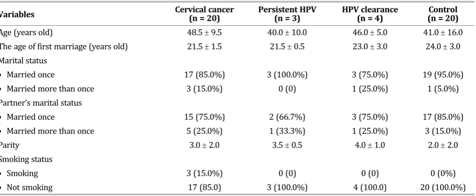
Over 2 years of period, from August 2013 to August 2015, forty-seven samples were obtained. The characteristics of the subjects were presented in Table 1.
According to the table, it could be seen that the average age of subjects with cervical cancer, cleared and persistent subclinical HR-HPV infection, and normal cervix (control) were 48.5, 40.0, 46.0, and 41.0 years old, respectively. The mean age of first marriage was regarded as the first sexual contact when relatively young, which all results showed for more than 20 years old. Most of the subjects and their partners were married only once; and most of them had low parity. Almost all subjects did not smoke. Based on these demographic characteristics, the subjects in this study had lower risk factors for HPV infection and cervical cancer.
Meanwhile, the deposit adequacy level of retinol was shown in Table 2. Of 47 total samples, retinol deposit sufficiency level in normal cervix, subclinical HPV infection clearance, persistent, and cervical cancer group was 85.0% (reference), 75.0% (OR 1.89), 33.3% (OR 11.33), and 75% (OR 1.89), respectively.
Table 1. Demographic Characteristics
Variables Cervical(n = 20)cancer Persistent(n = 3) HPV HPV(n clearance = 4) (nControl = 20)
Age (years old) 48.5 9.5 40.0 10.0 46.0 5.0 41.0 16.0 The age of first marriage (years old) 21.5 1.5 21.5 0.5 23.0 3.0 24.0 3.0 Marital status
Married once 17 (85.0%) 3 (100.0%) 3 (75.0%) 19 (95.0%)
Married more than once 3 (15.0%) 0 (0) 1 (25.0%) 1 (5.0%)
Partner’s marital status
Married once 15 (75.0%) 2 (66.7%) 3 (75.0%) 17 (85.0%)
Married more than once 5 (25.0%) 1 (33.3%) 1 (25.0%) 3 (15.0%)
Parity 3.0 2.0 3.5 0.5 4.0 1.0 2.0 2.0
Smoking status
Smoking 3 (15.0%) 0 (0) 0 (0) 0 (0%)
Statistically, there was no significant difference of retinol deposit sufficiency level between normal cervix and cleared subclinical HPV infection (p=0.628), normal cervix and persistent subclinical HPV infection (p=0.078), normal cervix and cervical cancer (p=0.433), cervical cancer and cleared subclinical HPV infection (p=1.000), cervical cancer and persistent subclinical HPV infection (p=0.430), persistent subclinical HPV infection and clearance (p=0.740).
As demonstrated on Table 2 and 3, there were not significant differences in the level of retinol adequacy deposits in all groups studied.
DISCUSSION
Statistically, there were no significant differences in the level of retinol deposit sufficiency level in all groups studied, such as between the normal cervix and cervical cancer, normal cervix and persistent subclinical HR-HPV infection, normal cervix and cleared subclinical HR-HPV infection (p=0.433; 0.078; 0.628); cervical cancer and clearance subclinical HR-HPV infection, cervical cancer and persistent HR-HPV subclinical infection (p=1.000; 0.430), and subclinical HR-HPV infection persistence and clearance (p=0.740).
In this study, we obtained the difference in the sufficiency level of retinol deposit in the group of subclinical infection with HR-HPV persistence, clearance, and normal cervix (control). This result was in accordance with predetermined clinical
justification. However, the result was far below clinical justification for cervical cancer group.
Until now, the study of retinol in the immune system related to the natural history of cervical cancer was inconsistent, especially when applied to population. There were many studies of endogenous retinoid in plasma level on the natural history of cervical cancer, there was still lack of observation on the adequacy of retinol deposit. This study showed that there was no statistically significant difference of sufficiency retinol deposit level in the group of normal cervix, cervical subclinical HR-HPV infection clearance, persistent, and cervical cancer. The results of this study were relatively consistent with study conducted by Siegel, et al.19 and Palan, et al.20 Siegel, et al. stated
that there was no significant association between endogenous retinoic acid and a clearance of HPV infection also cervical lesion regression. These results indicated that the role of retinoic acid as an HPV and carcinogenesis inhibitor in vitro could not be demonstrated in epidemiological studies on the setting of clinical population.19 Palan, et al. pointed
out that there were no significant differences in the mean level of plasma retinol on 235 subjects with normal cervix, cervical intraepithelial neoplasia (CIN), and cervical cancer. The study also found lower level of carotenoid and alfa-tocopherol in the plasma of CIN and cervical cancer patients. There was also significant linear trend in level of carotenoid and alfa-tocopherol to degree of cervical histopathological abnormalities.20 Another
Table 2. Sufficiency Rate of Retinol Deposit
Groups Deposit adequacy level of retinol OR IK (95%) pvalue
Less Normal
Cervical Cancer 5 15 1.89 0.39 to 9.27 0.433
Persistent HR-HPV 2 1 11.33 0.76 to 167.97 0.078
Cleared HR-HPV 1 3 1.89 0.14 to 24.79 0.628
Control 3 17 Ref
Chi-Square test
Table 3. Relationship between the Deposit Adequacy Level of Retinol Group
Group (Sufficiency Rate Deposit of Retinol) OR IK (95%) pvalue
Cervical Cancer vs Cleared HR-HPV 1.00 0.08-11.93 1.000
Cervical Cancer vs Persistent HR-HPV 0.17 0.01 to 2.26 0.430
Persistent HR-HPV vs Cleared HR-HPV 6.00 0.22 to 162.54 0.740
study by Giuliano, et al.21 stated that persistent
HPV infection could be inhibited by several anti-oxidant micronutrients. Consumption of papaya regularly at least once a week was associated with significant barrier to persistence of infection. Sedjo, et al.22 concluded that high consumption
of vegetables and fruits was related to 54% decreased risk of HPV persistence. Goodmann, et al.23 also found a significant correlation between
antioxidant level and the risk of CIN. Siegel, et al. from another study also revealed contrary result that retinol was not considered as a protective agent against persistent infection with oncogenic HPV on female population in Brazil.19 This
finding was also supported by Alvarez, et al.,24
who showed that there was no significant difference in the rate of cervical lesion regression in placebo, retinoid low dose, and high dose group. Retinol regulates kind of essential cells in the body. Retinoid plays an important role in the process of growth, cleavage, tissues maintenance, reproductive function, metabolism, differentiation, haematopoietic, bone formation, spermatogenesis, and embryogenesis. Deficiency of vitamin A will impact to unwanted effects.25,23 Some factors
significantly affecting the level of endogenous retinol include age, race, use o f o r a l c o n t r a c e p t i v e s , a n d n u m b e r o f pregnancies.26 The concentration of retinoid did
not differ significantly between fasting and non-fasting population.27
Dragnev, et al. stated that retinol had apoptosis effect, anti-proliferative, and regulator of cell differentiation, a chemo-preventive agent;28 thus,
it could be used in anti-cancer therapy. Retinol acts via nuclear receptor to activate target genes containing responsive element resulting to biological effect. Anti-cancer activity of retinoid is the result of three main mechanisms, such as cytodifferentiation, growth cessation, and apoptosis.28,29 Some retinoids are clinically
effective as chemo-preventive and anti-cancer therapy in promielocytic acute leukemia, but, it shows less effective against most solid tumors and results unwanted side effects.23,29,30 Headache is
the most frequent adverse events after retinoid treatment on approximately 74% of high doses of retinoid.24 In combination with IFN, retinoid has
potential effect in cervical SCC. Retinoic acid has the action as a radiosensitizer that does not require the function of p53 in vitro.31
N a r a y a n a n , e t a l . s h o w e d a n increased
expression of p53 and inhibition of E6/E7 transcription after retinoid administration. This finding suggested an important role of retinoic acid as a cell cycle regulator, chemo-preventive, and anti-viral agents.23,32,33
Our results had differences compared with previous epidemiological studies, in which the study showed the opposite relationship between the development of cancer and vitamin A-containing diet. Systemic administration of retinol can reduce the thickening of arterial intima layer significantly after endothelial injury in vivo. In vitro and in vivo, retinol has a pro-inflammatory effect and it can enhance the expression of TNF- as a cytokine that has an important role in acute inflammation.31-33 Immunomodulating
pharmaco-logical concentration of vitamin A can lower the incidence of tumor in experimental biochemistry. Some studies proved that natural and synthetic retinoids could inhibit the growth and development of various tumor types.31
Apoptotic process includes a series of action that is associated with retinoid. Geissmann, et al. showed that retinoid could induce apoptosis through its heterodimer receptor in spite of no signal inflammation. The existence of cross communication with inflammatory cytokines allows retinoid to activate DNA-binding-factor-KB
in the core of dendritic cells, triggers MHC class II, induces differentiation and maturation of dendritic cells, as well as improves specific T-cell response to antigens.34 Khan, et al. reported that human
keratinocytes (HKC), which had been immortalized due to transfection of HPV-16, was more sensitive to inhibition of retinoic acid compared with normal HKC. Retinoic acid also can inhibit mRNA expression of E6/ E7 and E2/ E5 HPV-16. R e t i n o i c a c i d b e c a m e b a r r i e r t o immortalization due to transfection of HPV-16 and it reached up to 95%.35
Retinol and vitamin A derivatives affect cell differentiation, proliferation, and apoptosis, and play an important physiological role in various biological processes. Retinol is primary obtained from animal products. Its intracellular bioavailability is regulated by specific and CRBP. The CRBP-1 as the most common CRBP isoform is a small 15 KDa cytosolic protein that is highly expressed in various tissues. It acts to regulate absorption, esterification, and bioavailability of retinol, also plays a major role in wound healing and remodeling process of arteries. In recent years, the role of retinoid signalling CRBP-1 during the development of cancer became the aim of several studies.37 Expression of CRBP-1
is associated with cervical epithelial cell differentiation. High amount of CRBP-1 could be found in columnar and epithelial cells.38 The drop
of CRBP-1 was coincided with the disappearance of retinol response in rat cervical epithelial cells.36
There are two types of retinol metabolism in the smooth muscle cell. Increased production of retinoic acid was found in the intima cell.37,39,40
Studies in vitro showed that the retinoid, in particular 9-cis-RA, could inhibit the growth of estrogen receptor through blocking the cell cycle.41
Synthetic retinoid is generally quite promising for the treatment of cancer and several clinical trials are also running, but only a few synthetic retinoid have been approved by Food and Drug Administration (FDA). Preclinical studies indicated that synthetic retinoid inhibited the growth of human cancer. Fenretinide (4-HPR) is one of the most promising retinoid clinically. It demonstrated significant cytotoxic activity of tumor cell through induction of apoptosis and non-apoptotic routes.37,42
The pattern of CRBP-1 on human epithelial endocervical is identic to those reported in mice.43
In addition, in humans, CRBP-1 was sufficient in myometrium of non-pregnant women along with protein CRABP; thus, they showed the role of ATRA in proliferation control of the myometrium in vivo. The level of CRBP-1 was down regulated on the upper and lower segment of uterus during the first and second trimesters of pregnancy.44 The CRBP-1
gene function in controlling the bioavailability of vitamin A suggested that it might have particular relevance in the inhibition of cancer transfor-mation. However, in human cancer, the presence and role of protein that specifically binded to retinol and retinoic acid had not been widely
investigated. The dysregulation of CRBP-1 occcured in some tumors, such as breast tumors, ovarian, endometrial, prostate, renal, cervical, larynx, nasopharynx, lymphoma, and gastrointestinal cancer. Furthermore, hypermetilation of CRBP-1 was responsible for the inactivation of some cancer cells. Thus, epigenetic disruption of CRBP-1 was a common event in human cancer and it might have important implication for cancer prevention and therapy of retinoid.37
Retinoid has long been used for the treatment of psoriasis and acne. Retinoid is effective in some pre-cancerous lesions, such as oral leukoplakia, actinic keratosis, and cervical dysplasia. It is able to delay the development of skin cancer in individual with xerodermapigmentosum; therefore, it shows chemopreventive potency. Moreover, several malignancies have been treated with retinoid-based therapy, as sole agent for pathological promielocytic including acute leukemia, Kaposi’s sarcoma, cutaneous T-cell lymphoma, leukemia mielogenus juvenile chronic, SCC, and kidney cancer.45,46
CONCLUSION
This study has proved that the normal cervix has sufficient level of retinol deposit. However, this study can not prove that cervical cancer and persistent HPV infection have less retinol deposit. These results provide important data on the contribution of some previous studies on the role of vitamin A that is still inconsistent as a chemo-preventive measure in the natural history of cervical cancer. Theoretically, retinol administration is expected to reduce the risk of HR-HPV p e r s i s t e n c e a n d p r o g r e s s i o n towards pre-cancerous lesion on the natural history of cervical cancer. However, result of this study would like to describe that there is no association between adequacy of retinol deposit and history of cancer. Therefore, based on the result of this study, supplementation of vitamin A can not become basic approach to primary prevention of cervical cancer in terms of nutrition.
RECOMMENDATION
relation to its sensitivity towards stimulation of retinylpalmitate to activate mechanisms through RAR. Retinol stimulation in vitro to activate several parameters of local immune response in cervical tissue is a promising study in the future.
Conflict
of
Interest
The authors hereby affirm that there is no conflict of interest in this study.
REFERENCES
1. Ferlay J SH, Bray F, Forman D, Mathers C, Parkin DM. Cancer incidence and mortality worldwide: IARC Cancer Based; GLOBOCAN 2008 [Internet]. Lyon, France: Int A for Res on Can; 2010.
2. Parkin DM. Global cancer statistics in the year 2000. Lancet Oncol. 2001; 2(9): 533-43.
3. World Health Organization. Information centre on HPV and cervical cancer (HPV Information Centre). Human Papillomavirus and Related Cancers in the World: Summary Report 2010. New Zealand: World Health Organization; 2010.
4. Aziz MF. Masalah kanker serviks. Cermin Dunia Kedokteran: 2001; 133: 5.
5. Campoli M, Ferrone S, Zea AH, Rodrigues PC, Ochoa AC. Mechanism of tumor evasion. In: Khleif S. Tumor immunology and cancer vaccines. New York: Kluwer Academic Publishers; 2005: 61-78.
6. Kresno SB. Imunologi tumor. Dalam: Ilmu Onkologi Dasar. Jakarta: Balai penerbit FKUI; 2010: 161-79.
7. Onon TS. History of human papillomavirus, warts, and cancer: What do we know today? Best Pract Res Clin Obstet Gynecol. 2011; 25(5): 565-74.
8. Narisawa-Saito M, Kiyono T. Basic mechanisms of high-risk human papillomavirus-induced carcinogenesis: Roles of E6 and E7 proteins. J Cancer Sci. 2007; 98(10): 1505-11. 9. Burd EM. Human papillomavirus and cervical cancer. Clin
Microbiol Rev. 2003; 16(1): 1-17.
10. Abbas AK, Lichtman AH, Pober JS. General Property of Immune Respons. In: Cellular and molecular immunology. 3rd ed. Phyladelphia: Saunders Company; 1997: 3-14.
11. Semba RD. On the ’discovery’ of vitamin A. Ann Nutr Metab. 2012; 61(3): 192-8.
12. Ball G. Vitamins in foods: Analysis, bioavailability, and stability. Florida: CRC press; 2005.
13. Dzhagalov I, Chambon P, He YW. Regulation of CD8+ T lymphocyte effector function and macrophage inflammatory cytokine production by retinoic acid receptor gamma. J Immunol. 2007; 178(4): 2113-21.
14. Basu P. Phase 2 randomized controlled trial of radiation therapy plus concurrent interferon-alpha and retinoic acid versus cisplatin for stage III cervical carcinoma. Int J Rad. 2016; 94(1): 102-10.
15. Visser J, van Baarle D, Hoogeboom BN, Reesink N, Klip H, Schuuring E, et al. Enhancement of human papilloma virus type 16 E7 specific T cell respones by local invasive procedures in patients with (pre)malignant cervical neoplasia. Int J Can. 2006; 118(10): 2529-37.
16. Ross AC. Vitamin A deficiency and retinoid repletion regulate the antibody response to bacterial antigens and the maintenance of natural killer cells. Clin Immunol Immunopathol. 1996; 80(32): S63-72.
17. Gariglio P, Gutierrez J, Cortes E, Vazquez J. The role of retinoid deficiency and estrogens as cofactors in cervical cancer. Med Res Arch. 2009; 40(6): 449-65.
18. McLaren DS, Frigg M. Sight and life manual on vitamin a deficiency disorders (VADD). 2nd ed. Switzerland: Task
Force Sight and Life; 2001.
19. Siegel EM, Salemi JL, Craft NE, Villa LL, Ferenczy AS, Franco EL, et al. No association between endogenous retinoic acid and human papillomavirus clearance or incident cervical lesions in Brazilian women. Cancer Prev Res. 2010; 3(8): 1007-14.
20. Palan PR, Mikhail MS, Goldberg GL, Basu J, Runowicz CD, Romney SL. Plasma levels of beta-carotene, lycopene, canthaxanthin, retinol, and alpha-tocopherol in cervical intraepithelial neoplasia and cancer. Am J Clin Cancer Res. 1996; 2(1): 181-5.
21. Giuliano AR. The role of nutrients in the prevention of cervical dysplasia and cancer. Nutr. 2000; 16(7-8): 570-3. 22. Bella MC, Grimmingerb DS, Jacobsend C, Chauhana SC,
Mahera DM, Buchwaldd DS. Risk factors for HPV infection among American Indian and white women in the Northern plains. Gynecol Oncol. 2011; 121(3): 532-6.
23. Goodman MT, Kiviat N, Duffie KM, Hankin JH, Hernandez B, Wi LR. The association of plasma micronutrients with the risk of cervical dysplasia in Hawaii. Cancer epidemiol Biomarkers Prev. 1998; 7(6): 537-44.
24. Alvarez RD, Conner MG, Weiss H, Klug PM, Niwas S, Manne U, et al. The efficacy of 9-cis-retinoic acid (alitretinoin) as a chemopreventive agent for cervical dysplasia: Results of a randomized double-blind clinical trial. Cancer Res. 2003; 12(2): 114-9.
25. Bollag W, Holdener EE. Retinoids in cancer prevention and therapy. Ann Oncol. 1992; 3(7): 513-26.
26. Sun SY, Lotan R. Retinoids and their receptors in cancer development and chemoprevention. Crit Revs Oncol Hematol. 2002; 41(1): 41-55.
27. Kane MA. Analysis, occurrence, and function of 9-cis-retinoic acid. Acta Biochim Biophys Sin. 2012; 1821(1): 10-20.
28. Dragnev KH, Petty WJ, Dmitrovsky E. Retinoid targets in cancer therapy and chemoprevention. Cancer Biol Ther. 2003; 2(4 Suppl 1): S150-6.
29. Chao TY, Jiang SY, Shyu RY, Yeh MY, Chu TM. All-trans retinoic acid decreases susceptibility of a gastric cancer cell line to lymphokine-activated killer cytotoxicity. Br J Cancer. 1997; 75(9): 1284-90.
30. Geissmann F, Revy P, Brousse N, Lepelletier Y, Folli C, Durandy A, et al. Retinoids regulate survival and antigen presentation by immature dendritic cells. J Exp Med. 2003; 198(4): 623-34.
32. Berlin GVM, Niranjali DS, Radhakrishnan PM, Devaraj H. HPV-induced carcinogenesis of the uterine cervix is associated with reduced serum ATRA level. Gynecol Oncol. 2006; 103(1): 113-9.
33. Levin AA. Receptors as tools for understanding the toxicity of retinoids. Toxicol Lett. 1995; 82-83: 91-7.
34. Vertuani S, Geer AD, Levitsky V, Kogner P, Kiessling R, Levitskaya J. Retinoids act as multistep modulators of the major histocompatibility class I presentation pathway and sensitize neuroblastomas to cytotoxic lymphocytes. Cancer Res. 2003; 63(22): 8006-13.
35. Khan MA, Jenkins GR, Tolleson WH, Creek KE, Pirisi L. Retinoic acid inhibition of human papillomavirus type 16-mediated transformation of human keratinocytes. Cancer Res. 1993; 53(4): 905-9.
36. Khuri LT, Talmage DA. Decreased cellular retinol binding protein expression coincides with the loss of retinol responiveness in rat cervical epithelial cells. Exp Cell Res: 1997; 230(1): 38-44.
37. Laurent CE, Coccia B, Krust AG. Hovanessian membrane-expressed HIV envelope glycoprotein heterodimer is a powerful inducer of cell death in uninfected CD4+ target cells. Res Virol: 1995; 146(1): 5-17.
38. Hail N Jr, Kim HJ, Lotan R. Mechanisms of fenretinide induced apoptosis. Apoptosis: 2006; 11(10): 1677-94. 39. Niles RM. Signaling pathways in retinoid chemoprevention
and treatment of cancer. Mutat Res: 2004; 555(1-2): 81-96.
40. Gidlo AC, Ocaya P, Olofsson PS, Torma H, Sirsjo A. Differences in retinol metabolism and proliferative response between neointimal and medium smooth muscle cells. J Vasc Res: 2006; 43(4): 392-8.
41. Jonsson AJ, Ares MP, Yan ZQ. Increased rate of apoptosis in intimal arterial smooth muscle cells through endogenous activation of TNF receptors. Arterioscler Thromb Vasc Biol: 2001; 21(12): 1909-14.
42. Zhao Z, Zhang ZP, Soprano DR, Soprano KJ. Effect of 9-cis-retinoic acid on growth and RXR expression in human breast cancer cells. Exp Cell Res: 1995; 219(2): 555-61. 43. Egawa K, Egawa N, Honda Y. Human
papillomavirus-associated plantar epidermoid cyst related to epidermoid metaplasia of the eccrine duct epithelium: A combined histological, immunohistochemical, DNA in situ hybridization and three-dimensional reconstruction analysis. Br J Dermatol: 2005; 152(5): 961-7.
44. Hillemanns P, Khuri LT, Koulos JP, Talmage D, Wright TC Jr. Localization of cellular retinoid-binding proteins in human cervical intraepithelial neoplasia and invasive carcinoma. Am J Pathol: 1992; 141(4): 973-80.
45. Tyson-Capper AJ, Cork DMW, Wesley E, Shiells EA, Loughney AD. Characterization of cellular retinoid-binding proteins in human myometrium during pregnancy. Mol Hum Reprod: 2006; 12(11): 695-701.