Case Report
Medical Journal of Islamic Republic of Iran, Vol. 26, No. 3, Aug. 2012, pp.143-146
__________________________________________________________________________________________________ 1. Associate Professor of Orthopedics, Orthopedics Department, Tabriz Medical University, Tabriz, Iran. hojjat.pourfeizi@gmail.com
2. Assistant of Orthopedics, Orthopedics Department, Tabriz Medical University, Tabriz, Iran. j-ganjpour@yahoo.com 3. (Corresponding author), Assistant of Orthopedics, Orthopedics department, Tabriz Medical University, Tabriz, Iran. elmimail@yahoo.com
3. Resident of Orthopedics, Orthopedics Department, Tabriz Medical University, Tabriz, Iran. ali.tab.ms@gmail.com
Osteoid osteoma of a scapula: a case report in a 34
years old woman
Hojjat Hossein Pourfeizi 1, Jafar Ganjpour Sales2, Asghar Elmi3, Ali Tabrizi3
Department of Orthopedics, Tabriz Medical University, Tabriz, Iran.
Received: 25 February 2012 Revised: 12 May 2012 Accepted: 23 May 2012
__________________________________________________________________________________________
Abstract
Osteoid osteoma is a benign bone tumor and accounts for 10% of benign tumors. Almost any bones can be involved but half of cases involving femur or tibia. Osteoid osteoma is a rare tumor of the scapula with only 18 reported cases in the literature. We presented a case of osteoid osteoma of the scapula in right shoulder in angle of coracoid and geloneid in a 34- year old woman. She had right radicular shoulder pain increased at night for 4 years. An important point about this case is that, patient was treated in long-term with miss diagnosis of cervicospinal discopathy. The key of diagnosis in this patient was paying attention to the nature of night increas-ing pain and performincreas-ing bone scan. After the operative curettage of the tumor, the pain dramatically stopped and a few weeks of physical therapy led to full range of motion in her shoulder.
Keywords: Osteoid osteoma, Scapula, Shoulder.
__________________________________________________________________________________________
Introduction
Osteoid osteoma is the third most common benign bone tumor. It usually affects the di-aphysis of long bones, especially the femur or the tibia, although many believe it is nonneoplastic. It was first described in 1935 by Jaffe (1). It accounts for 10% of benign bone tumors (2). It is twice as common in males as in female, and the highest incidence occurs in the second and third decades (3). After 30 years old, it is rare. The coracoid process is a rare location for a scapular mor. In a series of 243 bone tumors and tu-mor-like conditions of the scapula, only 18 cases involved the coracoid process (4).
The proximal femur is the most common
location followed by the tibia, posterior ele-ments of the spin and the humerus (5).
Pain is usually only symptom of disease presented in patients and is typically de-scribe as mild and intermittent of first con-stantly with increased severity at night. In-termittent and aching night pain readily re-lieves using salicylate which marks the early phase, but later the pain becomes constant and severe and may not respond to medica-tion (6).
The location of this neoplasm remains un-clear, though it has developed in the cora-coid process and the subglenoid region (7). Radiographic findings include sclerosis, but a nidus may be difficult to see on radiog-raphy. Bone scans and CT are often required
Osteoid Osteoma of a scapula
144 MJIRI, Vol. 26, No. 3, Aug 2012, pp. 143-146
Fig.1. CT scan shows hypodens lesion with same surrounding hyperdensity at coracoglenoid junc-tion.
Fig. 2. Bone scan shows marked increased up take at super lateral angle of scapula.
Fig. 3. Roberts approach of acromioclavicolar joint and coracoid process of scapula.
to localize the lesion accurately. Treatment includes surgical excision of the nidus (8).
Case report
The patient was a 34-year old woman with chief compliant of right radiculer shoulder pain for 4 years which referred to Shohada Training Hospital of Tabriz University of Medical Sciences.
The pain was decreased, with daily activity and increased at night.
At physical examination there was tender-ness around shoulder with joint movement restriction over 90ºof abduction with painful external rotation.
Shoulder plain x-rays were normal. The bone scan revealed obvious increased uptake at superolateral angle of scapula (Fig.1). The CT scan showed relatively ossified 1cm nidus with mild peripheral hyperdencity (Fig. 1). The lesion was at a few millimeters from subchondral bone of glenoid (Fig 2). Nerve Conduction Velocity (NCV) revealed neurogenic pattern at L5-L6 level muscles.
Surgical Technique: In the supine position with right shoulder elevation we performed a J shape incision such as the one used for coracoclavicular screw fixation. (Fig-3) An-terior insertion of deltoid to clavicle was in-cised with 1cm remnant.
After coracoid exposure, we performed drill hole with 2.7 drilbit on the tip of the coracoid. Coracoid was cut leaving its ten-don insertions attached. On the base of cora-coid next to the glenoid there was small dimpling. After corticotomy 1.51/5cm dense calcified tissue was removed and the hole completely curetted.
Coracoid was reattached using screw and deltoid sutured to clavicle. (Fig. 4) The orig-inal pain was subsided immediately after the surgery. In order to improve the range of motion of the shoulder, physical therapy was performed when the surgical pain relieved. Two months after surgery full range of mo-tion in shoulder was obtained. Histologic diagnosis was osteoid osteoma.
Discussion
Because the scapula is a rare site for
oste-oid osteoma, it is not often included in the differential diagnosis of chronic shoulder pain (7). The night pain seen is often at-tributed to rotator cuff pathology. However,
HH. Pourfeizi, et al.
145
MJIRI, Vol. 26, No. 3, Aug 2012, pp. 143-146
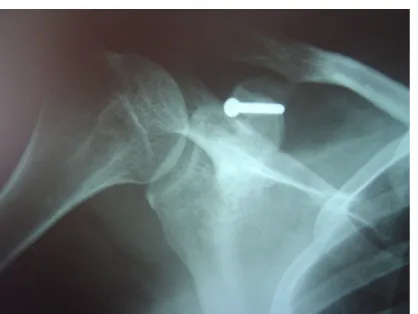
Fig. 4. Post up controlled shoulder plain x-ray.
the age range of the patients in these cases would make rotator cuff pathology less like-ly. Glanzmann et al reported osteoid osteoma presented by localized stiffness of the anterosuperior capsule which led to the chief complaint of painful restriction of ex-ternal rotation in the adducted arm position only (7). In fact, osteoid osteoma typically occurs in adolescence, whereas rotator cuff pathology would be unusual in that popula-tion. In particular, juxta-articular osteoid osteoma often presents a diagnostic dilemma secondary to referred pain, neurologic defi-cits, and global extremity weakness (8). The sensitivity of soft tissue radiographic tech-niques for the shoulder can also be problem-atic. Lesions in the labrum may be identified but may not be the cause of the patient’s symptoms (9). According to Ogose et al re-port, bone tumors of the coracoid process may be difficult to detect on plain radio-graphs. In the patient with persistent shoul-der pain unresponsive to the selected treat-ment, additional imaging studies should be considered to eliminate the possibility of a bone lesion (10).
Benign osseous lesion of the shoulder is un-common, osteoid osteoma and osteoblastoma occur in the proximal humerus or scapula in 10 to 15% of cases and when they do occur, favor the proximal humerus or glenoid (11). The en block excision in uncommon subglenoid re-gion can be problematic, since the surgical exposure is difficult, and shoulder Joint function can be affected if the lesion is subchondral (12).
Mosheiff et al reported a case of osteoid osteoma of the scapula with excision of the lesion by guided needle biopsy (8). In surgi-cal treatment by Ponali et al, the excision of the lesion and grafting was performed by a deltopectoral approach (10). One year after the surgery, the patient remains pain free and has full range of motion with no recurrence of the tumor. Another reported by Akpinar et al, the en bloc excision of the osteoid osteoma was managed by an anterior ap-proach using an osteotomy of the coracoid process had successful results (11).
Du ssaussois L et al reported a new
thera-peutic modality uses in a patient with an os-teoid osteoma of the scapula. They success-fully destroyed the nidus by percutaneous laser photocoagulation under CT guidance. Clinical improvement was manifested after 72 hours and the patient remained asympto-matic at months follow up (14). In Degreef et al case report, osteoid osteoma in the acromion was successfully treated by an acromioclavicular (AC) joint resection (4).
At our case the unusual site as well as age and gender of the patient and common com-pliant of radiculer neck and shoulder pain with mechanical nature caused long delayed diagnosis and treatment. Although osteoid osteoma is a very rare cause of radiculer shoulder pain but it's ignoring result in pro-longed relentless night pain. Paying attention to night increasing nature of pain is the key guide of diagnosis.
In osteoid ostema choice of treatment is ra-dio frequency ablation (R.F.A). This treat-ment was noninvasive and had good results (14). In RFA a minimum amount of bone is removed during the procedure and the pa-tient can return to normal function almost immediately (15). Another treatment of os-teoid osteoma is surgical excision of the nidus (9-12), but anatomic unusual site can produce some technical and rehabilitation difficulties. In our experience in training center of orthopedics, operation procedure was as successful as non-surgical treatment especially in patients with unusual anatomi-cal presentation.
Osteoid Osteoma of a scapula
146 MJIRI, Vol. 26, No. 3, Aug 2012, pp. 143-146 Conclusion
Osteoid osteoma of the scapula is a chal-lenging case to diagnose for several reasons. Because a differential diagnosis is unlikely and far-fetched, these tumors can be mis-diagnosed for long time and treated as cervi-cal radicular pain.
References
1.Jaffe H. Osteoma: a benign osteoblastic tumor composed of osteoid and atypical bone. Arch surg 1935; 31:709.
2.Swee RG, Mcleod RA, Beabout JW. Osteoid osteoma. Radiology 1979;130: 117-123.
3.Dahlin DC. Bone tumors: general aspects and da-ta on 6,221 cases. Spring field, Illinois: charles C Thomas, 1978: 43-569 75-85.
4.Degreef I, Verduyckt J, Debeer Ph, De Smet L. An unusual cause of shoulder pain: Osteoid osteoma of themacromion:A case report. J Shoulder Elbow Surg 2005; 14:643-644.
5.Bloem J. Kroon M. Osseous lesions, Radiologic clinics of North American,1993; 31(2):261-277.
6.Edeiken J, Dalinkam, karasick D. Edeiken’s roentgen diagnosis pf diseases of Bone .4th Baltimore,
Williams Wilkins;1990; p.44-64.
7.Glanzmann MC, Hinterwimme S, Woertler Klaus, Andreas B.Osteoid osteoma of the coracoid masked as localized capsulitis of the shoulder. J
Shoulder Elbow Surg 2011; 20,e4-e7.
8.Moshieff R, Leibergall M Zivi, Amirg, Segal D. Osteoid osteoma of the scapula. Clin Orthop 1991; 262:129-131.
9.Anne MK, Ronald M S, Erika L, O’Brien SJ and Drakos MC. Arthroscopic Removal of an Osteoid Osteoma of the Shoulder. The Journal of Arthroscop-ic and Related Surgery 2002; 18(7): 801–806.
10.Ogose A, Sim FH, OConnor MI, Unni KK. Bone tumors of the coracoid process of the scapula. Clin orthop 1999; 358:205-214.
11.Poyanli O Unay K Akan K Ozkan K Temiz D. Subchondral osteoid osteoma of the glenoid. Chir Organi Mov 2009; 93(1):79-81.
12.Akpinar S, Demirors H, Hersekli MA, Yildirim T, Barutcu O, Tandogan RN. Osteoid osteoma in the base of the coracoid process of the scapula. Excision by anterior approach: a case report. Bull Hosp Jt Dis 2001; 60(1):47-9.
13.Dussaussois L, Stelmaszyky, Golzariang. percu-taneous treatment of an osteoid osteoma of the scapu-la using a scapu-laser under scanner control. Acta orthop Belg 1998; 64(1):88-91.
14.Daria Motamedi, Thomas J. Learch, David N. Ishimitsu , Kambiz Motamedi, Michael D. Katz, Earl W. Brien, Lawrence Menendez. Thermal Ablation of Osteoid Osteoma: Overview and Step-by-Step Guide. RadioGraphics 2009; 29:2127–2141.
15.Akhlaghpoor S, Tomasian A, Arjmand SA, Ebrahimi M, Alinaghizadeh MR. Percutaneous oste-oid osteoma treatment with combination of radiofre-quency and alcohol ablation. Clinical Radiology
HH. Pourfeizi, et al.
147
MJIRI, Vol. 26, No. 3, Aug 2012, pp. 143-146 2007; 62, 268-e273.