Spring 2019
Characterization of the role of E6-AP in Angelman Syndrome and
Characterization of the role of E6-AP in Angelman Syndrome and
Similar Neurodevelopmental Disorders
Similar Neurodevelopmental Disorders
Rebecca Olsen
Iowa State University, rcolsen@iastate.edu
Follow this and additional works at: https://lib.dr.iastate.edu/creativecomponents
Part of the Medical Genetics Commons, and the Medical Neurobiology Commons
Recommended Citation Recommended Citation
Olsen, Rebecca, "Characterization of the role of E6-AP in Angelman Syndrome and Similar Neurodevelopmental Disorders" (2019). Creative Components. 178.
https://lib.dr.iastate.edu/creativecomponents/178
Characterization of the role of E6-AP in Angelman Syndrome
and Similar Neurodevelopmental Disorders
by
Rebecca Olsen
Presented to the graduate faculty in partial fulfillment of the requirements
for the degree of
MASTER OF SCIENCE
Major: Biomedical Sciences
Creative Component Committee:
Dr. Steven Carlson, Major Professor
Dr. Timothy Day
Dr. Michael Kimber
Iowa State University
Ames, Iowa
INTRODUCTION TO ANGELMAN SYNDROME
Angelman syndrome (AS) is a neurodevelopmental disorder first described by
Harry Angelman in 1965 [8]. Incidence of the disease is estimated to be between 1 and
4 in 40,000, with an estimated morbidity rate of 1 in 15,000 [5, 8]. 50% of cases of the
disease are initially misdiagnosed, therefore incidence rates may be higher than current
estimates [32].
The disease is a genetic malformation associated with loss of function of the
UBE3A gene; a gene located on chromosome 15q11-13 that is susceptible to genomic
imprinting [5,8]. Genetic material (DNA) contains coding regions called genes which are
arranged into chromosomes. Under normal conditions, humans have 46 chromosomes
(23 inherited from each parent) present within the nucleus of each cell. The 46
chromosomes are defined as 23 pairs of chromosomes, numbered as 1 through 22.
Two out of the 46 chromosomes are termed sex chromosomes meaning that they differ
between males (XY) and females (XX). The chromosome itself is composed of a short
arm (p) and a long arm (q) [12]. For specificity, the chromosomes are divided into
several numbered bands [27]. For example, chromosome 15q11-13 mentioned above,
specifies that UBE3A is located within chromosome 15 between bands 11 and 13 on
the q arm of the chromosome [27].
Gene expression can be regulated on the DNA level by DNA methylation,
addition of methyl groups to cytosine bases which has a negative effect on gene
expression. Under normal cellular conditions, each inherited copy of a gene is active
but some situations can cause only one of the copies to be active. Which is chosen to
or maternal inherited gene copy may be silent [30]. This process is called genomic
imprinting, one inherited copy of a UBE3A gene is silenced, while the other gene copy
remains active in the embryo and adult [12]. It occurs at the germ line level, meaning at
the level of egg and sperm cells before an embryo is formed. Imprinted genes are
usually located in small to large clumps. A specific imprinted clump can contain
expressed imprinted genes from both parents [31]. Within these clumps, there are CpG
islands (short sequences of DNA near the transcription start site of genes containing
large amounts of cytosine and guanine bases) that undergo DNA methylation only on
one of the two parental chromosomes, this modification is often passed on to offspring
[31]. Imprinting commonly appears as DNA methylation, but the process can also occur
through modifications in histones, insulator proteins, and long non-coding RNAs [31].
A gene contains both coding and non-coding regions. Only the coding portion of
the gene will undergo transcription and be translated into proteins. The UBE3A gene
contains 16 exons with the coding region being from exon 8 to exon 16 [8]. The highly
conserved HECT domain (homologous to the E6-AP carboxyl terminus) is at exon 16
and is composed of two lobes [8]. A catalytic site situated between the two lobes is a
common site for frameshift, nonsense, missense, and splice site mutations associated
with Angelman’s syndrome patients [8]. In addition, point mutations across the coding
region of UBE3A, but especially in exons 9 and 16, have also been identified in patients
diagnosed with this syndrome [8].
Due to the importance of the UBE3A gene in Angelman’s syndrome, it has been
extensively characterized and found to be differentially expressed in neural tissues.
brain and Purkinje cells of the cerebellum, it is also expressed, but not imprinted in
cultures of oligodendrocytes and astrocytes [8, 9]. The gene is not imprinted or
expressed in neural glial cells, or peripheral tissues of humans and mice [23]. The
paternal copy of UBE3A is silenced due to imprinting within neural tissues, specifically
within cells of the olfactory bulb and hippocampus, and within Purkinje neurons [7,8,20].
Silencing is controlled by an anti-sense UBE3A transcript specific to neurons, which
inhibits transcription of the gene from the paternal chromosome [9]. Imprinting of the
gene is not fully completed in utero, so some paternal UBE3A expression occurs for a
short period of time but declines as neurons mature [23]. The maternal copy of UBE3A
can also be disrupted in Angelman syndrome [8]. Maternal expression of the UBE3A
gene can be impacted by different mechanisms, and are currently classified into five
classes, as described below, based on their mechanisms affecting the UBE3A gene.
However, it should also be emphasized that in 5-10% of patients, no genetic defect in
the gene can be identified [3].
THE FIVE CLASSES OF ANGELMAN SYNDROME:
Class I: Deletion in Chromosome 15q11-13
This class is caused by interstitial de novo deletion of the maternal chromosome and
is the most common mutation associated with development of AS. Deletions,
typically 4-7 megabase pairs in size [4,8], are seen in approximately 70-75% of
patients. The deletions appear to be due to misbalanced crossing-over events
neurodevelopment symptoms, this class is also associated with hypopigmentation of
hair, eyes, and skin [17].
Class II: Uniparental paternal disomy
The second class is associated with uniparental paternal disomy (UPD) in
chromosome 15 [8]. Uniparental disomy is when two copies of a chromosome from
only one parent, rather than one copy from each parent, is present in the genome of
a child [11]. This mutation occurs in approximately 2% of AS patients, and almost
exclusively occurs when both copies of the UBE3A gene come from a paternal
source [8]. This mutation is influenced by age as incidence of UPD is correlated with
increased age of parents at the time of conception. Further research is needed to
define the mechanism causing UPD in offspring from older parents [8].
Class III: Methylation/imprinting defect
The third class is associated with atypical methylation of chromosome 15, a defect
present in approximately 3-5% of AS patients [8]. Through the process of genomic
imprinting, one inherited copy of a gene is silenced, while the other gene copy
remains active [12]. The imprinting centre located within chromosome 15q11-13
serves in regulating DNA methylation, gene expression, and chromatin structure
through cis-acting elements [8]. Roughly 50% of patients with atypical methylation
have a mutation located within the imprinting centre of chromosome 15q11-13 [8].
Class IV: Mutation in E6-AP (i.e. UBE3A)
This class of AS is associated with a mutation within thegene which codes for
papillomavirus E6-associated protein (E6-AP) [8]. This mutation arises in 5-10%
of AS cases, appearing as a random mutation in 20% of Class IV patients and
75% occurring through inheritance from defects in the parental genome [8]. All of
these patients have reduced E3 ligase activity, which is associated with E6- AP
function [17].
Class V: Absence of detectable genetic abnormality
The most controversial of the class designations is Class V, where there is no
identified chromosomal abnormality but patients express the clinical phenotype of
the disease. The existence of this class of patients is still under debate [8]. Some
experts believe that these individuals have a mutation of UBE3A within a region that
is not within the coding region or involves an unidentified gene within the ubiquitin
pathway that disrupts expression of UBE3A [8]. No mutations related to Class V
patients have been identified or defined, but the ATP10C gene is a potential suspect
due to its location within 200 kb of UBE3A and the fact that it is maternally imprinted
in the brain [8]. Individuals that lack a genetic abnormality in the UBE3A gene tend
to have mild or no AS symptoms. Patients in this class are often described as
having an Angelman-like syndrome [20].
Almost all mutations associated with AS can be detected through use of
methylation-sensitive DNA probes, since imprinting of genes associated with AS involves the
methylation of DNA [13].
Angelman syndrome (AS) often results in developmental insufficiencies and severe
learning disabilities. Traditionally, the disease is hallmarked by the presentation of
frequent, inappropriate laughter. Motor and cognitive symptoms associated with AS
tend to correlate to the severity of the associated genetic abnormality [4]. Some
physical characteristics include hypotonia (low muscle tone in the upper limbs),
hyperactive reflexes, ataxia (impaired muscle coordination), and a wide-based gait with
feet placed farther apart than usual [8, 17]. The gait of affected individuals is often fitful
with pronation at the ankles [17]. Many AS patients have distinct facial features such as
deep-set eyes, a prominent chin, and macrostomia (a wide mouth). These facial
features are noticeable because patients are known to have a cheerful demeanor and
tend to smile frequently [8]. Other disease characteristics include symptoms of epilepsy
(seizures), sleep deficiencies, and inability to speak [5, 8, 17]. Over 80% of AS
individuals develop seizures but frequency of seizures decreases with age [17]. Brain
morphology in patients generally appears normal on gross exam although a few cases
of microcephaly do occur; There may be some minor structural abnormalities in the
brain as well [11]. Individuals with smaller genetic abnormalities, such as point
mutations, tend to have milder symptoms [4].
Of interest is the observation that developmental symptoms of AS do not tend to
appear until 6-12 months of age [11]. This is hypothesized to be because paternal
UBE3A may be accessible during the initial stages of neurogenesis prior to inhibition of
the paternal allele, permitting regular brain development and cellular function for a brief
period of time [11]. This also suggests that imprinting of the UBE3A gene is incomplete
studies have shown that the paternal UBE3A allele is not silenced within the brain
cortex at birth, while it is silenced in the rest of the brain [11].
INFLUENCE OF MUTATIONS IN E6-AP ON MOLECULAR PATHWAYS &
CELLULAR FUNCTIONS
1. DEFECTS IN HUMAN PAPILLOMAVIRUS E6-ASSOCIATED PROTEIN (E6-AP)
Defects in human papillomavirus E6-associated protein (E6-AP, or also known as
ubiquitin protein ligase E3A (UBE3A)) are thought to be the primary cause for the
pathogenesis of Angelman syndrome [17]. E6-AP serves many cellular functions; it
controls translation in protein synthesis, replication of DNA, intracellular trafficking
through manipulation of the PI3K/Akt pathway and other protein kinase cascades, in
addition to being reported to play a role in the pathogenesis of other diseases including
cervical cancer, prostate cancer, Prader-Willi syndrome, Dup15q syndrome, and Autism
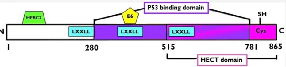
spectrum disorders [15]. The E6-AP protein is 865 amino acids in length [2, 5, 10],
functions as a HECT E3 ubiquitin ligase protein (on the carboxyl terminus), and is
encoded by the UBE3A gene on chromosome 15q11-13 in humans and chromosome 7
in mice [3, 2, 5]. The neuroactivity of the gene is determined by monoallelic expression
of the maternal allele, but its function is dependent on the parent-of-origin, as the
paternal or maternal inherited gene copy may be silent [23]. The gene contains five
functional domains: a p53 binding domain, catalytic HECT domain, an E6 binding
domain, three nuclear receptor response domains (LXXLL) and an activation domain
The catalytic HECTdomain located at the carboxyl terminus of E6-AP, contains two
lobes (C and N), and is roughly 45 kDa in size [15,20]. The catalytic domain is
conserved within the family of HECT E3 ligases [15, 20]. The C-lobe of the HECT
domain contains a catalytic cysteine, which forms a thioester bond with the E2-ubiquitin
complex when the E2 enzyme binds to the N lobe of the domain [15, 20]. The exosite
region of E6-AP, which is a secondary binding site outside of the active site, is critical
for formation of isopeptide bonds and elongation of ubiquitin chains, and can impact
binding of ubiquitin to substrates [15]. E6-AP also contains three Leu-Xaa-Xaa-Leu-Leu
(LXXLL) motifs, which serve as nuclear receptor interaction domains and are important
for protein-protein interactions related to regulation of transcription, activation of steroid
receptors, and regulatory control of proteins such as HPV E6 protein preventing p53
degradation and tumorigenesis [20, 33]. Because of its role in regulation of cellular
activities, modification of the E6-AP protein can alter function within cells, particularly
within the neural system.
[image:10.612.74.540.72.182.2]2. E6-AP & THE UBIQUITIN-PROTEASOME PATHWAY
In a complex with E6-AP, ubiquitin plays an important role in targeting proteins
for degradation within the proteasome of cells. Binding of E6-AP to ubiquitin requires
the linkage of two ubiquitin molecules catalyzed by E2 or E3 enzymes. One ubiquitin
molecule (“donor”) is covalently linked to the active cysteine of the E2 enzyme or bound
to the HECT domain of the E3 enzyme. This allows the donor ubiquitin to attach to one
of seven lysine residues on the accepting ubiquitin molecule [16]. The hydrophilic
region located near Lys-48, is necessary for the acceptor ubiquitin’s to interact with
E6-AP [15]. Binding between ubiquitin and the HECT domain of E6-E6-AP is by a thioester
bond is catalyzed by NEDD4 (neural precursor cell-expressed, developmentally
downregulated 4)-type ligases, a subtype of HECT ligases [15, 20]. Ubiquitination is
highly conserved throughout the HECT protein family [2].
An 18-amino acid region (amino acids 391-408) in E6-AP serves as a binding
site for the E6 protein, and is necessary for the formation of the E6/E6-AP complex [2,
6]. The carboxyl terminal end of E6-AP and ubiquitin’s “canonical” hydrophobic patch
both contain Leu-8, Ile-44, and Val70 residues, which aid the ubiquitin chain elongation
function of E6-AP [2, 14, 15]. After formation of the E6/E6-AP complex, the
hydrophobic patch of ubiquitin is no longer necessary because a conformational change
allows substrates to bind, thus E6-AP serves as an allosteric activator [20].
The E6/E-AP complex uses Lys48 to add polyubiquitin chains to specific proteins
to target them for degradation by proteasomes [2, 6, 15]. Ubiquitination of proteins
requires three cellular enzymes (E1, E2, and E3). E1 functions as a ubiquitin-activator
which forms a thioester bond between cysteine and the C-terminus of ubiquitin [20]. E2
formation of an isopeptide bond between the C-terminus of ubiquitin and a lysine
residue of the targeted substrate [20]. The combined activity of the three enzymes
leads to polyubiquitination of proteins, allowing them to be recognized for degradation
by the 26S proteasome [2, 15]. The Lys48-linked polyubiquitination catalyzed by E6-AP
also serves an important role in stimulation of p97, an ATPase associated with various
cellular activities [16]. P97 recognizes Lys48-linked polyubiquitin chains by an unknown
mechanism, resulting in tagged substrates being transferred to the proteasome [16] and
also plays a cellular role in the breakdown of misfolded proteins within the endoplasmic
reticulum [16]. Weak enzymatic interactions are required for transfer of ubiquitin and
E6-AP recognition of substrates targeted for degradation [15].
Examples of substrates targeted for ubiquitination and degradation by E6-AP
include Mcm-7 (minichromosome maintenance protein 7), p27 (a cell cycle protein
kinase inhibitor), IL- β1 (a cytokine synthesized by mononuclear macrophages), Sox9 (a
gene associated with controlling cartilage generation in early fetal development) [5], and
the p53 tumor suppressor protein [12]. For the P53 protein, E6-AP forms a high-energy
thioester bond between the C-terminus of ubiquitin and the active site of the ligase [15].
A E6-AP-E6-p53 enzyme-substrate complex is formed and additional ubiquitin is added
in the presence of the human papillomavirus (HPV) E6 gene product to target p53 for
degradation by the proteasome [2, 10]. Loss of p53 function is associated with many
cancers including HPV-induced cervical carcinogenesis, as genetically damaged cells
avoid programmed cell death and continue to proliferate, driving cervical cancer [12,
regulation of transcription through the modification of histones and transcription factors
[6, 4].
Altered E6-AP function could influence the ubiquitin pathway of protein
degradation leading to accumulation of proteins within cells, alteration of physiological
processes, and ultimately cause metabolic dysfunction at the cellular level. Because of
the concentration of E6-AP activity in neural cells, this could contribute to the
pathophysiology that causes the clinical symptoms of AS.
INTERACTION BETWEEN HERC2 & E6-AP
The E3 ubiquitin ligase activity of E6-AP is impacted by HPV E6 oncoprotein and
HERC2, a large HECT and RCC1-like E3 ubiquitin protein ligase. The HERC2 protein
contains three RCC1-like (Regulator of Chromosome Condensation 1) domains, and a
HECT domain at its C-terminus allowing for E3 ligase function [14, 17,20]. Both HERC2
and HPV E6 proteins bind near the N-terminus of E6-AP to enhance the protein’s E3
ubiquitin ligase activity [12]. HERC2 serves as an allosteric activator of E6-AP and
drives a conformational change to make the protein more or less active [14]. Mutations
in HERC2, can significantly decrease expression of the HERC2 protein resulting in
dose-dependent reductions in E6-AP function. This interaction has been shown to
result in a neurodevelopmental disease phenotype in humans in an Amish community
that is similar but of reduced clinical severity as compared to AS. These patients
present with hypotonia and behavioral effects, but less extreme language difficulties
were missing part of their posterior corpus callosum, an area of the brain associated
with motor, sensory, and mental functions [17]. As decreased E6-AP levels have been
linked to HERC2 gene mutations that cause enhanced break down of the HERC2
protein, this type of mutation may also be related to AS symptoms [12, 14].
INFLUENCE OF E6-AP ON THE PI3K/AKT PATHWAY
E6-AP is an important regulator of protein kinase cascades, specifically the
PI3K-Akt-GSK3 signaling pathway, which controls functions of the endoplasmic reticulum
actions through phosphorylation thereby promoting neuronal cell growth and survival
during development [20]. PI3K is an important regulator of transcription and translation.
Both PI3K and the MAP kinase pathway are frequently disrupted in neoplastic cells
suggesting a role in promoting carcinogenesis [20]. Although the mechanism its
influence on the activation of the PI3K/Akt pathway is unknown, it has been
hypothesized that E6-AP directly activates RhoA or increases concentrations of reactive
oxygen species that activate the PI3K/Akt pathway. RhoA is a small GTPase that
stimulates actin filament formation and functions in contraction of the myosin, actin
filament ring located between cells during the cytokinesis stage of the cell cycle,
allowing for separation into two individual cells [12]. However, additional studies are
needed to define the mechanistic interaction between E6-AP and the PI3K/Akt pathway
[20].
E6-AP serves an important role in regulating excitatory synapse frequency in the
cortex and hippocampus through ephexin5 [5, 7]. Ephexin5 (E5) inhibits creation of
excitatory synapses and is controlled and targeted for degradation by E6-AP, [7, 4]. E5
function requires binding of EphrinB and EphB (a receptor tyrosine kinase), which
results in the phosphorylation of the N-terminal end of E5 at Tyr-361 [7]. In contrast to
E5, EphB promotes excitatory synapse development [7]. The interaction of E5and
EphBare thought to be important for contact between incoming axons and postsynaptic
dendrites that lead to creation of excitatory synapses during brain development [7].
This can greatly impair learning, memory, and overall development as communication
between neurons is critical for many different processes throughout the human lifespan.
Normally Ephexin5 is able to be degraded by EphrinB, which acts to oppose the action
of Ephrexin5. But, when E6-AP is knocked out in mouse models, EphrinB treatment is
unable to degrade or oppose the action of Ephexin5, thus the disruption in development
of synapses remains [7].
INTERACTION BETWEEN E6-AP & ARC
The E6-AP protein also influences ARC, an important neuro protein vital for
synaptic plasticity, learning, and memory whose transcription is stimulated by the
presence of the steroid hormone estradiol. Arc controls post-synaptic internalization of
AMPA-type glutamate receptors through clathrin-mediated endocytosis [20]. In mouse
models, Arc is expressed at high levels when E6-AP expression is absent [3]. Some
data suggests that Arc can be ubiquitinated and targeted for degradation by E6-AP,
transcription [3, 20]. A role for Arc and E6-AP has been demonstrated in mouse models
for long-term effects on strengthening of synapses between nerve cells and neuronal
synapse firing [4]. The mechanism for how E6-AP may negatively affect transcription of
ARC and its role in the pathogenesis of E6-AP-related diseases has yet to be
determined [3,24]. Many believe that the pathogenesis of Angelman syndrome in
neural tissues is directly related to modification of transcription of Arc and other genes
and the influence of E6-AP on steroid hormone function [20].
INFLUENCE ON E6-AP ON NEURAL FUNCTION
1. E6-AP MUTATION EFFECT ON BRAIN STRUCTURE
Reduced levels or function of E6-AP can alter neural architecture. Microcephaly
is thought to be related to microdeletions of the genomic area of the UBE3A mutation
[17]. Diffusion tensor imaging studies, utilizing MRI-based neuroimaging techniques,
have demonstrated altered architecture within the white matter tracts, thinning of the
corpus callosum, which connects the two hemispheres of the brain and delayed
myelination in AS patients [11]. Myelination of nerve cells, a protein that coats the
axons and enhance transmission of electrical impulses, is disrupted in AS mouse
models. Astrocytes are star-shaped glial cells of the central nervous system located
within the brain and spinal cord. These cells are the most abundant glial cell type in the
brain play an essential role in maintenance of synaptic activity including control of
signaling between neurons, guidance of axons to correct targets, and regulation of the
is the one of the most susceptible cell types of the central nervous system due to the
fact that the high production rate of myelination consumes massive amounts of oxygen
and ATP, forming hydrogen peroxide and reactive oxygen species byproducts [29].
Some of the myelination enzymes require iron as a co-factor, which has the potential to
accumulate leading to free radical formation and lipid peroxidation [29]. Both of which
have the potential to be toxic and damaging to oligodendrocytes. In murine AS models,
oligodendrocytes showed no reduction in expression of paternal E6-AP, suggesting that
UBE3A is not imprinted in oligodendrocytes [11]. However, it is interesting that in
primary astrocyte and oligodendrocyte cultures from murine models of AS, E6-AP
expression occurs [11]. Although astrocytes from AS mice had reduced E6-AP
expression as compared to the wild-type mice, this level of protein expression suggests
that UBE3A is not imprinted in astrocytes [11]. In summary, data suggests that E6-AP
mutations cause decreased expression in astrocytes, modify white matter tracts,
diminish the size of the corpus callosum, and slow the process of myelination within the
central nervous system thereby leading to disruption of normal synapse physiology.
2. ABSENCE OF E6-AP FUNCTION AT THE NEURON LEVEL
The absence the E6-AP function leads to dysfunction at both cellular and
physiological levels that can lead to development of disease symptoms. Lack of E6-AP
function elevates the concentration or continuous existence of substrates normally
targeted for degradation within the proteasome [4], and can promote tumor formation.
ubiquitinated and degraded thereby promoting neoplasia. Reports in the literature have
linked E6-AP disruption to the occurrence of breast, prostate, and cervical cancers [5].
Lack of E6-AP activity can be detrimental on both a cellular and physiological
level. Absence of E6-AP function contributes to reduced formation of excitatory
synapses in the brain (probably through Ephexin5) and impacts dendrite structure
during early neurogenesis causing reduced development of communications between
neurons thereby leading to impaired learning, memory, motor deficits, and lack of
cognitive development. Loss of function can impact transmission of electrical signals in
the brain and alter brain structure. Lack of E6-AP function may also impact nervous
tissues outside the central nervous system as a Drosphilia model demonstrated a
correlation between lack of E6-AP expression and decreased growth and branching of
dendrites in peripheral nerves [11]. It has been hypothesized that decreased levels of
E6-AP reduce the threshold for a seizure response [17].
AS is known to involve genetic mutations resulting in decreased expression or
altered forms of E6-AP within neurons including mutated forms of E6-AP, decreased
activity of E3, or lack of expression of E6-APl [3]. However, little is known about the
control of E6-AP at the post-translational level in neurons [14]. The amount of substrate
proteins utilized by E6-AP within neurons may be important in development of AS [3].
.
3. EFFECTS OF E6-AP MUTATION ON SYNAPTIC TRANSMISSION
AMPA receptors are ionotropic glutamate receptors that control sodium
receptors are normally present in high numbers within the cortex, sensory pathways,
and basal ganglia. However, mutations in E6-AP decrease the numbers of synaptic
AMPA receptors in these areas [18, 4] leading to a negative effect on synaptic plasticity
and long-term potentiation, functions important for learning and memory functions (LTP)
[4,19]. Long-term potentiation is a process in which synaptic connections between
neurons become strengthened with frequent activation due to addition of more AMPA
receptors resulting in the cell becoming more sensitive to glutamate. [4,19]. Addition of
new AMPA receptors is lacking when E6-AP is absent [4,19]. This may be influenced
by the relationship between the loss of E6-AP function and Arc, as the Arc protein is
believed to contribute to reduced synaptic activity during AS [3]. Overall, E6-AP activity
plays a critical role in the development, organization, and maintenance of neuronal
connections in the brain, including experience-dependent plasticity within the cerebral
cortex [23].
4. E6-AP MUTATIONS IMPACT DENDRITIC SPINES AND ACTIN FILAMENTS
Lack of E6-AP reduces the functionality of actin filaments [4], cytoskeletal
filaments that serve an important role in cell adhesion, muscle contraction, cell motility,
cytoskeletal remodeling, and durability of dendritic spines [19]. Lack of E6-AP function
has negative effects on long term potentiation and decreased density of dendritic
protrusions (spines) [4]. Reduced density of dendritic correlates with lower neuronal
excitability and slower excitatory synaptic transmission [26]. These effects could impact
vulnerable during early neuronal development and impaired neural activity can impact
the hippocampus functions [26].
CLINICAL SYNDROMES OF E6-AP
1. AUTISM SPECTRUM DISORDERS
Although many genes and environmental aspects are thought to also play a role,
duplication of the UBE3A gene is one of the most prevalent genetic mutations in Autism
Spectrum Disorders (ASD) [7, 20]. This group of heterogenous neurodevelopmental
conditions predominately affects males and is defined by social interaction
complications, communication difficulties, and the presence of repetitive actions [20].
Experimental studies using mouse and fruit fly models have suggested that autistic
phenotypes are associated with the presence of increased E6-AP function after
duplication or triplication of the UBE3A gene (similar to those that give rise to ASD),
leading to overexpression of E6-AP [12, 20]. Elevated E6-AP levels leads to
ubiquitination and degradation of X-linked inhibitor of apoptosis protein (XIAP) resulting
in impairment of growth and branching in dendrites [25]. As a result, there is increased
caspase activity, lessening of the size and branching ability of the dendrites, leading to
local deterioration and loss of neural function [25]. These autistic patient’s neurons
have difficulty receiving information from neighboring neurons, thereby impacting overall
brain function.
Duplication of the chromosomal region containing UBE3A also occurs in Dup15q
syndrome, a developmental disorder related to autism [12]. This neurodevelopmental
disorder arises from duplications of portions of the 15q11,2-13.1 chromosome that lead
to approximately twice as much expression of UBE3A [4]. The duplication events
predominately originate from the maternal copy of UBE3A during the course of
oogenesis [24] and can be further specified into two categories based on whether the
duplication events are interstitial or isodicentric 15q duplications [24]. The isodicentric
15q duplication contain three copies of the maternal allele and one copy of the paternal
allele, whereas individuals with interstitial duplications have an additional copy of the
15q11.2-q13.3 region within the q arm of the maternal copy of chromosome 15 [24].
Symptoms of this disorder are similar to AS and may include developmental disabilities,
speech impairments, developmental hypotonia, excessive drooling, seizures, and mild
ambulatory problems, although most Dup15q patients have the ability to walk without
assistance [24].
3. PRADER-WILLI SYNDROME
The imprinting properties of UBE3A and the genes that surround it are regulated
by a control center, the Prader-Willi syndrome-AS imprinting center, within an area of
chromosome 15q11-q13 in humans. This center is positioned upstream of the SNURF
(SNRPN upstream reading frame)-SNRPN (small nuclear ribonucleoprotein-associated
protein N) gene [23]. Prader-Willi Syndrome is caused by deletion or imprinting
mistakes in the imprinting region of chromosome 15, causing cognitive and sexual
chromosome 15q is silenced in this disorder whereas in AS, the paternal copy of the
chromosome is deleted [13]. The mechanism for inactivation of chromosome 15 is
thought to be similar for both disorders and both could be the result of uniparental
disomy, depending on the parent of origin for the chromosome [13].
4. RETT SYNDROME
Rett syndrome is brain disorder, most commonly affecting females, that is
X-linked dominant to abnormalities of the MECP2 (Methyl CpG binding protein 2) gene on
chromosome Xp28. Mutations at this locus disrupt gene expression through the binding
of methylated DNA [20, 21]. Symptoms include severe developmental problems after
approximately 18 months of age, stunted growth, language and learning impairments,
coordination problems, and loss of hand function [21]. Other common symptoms
include seizures, scoliosis, and respiratory difficulties [21]. The MECP2 gene is thought
to regulate synapses between neurons thereby influencing communication between
neurons [21]. The influence of E6-AP on MECP2 function has been suggested, but is
not currently proven. Some scientists believe that MECP2 activates UBE3A
transcription [20]. This syndrome is similar to AS in that mutations that result in the
absence of MECP2 function induce lower expression of E6-AP [20]. Although both AS
and Rett syndrome have similar clinical symptoms and neurodevelopmental
impairments, AS is often diagnosed at an earlier age than Rett syndrome.
In contrast to Rett syndrome, MECP2 duplication syndrome occurs almost
exclusively in males. In this syndrome, two copies of the MECP2 gene arise due to a
gene duplication event on the q arm of the X chromosome [22]. These patients have
increased MECP2 protein production which disrupts gene regulation and neuron
activity, resulting in moderate to severe intellectual disability and a broad autism
phenotype [22, 20]. As with Rett syndrome, these patients have difficulties with motor
coordination, breathing, speech, muscle tone, and many individuals also experience
seizures [22]. In contrast to Rett syndrome, individuals with MECP2 duplication
syndrome are more likely to learn to walk, but roughly one third of these individuals will
require assistance [22]. As these patients are prone to respiratory symptoms, half of
affected individuals will not live to see 25 years of age due to susceptibility to recurrent
respiratory infections [22]. It is unknown if E6-AP is affected in this disease. However,
the establishment of autistic attributes is correlated to the duplication of the UBE3A
gene and both genes are important for neurodevelopment [20].
OTHER CLINICAL EFFECTS ASSOCIATED WITH E6-AP
1. E6-AP INFLUENCES REPRODUCTIVE DEVELOPMENT AND STEROID
HORMONE RECEPTORS
E6-AP is involved in reproductive organ development and puberty, and also
functions in cellular signaling pathways and serves as a transcriptional co-activator of
steroid hormone receptors [5,4]. As a steroid hormone receptor co-activator, E6-AP can
enhance activation of androgen, estrogen, and progesterone receptors, as well as
gland, E6-AP can influence steroid hormone receptor cells by blocking apoptosis,
enhancing cell division, and increasing cell growth thereby promoting tumorigenesis [5].
2. RELATIONSHIP BETWEEN AS MUTATIONS & OBESITY
Cases of Angelman syndrome that do not have extensive deletions within the
15q11-q13.3 locus tend to have an abnormal appetite and are prone to morbid obesity.
Symptoms resulting in an excess of fat mass are linked to Prader-Willi syndrome and
some cases of Dup15q syndrome [24]. In fact, Prader-Willi syndrome is presently
considered to be the most prevalent genetic cause of morbid obesity in adolescents
[38]. Obesity affects 600 million people worldwide and is also the leading cause of
morbidity and mortality in individuals with Prader-Willi syndrome [34, 37]. It has been
proposed that uneven amounts of paternal and maternal genetic transcripts in this
region of chromosome 15 could cause problems with maintenance of metabolism [24].
The 15q11-q13.3 region that contains the UBE3A gene also contains several GABA
(gamma aminobutyric acid) receptor subunits, which are associated with the paternal
allele [38]. When the paternal allele is absent, the amount of GABA, an important
inhibitory neurotransmitter associated with control of appetite, metabolism, compulsive
behavior, and memory is decreased [38]. The cellular mechanisms relating obesity to
AS is not known. In general, the lack of motor coordination associated with AS makes it
difficult for individuals with the disease to live an active lifestyle, likely increasing the risk
for development of obesity.
Malignant neoplasms of the cervix or inferior portion of the uterus is often brought
on by a sexually transmitted, human papillomavirus (HPV) infection [20]. All cervical
neoplasms contain a minimum of one integrated copy of the virus’ DNA within the
genome resulting in constitutive expression of early viral genes and production of E6
and E7 oncoproteins [20]. On a molecular level, the presence of HPV E6 oncoprotein
can overpower E6-AP during formation of the E6/E6AP complex. This occurs because
two zinc binding domains located at the N-terminus of the E6 oncoprotein overwhelms
the activity of the LXXLL motif of E6-AP [20] and leads to polyubiquitination and
degradation of p53. It should be noted that p53 is not normally a substrate of E6-AP. In
the absence of p53 activity, proliferation of cancerous cells and prevention of apoptosis
is promoted. There are differences between HPV viruses as high-risk HPV virions
promote the degradation of p53, whereas low-risk forms of the virus produce E6
proteins that can bind to E6-AP but do not degrade p53 [20]. As high activity of E6-AP
is hypothesized to contribute to neoplastic growth, cancer proliferation is suppressed
and apoptosis is promoted when the activity of E6-AP is knocked out [5]. Therefore,
E6-AP could potentially be a good target for cervical cancer treatment [5]. Data
suggests that overexpression of E6-AP is also involved in the growth, proliferation,
Works Cited
1. Daily, Jennifer L et al. “Adeno-associated virus-mediated rescue of the cognitive
defects in a mouse model for Angelman syndrome.” PloS One, vol. 6, no.12,
2011. PubMed Central, www.ncbi.nlm.nih.gov/pmc/articles/PMC3235088.
2. Talis, A. L., et al. “The Role of E6AP in the Regulation of p53 Protein Levels in
Human Papillomavirus (HPV)-Positive and HPV-Negative Cells.” Journal of
Biological Chemistry, vol. 273, no. 11, 1998, pp. 6439-45. Pubmed,
www.ncbi.nlm.nih.gov/pubmed/9497376.
3. Kühnle, Simone, et al. “Role of the Ubiquitin Ligase E6AP/UBE3A in Controlling
Levels of the Synaptic Protein Arc.” PNAS, vol. 110, no. 22, 2013, pp.
8888-8893. www.pnas.org/content/110/22/8888.
4. Bailus, Barbara J., and David J. Segal. “The prospect of molecular therapy for
Angelman syndrome and other monogenic neurologic disorders.” BMC
Neuroscience, vol. 15, no. 76, 2014. doi:10.1186/1471-2202-15-76.
5. Zhou, Xiaofang, et al. “The Function of Ubiquitin Protein Ligase E3A and its
Roles in Human Diseases.” Journal of Biochemistry and Molecular Biology
Research, no.1, 2015, pp.14-18. ResearchGate,
doi:10.6051/j.issn.2313-7177.2015.01.2.
6. Haglund, Kaisa and Ivan Dikic. “Ubiquitylation and cell signaling.” EMBO journal,
vol. 24, no. 19, 2005, pp. 3353-3359. Pubmed Central,
www.ncbi.nlm.nih.gov/pmc/articles/PMC1276169/.
7. Margolis, Seth S., et al. “EphB-mediated degradation of the RhoA GEF Ephexin5
no. 3, 2010, pp. 442-455. Pubmed Central,
www.ncbi.nlm.nih.gov/pmc/articles/PMC2967209/.
8. Clayton-Smith, J., and L. Laan. “Angelman syndrome: a review of the clinical and
genetic aspects.” Journal of Medical Genetics, vol. 40, no. 2, 2003, pp. 87-95.
Pubmed central,
www.ncbi.nlm.nih.gov/pmc/articles/PMC1735357/pdf/v040p00087.pdf.
9. Grier, Mark D et al. “Toward a Broader View of Ube3a in a Mouse Model of
Angelman Syndrome: Expression in Brain, Spinal Cord, Sciatic Nerve and Glial
Cells.” PloS One, vol. 10, no. 4, 2015, doi:10.1371/journal.pone.0124649.
10. Sailer, Carolin et al. “Structural dynamics of the E6AP/UBE3A-E6-p53
enzyme-substrate complex.” Nature Communications, vol. 9, no. 4441, 2018,
doi:10.1038/s41467-018-06953-0.
11. Freeborn, Donna, and Chad Haldeman-Englert. “Uniparental Disomy:
Prader-Willi Syndrome, Angelman Syndrome.” Health Encyclopedia - University of
Rochester Medical Center,
www.urmc.rochester.edu/encyclopedia/content.aspx?ContentTypeID=90&Conten
tID=P02159.
12. Alberts, Bruce. Molecular Biology of the Cell. Garland Science, Taylor & Francis,
2008.
13. Cassidy, SB., and Schwartz, S. “Prader-Willi and Angelman syndromes.
Disorders of genomic imprinting.” Medicine (Baltimore), vol. 77, no. 2, 1998,
14. Mortensen, Franziska et al. “Role of ubiquitin and the HPV E6 oncoprotein in
E6AP-mediated ubiquitination.” Proceedings of the National Academy of
Sciences of the United States of America, vol. 112, no. 32, 2015, pp. 9872-7.
PubMed Central, doi: 10.1073/pnas.1505923112.
15. Ries, Lena K., et al. “Analysis of Ubiquitin Recognition by the HECT Ligase E6AP
Provides Insight into Its Linkage Specificity.” Journal of Biological Chemistry,
2019, doi: 10.1074/jbc.ra118.007014.
16. Li, Wei and Yihong Ye. “Polyubiquitin chains: functions, structures, and
mechanisms.” Cellular and Molecular Life Sciences: CMLS, vol. 65, no.15, 2008,
pp. 2397-2406. PubMed Central,
www.ncbi.nlm.nih.gov/pmc/articles/PMC2700825/.
17. Harlalka, Gaurav V., et al. “Mutation of HERC2 causes developmental delay with
Angelman-like features.” Journal of Medical Genetics, vol. 50, no. 2, 2012, pp.
65-73. BMJ, doi:10.1136/jmedgenet-2012-101367.
18. Rang, Humphrey P. Rang and Dales Pharmacology. Churchill Livingstone,
Elsevier, 2001.
19. Krucker, T. et al. “Dynamic Actin Filaments Are Required for Stable Long-term
Potentiation (LTP) in Area CA1 of the Hippocampus.” Proceedings of the
National Academy of Sciences of the United States of America, vol. 97, no.12,
2000, pp. 6856-61. PubMed Central,
20. Mortensen, F., et al. “Role of ubiquitin and the HPV E6 oncoprotein in
E6AP-mediated ubiquitination.” Proc. Natl. Acad. Sci. vol. 112, no. 32, 2015, pp.9872–
9877. http://d-nb.info/1138566217/34.
21. U.S. National Library of Medicine. “Rett syndrome.”Genetics Home Reference,
16 Apr. 2019, www.ghr.nlm.nih.gov/condition/rett-syndrome#inheritance.
22. U.S. National Library of Medicine. “MECP2 Duplication syndrome.”Genetics
Home Reference, 16 Apr. 2019,
ghr.nlm.nih.gov/condition/mecp2-duplication-syndrome.
23. Sato, Masaaki. “Early Origin and Evolution of the Angelman Syndrome Ubiquitin
Ligase Gene Ube3a.” Frontiers in Cellular Neuroscience, vol. 11, no. 62, 2017,
doi:10.3389/fncel.2017.00062.
24. Lasalle, Janine M., et al. “Epigenetic Regulation of UBE3A and Roles in Human
Neurodevelopmental Disorders.” Epigenomics, vol. 7, no. 7, 2015, pp.1213–
1228. PubMed Central, doi:10.2217/epi.15.70.
25. Khatri, Natasha, et al. “The Autism Protein Ube3A/E6AP Remodels Neuronal
Dendritic Arborization via Caspase-Dependent Microtubule Destabilization.”
Journal of Neuroscience, vol. 38, no. 2, 2018, pp.363-378. PubMed Central,
doi:10.1523/JNEUROSCI.1511-17.2017.
26. Casanova, Jose R., et al. “Impact of Seizures on Developing Dendrites:
Implications for Intellectual Developmental Disabilities.” Epilepsia, vol. 53, no. s1,
27. National Organization for Rare Disorders. “Angelman Syndrome.” National
Organization for Rare Disorders, 2018.
www.rarediseases.org/rare-diseases/angelman-syndrome/.
28. Blackburn, Daniel, et al. “Astrocyte Function and Role in Motor Neuron Disease:
A Future Therapeutic Target?” Glia, vol. 57, no. 12, 2009, pp.1251–1264.
PubMed, doi:10.1002/glia.20848.
29. Bradl, Monika, and Hans Lassmann. “Oligodendrocytes: Biology and Pathology.”
Acta Neuropathologica, vol. 119, no. 1, 2009, pp.37-53. PubMed,
doi:10.1007/s00401-009-0601-5.
30. Gabriel, J. M., et al. “A Model System to Study Genomic Imprinting of Human
Genes.” Proceedings of the National Academy of Sciences of the United States
of America, vol. 95, no. 25, 1998, pp.14857-62. PubMed Central,
www.ncbi.nlm.nih.gov/pmc/articles/PMC24540/.
31. Li, Yufeng, and Hiroyuki Sasaki. “Genomic Imprinting in Mammals: Its Life Cycle,
Molecular Mechanisms and Reprogramming.” Cell Research, vol. 21, no. 3,
2011, pp.466–473. Natureresearch, doi:10.1038/cr.2011.15.
32. Angelman Syndrome Foundation. Angelman Syndrome Foundation,
www.angelman.org/.
33. Mcinerney, E. M., et al. “Determinants of Coactivator LXXLL Motif Specificity in
Nuclear Receptor Transcriptional Activation.” Genes & Development, vol. 12, no.
21, 1998, pp.3357–3368. ColdSpring Harbor Laboratory Press,
34. Kalsner, Louisa, and Stormy J. Chamberlain. “Prader-Willi, Angelman, and
15q11-q13 Duplication Syndromes.” Pediatric Clinics of North America, vol. 62,
no. 3, 2015, pp.587-606. PubMed, doi:10.1016/j.pcl.2015.03.004.
35. Huvenne, Hélène, et al. “Rare Genetic Forms of Obesity: Clinical Approach and
Current Treatments in 2016.” Obesity Facts, vol. 9, no. 3, 2016, pp.158-73.
PubMed, doi:10.1159/000445061.
36. Butler, Merlin G. “Prader-Willi Syndrome: Obesity due to Genomic Imprinting.”
Current Genomics, vol. 12, no. 3, 2011, pp.204-15. PubMed,