REVIEW
Estrogen alpha receptor antagonists
for the treatment of breast cancer: a review
Deepika Sharma, Sanjiv Kumar and Balasubramanian Narasimhan
*Abstract
Background: Cancer is at present one of the leading causes of death in the world. It accounts for 13% of deaths occurred worldwide and is continuously rising, with an estimated million of deaths up to 2030. Due to poor avail-ability of prevention, diagnosis and treatment of breast cancer, the rate of mortality is at alarming level globally. In women, hormone-dependent estrogen receptor positive (ER+) breast cancer making up approximately 75% of all breast cancers. Hence, it has drawn the extensive attention of researchers towards the development of effective drugs for the treatment of hormone-dependent breast cancer. Estrogen, a female sex hormone has a vital role in the initiation and progression of breast malignancy. Therefore, estrogen receptor is the central target for the treatment of breast cancer.
Conclusion: In this review, we have studied various classes of antiestrogens that have been designed and synthe-sized with selective binding for estrogen alpha receptor (ER). Since estrogen receptor α is mainly responsible for the breast cancer initiation and progression, therefore there is need of promising strategies for the design and synthesis of new therapeutic ligands which selectively bind to estrogen alpha receptor and inhibit estrogen dependent prolifera-tive activity.
Keywords: Estrogen receptor alpha, Antiestrogens, Relative binding affinity, Molecular docking, Breast cancer
© The Author(s) 2018. This article is distributed under the terms of the Creative Commons Attribution 4.0 International License
(http://creat iveco mmons .org/licen ses/by/4.0/), which permits unrestricted use, distribution, and reproduction in any medium,
provided you give appropriate credit to the original author(s) and the source, provide a link to the Creative Commons license, and indicate if changes were made. The Creative Commons Public Domain Dedication waiver (http://creat iveco mmons .org/
publi cdoma in/zero/1.0/) applies to the data made available in this article, unless otherwise stated.
Background
Global scenario of breast cancer
According to breast cancer statistics obtained from the global cancer project (GLOBOCAN, 2012), it was observed that 5,21,907 approx deaths cases recorded worldwide in 2012 were due to breast cancer. With the increase in age, the risk for breast cancer and death rates due to it generally increases [1]. The highest incidence of breast cancer was in Northern America and Oceania and the lowest incidence in Asia and Africa. In non-Hispanic white (NHW) and non-Hispanic black (NHB) women the frequency of occurrence and death due to breast cancer are higher than other racial groups. Global differences in the rates of breast cancer are affected by changes in risk factors prevalence and poor diagnosis of it. Adaptation of western lifestyle [2, 3] and delayed childbearing [4,
5] has increased the risk of breast cancer among Asian and Asian American women [2]. The extent of events of breast cancer increases among Hispanic and Hispanic American women especially due to delayed childbearing [2]. In contrast, African countries show approximately 8% new cases of breast cancer; most of the deaths occur due to the limited treatment and late stage diagnosis. According to World Health Organization (WHO 2015) reports, the highest incidence rates of breast cancer were recorded in Malaysia and Thailand [6]. In light of above, in the present review we have covered the role of estro-gen receptor α antagonists as anticancer aestro-gents against breast cancer especially over the past decade as there was no such extensive report is found in the literature.
Role of estrogen alpha in breast cancer
Estrogen, a female sex hormone, related physiological functions are exhibited mostly by the estrogen recep-tors subtypes’ ER-α and β. The estrogen receptor alpha has leading role in uterus and the mammary gland.
Open Access
*Correspondence: naru2000us@yahoo.com
Aromatase enzyme synthesizes 17β–estradiol from andostenindione. This synthesized estradiol (E2) binds to the estrogen receptor which is located in the cytoplasm undergoes receptor dimerization and this estradiol-ER complex translocated into the nucleus where this com-plex further bind to DNA at specific binding sites (estro-gen response element). In response to estradiol hormone binding, multiprotein complexes having coregulators
assemble and activate ER− mediated transcriptional
activity via ER designated activation functions AF1 and AF2 to carry out the estrogenic effects. The deregulation in the functioning of these various coregulators such as alteration in concentration of coregulators or genetic dysfunctionality leads to uncontrolled cellular prolifera-tion which results into breast cancer. Such as loss of the epithelial adhesion molecule Ecadherin leads to metas-tasis by disrupting intercellular contacts. Deregulation of MTA1 coregulator, enhances transcriptional repression of ER, resulting in metastasis. The AIB1 (ERα coregula-tor) get amplified, results in the activation of PEA3-medi-ated matrix metalloproteinase 2 (MMP2) and MMP9 expression which cause metastatic progression. Another ER coregulator SRC-1, has promoted breast cancer inva-siveness and metastasis by coactivating PEA3-mediated Twist expression. In recent study, PELP1 overexpression results into ERα- positive metastasis. Collectively, these studies showed that ERα coregulators modified expres-sion of genes involved in metastasis [7, 8].
Mechanism of action of estrogen alpha receptor antagonists
Endocrine therapy is first choice treatment for the most of the ER+ve breast cancer patients. Currently, three classes of endocrine therapies are widely used.
• Aromatase inhibitors (AIs): Letrozole and anastro-zole decrease the estrogen production by inhibiting the aromatase enzyme thus suppressing the
circulat-ing level of estrogen [8].
• Selective estrogen receptor down regulators (SERDs): Fulvestrant, competitively inhibits estradiol binding to the ER, with greater binding affinity than estradiol. Fulvestrant–ER binding impairs receptor dimeri-sation, and energy-dependent nucleo-cytoplasmic shuttling, thus blocking nuclear localisation of the
receptor [9].
• Selective estrogen modulator: Tamoxifen competi-tively bind with the estrogen receptor and displaces estrogen and thus inhibits estrogen function in breast cells. The co-activators are not binding but, inhibiting the activation of genes that enhance cell proliferation
[8]. The flow diagram of role of estrogen receptor and
estrogen receptor antagonist is as shown in Fig. 1.
Efforts have been aided for estrogen receptor sub-type-selectivity by making changes in the structural configuration of estrogen receptors to develop specific
ER− pharmacophore models. The newly developed
anti-estrogens should not only have good binding affinity with particular receptor but it also must have selective activa-tion for that receptor which expressed in breast cancer progression. Therefore, selective ER α antagonists may be helpful for the breast cancer treatment [10].
Rationale of study
Currently, a number of breast cancer drugs are available in Fig. 2 [11, 12] namely: tamoxifen (i), raloxifene (ii), toremifene (iii) and fulvestrant (iv) but they have follow-ing limitations:
I. Tamoxifen is the drug of choice to treat patients with estrogen related (ER) breast tumors. Resist-ance to tamoxifen develops after some years of treatment due to change in its biocharacter from antagonist to agonist and it is also responsible for
the genesis of endometrial cancer [9].
II. Women who take toremifene for a longer period to treat breast cancer are at higher risk of develop-ment of endometrial cancer.
III. Raloxifene an oral selective estrogen receptor mod-ulator increases the incidence of blood clots, deep thrombosis and pulmonary embolism when taken by breast cancer patients.
IV. Fulvestrant down regulates the ER α but it has poor pharmacokinetic properties i.e. low solubility in water.
Various heterocyclic analogues as estrogen alpha receptor antagonists
Dibenzo[b, f]thiepines analogues
Ansari et al. [13], developed some molecules of
dibenzo[b,f]thiepine and evaluated their
antiprolif-erative potential against ER + ve (MCF-7) cancer cell
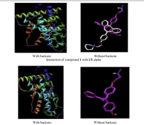
line using MTT assay. Among synthesized derivatives, compound 1, (Fig. 3)] exhibited the potent anticancer activity with IC50 value 1.33 µM against MCF-7 tumor cell line, due to arrest in G0/G1phase of cell cycle. Molecular docking studies carried out by MGL Tools 1.5.4 revealed that the tricyclic core of the compound
1 occupied the same binding space in the ER-α pocket
as tamoxifen. The most active compound 1 showed
significant homology with tamoxifen while interacting with amino acids (GLY390, ILE386, LEU387, LEU391,
LEU403, GLU353, LYS449 and ILE326) of ER-α but the
to that of tamoxifen (Fig. 4). Thus, it showed that com-pound 1 exhibited the better binding affinity with ER alpha as compared to tamoxifen (9.6 ± 2.2 µM) and this improved binding might be responsible for good anti-estrogenic potential.
Diphenylmethane skelon
Maruyama et al. [14], synthesized some derivatives of diphenylmethane as estrogen antagonist that would bind to the estrogen receptor similar as estradiol. The antagonistic activity of synthesized derivatives was
i
ii
iii
iv
Fig. 2 Marketed drugs for breast cancer
1
2
(3-7)
8
9
10
evaluated by AR reporter gene assay. Among the
syn-thesized compounds, compound 2, [4,4′
-(heptane-4,4-diyl)bis(2-methylphenol) (Fig. 3)] was found to be potent one and displayed 28-times more selectivity for estrogen receptor alpha (IC50= 4.9 nM) over estrogen receptor beta (IC50= 140 nM). The binding interactions
of compound 2 were determined computationally using
AutoDock 4.2 program into ER-α (PDB ID: 3UUC).
Docking study showed that phenol group of compound 2 interacted with the amino acid E353 of ER-α through
H-bonding and the bulky side chain (n-Propyl)
pre-sent at the central carbon atom of bisphenol A directed towards the amino acid M421 of ER-α.
SAR: Thus, introduction of alkyl chains at central car-bon atom switched it from agonist to antagonist and presence of two methyl groups at the 3 and 3′-positions improved the antagonistic activity and selectivity for ER-α over ER-β (Fig. 5).
Conjugated heterocyclic scaffolds
Parveen et al. [15], developed new conjugates of pyrimi-dine-piperazine, chromene and quinoline. Antiprolifera-tive activity of the synthesized conjugates was determined against (MCF-7) tumor cell line using MTT assay.
Among these conjugates, compound 3,
(2-(4-(2-methyl-With backone
Without backone
Interaction of compound
1
with ER alpha
With backone
Without backone
Interaction of tamoxifen with ER alpha
6-((4-p-tolyl-1,4-dihydroquinolin-7-yloxy)methyl) pyridin-4-yl)piperazin-1-yl) ethanol), 4, (2-(4-(2-methyl-6-((4-phenyl-1,4-dihydroquinolin-7-yloxy)methyl) pyridin-4-yl) piperazin-1-yl ethanol), 5, (2-(4-(2-methyl-6 - ( ( 4 - p h e n y l - 4H- c h r o m e n - 7 - y l o x y ) m e t h y l ) pyridin-4-yl)piperazin-1-yl)ethanol), 6, (2-(4-(2-methyl-6 - ( ( 4 - ( 4 - n i t ro p h e ny l ) - 4H- c h ro m e n - 7 - y l ox y ) methyl)pyridin-4-yl)piperazin-1-yl) ethanol) and 7,
(2-(4-(2-methyl-6-((4-p-tolyl-4H-chromen-7-yloxy)
methyl)pyridin-4-yl)piperazin-1-yl)ethanol) showed good anti-proliferative activities as compared to standard
Fig. 5 Structure activity relationship study of compound 2
Table 1 Anticancer activity (IC50= µM) results of conjugates 3–7
Compound No. Cancer cell
line MCF-7
3 48 ± 1.70
4 65 ± 1.13
5 92 ± 1.18
6 30 ± 1.17
7 16 ± 1.10
Curcumin 48 ± 1.11
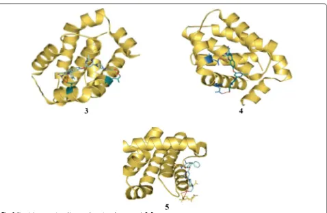
curcumin (Table 1, Fig. 3). Molecular docking of most active compounds 3, 4 and 5 against 3D structure of Bcl-2 protein was performed using Autodock 4.2 (Fig. 6). The Lamarckian genetic algorithm (LGA) was applied to study the protein-ligands interactions. The p-tolyl
pre-sent in compound 3 and phenyl group present in
com-pound 4 formed three hydrogen bond one with amino
acid Asp100 and two with amino acid Asp108
respec-tively. The chromene ring in compound 5 formed four
hydrogen bond with Glu133, Ala146, Arg136 and Asp137 with good binding interaction having binding energy (∆G) − 7.70 kcal/mol, Ki = 2.26 µM). The most favora-ble binding within the active sites of BCL-2 was shown
by compounds 3 and 4 with minimum binding energy
(∆G) =−9.08 kcal/mol and (∆G) =− 8.29 kcal/mol, respectively.
SAR: Structure–activity relationship study showed that the anticancer potential improved when chromene and quinoline nucleus combined with piperazine and pyrimi-dine rings.
Aromatase inhibitors/selective estrogen receptor modulator
Zhao et al. [16], designed and synthesized selective estrogen receptor modulators (SERMs) based on diphe-nylmethylene scaffold by incorporating some of the
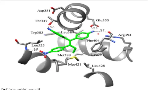
structural features of the aromatase inhibitor letro-zole into lead compound (norendoxifen) by bis-Suzuki coupling to generate a series of selective anti-breast cancer agents to address the problem of E, Z isomeri-zation related with norendoxifen. The functional cel-lular assay method was employed on MCF-7 cancer cells to evaluate the aromatase inhibitory potential indi-cated that compound 8, (Fig. 3) was the most active one (IC50= 62.2 nM). The binding pattern of the most active one (8) was determined using docking software GOLD3.0
In compound 8, the amino substituent present on the
phenyl ring that is cis conformation to the nitrophenyl nucleus formed H- bond with the OH group of Thr347 while the other amino substituent formed H-bond to the carboxylate of amino acid Glu353 and the backbone
bonded to the carbonyl of Phe404 of ER-α (PDB-3ERT)
as shown in Fig. 7. The binding affinity of compound 8 for both ER-α and ER-β was found to be (EC50= 72.1 nM) and (EC50= 70.8 nM), respectively.
Furan derivatives
Zimmermann et al. [17], prepared estrogen
antago-nists by incorporating side chains having amino or sul-fur functional groups linked at 3rd position of sul-furan for the breast cancer therapy. The synthesized furan deriva-tives were determined for their anticancer potential
against MCF-7/2a breast cancer cells line. The degree of alpha selectivity increased from 2.5 to 236 times when alkyl group attached at 4th position of furan nucleus. Especially, compound 9, (4,4′ -(3-ethyl-4-(6-(methyl(3-(pentylthio)propyl)amino)hexyl)furan-2,5-diyl) diphe-nol showed the strongest antiestrogenic effect (Table 2, Fig. 3). It was found that 2,5-bis(4-hydroxyphenyl)furans with two short alkyl chains have better binding interac-tions with ER α than that for ER β.
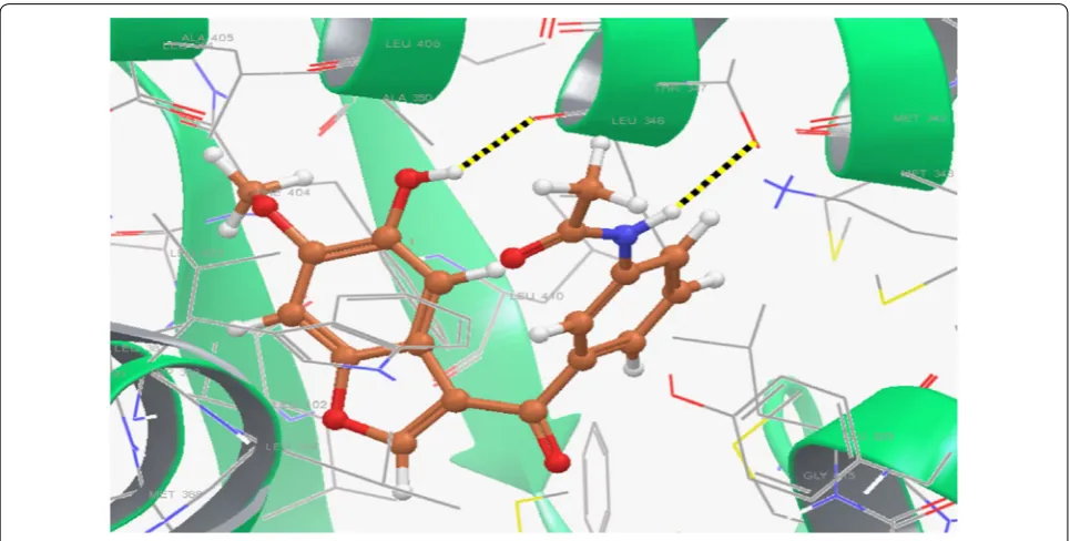
Li et al. [18], prepared new library of 3-acyl-5-hydroxy-benzofuran derivatives by microwave-assisted method and evaluated its antineoplastic potential against MCF-7 cell line. Compound 10, [(N
-(3-(5-hydroxy-6-methoxy-benzofuran-3-carbonyl)phenyl) acetamide), (Fig. 3)]
exhibited promising antineoplastic activity against MCF-7 (IC50= 43.08 µM) compared to tamoxifen using as positive control as evaluated by MTT assay. A quan-tum mechanics polarized ligand docking (QPLD) study using (PDB code: 1A52) was carried out to interpretate
the binding mode between the synthesized molecules
and ER-α using Schrödinger Suite 2010. Structural
analysis of the most active compound 10 showed that
(Fig. 8) it bound to amino acid residues 5-OH/Leu346, N–H/Thr347 of ER-α through H-bonding (− 1.297 kcal/ mol) and formed pi–pi conjugate interactions with the benzofuran nucleus and amino acid Phe404. Thus,
compound 10 showed the best calculation score (G
score =− 10.138 kcal/mol) as compared to other synthe-sized derivatives.
Coumarin conjugates
Kirkiacharian et al. [19], synthesized a library of estro-gen antagonists based on coumarin scaffold with various substitution patterns and their relative binding affinities (RBA) were evaluated for estrogen alpha and beta recep-tor in Cos cells. Anticancer results showed that com-pounds substituted at position 3rd and 4th with phenyl group have higher selectivity for ER-α than ER-β. In this
study, compound, 11,
[(3,4-diphenyl-7-hydroxycou-marin), (Fig. 9)] showed 13.5 times higher selectivity for estrogen alpha receptor than estrogen beta receptor.
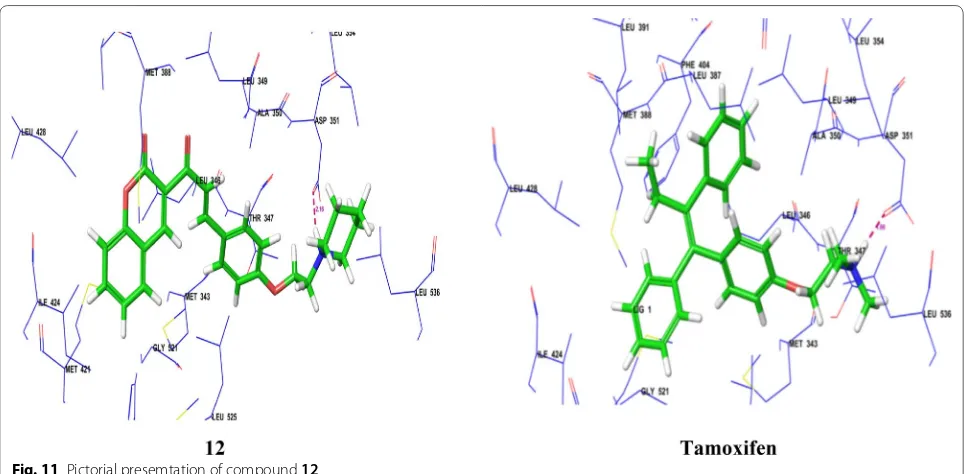
Mokale et al. [20], synthesized a class of coumarin-chalcone hybrids by fusing various pharmacophores and determined their antineoplastic activity against MDA-MB-435 MCF-7 breast cancer cell lines using
Sul-forhodamine B assay. The compound 12, showed
high-est antineoplastic potential compared to standard drug (tamoxifen). Anticancer potential demonstrated that the Table 2 Antiestrogenic and antiproliferative activity
of compound 9
Compound No. (IC50= µM)
Antiestrogenic activity Antiproliferative activity (MCF-7)
9 0.050 0.022
Fulvestrant 0.003 0.004
compound having amine side chain with piperidine ring have good binding affinity (Table 3, Figs. 9 and 10). Dock-ing study was performed usDock-ing Glide v5.8 (SchrödDock-inger, LLC) to explore binding interactions of synthesized com-pounds with estrogen receptor alpha. Coumarin nucleus
and 4-ethoxy piperidine side chain of compound 12
interacted deeply within the hydrophilic pocket of ER-α
and formed strong H-bonding with Asp351 similar to standard tamoxifen and raloxfiene (Fig. 11). In addition,
11
12
13
14
15
16
17
18
19
Fig. 9 Molecular structures of compounds (11–19)
Table 3 In vitro antiproliferative activity (IC50 = µg/ml) of compound 12
Compound No. Cancer cell lines
MCF-7 MDA-MB-435
12 LC50 TGI GI50 LC50 TGI GI50
74.5 40 < 10 > 80 78.2 75.3
Tamoxifen 29.5 11.2 < 10 54.2 21.5 < 10
compound 12 also showed pi–pi stacking interactions with Phe404 similar to tamoxifen.
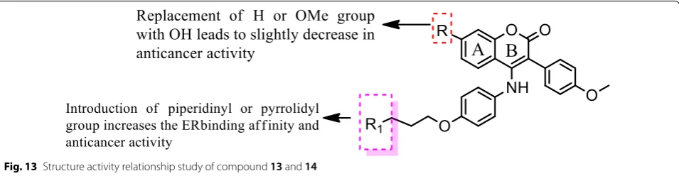
Luo et al. [21], prepared new class of chromene deriva-tives as potential selective antagonists for ER subtypes.
The anticancer results indicated that piperidyl substi-tuted compounds, 13 and 14 exhibited potent antineo-plastic activity against MCF-7 and Ishikawa tumor cell lines by MTT assay and showed good ER-α binding affin-ity (Table 4, Fig. 9). Molecular docking, a deeper binding mode analysis was performed on the promising com-pounds 13 and 14 having structural diversities on the C-7 position of coumarin skeleton using Discovery
Stud-ies 3.0/CDOCKER protocol targeting ER-α. The basic
side chains of compounds 13 and 14 pointed toward
Asp351 to generate an antagonistic conformation simi-lar to Tamoxifen as shown in (Fig. 12). The two methoxy
groups containing compound 13 formed two hydrogen
Fig. 11 Pictorial presemtation of compound 12
Table 4 In vitro anticancer results of 13–14
Compound No. Tumor cell lines (IC50= µM)
MCF-7 Ishikawa
13 4.52 ± 2.47 11.58 ± 3.81 14 7.31 ± 2.12 8.43 ± 1.06 Tamoxifen 11.35 ± 3.13 16.47 ± 2.04
bonds with Arg394 and His524, respectively. The plausi-ble binding mode of 14 was that it formed two H- bonds with Glu353 and Arg394 amino acid residues in the hinge region of estrogen receptor alpha through 7-OH.
SAR: From this series, compound 14 containing
hydroxyl group displayed the best ER-α binding affin-ity (RBA = 2.83%), while compound 13 bearing methoxy
group displayed the best in vitro antineoplastic potential against MCF-7 carcinoma cell line (Fig. 13).
Inverse agonist
ERR α is the orphan nuclear receptor (ONR) which is
identified homologous to estrogen receptor alpha at
DNA-binding domain, indicated that ERR α inflect the
actions of estrogen alpha receptor. Thus, ERR α act as a prognostic marker in breast malignancy.
Ning et al. [22], synthesized a novel compound as a selective inverse agonist of estrogen-related receptor and determined for its anticancer activity against triple nega-tive breast cancer cells (MDA-MB-231) and found that
compound 15
[(1-(4-(methyl-sulfonamido)-2,5-diprop-oxybenzyl)-3-(3-bromophenyl)urea), (LingH2-10), (Fig. 9)] as a potential ligand that selectively inhibited
the ERR α transcriptional activity and inhibited the can-cer cell growth both in vitro and in vivo. The 3D docking simulations of compound 15 (LingH2-10, Fig.14) demon-strated within the binding pocket of ERR α using surflex-dock geomx program (Sybyl X2.0). The 3-bromo-phenyl group in LingH2-10 occupied the position interacted with the receptor ERR through hydrophobic interac-tions. One of the amino in the ureido group in LingH2-10 formed H- binding interaction with the residue Gly397
of ERR α receptor. The methane sulfonamide group at
the end of LingH2-10 stretched downwards into the cav-ity formed by the residues Phe495 and Gly397 possibly with some polarity interactions. In order to carry out the in vivo studies, breast tumor xenografts were developed
in nude mice. The 10 doses of compound 15 (30 mg/kg)
were given on alternate days. After the treatment, the results demonstrated that there is 42.20% inhibition of tumor growth such as in mice the volume of tumor in treated xenografts was 810 mm3 while in control it was
1397 mm3. These results demonstrated that the
com-pound 15 might act as lead molecule.
Steroidal analogs
Alsayari et al. [23], synthesized a new class of estrone based analogs were investigated for their anticancer activity using MTT assay. Compounds, 16 and 17 (Figs. 9 and 15) exhibited significant inhibitory estrogenic pro-file. In silico molecular docking simulations carried out by competitive binding assay revealed that compound 16 has very similar binding mode (IC50= 5.49 µM) to estradiol (IC50= 0.0069 µM) on estrogen alpha receptor through H-bonding interaction between the methoxy group present at 3rd position in steroidal nucleus and amino acid residue in ARG: 394.
Reseveratrol (phytoestrogen) analogs
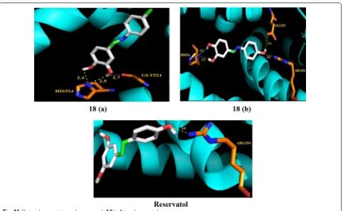
Siddqui et al. [24], synthesized a library of reseveratrol analogs and evaluated its anticancer potential against T47D, MDA-MB-231 breast tumor cells using MTT
Fig. 13 Structure activity relationship study of compound 13 and 14
assay. The molecular docking study showed the binding pattern of aza-resveratrol analogs with estrogen recep-tor alpha indicated the presence of additional hydrogen bonding and tight binding interactions with active sites of protein cavity of estrogen receptor alpha. Among the synthesized compounds, 18 (a, ((E)-4-(1-(p-tolylimino) ethyl)benzene-1,2-diol) and (b, ((E )-4-(1-(4-hydroxy-phenylimino)ethyl)benzene-1,2-diol)) exhibited potent
Fig. 15 Visual presentation of compound 16 and 17 with receptor ER α. Dotted red lines show the hydrogen bond formation
Table 5 Anticancer activity (IC50= µM) results of reseveratrol analogs 18 (a and b)
Compound No. Cancer cell lines
MDA-MB-239 T47D
a 21 32
b 29 44
Resveratrol 66 76
antibreast cancer activity as compared to resveratrol against both cell lines (Table 5, Fig. 9). The anticancer results demonstrated that incorporation of the imino-group in the parent resveratrol enhanced its anticancer
potential. Molecular docking of the most active synthe-sized resveratrol analogs a and b was performed in estro-gen receptor alpha protein cavity to observe their binding pattern as shown in Fig. 16. The vicinal hydroxyl groups
on ring A of compound b undergo H-bonding with
HIS524 residues while methyl group interacted with ARG394 and GLU354 residues, respectively. The 3, 4-dihydroxyl groups on ring A in compounds 18 (a and
b) favored Van der Waals interactions with amino acid
residues in the ER-α protein leading to stabilization of these ligands into the protein cavity. Compounds 18 (a
and b) displayed potent activity against MDA-MB-231
(with 65–75% cytotoxicity) and T47D cells (with 40–60% cytotoxicity), while resveratrol induced only 40% cyto-toxicity against both tested cell lines.
Resveratrol, a natural phytoestrogen, have potent anti-neoplastic properties but its poor efficacy and bioavail-ability have limited its clinical applications. In order to overcome these difficulties, Ronghe et al. [25] synthe-sized aza-resveratrol analogs and tested for their antineo-plastic activity against MDA-MB-231, T47D and MCF-7 breast tumor cells using MTT assay. The in vitro antican-cer results showed that compound 19, [4-(E)-{(p-tolyl imino)-methylbenzene-1,2-diol}, Figs. 9 and 17] showed better anticancer properties than parent resveratrol [19].
Triarylethylene analogs
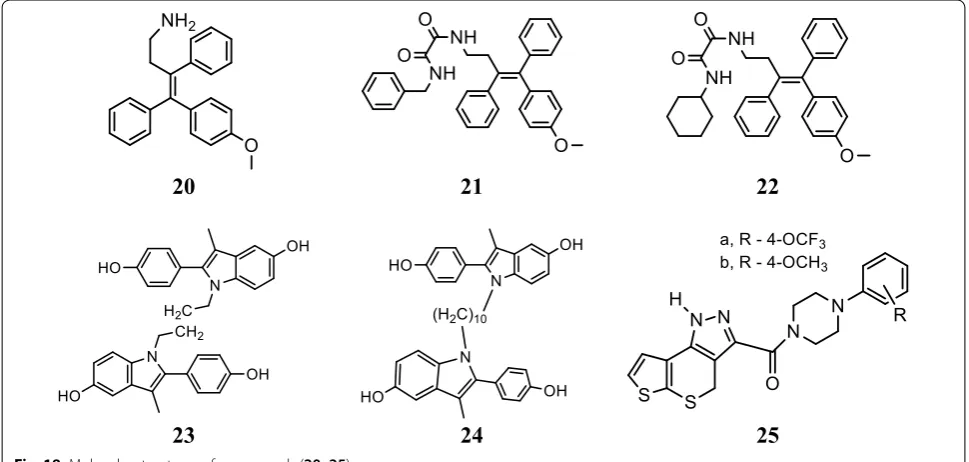
Kaur et al. [26], developed novel derivatives of tri-arylethylene and determined their in vitro cytotoxic
potential against ER− (MDAMB-231) and ER+
(MCF-7) human breast cancer cell using MTT assay.
Fig. 17 Pictorial pesentation of compound 19
Table 6 Cytotoxicity (IC50= µM) of triarylethylene analogs (20–22)
Compound No. Cancer cell lines
MDA-MB-231 MCF-7
20 11.4 ± 4.2 16.9 ± 7.7
21 16.9 ± 7.7 > 50
22 12.2 ± 5.3 > 50
Tamoxifen > 50 50
Ospemifene > 50 > 50
20
21
22
23
24
25
Compounds 20, 21 and 22 displayed better anticancer activity than standard drug (tamoxifen, ospemifene) (Table 6, Fig. 18). Especially, compound 20 suppressed the expression of c-Myc, MMP-9 and caveolin in both MDA-MB-231 and MCF-7 cells. In silico, docking sim-ulations performed using the CDocker docking
algo-rithm indicated that compound 20 have good binding
affinity with estrogen receptors (ERs).
SAR: The structure activity relationship study dem-onstrated that the presence of amino or oxalamido sub-stituents on 20, 21 and 22 increases the potency and selectivity against both ER− and ER+ tumor cell lines.
Indole derivatives
Kelley et al. [27], prepared a library of selective estro-gen receptor modulators based on the 2-arylindole scaffolds to selectively target the estrogen receptor in hormone positive breast cancers (MCF-7). Among
the synthesized compounds, compounds 23 and 24
(Table 7, Fig. 18) demonstrated strong estrogen recep-tor (ER) binding (Fig. 19) as evaluated by Fred 3.0.1. and also exhibited good anticancer potential in ER responsive MCF-7 cell with minimal residual effects as evaluated by AlamarBlue assay.
Pyrazole derivatives
Sun et al. [28], synthesized a new class of 1,4-dihy-drothieno[3′,2′:5,6]thiopyrano[4,3-c ]pyrazole-3-car-boxylic amides and assessed their anticancer potential against MCF-7 tumor cell line by MTT method and compared to positive control (tamoxifen). Among the target compounds, compounds 25 (a and b) were found to be more active against selected cell line (Table 8, Fig. 18).
SAR: The structure activity relationship study showed that compounds 25 (a and b) having substitution (OCF3 and OCH3) at 4th position of benzene ring plays a vital role in antitumor activity.
Stauffer et al. [29], developed a new class of pyrazoles and evaluated their antiproliferative activity by cell-based transfection assay. N-piperidinyl-ethyl chain was introduced at all the four sites of substitution on the pyrazole ring to observe the binding mode in the ER
ligand binding pocket. Piperidinyl-ethoxy-substituted pyrazole at 5th position of 26 (Fig. 20)] was found to be the most active one (IC50= 20 nM) against lamb uter-ine cytosol. Docking studies carried out using Flexi-dock routine within SYBYL 6.5.2 demonstrated that compound 26 (Fig. 21) showed 20-fold higher selectiv-ity and binding affinselectiv-ity for ER-α (11.5 ± 1) than ER-β
(0.650 ± 0.02).
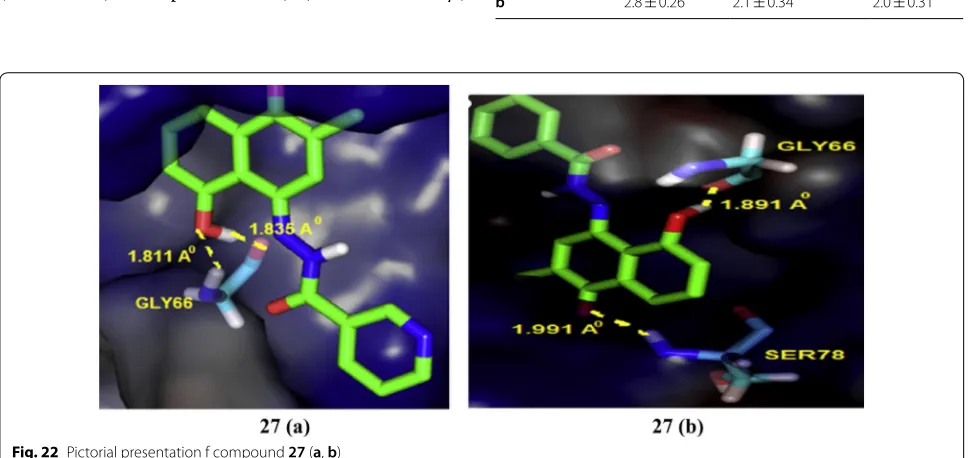
Hydrazones
Dadwante et al. [30], prepared plumbagin hydrazonates and screened for their cytotoxic potential against MCF-7
(ER+ ve) and triple negative MB-231and
MDA-MB-468 breast tumor cell lines by MTT assay. The hydroxyl group of plumbagin was found to be essential for the inhibition of histone acetyltransferase activity of p300/CBP, which is a transcriptional activator of ER-α.
In particular, compound 27 (a
(5-hydroxy-2-methyl-4-(2-(1-(pyridin-2-yl)vinyl)hydrazono) naphthalen-1(4H )-one)) and (b (5-hydroxy-2-methyl-4-(2-(1-phenylvinyl)
hydrazono) naphthalen-1(4H)-one)) was found to be
more effective in inhibiting NF-ḵB expression. Molecu-lar docking studies carried out with the help of Auto-dock 4.0 to analyze ligand interactions (Fig. 22) with the crystal structure binding site of p50-NF- ḵB obtained from PDB ID (1NFK) demonstrated that OH-groups on plumbagin and hydrazonate side chain favor additional Table 7 Anticancer results (IC50 = µM) of indole analogs
(23–24)
Compound No. Cancer cell
line MCF-7
23 2.71
24 1.86
Fig. 19 Pictorial presentation of compound 23 and 24
Table 8 Cytotoxic results of pyarzole derivatives 25 (a and b)
Compound No. MCF-7 cancer cell line
Inhibition rate % IC50= µmol/L
a 71.09 90.63
b 88.86 72.55
H-bonding with amino acid which may be responsi-ble for the improved anticancer potential. The binding energies were in the range of − 7.43 to − 7.88 kcal/mol
which are greater than that of the parent plumbagin com-pound, indicated strong binding interactions in the active site of p50-subunit of NF-ḵB protein enhanced through H-bonding interaction with GLY66 and HIS64 amino acid, respectively (Table 9, Fig. 20)
Isoquinoline derivatives
Tang et al. [31], synthesized and structurally character-ized a series of 6-aryl-indeno isoquinolone inhibitors
targeting ER α to improve efficacy as compared to
tamox-ifen. The synthesized derivatives presented good ER α
binding affinity and antagonistic activity and also showed excellent anticancer activity against MCF-7 using MTT assay. In this series, compound 28, (Fig. 20)] exhibited promising anticancer activity (IC50= 0.5 µM) which is 27-times greater anticancer potential than the reference drug tamoxifen (IC50= 13.9 µM). Docking studies car-ried out with Discovery Studio.2.5/CDOCK protocol to explore binding pattern of compound 28 in ER-α indi-cated that compound 28 favorably docked with the active sites of ER-α (Fig. 23). The hydroxyl group present at 9th
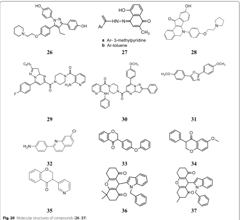
26
27
28
29
30
31
32
33
34
35
36
37
position in 28 interacted with Glu353 and Arg394 which imitate with the A-ring phenol of estradiol while the hydroxyl group at 3rd position interacted with His524 with similar binding mode as 17β-OH of estradiol. The basic side chain of 28 was oriented to Asp351 such as to generate antagonistic conformation similar to tamoxifen.
Anilinonicotinyl linked pyrazolo[1,5-a]pyrimidine conjugate
A library of aniline nicotinyl linked pyrazolo[1,5-a] pyrimidine conjugates was prepared by Kamal et al.
[32] and evaluated against MCF-7 cancer cell line
using MTT assay and compared to standard drug
(doxorubicin). Compound 29, (4-(2-aminonicotinoyl)
piperazin-1-yl)(7-(4-fluorophenyl)-2-phenyl-3,3a
-dihydropyrazolo[1,5a]pyrimidin-5-yl) methanone) and
compound 30, ((7-(4-methoxyphenyl)-2-phenyl-3,3a
-dihydropyrazolo[1,5-a
]pyrimidin-5-yl)(4-(2-(phe-nylamino)nicotinoyl)piperazin-1-yl)methanone), (Table 10, Fig. 20) possessed significant antiprolifera-tive potential against breast carcinoma cells (MCF-7) by affecting interaction between ERE–ER α.
Bis(hydroxyphenyl)azoles
Bey et al. [33], synthesized bis(hydroxyphenyl) azoles and evaluated as selective non-steroidal inhibitors of
17β-HSD1 for the therapy of estrogen-dependent
dis-eases and the molecular docking was carried out by automated docking program GOLD 3.0, the docked
com-pound 31 shown as yellow within 17β-HSD1-binding
pocket (green amino acids) (Fig. 24). In this series, com-pound 31, [(IC50= 0.31 µM), (Fig. 20)] showed good anticancer potential with higher selectivity for ER α with regard to 17β-HSD2 as evaluated by cell free assay. The
p-hydroxyphenyl substituent lay in the same plane while
m-hydroxyphenyl substituent of compound 31 laid 32o out of this plane, respectively. This conformation allowed 31 to create H-bond interactions (shown by violet lines in Fig. 24, distances were expressed in Å) with His221/
Glu282 and Ser142/Tyr155 with p-hydroxyphenyl
nucleus and m-hydroxyphenyl nucleus, respectively.
Fig. 21 Pictorial presentation of compound 26
Fig. 22 Pictorial presentation f compound 27 (a, b)
Table 9 Anticancer results of compounds 27 (a and b)
Compound No. Tumor cell lines (IC50= µM ± S.E.)
MCF-7 MDA-MB-231 MDA-MB-468
Quinoline analogues
A novel library of quinoline-based analogs was synthe-sized by microwave assisted method and its antican-cer activity was evaluated against ER α positive human
cancer cells by Bharathkumar et al. [34]. Among the
synthesized compounds, compound 32,
[(4-(7-chloro-quinolin-2-yl)benzenamine), (Fig. 20)] hold significant
antineoplastic potential. Compound 32 displayed
sig-nificant anticancer potential against HepG2 and MCF-7 tumor cells having IC50 value of 6 µM and 11 µM, respec-tively. The structure activity relationship study of com-pound 32 as displayed in Fig. 25.
Isoflavone derivatives as aromatase inhibitor
Bonfield et al. [35], designed and synthesized 3-phenyl-chroman-4-one (isoflavone) derivatives and evaluated their anticancer potential by fluorescence-based assay using recombinant human aromatase using ketoconazole as positive control. Compounds, 33, 34 and 35 (Table 11, Figs. 20 and 26) displayed effective inhibitory activity against aromatase. Docking study was carried out using program GOLD (version 5.0.1.) to observe H-bonding and hydrophobic interactions.
SAR: The structure activity relationship results showed
that presence of functional groups (-OCH3 (34), -OPh
(33) and C6H-5N (35)) displayed good inhibitory activi-ties against aromatase, showing that the non-planarity configuration of the isoflavanone analogs might play vital
role in enzyme–ligand binding. Compound 34 having
methoxy substitution at 6th position of coumarin nucleus was found to be the most active one.
Singla et al. [36], synthesized indole-xanthendione ana-logs and screened their anticancer potential and estrogen receptor alpha binding affinity utilizing ER α responsive T47D breast cancer cell line. Compounds 36 and 37 dis-played most promising anticancer potential targeting on ER-α (Table 12, Fig. 20). RT-PCR and Western blotting experiments indicated that these derivatives 36 and 37 exhibited their anticancer activity by altering the m-RNA and ER-α receptor expression, thus inhibiting further transactivation and signaling in T47D cancer cells. Gli-deXP (Glide Extra precision) with vdW scaling 0.8 was
Fig. 23 Pictorial presentation of compound 28 in ER alpha (a) and tamoxifen in ER alpha (b)
Table 10 Anticancer potential of pyrazolo[1,5-a] pyrimidine conjugate (29–30)
Compound No. IC50= µM
MCF-7 Cancer cell line
29 1.79
30 2.16
Doxorubicin 0.473 µM
employed to carry out molecular docking and then ranked them based on the GlideXP score. Induced fit simulation was employed to analyze the binding pattern of compounds 36 and 37 with estrogen receptor alpha (PDB: 4XI3) and it showed that these compounds bind in the shallow binding site of the ER-α receptor in simi-lar docking pose as that of the bazedoxifene with strong binding affinity of − 12.51 kcal/mol and − 12.06 kcal/
mol, respectively that is comparable to the bazedoxifene (− 9.33 kcal/mol). The indole moiety present in
com-pounds anchored the xanthendione nucleus in the hydro-phobic cavity. These compounds showed hydrogen bond interaction with Arg 394, Lys 529 and Asn 532, respec-tively (Fig. 27). Compounds 36 and 37 showed extensive Van der Waals forces of interaction with various amino acids listed in Table 12.
SAR: Further, from the structure activity relationship studies it was concluded that increasing the substitution at xanthendione moiety decreases the anticancer activity of the synthesized derivatives.
Singla et al. [37], synthesized indole benzimidazole hybrids to develop novel selective estrogen receptor modulators and investigated their antibreast cancer potential via ER-α (+) T47D cariconoma cell line using MTT assay. From these hybrids, bromo substituted
compounds, 38 and 39 were found to be most
effec-tive in targeting ER-α. RT-PCR and Western blotting
experiments results showed that both the hybrid com-pounds 38 and 39 altered the mRNA and ER-α recep-tor protein expression, thus preventing the further
transcriptional activation and signaling pathway in can-cer cell line (Table 13, Figs. 28 and 29). GlideXP (Glide Extra precision) with vdW scaling 0.8 was used to carry out molecular docking and ranked them based on the GlideXP score. Induced fit simulation was employed to anlayse the binding interaction pattern of both the
compounds with receptor ER-α (PDB: 4XI3) and it
showed that these derivatives bind in the shallow bind-ing site of the ER-α receptor in similar docking pose as that of the bazedoxifene with strong binding affinity of − 12.51 kcal/mol and − 12.06 kcal/mol respectively that is comparable to the bazedoxifene (− 9.33 kcal/ mol). These compounds showed H-bond interaction with Asp 351, Leu 346, Asn 532, Val 533, respectively.
Compounds 38 and 39 showed extensive van der Waals
forces of interaction with various amino acids listed in Table 13.
Perron et al. [38], synthesized two new molecules of 17β-estradiol-linked platinum (II) complexes by linking alkyl chain at position 16th of the steroid nucleus. The anticancer potential of these prepared derivatives was determined on estrogen dependent and independent (ER+ and ER−) human breast tumor cell lines: MCF-7
and MDA-MB-231. by Sulforhodamine B colorimetric assay. The compound 40, (Fig. 28) showed potent cyto-toxicity against both tumor cell line and also displayed high affinity for ER-α as evaluated by HitHunter EFC Estrogen Fluorescence assay kit.
Lappano et al. [39], synthesized indole derivative, com-pound 41 (Fig. 28) and its anticancer properties were exerted through ER-α and GPER receptor in breast can-cer cells as determined by RT-PCR, western blotting assay. The simultaneous antagonistic action exhibited
on both GPER and ER-α by 41 showed a new
pharma-cological approach for targeting breast tumors which express one or both receptors during cancer progres-sion. Docking study carried out with the help of GOLD 5.0.1., program using a genetic algorithm illustrated that compound 41 bind to ER-α in similar manner as OHT as shown in Fig. 30.
Mortensen et al. [40], developed a library of
3-alkyl-2,4,5-triarylfurans derivatives whose selectiv-ity for ER alpha receptor increased due to presence of basic side chain on the 4th position of phenol. From syn-thesized compounds, the structure activity relationship evaluation of compound 42 (Fig. 28) which was found to be the most active and selective antagonist is shown in (Fig. 31). A dose–response curve for 42 showed that (at concentration 0.1 µM) it wholly suppressed the tran-scriptional activity of estradiol via ER-α, without affect-ing ER-β. The IC50 values approximately 6.5 × 10⁻⁸ and 4.8 × 10⁻⁷ M of compound 42 on ER-α and ER-β are
Fig. 25 Structure activity relationship of compound 32
Table 11 Aromatse inhibitory activity of isflavaone derivatives (33–35)
Compound No. Aromatase
inhibitory activity IC50= µM
33 2.4
34 0.26
Fig. 26 Pictorial presentation of compounds 33–35
Table 12 Anticancer activity and binding affinity of the synthesized derivatives 36–37
Compound no Cancer cell line
(IC50= µM)
Binding affinity (nM)
T47D ER-α
36 16.51 ± 0.75 55 ± 1.97
37 17.94 ± 1.0 16.55 ± 1.95
Bazedoxifene 16.43 ± 0.94 31.71 ± 1.41
Amino acid residues
respectively, indicated tenfold antagonist selectivity for ER-α over ER-β.
Genistein, a soy isoflavone, has structure analogous to estrogen and can exhibit antiestrogenic activity at high concentration. To make it effective and selective estro-gen alpha antagonists at lower concentration, Marik et al. [41], designed and synthesized new genistein scaffolds by introducing stiffer and bulkier side chain that restrain the agonist binding by steric hindrance as evaluated by eHiTS docking program (SymbioSys Inc., Nashua, NH).
Among these compounds, compounds 43, 44 and 45
showed antiproliferative activity as evaluated against ER responsive breast cancer cell lines (T47D, 21PT and
Fig. 27 Pictorial presentation of compound 36 and 37
Table 13 Anticancer results (IC50= µM) of the synthesized derivatives 38–39
Compound No. Cancer cell line
T47D
38 15.48 ± 0.10
39 4.99 ± 0.60
Amino acid residues
38
39
40
41
42
43
44
45
46
MCF-7) by MTT assay (Table 14, Fig. 28). Compounds 43, 44 and 45 exhibited anticancer effect by inhibiting ER
α messenger RNA expression.
Diphenylheptane skeleton
Eto et al. [42], synthesized a novel library of 4-hetero-cycle-4-phenylheptane analogues and evaluated their estrogen receptor antagonistic activity. Compound 46, [ethyl 5-(4-(4-hydroxy-3-methyl-phenyl)heptan-4-yl)-1H-pyrrole-2-carboxylate], (Fig. 28 and SAR Fig. 32)] containing the pyrrole ring displayed the highest bind-ing affinity (195 nM) for ER alpha as observed by Fluo-rescence polarization assay and exhibited anticancer potenial by suppression of ER alpha transcriptional activity having IC50 value of 450 nM. It was observed that the amine of pyrrole ring form H-bond with the
Fig. 29 Pictorial presentation of compound 38 and 39
vicinal carbonyl group and fixed the orientation of the ethyl ester, resulting in H-bond formation with Thr347 and increases estrogen receptor antagonistic effect.
3, 2′-Dihydroxy-19-norpregna-1, 3, 5(10)-trienes analogs Kuznestov et al. [43], prepared a library of ER-α
antag-onists based on 3,2′-dihydroxy-19 norpregna-1,3,5
(10)-trienes scaffolds and evaluated their cytotoxicity against MCF-7 cell line using MTT assay. 3,2′-Dihydroxy steroids containing the six-membered ring D´ was found
to be the most effective ER α inhibitors. Compound
47 (Table 15, Fig. 33) was found to be potent one and comparable to that of tamoxifen. The molecular dock-ing study showed that the target compound can bind to estrogen receptor in manner similar to estradiol (Fig. 34).
Suresh et al. [44], synthesized tetrahydroisoquinoline (THIQs) derivatives and determined their cytotoxicity
against ER (+) MCF-7 (breast), MDA-MB-231 (breast)
and Ishikawa (endometrial) tumor cell lines using CellTi-ter-Glo luminescent cell viability assay. In this study, compounds 48, 49 and 50 were found to be most active ones compared to tamoxifen (Table 16, Fig. 33). The
syn-thesized compounds were also docked with ER α and
ER β to find out their favorable bioactive conformations (Figs. 35 and 36)
Jiang et al. [45], designed and synthesized new analogs of estrogen receptor antagonists of 17β-estradiol (E2) by coupling reactions and determined their antiprolifera-tive potential against breast tumor cells (MCF-7). Among the synthesized analogs, compounds, 51, 52, 53 and 54 (Table 17, Fig. 37) was found to have profound inhibitory activity for ER α transactivation as evaluated by luciferase reporter assay. Computational docking studies conducted
Fig. 31 Structure activity relationship study of compound 42
Table 14 Cytotoxicity of genistein derivatives 43–45
Compound No. Cancer cell lines (IC50= μM)
MCF-7 T47D 21PT
43 1.0 1.1 2.6
44 0.8 0.9 0.9
45 1.2 1.2 0.9
Genistein 14 15 16.4
Fig. 32 Structure activity relationship study of compound 46
Table 15 Anticancer evaluation of compound 47
Compound No. Cancer
cell line (IC50= µM)
MCF-7
47 6.8 ± 0.7
using InsightII modeling software (Version 2005, Accelrys Inc. San Diego, CA) also supported their binding with ER
α in a manner similar to raloxifene.
47
48
49
50
Fig. 33 Molecular structures of compounds (47–50)
Fig. 34 Pictorial presentation and surface view of compound 47
Table 16 In vitro antiproliferative activity
of tetrahydroisoquinoline derivatives 48–50
Compound No. Tumor cell lines (IC50= µg/ml)
Ishikawa MCF-7 MDA-MB-231
48 0.08 0.2 0.13
49 0.09 0.61 1.36
50 0.11 0.25 0.23
Tamoxifen 7.87 3.99 7.85
SAR: The structure activity relationship study pre-sented that compounds having two nearly—placed rings and the presence of oxygen and nitrogen atoms in the side chain of estradiol ring were essential for the antago-nistic activity.
Ohta et al. [46], designed and prepared estrogen recep-tor antagonists by doing structural modifications in the diphenylamine estrogen agonist structure by introducing a basic alkylamino side chain at one of the phenol groups. Among evaluated compounds, compound bearing cyclic alkylamine chain showed potent estrogen receptor
antagonistic activity than the respective acyclic deriva-tives as evaluated by cell proliferation assay using MCF-7 cancer cell line. Compound 55, [4-(hexyl(4-(2-(piperidin-1-yl)ethoxy)phenyl) amino)phenol], (Fig. 37)] showed the higher antiestrogenic activity (IC50= 1.3 × 10⁻⁷ M), being 10 folds potent than standard drug (tamoxifen). The alkylamino chains in diphenylamine derivatives played vital job in the exhibition of anticancer activity by means of H-bond formation with Asp351 of the ER α. The phe-nolic hydroxyl group present in compound 55 interacted strongly with Arg394 and Glu353 group of amino acids of the estrogen receptor α to exhibit its antiproliferative activity.
Lao et al. [47], developed a class of 11α-substituted 2-methoxyestradiol analogs. Anticancer activity of these analogs was determined against ER dependent
breast cancer cell line targeting ER-α by MTT assay.
The anticancer results displayed that compounds 56
(IC50= 2.73 mM) and 57 (IC50= 7.75 mM) (Fig. 38) exhibited good anticancer activity by inducing G2/M cell cycle arrest by disrupting normal microtubule functions.
Marinero et al. [48], prepared a library of organo-metallic scaffolds having side chains of various lengths
Fig. 36 Strcuture activity relationship study of compound 36
Table 17 Anticancer activity results of synthesized compounds 51–54
Compound No. MCF-7 cancer
cell (IC50= nm)
51 50
52 50
53 100
54 50
Tamoxifen 200
51
52
53
54
55
Fig. 37 Molecular structures of compounds (51–55)
56
57
and functional groups. These developed derivatives were screened against hormone dependent MCF-7 breast cancer cells. Anticancer results displayed that
compound 58 (Fig. 39) was found to be potent one
(IC50= 1.06 µM) against MCF-7 carcinoma cells, exhib-ited its antagonist effect through estrogen receptor alpha. The good antiproliferative activity displayed by
compound 58 against MCF-7 cells was found to be due
to steric effect exerted by the succinimide group and its potential ability to bind with Trp-383, Thr-347 and Ala-350 amino acids (Fig. 40).
Conclusion
As estrogens are well known to play vital role in breast cancer development, considerable research efforts have been done to block their progression. In this article, we reviewed various classes of compounds that can be act as promising lead for future development of novel anti-breast cancer agents. Since estrogen receptor α is mainly responsible for the breast cancer initiation and progres-sion, therefore there is need of promising strategies for the design and synthesis of new therapeutic ligands which selectively bind to estrogen alpha receptor and inhibit estrogen dependent proliferative activity. Con-densed information of the discussed compounds is given in Table 18.
58
Fig. 39 Molecular structure of compound (58)
Table 18 C ondensed inf orma tion of v arious het er oc
yclic analogues as
estr
ogen alpha r
ec ept or an tagonists Sr . No . C omp . Br east canc er c ell lines/struc tur al similarit y IC50 v alues Ref er enc
e drugs with
IC 50 v alue M olecular dock ing In vitr o/viv o study M echanism Ref er enc es D iphenylmethane , D iphenylmethy elene , D iphenylheptane , D
iphenyl amine analogs and triar
ylethylene analogs
1.
1
On ER-α 4.9
nM
In pr
esence of 0.5
nM 17 β -estradiol A ut odock pr og ram 4.2 In silico M aur yama et al . [ 14 ] 2. 8 MCF-7 (62.2 nM) ( E, Z
) nor endof
exin (10.22 ± 32.7) GOLD 3.0 In vitr o Antagoniz
e the PGR mRNA
expr ession le vel Zhao et al . [ 16 ] 3. 20 – 22 MCF-7 Tamo xif en, (> 50) CDOCKER dock ing algor ithm In vitr o Suppr
essed the expr
ession of
c-m
yc
, MMP
-9 and ca
veolin Kaur et al . [ 26 ] 20 11.4 ± 4.2 µM 21 16.9 ± 7.7 µM 22 12.2 ± 5.3 µM 4. 46 MCF-7 (450 nM)
17 β estradiol
M
olecular operating en
vir on -ment In vitr o Suppr
ession of ER alpha tran
-scr iptional ac tivit y Et o et al . [ 42 ] 5. 55 MCF-7 (1.3 × 10 −
7 M)
Tamo xif en (2.1 × 10 −
6 M)
– In vitr o – Ohta et al . [ 46 ]
Coumarin analogs 6.
12 MCF-7, GI50 < 10 Tamo xif en (29.4 µg/ml)
Glide v 5.8
In vitr
o
Inhibit ER func
tional ac tivit y M ok ale et al . [ 20 ] 7. 13 – 14 MCF-7 Tamo xif en (11.35 ± 3.13 µM) Disco ver studies3.0/CDOCKER pr ot ocol In vitr o Antagonistic confir mation as
that of OHT
Luo et al . [ 21 ] 13 4.52 ± 2.47 14 7.31 ± 2.12
Steriodal analogs 8.
16 MCF-7, 5.49 µM Tamo xif en (0.0075 µM) – In vitr o – Alsa yar i et al . [ 23 ] 9. 51 – 54 MCF-7 (nm) Tamo xif en (200 nm) F ulv estrant (2 nm) Insight II modeling sof twar e In silico Inhibit or y ac tivit y f or ER α trans -ac tivation Jiang et al . [ 45 ] 51 50 52 50 53 100 54 50 10. 18 18 MD A- MB-239) T47D Reser vat ol Computational dock ing modeling In vitr o H–bonding int erac tions and
tight binding with ac
tiv
e sit
es
of ER alpha
Siddqui et al . [ 24 ] a 21 µM 32 µM 66 µM b 29 µM 44 µM 76 µM 11. 47 MCF-7 (6.8 ± 0.7 µM) Tamo xif en, 5.3 ± 0.6 µM DOCK 6.5 In vitr o
Inhibit ER transcr
iptional ac tivit y Kuz estno v et al . [ 43 ] 12. 56 – 57 MCF-7 2-metho
xy estradiol (6.01
µM)
–
In vitr
o
G2/M cell c
ycle ar rest b y dis -rupting nor mal micr otubule func tions Lao et al . [ 47 ] 56 2.73 µM 57 7.75 µM Q uinoline
, Isoquinolne and Isoflav
Table 18 (c on tinued) Sr . No . C omp . Br east canc er c ell lines/struc tur al similarit y IC50 v alues Ref er enc
e drugs with
IC 50 v alue M olecular dock ing In vitr o/viv o study M echanism Ref er enc es 15. 33 – 35 Ar omatase inhibit or y ac tivit y Ket oconaz ole GOLD 5.0. In vitr o Inhibit or y ac tivit y against ar omatase Bonfield et al . [ 35 ] 33 2.4 µM 34 0.26 µM 35 5.8 µM 16. 43 – 45 MCF-7 G enist ein (14 µM) eH iT S dock ing pr gram In vitr o Inhibiting ER α messenger RNA expr ession M ar ik et al . [ 41 ] 43 1.0 µM 44 0.8 µM 45 1.2 µM 17. 48 – 50 MCF-7 (µg/ml) Tamo xif en (3.99 µg/ml) HYBRID V 3.01 In vitr o M icr otubule destabilizing ag reement Sur esh et al . [ 44 ] 48 0.2 49 0.61 50 0.2
Indole analogs 18.
36–37 T47D Baz edo xif ene (16.43 ± 0.94 µM)
Glide XP with v
dW 0.8
In vitr
o
Alt
er
ing the m-RNA and
ER-α recept or expr ession,thus inhibiting fur ther transac tiva
-tion and sig
naling Singla et al . [ 36 ] 36 16.51 ± 0.75 µM 37 17.94 ± 1.0 µM 19. 38 – 39 T47D Baz edo xif ene (16.43 ± 0.94 µM)
Glide XP with v
dW 0.8
In vitr
o
Alt
er
ed the mRNA and
ER-α recept or pr ot ein expr ession, thus pr ev
enting the fur
ther transcr iptional ac tivation and sig naling path wa y Singla et al . [ 37 ] 38 4.99 ± 0.60 µM 39 15.48 ± 0.10 µM 20. 23 – 24 MCF-7 Fr ed 3.0.1 In vitr o Inducing apopt osis Kelle y et al . [ 27 ] 23 2.7 µM Tamo xif en 24 1.8 µM Comberstatin 21. 41 MCF-7 Tamo xif en ( OHT ) GOLD 5.0.1 In vitr o
Inhibit ER transcr
iption ac
tivit
y
and gene expr
ession Lappano et al . [ 39 ] Fur an deriv ativ
es and Bis(hy
dr ox yphenyl) az oles 22. 9 MCF-7, (0.022 µM) Fulv estrant, (0.004 µM) – In vitr o – Zimmer mann et al . [ 17 ] 23. 10 MCF-7, (43.08 µM) Tamo xif en (12.35 µM) Schr odinger suit e 2010 In vitr o pi – pi conjugat e int erac tins Li et al . [ 18 ] 24. 31 T47D , (0.31 µM) – GOLD 3.0 In vitr o Non st er oidal inhibit ors of 17 β -HSD1 Be y et al . [ 33 ] 25. 42
ER alpha, (6.5
×
10
−
8 M)
Tamo xif en – In vitr o
Inhibit the trans cr
ipt
onal ac
tiv
-ity of estradiol
Table 18 (c on tinued) Sr . No . C omp . Br east canc er c ell lines/struc tur al similarit y IC50 v alues Ref er enc
e drugs with
IC 50 v alue M olecular dock ing In vitr o/viv o study M echanism Ref er enc es 27. 26 hER alpha SYBYL 65.2 Stauff er et al . [ 29 ] 28. 29 – 30 MCF-7 D ox orubicin (0.473 µM) – In vitr o By aff ec ting int erac tion bet w
een ERE-ER alpha
Kamal et al . [ 32 ] 29 1.76 µM 30 2.16 µM
Metal based analogs 29.
40 MCF-7, (0.50 µM) Cisplatin (16.1 µM) – In vitr o – Per ron et al . [ 38 ] 30. 58 MCF-7, (1.06 µM) – In vitr o Inhibit hist one deacet ylase M ar iner o et al . [ 48 ] Inv erse agonist 31. 15
ERR alpha pr
ot
ein in MD
A-MB-231br
east
Cancer cell line 0.64
± 0.12 µM – Syb yl x2.0 In vitr o
Inhibit ERR alpha transcr
ip -tional ac tivit y thr ough PDK4, Ost
eopontin and pS2
N ing et al . [ 22 ] M ice (MD A-MB-231,br
east tumor x
eno
-graf
ts) 42.02% inhibition
Untr
eat
ed g
ro
wth tumor cell
In viv
Authors’ contributions
Authors BN, DS and SK have designed and prepared the manuscript. All authors read and approved the final manuscript.
Competing interests
The authors declare that they have no competing interests.
Availability of data and materials
Present in manuscript.
Funding
Not applicable.
Publisher’s Note
Springer Nature remains neutral with regard to jurisdictional claims in pub-lished maps and institutional affiliations.
Received: 26 July 2018 Accepted: 5 October 2018
References
1. American Cancer Society (2017) Breast cancer facts & figures 2017–2018. American Cancer Society, Atlanta, pp 3–4
2. Althuis MD, Dozier JM, Anderson WF, Devesa SS, Brinton LA (2005) Global trends in breast cancer incidence and mortality 1973–1997. Int J Epide-miol 34:405–412
3. Lukong KE (2017) Understanding breast cancer—the long and winding road. BBA Clin 7:64–77
4. Fan L, Goss PE, Weippl KS (2015) Current status and future projections of breast cancer in Asia. Breast Care 10:372–378
5. Varughese J, Richman S (2010) Cancer care inequity for women in resource-poor countries. Rev Obstet Gynecol 3:122–132
6. Ghoncheh M, Pournamdar Z, Salehiniya H (2016) Incidence and mortality and epidemiology of breast cancer in the world. Asian Pac J Cancer Prev 17:43–46
7. Saha Roy S, Vadlamudi RK (2011) Role of estrogen receptor signaling in breast Cancer metastasis. Int J Breast Cancer 2012:1–8
8. Ali S, Rasool M, Chaoudhay H, Pushparaj PN, Jha P, Hafiz A, Mahfooz M, Sami GA, Kamal MA, Bashir S, Ali A, Jamal MS (2016) Molecular mecha-nisms and mode of tamoxifen resistance in breast cancer. Bioinformation 12(3):135–139
9. Osborne CK, Wakeling A, Nicholson RI (2004) Fulvestrant: an oestrogen receptor antagonist with a novel mechanism of action. Br J Cancer 90:S2–S6
10. Paterni I, Granchi C, Katzenellenbogen JA, Minu F (2014) Estrogen recep-tors alpha (ER α) and beta (ER β): subtype-selective ligands and clinical potential. Steroids 90:13–29
11. Mukherjee S, Majumder D (2009) Computational molecular docking assessment of hormone receptor adjuvant drugs: breast cancer as an example. Pathophysiology 16:19–29
12. Pavlin M, Spinello A, Pennati M, Zaffaroni N, Gobbi S, Bisi A, Colombo G, Magistrato A (2018) A computational assay of estrogen receptor α
antagonists reveals the key common structural traits of drugs effectively fighting refractory breast cancers. Sci Rep 8(649):1–11
13. Ansari MI, Hussain MK, Arun A, Chakravarti B, Konwar R, Hajela K (2015) Synthesis of targeted dibenzo[b, f ]thiepines and dibenzo[b, f ]oxepines as potential lead molecules with promising anti-breast cancer activity. Eur J Med Chem 99:113–124
14. Maruyama K, Nakamura M, Tomoshige S, Sugita K, Makishima M, Hashi-moto Y, Ishikawa M (2013) Structure–activity relationships of bisphenol A analogs at estrogen receptors (ERs): discovery of an ER α-selective antagonist. Bioorg Med Chem Lett 23:4031–4036
15. Parveen I, Ahmed N, Idrees D, Khan P, Hassan MI (2017) Synthesis, estrogen receptor binding affinity and molecular docking of pyrimidine-piperazine-chromene and quinoline conjugates. Bioorg Med Chem Lett 27(18):4493–4499
16. Zhao LM, Jin HS, Liu J, Skaar TC, Ipe J, Lv W, Flockhart DA, Cushman M (2016) A new suzuki synthesis of triphenylethylenes that inhibit aromatase and bind to estrogen receptors α and β. Bioorgan Med Chem 24:5400–5409
17. Zimmermann J, Liebl R, Angerer EV (2005) 2, 5-Diphenylfuran-based pure antiestrogens with selectivity for the estrogen receptor α. J Steroid Biochem Mol Biol 94:57–66
18. Li XY, He BF, Luo HJ, Huang NY, Deng WQ (2013) 3-Acyl-5-hydroxyben-zofuran derivatives as potential anti-estrogen breast cancer agents: a combined experimental and theoretical investigation. Bioorg Med Chem Lett 23:4617–4621
19. Kirkiacharian S, Lormier AT, Chidiack H, Bouchoux F, Cerede E (2004) Syn-thesis and binding affinity to human α and β estrogen receptors of vari-ous 7-hydroxycoumarins substituted at 4- and 3,4-positions. Il Farmaco 59:981–986
20. Mokale SN, Begum A, Sakle NS, Shelke VR, Bhavale SA (2017) Design, synthesis and anticancer screening of 3-(3-(substituted phenyl)acryloyl)-2H-chromen-2ones as selective anti-breast cancer agent. Biomed Pharmacother 89:966–972
21. Luo G, Chen M, Lyu W, Zhao R, Xu Q, You Q, Xiang H (2017) Design, synthesis, biological evaluation and molecular docking studies of novel 3-aryl-4-anilino-2H-chromen-2-one derivatives targeting ER α as anti-breast cancer agents. Bioorgan Med Chem Lett 27:2668–2673 22. Ning Y, Chen H, Dua Y, Ling H, Zhang L, Chen L, Qi H, Shi X, Li Q (2017) A
novel compound LingH2-10 inhibits the growth of triple negative breast cancer cells in vitro and in vivo as a selective inverse agonist of estrogen-related receptor a. Biomed Pharmacother 93:913–922
23. Alsayari A, Kopel L, Ahmed MS, Pay A, Carlson T, Halaweish FT (2017) Design, synthesis and biological evaluation of steroidal analogs as estro-genic/anti-estrogenic agents. Steriods 118:32–40
24. Siddiqui A, Dandawate P, Rub R, Padhye S, Aphale S, Moghe A, Jagyasi A, Swamy KV, Singh B, Chatterjee A, Ronghe A, Bhat HK (2013) Novel aza-resveratrol analogs: synthesis, characterization and anticancer activity against breast cancer cell lines. Bioorgan Med Chem Lett 23:635–640 25. Ronghe A, Chatterjee A, Singh B, Dandawate P, Abdalla F, Bhat NK, Padhye
S, Bhat HK (2016) 4-(E)-{(p-Tolylimino)-methylbenzene-1,2-diol a novel resveratrol analog, differentially regulates estrogen receptors α and β in breast cancer cells. Toxicol Appl Pharmacol 301:1–13
26. Kaur G, Mahajan MP, Pandey MK, Singh P, Ramisetti SR, Sharma AK (2016) Design, synthesis and anti-breast cancer evaluation of new triarylethyl-ene analogs bearing short alkyl- and polar amino-/amido-ethyl chains. Bioorg Med Chem Lett 26:1963–1969
27. Kelly PM, Bright SA, Fayne D, Pollock JK, Zisterer DM, Williams DC, Meegan MJ (2016) Synthesis, antiproliferative and pro-apoptotic activity of 2-phe-nylindoles. Bioorgan Med Chem 24:4075–4099
28. Sun R, Song J, Liu SJ, Zhao H, Yan CL, Zhang AJ, Koirala D, Li DW, Hu C (2011) Design, synthesis and biological evaluation of 1,4-dihydrothieno [3′,2′:5,6]thiopyrano [4,3-c]pyrazole-3-carboxylic amide derivatives as potential estrogen receptor antagonists. Chin Chem Lett 22:256–259 29. Stauffer SR, Huang YR, Aron ZD, Coletta CJ, Sun J, Katzenellenbogen BS,
Katzenellenbogena JA (2001) Triarylpyrazoles with basic side chains: development of pyrazole-based estrogen receptor antagonists. Bioorgan Med Chem 9:151–161
30. Dandawate P, Khan E, Padhye S, Gaba H, Sinha S, Deshpande J, Swamy KV, Khetmalas M, Ahmad A, Sarkar FH (2012) Synthesis, characterization, molecular docking and cytotoxic activity of novel plumbagin hydrazones against breast cancer cells. Bioorg Med Chem Lett 22:3104–3108 31. Tang Z, Wu C, Wang T, Lao K, Wang Y, Liu L, Muyaba M, Xu P, He C, Luo
G, Qian Z, Niu S, Wang L, Wang Y, Xiao H, You Q, Xiang H (2016) Design, synthesis and evaluation of 6-aryl-indenoisoquinolone derivatives dual targeting ER α and VEGFR-2 as anti-breast cancer agents. Eur J Med Chem 118:328–339
32. Kamal A, Faazil S, Hussaini SMA, Ramaiah MJ, Balakrishna M, Patel N, Push-pavalli SNCVL, Bhadra MP (2016) Synthesis and mechanistic aspects of 2-anilinonicotinyl-pyrazolo[1, 5-a]pyrimidine conjugates that regulate cell proliferation in MCF-7cells via estrogen signaling. Bioorgan Med Chem Lett 26:2077–2083
•fast, convenient online submission •
thorough peer review by experienced researchers in your field • rapid publication on acceptance
• support for research data, including large and complex data types •
gold Open Access which fosters wider collaboration and increased citations maximum visibility for your research: over 100M website views per year •
At BMC, research is always in progress.
Learn more biomedcentral.com/submissions
Ready to submit your research? Choose BMC and benefit from: (17β-HSD1) for the treatment of estrogen-dependent diseases. Bioorgan
Med Chem 16:6423–6435
34. Bharathkumar H, Mohan CD, Ananda H, Fuchs JE, Li F, Rangappa S, Surender M, Bulusu KC, Girish KS, Sethi G, Bender A, Basappa Rangappa KS (2015) Microwave-assisted synthesis, characterization and cytotoxic studies of novel estrogen receptor a ligands towards human breast cancer cells. Bioorg Med Chem Lett 25:1804–1807
35. Bonfield K, Amato E, Bankemper T, Agard H, Steller J, Keeler JM, Roy D, McCallum A, Paula S, Ma L (2012) Development of a new class of aromatase inhibitors: design, synthesis and inhibitory activity of 3-phenylchroman-4-one (isoflavanone) derivatives. Bioorgan Med Chem 20:2603–2613
36. Singla R, Gupta KB, Upadhyay S, Dhiman M, Jaitak V (2018) Design, syn-thesis and biological evaluation of novel indole-xanthendione hybrids as selective estrogen receptor modulators. Bioorgan Med Chem 26:266–277 37. Singla R, Gupta KB, Upadhyay S, Dhiman M, Jaitak V (2018) Design,
syn-thesis and biological evaluation of novel indole-benzimidazole hybrids targeting estrogen receptor alpha (ER-α). Eur J Med Chem 146:206–219 38. Perron V, Rabouina D, Asselina E, Parenta S, Gaudreaultb RC, Berube G
(2005) Synthesis of 17β-estradiol-linked platinum (II) complexes and their cytocidal activity on estrogen-dependent and-independent breast tumor cells. Bioorgan Chem 33:1–15
39. Lappano R, Santolla MF, Pupo M, Sinicropi MS, Caruso A, Rosano C, Mag-giolini M (2012) MIBE acts as antagonist ligand of both estrogen receptor a and GPER in breast cancer cells. Breast Cancer Res 14(R12):1–13 40. Mortensen DS, Rodriguez AL, Sun J, Katzenellenbogen BS,
Katzenellen-bogena JA (2001) Furans with basic side chains: synthesis and biological evaluation of a novel series of antagonists with selectivity for the estro-gen receptor alpha. Bioorg Med Chem Lett 11:2521–2524
41. Marik R, Allu M, Anchoori R, Stearns V, Umbricht CB, Khan S (2011) Potent genistein derivatives as inhibitors of estrogen receptor alpha-positive breast cancer. Cancer Biol Ther 11(10):883–892
42. Eto R, Misawab T, Yachidec TN, Ohokad N, Kuriharae M, Naitod M, Tanakaa M, Demizu Y (2018) Design and synthesis of estrogen receptor ligands with a 4-heterocycle-4-phenylheptane skeleton. Bioorgan Med Chem 26(8):1638–1642
43. Kuznetsov YV, Levina IS, Scherbakov AM, Andreeva OE, Fedyushkina IV, Dmitrenok AS, Shashkov AS, Zavarzin IV (2018) New estrogen receptor antagonists. 3, 2′-dihydroxy-19-norpregna-1,3,5(10)-trienes: synthe-sis, molecular modeling, and biological evaluation. Eur J Med Chem 143:670–682
44. Suresh VKE, Madhavi G, Bereket M, Nelly M, Knife KR (2017) Synthesis and biological evaluations of ring substituted tetrahydroisoquinolines (THIQs) as anti-breast cancer agents. J Cancer Sci Ther 9(7):528–540
45. Jiang SR, Wang P, Smith CL, Zhu BT (2013) Synthesis of novel estrogen receptor antagonists using metal-catalyzed coupling reactions and characterization of their biological activity. J Med Chem 56(7):2779–2790 46. Ohta K, Chib Y, Kaise A, Endo Y (2015) Structure–activity relationship
study of diphenylamine-based estrogen receptor (ER) antagonists. Bioor-gan Med Chem 23:861–867
47. Lao K, Wang Y, Chen M, Zhang J, You Q, Xiang H (2017) Design, synthesis and biological evaluation of novel 2-methoxyestradiol analogs as dual selective estrogen receptor modulators (SERMs) and anti-angiogenic agents. Eur J Med Chem 139:390–400