UDC 577.115.3.[616.39-056.257]-092.9
The effecT of N-sTearoyleThaNolamiNe
oN plasma lipid composiTioN iN raTs
wiTh experimeNTal iNsuliN resisTaNce
O. V. ONOpcheNkO, G. V. kOSIakOVa, V. M. klIMaSheVSky, N. M. hUla palladin Institute of Biochemistry, National academy of Sciences of Ukraine, kyiv;
e-mail: onop.89.av@mail.ru
a model of insulin resistance (IR), induced by prolonged high fat diet with high content of saturated fats was used to investigate the effect of N-stearoylethanolamine (NSe) on the composition of free fatty acids (FFA), plasma lipoprotein spectrum and content of proinflammatory cytokine TNFα in rats. The results of this work showed a rise in the content of monounsaturated fatty acids (18:1 n-9) and a reduction in the level of polyunsaturated fatty acids (20:4 n-6) in plasma of rats with experimental IR. These findings are accompa -nied by the increased TNFα production and significant changes in plasma lipoprotein profile of rats with the fat overload. particularly, a decreased high-density lipoprotein (hDl) cholesterol level and increased low-density (LDL) and very low-low-density lipoprotein (VLDL) cholesterol level were detected. The NSE administra -tion to obese rats with IR restored the content of mono- and polyunsaturated FFa, increased hDl cholesterol content and reduced lDl cholesterol level. In addition, the IR rats treated with NSe showed normalization in the serum TNFα level. Our results showed the restoration of plasma lipid profile under NSE administration in rats with obesity-induced IR. considering the fact that plasma lipid composition displays the lipid metabolism in general, the NSE actions may play a significant role in the prevention of IR-associated complications.
k e y w o r d s: N-stearoylethanolamine, blood plasma of rats, free fatty acids composition, lipoprotein
spec-trum, TNFα, obesity, experimental insulin resistance.
I
n numerous experimental and clinical studies it was found that the excessive consumption of saturated fats favors the development of obesity and dyslipidemia, which causes insulin resistance as well as the development of a chronic inflammatory process. Thus, the obesity-induced increase of the free fatty acids (FFA) pool leads to evaluated pro-duction of malonyl-KoA – an allosteric inhibitor of the protein responsible for the transport of fatty ac-ids (FA) to mitochondria and as a result, inhibition of FA β-oxidation [1]. The enhanced pool of satu -rated FFA through activation of Toll-like receptors triggers the intensification of proinflammatory cy -tokines formation. Simultaneously, intensification of oxygenase pathway of polyunsaturated FA (PUFA) metabolism causes the formation of eicosanoids and leukotrienes, a considerable part of which displays pro-inflammatory properties [2].It is known that an increase of saturated FFA pool that inhibits insulin signaling by reducing the
activity of phosphatidylinositol-3-kinase, Akt-kinase and by decreasing the insulin receptor expression [3]. The elevated FFA pool induces the accumula-tion of triacylglycerols (TAG) and conside rable changes in phospholipid fatty acid composition, thus, influen cing membrane properties and activity of membrane-bound receptors [4]. The insulin re-sistance (IR) progression causes the disturbance of plasma lipoprotein profile that triggers the develop -ment of cardiovascular diseases. Primarily, the over-production of very low density lipoproteins (VLDL) enriched in TAG initiate significant changes in other lipoproteins ratio (the increase of low-density protein (LDL) and the decrease of high-density lipo-protein (HDL) level) [5].
mono unsaturated (N-oleylethanolamine) and satu-rated NAEs (N-palmitoyletanolamine, N-stearoyle-thanolamine) exert opposite effects on lipogenesis. In particular, anandamide activates CB1 receptors and stimulates synthesis of fatty acids in hepato-cytes of rats with obesity, while monounsaturated and satu rated NAE by activation of peroxisome prolifera tor-activated receptors (PPARα) stimulate lipoly sis in the liver and lowers TAG content in blood plasma [6]. Earlier it was found that saturated N-stearoylethanolamine (NSE) inhibits the expres-sion of hepatic stearoyl-CoA-desaturase, the key enzyme in lipid biosynthesis that causes the reduc-tion of TAG formareduc-tion [7]. Moreover, in our previous work on the model of obesity-induced IR we showed that NSE normalizes the liver phospholipid and fatty acid composition that was accompanied by the de-crease of plasma insulin content, improvement of glucose tolerance and insulin sensitivity [8, 9].
Therefore, the aim of our study was to investi-gate the effect of NSE on plasma FFA composition, li-poprotein spectrum and anti-inflammatory cytokines (TNF-α) level from rats with experimental IR.
materials and methods
Experimental model was induced on white out-bred male rats with initial body weight 200-220 g. In the process of the experiment the animals were
kept in standard cages with free access to food and water in accordance with General Ethical Principles of Experiments on Animals (Ukraine, 2001). Insulin resistance was induced by the diet with prevailing content of fats (58% of the diet) during 6 months. The percentage of fat was increased by adding vis-ceral lard to the diet of rats. Table 1 presents the fatty acid composition of the fat, where the amount of saturated and monounsaturated FA (MUFA) was, respectively, by 38% and by 35% higher compared to these indices in the content of granulated chow at the expense of increase of the amount of palmitic, stearic and oleic FA, while the content of cholesterol in the fat was only 0.57 mg/g. The control rats re-ceived standard pellet diet, which contained 4% of fats of the total diet.
The peroral glucose tolerance test was carried out after 24 weeks [10]. According to the results of the test rats with impared glucose tolerance (glucose level remained at a high level (> 5 mmol) during 150 min after loading) were selected and divided in two groups: IR (n = 9) and IR+NSe (n = 10). The control rats with normal glucose tolerance (150 min after loading the glucose level was decreased to 3.8 mmol) were divided into groups control (n = 10) and NSe (n = 7). The rats of the NSe and IR+NSe groups were given daily water suspension of NSE per os at a dose of 50 mg/kg of the body weight
Ta b l e 1. Fatty acid composition (% of total amount of fatty acids), content of cholesterol (mg/g) of lard and pellet diet
Fatty acids Formula Lard Pellet diet
Saturated FA 52.56 38.4
Unsaturated FA 47.32 60.7
Sat./Unsat. 1.11 0.63
MUFA 37.92 28.12
DUFA 8.79 20.9
PUFA 0.61 11.57
Palmitic 16:0 24.21 14.76
Palmitoleic 16:1n-9 5.24 3.31
Stearic 18:0 23.44 13.52
Oleic 18:1n-9 30.17 18.92
Linoleic 18:2n-6 8.37 20.32
Linolenic 18:3n-6 0.56 6.43
Eicosatrienoic 20:3n-6 – 0.71
Arachidonic 20:4 – 4.27
during two weeks. The IR development in rats with fat overload was confirmed by the results of plasma insulin test (6.3 ± 0.4; Р < 0.05, compared with the control group of rats 2.3 ± 0.09), glycosylated he-moglobin (0.61 ± 0.05; Р < 0.05 compared with the control group of rats 0.46 ± 0.04) level and the cal-culation of HOMA-IR index (1.34 ± 0.12; Р < 0.05 compared with the control group of rats 0.39 ± 0.01) [9].
At the end of the experiment rats were decapi-tated under nembutal anesthesia. For biochemical research plasma was isolated from erythrocytes by centrifugation during 5 min at 450 g and at 4 °С. The obtained plasma was taken into plastic Eppendorf tubes and immediately frozen.
Insulin content in plasma was determined using a set of reagents for solid phase immunoenzyme analysis (ELISA) of DRG Company (Germany). Measurements were performed by the Stat Fax 2100 microplate reader.
The lipid extraction by Bligh and Dyer method [11] was performed to estimate lipid composition of plasma, fat and pellet diet. The content of general lipids in plasma was determined by weighing dry residue.
After that lipid extracts were separated by thin-layer chromatography in the system of solvents he-xan : diethyl ether : glacial acetic acid (85 : 15 : 1) a fraction of free fatty acids was methylated with 3 М HCl in methanol [12].
Quantitative analysis of methyl esters was per-formed by gas-liquid chromatograph HRGC 5300 Carlo Erba Instruments (Italy). Individual fatty acids were identified using standards of the firms Sigma and Serva (Germany). The content of fatty acids was calculated in percents of their total quantity.
The content of free and etherified cholesterol in the rat plasma was determined by enzymatic method, using a standard set of the firm Philicite-Diagtostics (Dnipropetrovsk, Ukraine).
The LDL and HDL content in the plasma was determined by the commercial kit (Philicite-Diag-tostics, Dnipropetrovsk, Ukraine), while the level of cholesterol in lipoproteins of IDL+VLDL – by the difference in the level of total cholesterol and in the composition of LDL and HDL.
The content of TNFα in the rat plasma was de -termined using a set of reagents for solid phase im-munoenzyme analysis (Platinum ELISA) of the firm eBioscience, USA.
Experimental data were processed by generally accepted methods of variation statistics with the use of Student’s t-test. The results were considered sta-tistically significant, if p <0.05.
results and discussion
The long-term saturated fats overload, which causes the increase of the FFA pool and their in-flux to the liver, favors the disturbance of lipopro -tein profile of rat plasma (Table 2). It is known that FFA are a source of production of VLDL with high level of TAG, that is agreed with the obtained data concerning the increase of cholesterol content in the fraction VLDL+IDL (Table 2). The increase of ApoB-lipoproteins secretion in the mice liver ac-cording to the study of Zhang y. L., et al. was asso-ciated with infusion of high concentrations of oleic acid (18:1n-9) [13]. Thus, we suggest that the high level of 18:1n-9 in the fat diet (Table 1) is one of the causes of increased VLDL level in IR group of rats. In the previous research we have fixed a considerab le compensatory increase of free cholesterol level as a result of the increased content of both free mono-enoic FA (MUFA) and those in the composition of liver phospholipids from rats with obesity [9, 15].
It is well-known that activation in the liver of VLDL production and/or disturbance of their re-moval from the blood flow also leads to formation of small LDL. Thus, in Table 2 it is shown that the level of LDL cholesterol in the IR rat plasma is by 55% higher compared with Control group of rats. Simultaneously, a tendency to the decrease of HDL content in the IR rat plasma was revealed in our research (Table 2); this may be connected with the decrease of TAG-enriched lipoproteins clearance and the increase of cholesterol esters transport from HDL to VLDL in exchange for TAG by cholesterol-transporting protein (ChTP). Thus, under IR-state the increase of ChTP activity results in production of TAG-enriched HDL, which are then modified with participation of liver lipase, and this leads to disso-ciation of structurally important protein ApoA-1 and to a decrease of HDL content [16].
ex-cessive income to the liver, where they are involved in the TAG synthesis [17, 18]. Beside the activation of TAG synthesis in the liver, the overproduction of TNFα leads to the decrease of TAG-enriched VLDL clearance that is associated with its inhibiting effect on lipoprotein lipase [19]. Interesting to submit that changes in the TNFα content are related to the insu -lin resistance level. It is shown in the in vitro studies that TNFα inhibits the expression of several proteins involved in the insulin signaling pathway (IRS-1, glucose-4 transporter) and also PPARγ receptor and adiponectin [20].
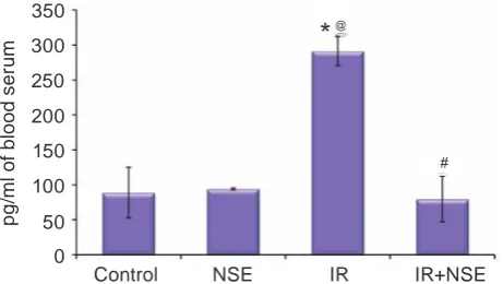
The NSE administration to rats with fat over-load caused normalization of TNFα content in blood serum (Fig. 2), that was accompanied by the increase of the content of plasma HDL and the decrease of LDL level compared to those in IR group of rats (Ta-ble 2). Simultaneously, the decrease of VLDL+IDL level in the rat plasma under the effect of NSE was found (Table 2).
The data presented in the work of A. Artmann and co-authors [21] shows the capacity of NSE to activate PPARα. It is known that activation of PPAR under dyslipidemic condition modulates the expres-sion of transcriptional factors SEBP1-c (sterol regula-tory element binding protein-1) and ChREBP (carbo-hydrate regulatory element binding protein), which regulate almost all genes involved in biosynthesis of FA, TAG and phospholipids [22, 23]. According to the data that we have already obtained (compen-satory action of NSE on the liver phospholipid and fatty acid composition of rats with IR), we suggest
Ta b l e 2. Content of total lipids and cholesterol of lipoprotein fractions in rat plasma (M ± m; n = 7–10)
Parameter Group of animals
Control NSE іR іR+NSE
Content of total lipids
in plasma, mg/ml 4.5 ± 0.8 7.29 ± 0.86 10.67 ± 1.24*,@ 3.25 ± 0.59@,# Content of total cholesterol
in plasma, mM 0.880 ± 0.032 0.720 ± 0.049* 1.21 ± 0.14*,@ 1.140 ± 0.058*,@ Content of HDL in plasma,
mM of cholesterol 0.080 ± 0.007 0.080 ± 0.026 0.06 ± 0.006 0.100 ± 0.007# Content LDL in plasma,
mM of cholesterol 0.090 ± 0.015 0.060±0.008 0.140 ± 0.017*,@ 0.110 ± 0.004@,# Content VLDL+IDL in
plasma, mM of cholesterol 0.790 ± 0.004 0.064 ± 0.040 1.060 ±0.106@ 0.790 ±0.106 Values represented mean ± SEM. Values in “Control” (n = 10), “NSE” (n = 7), “IR” (n = 9), “IR+NSE” (n = 10) were comparable. *p < 0.05, compared to the “Control” rats; @Р < 0.05, compared to the “NSE” grop; #p < 0.05, compared to
the “IR” group
that NSE restores the distribution of cholesterol in lipoprotein fractions of rat plasma by influencing on the processes of hepatic de novo lipogenesis .
Previously on the model of nonspecific in-flammation we found the decreased content of anti-inflammatory cytokines under the NSE effect [24]. According to the fact that activation of PPARα and PPARγ under іR inhibits activity of the transcrip -tion factor NF-kB, which stimulates produc-tion of inflammatory mediators [25, 26], the decrease of TNFα content under the effect of NSE may be one of confirmations of its interaction with PPARs.
Fig. 1. Content of TNFα (pg/ml) in rat blood serum (М ± m, n = 7–10). Here, on Fig. 2–3 * Values represented mean ± SEM. Values in “Control” (n = 10), “NSE” (n = 7), “IR” (n = 9), “IR+NSE” (n = 10) were comparable. *P < 0.05, compared to the “Control” rats; @ Р< 0.05, compared to the
“NSE” grop; #P < 0.05, compared to the “IR” group
Control NSE IR IR+NSE 350
@
*
#
250
150
50 300
200
100
0
p
g
/m
l o
f b
lo
o
d s
e
ru
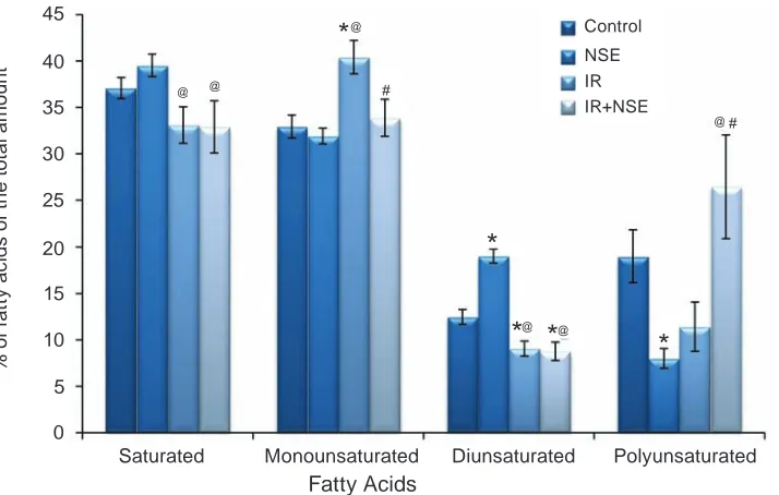
The investigation results have shown that the long-term high fat overload causes considerable changes in the fatty acid composition of rat plasma (Fig. 2), in particular, the increase of the content of MUFA (Fig. 2), primarely 18:1n-9, in plasma of IR group of rats. The decreased content of palmitic (16:0) and increased stearic (18:0) FFA level, the pre-cursor for 18:1n-9 synthesis, suggest the activation of n-9 FA synthesis pathway in rat liver with fat over-load. The administration of NSE to rats with experi-mental IR causes normalization of 18:0 and 18:1n-9 FFA content in the rat plasma (Fig. 3) that correlates with the change of FFA composition in the liver of rats with IR [15].
The increased pool of 18:1n-9 FFA, the main substrate for TAG synthesis, is accompanied by the increase of TNFα production, the decrease of PPARα expression, activation of the lipid peroxidation pro-cesses and apoptosis in hepatocyte cells [27]. Ac-cordingly, for the leading role of monounsaturated FFA in TAG synthesis, the decrease of their level un-der the effect of NSE can evidence for its protecting influence on tissues of rats with dyslipidemia.
The investigation of PUFA composition in the rat plasma has shown the increase of linolenic (18:3n-6), eicosatrienoic (20:3n-6) level, the decrease of linoleic (18:2n-6) and a tendency to decrease of arachidonic FFA content in IR group compared to Control (Fig.3). This finding is accordingly in sup
-port of the literature data [28] and results of clinical research, where a considerable decrease of 18:2n-6 and 20:4n-6 FFA level in the plasma of children with obesity was shown [29]. The increase of 18:3n-6 and 20:3n-6 content – the precursors for 20:4n-6 syn-thesis, with simultaneous decrease of the product was found, suggesting the disturbance of 20:4n-6 biosynthesis under experimental IR. Meanwhile, the decrease of 20:4n-6 in the rat plasma may be related to activation of oxidative processes and synthesis of pro-inflammatory eicosanoides under IR conditions [30]. The increased amount of lipid and protein per-oxidation products in the rat liver with experimental IR was found in our previous study [9, 15].
The NSE administration to rats with IR caused the increase of 20:4n-6 level (Fig. 3) that may be associated with its antioxidant activities and its influen ce on FA metabolism. It is known that a de -crease of 20:4n-6 content may be followed by the development of IR, because the Δ5-desaturase ac -tivity that catalyzes the 20:4n-6 formation is regu-lated by insulin [31]. Thus, the increase of 20:4n-6 content under the NSE action correlated with the improvement of insulin sensitivity and normaliza-tion of 20:4n-6 biosynthesis. On the other hand, this effect of NSE may be associated with the inhibition of polyunsaturated lipids peroxidation [9].
Thus, the decrease of MUFA level in blood plasma of IR rats treated by NSE was accompa-Fig. 2. Saturated and unsaturated (monounsaturated, diunsaturated, polyunsaturated) fatty acids in the phos-pholipid composition (% of the total amount of fatty acids) in the rat plasma (M ± m, n = 7–10)
Saturated Monounsaturated Diunsaturated Polyunsaturated
35 #
25 45
15 30
20
10
0
% o
f f
at
ty a
c
id
s o
f t
h
e t
ot
a
l a
m
o
u
n
t
Fatty Acids
5 40
@
*
@ @
* *
*
*
#
@ @
@
Control
Fig. 3. The main free fatty acids (% of the total amount) in the rat plasma (M ± m, n = 7–10)
16:00 18:00 18:1n-9 18:2n-6 18:3n-6 20:3n-6 20:4n-6 35
#
25
15 30
20
10
0
% o
f f
at
ty a
c
id
s o
f t
h
e t
ot
a
l a
m
o
u
n
t
5
@
*
@ @
*
*
*
*
#
@ @
@
*
*
#Control NSE
IR
IR+NSE
nied by the reduction of pro-inflammatory cytokine TNFα and the restoration of cholesterol in lipopro -tein fractions. Therefore, this effect of NSE may pre-vent the development of complications connected to IR.
ВплиВ N-стЕароїлЕтаноламіну на ліпідний сКлад
плазми КроВі щуріВ з ЕКспЕримЕнтальною інсулінорЕзистЕнтністю
О. В. Онопченко, Г. В. Косякова, В. М. Клімашевський, Н. М. Гула
інститут біохімії ім. о. В. палладіна нан України, київ;
e-mail: onop.89.av@mail.ru
на моделі інсулінорезистентності (ір), індукованої довготривалим жировим наванта -женням із переважним вмістом насичених та мононенасичених жирів, було вивчено вплив N-стеароїлетаноламіну (NSE) на склад вільних жирних кислот (ВЖк), ліпопротеїновий спектр плазми крові та вміст прозапального цитокіну TNFα в організмі щурів. За результатами прове -дених досліджень встановлено зростання рівня мононенасичених ВЖк (18:1n-9) та зниження
поліненасичених ВЖк (20:4n-6) у плазмі крові щурів за експериментальної ір. Ці процеси су -проводжувались збільшенням вмісту цитокіну TNFα та значними змінами у ліпопротеїновому профілі плазми крові. Зокрема, зафіксовано зниження вмісту холестеролу ліпопротеїнів високої щільності (лВЩ), зростання холестеро -лу ліпопротеїнів низької (лнЩ) та дуже низької щільності (лДнЩ). Введення NSE щурам з індукованою ожирінням ір сприяло відновленню рівня моно- та поліненасичених ВЖк, зростан -ню вмісту холестеролу лВЩ та знижен-ню – хо -лестеролу лнЩ. крім того, за ір в щурів, які отримували NSE, показана нормалізація рівня TNFα в сироватці крові. одержанні результати свідчать про відновлення за дії NSE ліпідного профілю плазми крові щурів з ір, індукованою аліментарним ожирінням. оскільки ліпідний спектр плазми крові відображає метаболізм ліпідів у цілому, встановлений ефект NSE може мати певне значення для попередження усклад -нень, пов’язаних із розвитком ір.
к л ю ч о в і с л о в а: N-стеароїлетаноламін,
плазма крові щурів, склад вільних жирних кис
ВлияниЕ
N-стЕароилэтаноламина на липидный состаВ плазмы КроВи Крыс с эКспЕримЕнтальной
инсулинорЕзистЕнтностью
А. В. Онопченко, Г. В. Косякова, В. М. Климашевский, Н. М. Гулая
институт биохимии им. а. В. палладина нан Украины, киев;
e-mail: onop.89.av@mail.ru
на модели инсулинорезистентности (ир), вызванной длительной жировой нагрузкой с преимущественным содержанием насыщен -ных и мононенасыщен-ных жиров, было изуче -но влия ние N-стеароилэта-ноламина (NSE) на состав свободных жирных кислот (СЖк), ли -попротеиновый спектр плазмы крови и содер -жание провоспалительного цитокина TNFα в организме крыс. B проведенных исследованиях установлено повышение уровня мононенасы -щенных СЖк (18:1 n-9) и снижение уровня по -линенасыщенных СЖк (20:4 n-6) в плазме кро -ви крыс с экспериментальной ир. Эти процессы сопровождались увеличением содержания ци -токина TNFα и значительными изменениями липопротеинового профиля плазмы крови крыс. В частности, зафиксировано снижение содержа -ния холестерола липопротеинов высокой плот -ности (лВп) и увеличение уровня холестерола липопротеинов низкой (лнп) и очень низкой плотности (лонп). Введение NSE крысам с ир способствовало восстановлению уровня моно- и полиненасыщенных СЖк, увеличению содер -жания холестерола лВп и снижению – холесте -рола лнп. кроме того, у крыс с ир, получавших NSE, отмечена нормализация уровня TNFα в сы -воротке крови. полученные результаты свиде -тельствуют о восстановлении при действии NSE липидного профиля плазмы крови крыс с ир, индуцированной алиментарным ожирением. принимая во внимание, что липидный состав плазмы крови отражает метаболизм липидов в целом, установленный эффект NSE может иметь определенное значение для предупреждения ос -ложнений, связанных с развитием ир.
к л ю ч е в ы е с л о в а:
N-стеароил-этаноламин, плазма крови крыс, состав свобод
-ных жир-ных кислот, липопротеиновый спектр,
TNFα, ожирение, экспериментальная инсулино
-резистентность.
references
1. McGarry J. D. Banting lecture 2001: dysregulation of fatty acid metabolism in the etiology of type 2 diabetes. Diabetes. 2002;51(1):7-18.
2. Teng K. T., Chang C. y., Chang L. F., Nesaret-nam K. Modulation of obesity-induced inflam-mation by dietary fats: mechanisms and clinical evidence. Nutr. J. 2014;13:12.
3. Ruddock M. W., Stein A., Landaker E., Park J., Cooksey R. C., McClain D., Patti M. E. Saturated fatty acids inhibit hepatic insulin action by modulating insulin receptor expression and post-receptor signaling. J. Biochem. 2008;144(5):599-607.
4. Storlien L. H., Jenkins A. B., Chisholm D. J., Pascoe W. S., Khouri S., Kraegen E. W. Influence of dietary fat composition on development of insulin resistance in rats. Relationship to muscle triglyceride and omega-3 fatty acids in muscle phospholipid. Diabetes. 1991;40(2):280-289. 5. Adiels M., Olofsson S. O., Taskinen M. R.,
Borén J. Overproduction of very low-density lipoproteins is the hallmark of the dyslipidemia in the metabolic syndrome. arterioscler. Thromb. Vasc. Biol. 2008;28(7):1225-1236. 6. Matias I., Gonthier M. P., Petrosino S., Docimo L.,
Capasso R., Hoareau L., Monteleone P., Roche R., Izzo A. A., Di Marzo V. Role and regulation of acylethanolamides in energy balance: focus on adipocytes and beta-cells. Br. J. pharmacol. 2007;152(5):676-690.
7. Terrazzino S., Berto F., DalleCarbonare M., Fabris M., Guiotto A., Bernardini D., Leon A. Stearoylethanolamide exerts anorexic effects in mice via down-regulation of liver stearoyl-coenzyme A desaturase-1 mRNA expression. FaSeB J. 2004;18(13):1580-1582.
8. Onopchenko O. V., Kosiakova G. V., Goridko T. M., Berdyschev A. G., Meged O. F., Hula N. M. The effect of N-stearoyl ethanolamine on the content of lipid peroxidation products, activity of antioxidant enzymes and the level of nitric oxide in the liver and plasma blood of rats with induced insulin-resistance. Ukr. Biokhim. Zhurn. 2013;85(5):88-96. (In Ukrainian).
phospholipid composition of rats with high-fat diet-induced insulin resistance. Ukr. Biochem. J. 2014;86(1):101-110. (In Ukrainian).
10. Collier G. R., Chisholm K., Sykes S., Dryden P. A., O’Dea K. More severe impairment of oral than intravenous glucose tolerance in rats after eating a high fat diet. J. Nutr. 1985;115(11):147-146. 11. Bligh E. G., Dyer W. I. A rapid method of
total lipid extraction and purification. can. J. Biochem. physiol. 1959;37:911-917.
12. Ichihara K., Fukubayashi y. Preparation of fatty acid methyl esters for gas-liquid chromatography. J. lipid. Res. 2010;51(3):635-640.
13. Zhang y. L., Hernandez-Ono A., Ko C., yasunaga K., Huang L. S., Ginsberg H. N. Regulation of hepatic apolipoprotein B-lipo-protein assembly and secretion by the availability of fatty acids. I. Differential response to the delivery of fatty acids via albumin or remnant-like emulsion particles. J. Biol. chem. 2004;279(18):19362-19374.
14. Dashti N. The effect of low density lipoproteins, cholesterol, and 25-hydroxycholesterol on apolipoprotein B gene expression in HepG2 cells. J. Biol. chem. 1992;267(10):7160-7169. 15. Onopchenko O. V., Kosiakova G. V., Meged E. F.,
Klimashevsky V. M., Hula N. M. The effect of N-stearoylethanolamine on cholesterol content, fatty acid composition and protein carbonylation level in rats with alimentary obesity-induced insulin resistance. Ukr. Biochem. J. 2014;86(6):119-128.
16. Le Goff W., Guerin M., Chapman M. J. Pharmacological modulation of cholesteryl ester transfer protein, a new therapeutic target in atherogenic dyslipidemia. Pharmacol. Ther. 2004;101(1):17-38.
17. Chen X., Xun K., Chen L., Wang Y. TNF-α, a potent lipid metabolism regulator. cell. Biochem. Funct. 2009;27(7):407-416.
18. Browning J., Horton J. Molecular mediators of hepatic steatosis and liver injury. J. clin. Invest. 2004;114(2):147-152.
19. Feingold K. R., Marshall M., Gulli R., Moser A. H., Grunfeld C. Effect of endotoxin and cytokines on lipoprotein lipase activity in mice. Arterioscler. Thromb. 1994;14(11):1866-1872.
20. Hotamisligil G. S., Murray D. L., Choy L. N., Spiegelman B. M. Tumor necrosis factor alpha inhibits signaling from the insulin receptor.
proc. Natl. acad. Sci. USa. 1994;91(11):4854-4858.
21. Artmann A., Petersen G., Hellgren L. I., Boberg J., Skonberg C., Nellemann C., Hansen S. H., Hansen H. S. Influence of dietary fatty acids on endocannabinoid and N-acylethanolamine levels in rat brain, liver and small intestine. Biochim. Biophys. acta. 2008;1781(4):200-212.
22. Boergesen M., Poulsen L. I., Schmidt S. F., Frigerio F., Maechler P., Mandrup S.ChREBP mediates glucose repression of peroxisome proliferator-activated receptor alpha expression in pancreatic beta-cells. J. Biol. chem. 2011;286(15):13214-13225.
23. Fernández-Alvarez A., Alvarez M. S., Gonzalez R., Cucarella C., Muntané J., Casado M. Human SREBP1c expression in liver is directly regulated by peroxisome proliferator-activated receptor alpha (PPARalpha). J. Biol. chem. 2011;286(24):21466-21477.
24. Zhukov A. D., Berdyshev A. G.,
Kosiakova G. V., Klimashevskiy V. M., Gorid’ko T. M., Meged O. F., Hula N. M. N-stearoylethanolamineeffect on thelevelof11-hydroxycorticosteroids, cytokines IL-1β, IL-6 and TNFαin rats with nonspecific inflammation caused by thermal burn of skin.Ukr. Biochem. J. 2014;86(3):88-97. (In Ukrainian).
25. Qatanani M., Lazar M. A. Mechanisms of obesity-associated insulin resistance: many choices on the menu. Genes. Dev. 2007;21(12):1443-1455. 26. Romics L. Jr., Kodys K., Dolganiuc A.,
Graham L., Velayudham A., Mandrekar P., Szabo G. Diverse regulation of NF-kappaB and peroxisome proliferator-activated receptors in murine nonalcoholic fatty liver. hepatology. 2004;40(2):376-385.
27. Cui W., Chen S. L., Hu K. Q. Quantification and mechanisms of oleic acid-induced steatosis in HepG2 cells. Am. J. Transl. Res. 2010;2(1):95-104.
28. Fukuchi S., Hamaguchi K., Seike M., Himeno K., Sakata T., yoshimatsu H. Role of fatty acid composition in the development of metabolic disorders in sucrose-induced obese rats. exp. Biol. Med. (Maywood). 2004;229(6):486-493. 29. Okada T., Sato N. F., Kuromori y., Miyashita M.,
30. Hardwick J. P., Eckman K., Lee y. K., Abdelmegeed M. A., Esterle A., Chilian W. M., Chiang J. y., Song B. Eicosanoids in metabolic syndrome. J. adv. pharmacol. 2013;66:157-266.
31. Galgani J. E., Aguirre C. A., Uauy R. D., Díaz E. O. Plasma arachidonic acid influences insulin-stimulated glucose uptake in healthy adult women. ann. Nutr. Metab. 2007;51(5):482-489.