Iranian Veterinary Surgery Association
IRANIAN JOURNAL OF VETERINARY SURGERY
Journal homepage: www.ivsajournals.com
ORIGINAL ARTICLE
Radiological and Histological Assessment of the Ossification Centers of
Hind Limb after Hatching in Pigeon
Mohammad Reza Ojaghloo
1, Mehdi Rezaei
2,*, Siamak Alizadeh
21 Graduate of Veterinary Medicine, Faculty of veterinary medicine, Urmia Branch, Islamic Azad University, Urmia, Iran.
2 Department of Clinical Sciences, Faculty of Veterinary Medicine, Urmia Branch, Islamic Azad University, Urmia, Iran.
Received: 7 February 2018 Accepted: 15 May 2018 Available Online: 4 August 2018
Keywords: Histology; Hind Limb;
Ossification Centers; Pigeon;
Radiology.
Abstract
Objective- The aim of this study was to determine the age of physical maturity and evaluation of radiology and histology of hind limb ossification centers in pigeon.
Design- Fundamental study.
Animals- 14 pigeons.
Procedures- These pigeons were cultivated in identical and standard conditions and radiological and histological tests performed every 7 days to 91 days.
Results- Based on the results of radiology and histology, the hind limb skeletal differentiation in pigeons with the appearance of centers of immature cartilages in diaphyses of the femur, tibiotarsus and tarsometatarsus at the end of the first week and the fibula bone at the end of the third week began. Growth sequences in the femur, tibiotarsus, fibula, tarsometatarsus and digits were observed during different stages. The maximum growth of these bones was related to the periods of maximum cartilage activity and their bone formation, and the femur holds steady its growth relation to the length of the skeletal body of the hind limb, although at the end of the fifth week its growth slowed down. The histological findings were based on the examination of the proximal extremity of the femur. The tissue samples at one day were lack of bone marrow and the bone marrow begins to form at the end of the first week. The presence of epiphyseal growth plate in samples and its bone formation was confirmed on the basis of radiology early in the fifth week.
* Correspondence to: Mehdi Rezaei, Department of Clinical Sciences, Faculty of Veterinary Medicine, Urmia Branch, Islamic Azad University, Urmia, Iran.
E-mail: mehdi217mr@yahoo.com
www.ivsajournals.com© Iranian Journal of Veterinary Surgery, 2018
Conclusion and Clinical Relevance- According to this study, the best time to complete the development of bone formation and the formation of all parts of the hind limb skeleton of the pigeon is probably 35 days after the hatch.
1. Introduction
Skeletal development in birds has always been a topic of concern. The first studies on histogenesis were made by the bracket,1 but generally done before and after the hatch.8,32 leg skeleton in birds is considerably less noticeable than the skeleton of the wings due to the five-legged position (pentadactyl limb), which is probably why the amount of information about the natural growth of the skeleton is scarce. A notable exception is the study by Johnson (1883) about the development of leg in the chick that the early stages of skeletal development were described in detail, but the subsequent stages were ignored.16 Most investigations about embryonic development of leg in birds are about chickens, except for some on zebra parakeet, ostrich, duck, and larus.2,29,33 Many studies are done on chicken with special aspects of leg skeletal development, like knee joint development24,25 or development of a certain bone, for example tibiotarsus or tarsometatarsus,8,28 with only a now-and-then mentioning of other parts of the skeletal system. Many different investigations are done about the ossification centers in embryonic period that are about all mesenchymal, cartilaginous and ossification stages.12,27Also the natural growth of leg skeleton in pigeon embryo from the primary mesenchymal changes to last stages of embryonic period are investigated thoroughly. Most of the studies are about the embryonic period and the best studies about hatching and after hatching are mostly done on gallus domesticus.14 There is no citable study until now about the ossification centers in pigeon after hatching and this investigation studies the ossification centers in pigeon after hatching for the first time.
2. Materials and Methods
In this study, the growth and differentiation of pelvic girdle, hind limb and ossification centers in these areas were investigated in post-hatching period in pigeons. The 14 hatching pigeons were cultivated in identical and standard conditions and radiological and histological tests performed every 7 days to 91 days. These birds were grown under the standard conditions related to diet, temperature, humidity and light. The moisture content of the storage medium was about 70%, the brightness was 13 hours, and the temperature of the storage medium was 34-36°C in the first few days, which gradually decreased to about 25-27°C from 14 days afterwards and then the
temperature was kept constantly. In this study, the methods used included the provision of radiographic stereotypes, histological sections and tissue staining.
After each stage of radiography, a sample was taken to examine the upper and lower centers of the femur and remove the tissue sections. Obviously, the number of specimens after the first radiography from 14 to 13 and from then on, also decreased and the samples were removed from the tissue sections. The radiography machine was Dean 44 X-Ray machine, KV 40 and mAs 3.6.For radiography the lateral and ventrodorsal positions were used. The radiographs of the samples were made according to a regular sequence, so that radiographs were taken every day from the pigeons (in the first week, twice (day one and seventh day), and then at the end of the second to the thirteenth weeks, radiography was performed once).
At the end of each week following a radiograph, a pigeon was randomly selected and with pentobarbital sodium was euthanized and the detached leg was placed in 10% formalin and then sent to a histological laboratory. In the laboratory, after the tissue passage, 5 micron thick sections of the proximal end of the thigh bone were prepared and then the samples were stained with Hematoxylin and Eosin staining.
3. Statistical Analysis
Statistical analyses were done by SPSS package. The data were described by Mean, Std. Deviation and Std. Error Mean. Statistics are based on cases with no missing values for any dependent variable or factor used. 95% Confidence Interval of the Mean Difference was statistically considered significant.
4. Results
Ossification time of pelvic girdle bones based on
radiological findings: ilium, ischium, pubis
Table 1. Ossification time of pelvic girdle bones.
Bone
Days after hatching
1 7 14 21 28 35 42 49 56 63 70 77 84 91
Ilium - - - + + + + + + + + + + +
Ischium - - - + + + + + + + + + + +
Pubis - - - + + + + + + + + + + +
Figure 1. Ilium bone observed in all specimens. 21st day.
Ossification time of hind limb bones based on
radiological findings
Hind limb bones were not observed in any of the specimens on the first day after hatching due to cartilage of these bones. From 7 days later, Femur, Tibiotarsal bone, Tarsometatarsal bone, Trochlea of tarsometatarsal bone, Digit I, Digit II, Digit III, Digit IV, Phalanges, Metatarsal I and from 14 days later, Condyle of femur, Condyle of tibiotarsal bone were seen in all specimens; from 21 days later, Head of femur, Trochanter of femur and Fibula were Suspected in specimens. From 28 days later, Head of femur, Trochanter of femur, Fibula, Patella and Hypotarsal crest of tarsometatarsal bone in all specimens, they were seen (Table 2).
Histological results
Histological results are based on the examination of the histological sections of the proximal extremity of the femur. During this study, two samples were selected at each stage and the femur of each sample was isolated and the histochemical process was performed on them.
The tissue samples have no bone marrow at one day and the bone marrow begins to form at the end of the first week (7 days of age). The presence of the growth plate in samples and their closure was confirmed on the basis of radiology in the early fifth week. The increase in bone length seems to be due to the alteration of the epiphyseal cartilage to the bone at the diaphyseal side. With age, shape and form of bone blades are formed, and the density and volume of blades are reduced.
Figure 2. Fibula. 28th day.
Table 2. Ossification time of hind limb bones
Figure 4. Hock joint. 14th day.
Investigating the process of converting cartilage
tissue into bone tissue in the proximal region of
the femur on the basis of microscopic
observations
1-day old: Cartilages that have previously been mesenchymal tissue are seen and these cartilages have been hypertrophic. The matrix surrounding them has been reduced and calcified, which causes the cartilage or chondrocyte cells to disappear. When the chondrocyte cell is destroyed, from the periosteum (perichondrium), a series of nerves and vessels attack the place where the cartilage is destroyed. With blood vessels and nerves, osteocytes and
osteoblasts also go there to replace chondrocytes. The nucleus of the cells is lost during its destruction and its normal state is lost in the corner and it has a crushy state. 7 days old: There is cartilage, but on the side lines of the cartilage cell, there are osteoblasts with an epithelial form that secrete the bone. Also, on the seventh day, we see osteoblasts that secrete the initial bone instead of the cartilage. There is no calcium in the initial bone and after a few days, calcium is deposited on collagen.
14 days old: Both cartilage and bone appear, but the thickness of the bone tissue has increased. Osteoblasts are seen that have changed from cubic or squamous to cylindrical state, and the height of osteoblasts has increased. Cartilage tissue is more restricted and bone tissue volume has increased. There are nests inside the bone tissue. There is one osteocyte cell in the bone tissue and within each cell nest. When osteoblasts secrete the bone, the osteoblast cell floats inside the bone tissue and is called osteocyte.
21 days old: Central bone channel is formed. Bone tissue is limited to the bone margin and not completely regular (woven bone). The osteon or haversian system is not completely formed and the cartilage tissue is very low. 28 days old: It looks like the 21st day, but the haversian system has been identified. The woven bone is still there. Non-haversian systems also exist.
Bone
Days after hatching
1 7 14
21
28
35
42
49
56
63
70
77
84
91
Head of femur
- -
-
+-
+
+
+
+
+
+
+
+
+
+
Trochanter of femur
- -
-
+-
+
+
+
+
+
+
+
+
+
+
Femur
- + +
+
+
+
+
+
+
+
+
+
+
+
Patella
- -
-
-
+
+
+
+
+
+
+
+
+
+
Condyle of femur
- -
+
+
+
+
+
+
+
+
+
+
+
+
Fibula
- -
-
+-
+
+
+
+
+
+
+
+
+
+
Tibiotarsal bone
- + +
+
+
+
+
+
+
+
+
+
+
+
Condyle of tibiotarsal bone
- -
+
+
+
+
+
+
+
+
+
+
+
+
Hypotarsal crest of tarsometatarsal bone
- -
-
-
+
+
+
+
+
+
+
+
+
+
Tarsometatarsal bone
- + +
+
+
+
+
+
+
+
+
+
+
+
Trochlea of tarsometatarsal bone
- + +
+
+
+
+
+
+
+
+
+
+
+
Digit I
- + +
+
+
+
+
+
+
+
+
+
+
+
Digit II
- + +
+
+
+
+
+
+
+
+
+
+
+
Digit III
- + +
+
+
+
+
+
+
+
+
+
+
+
Digit IV
- + +
+
+
+
+
+
+
+
+
+
+
+
Phalanges
- + +
+
+
+
+
+
+
+
+
+
+
+
58
Figure 5. Histological section of the proximal extremity of the femur. A. The region of cartilage conversion into bone (1 days old). B.
Osteoblasts are visible (7 days old). (10×H&E) C. Osteoblasts have been transformed from cubic or squamous to cylindrical state, and there is an osteocyte within each cell nest (14 days old). D. The central bone canal is composed and the woven bone is seen (21 days old).
E. The haversian and non-haversian systems are visible (28 days old). (40×H&E).
35 days old: The bone has a mature state. The non-haversian system isn’t there and there is a complete haversian system. The cartilage has completely disappeared and there is no effect of the cartilage model (Figure 5).
Calculation results of bone layers by graded
lens
Total diameter of the bone: Based on the results, the lowest total diameter of the specimens was obtained in one day
(1626.625 μm) and the highest overall diameter was in 91 days old (4513.5 μm).In the statistical analysis of the data, the increase in overall diameter after one day is accompanied by a gradual and regular increase and has a linear gradient (Chart 1).
Inner diameter of the bone: Based on the statistical results,
the inner diameter increased from 1 to 14 days old at a
gentle speed and from 14 to 21 days old in a high speed.
decrease and then had increased from 28 days later. This
increase was followed from the age of 35 to 42 days at a
high speed and from 42 days of age by a gentle song and
reached to its maximum at 91 days of age. In fact, it can be
stated that the growth of the inner diameter of the bone has
3 stages of increase (1 to 21 days old), decrease (21 to 28
days old) and increase (28 to 91 days old) (Chart 2).
Osteocyte thickness of the bone: Osteocyte thickness
during the period from day 1 to day 35 increased
significantly so that the peak of bone thickness at 35 days
old was 702.1 micrometers. After that, the thickness of the
bone tissue was lowered, so that the minimum thickness of
the bone tissue was 401.2 micrometers in 91-day
specimens (Chart 3).
Periosteum thickness of the bone: According to the data
analysis, the thickness of the periosteum increases from 1
to 35 days old. This increase in periosteal thickness from 1
to 7 days old has a significant increase then increases from
7 to 21 days old at a gentle speed and from 21 to 35 days
old at a high speed. The thickness of the periosteum
decreases gradually over a period of 35 to 56 days of age at
a gentle speed, and then from 56 to 63 days of age, this
downtrend continues at a high rate. From 63 to 70 days of
age, the reduction of periosteum thickness was subtle, so
there was no difference in the thickness of the periosteum
(35.105 μm).From 70 to 77 days old, this decrease in
thickness continued and then remained constant until 91
days of age. The thickness of the periosteum in 77, 84 and
91 days old was 32.5975 micrometers (Chart 4).
Endosteum thickness of the bone: Thickness of endosteum
increased from 1 to 35 days old. The highest speed of
increasing in endosteum thickness was observed between
14 to 21 days old, with the highest thickness of endosteum
in 35-day specimens, which was 41.2928 micrometers. The
thickness of endosteum was lowered in specimens after 35
days old, and this decrease alternatively continued for up
to 91 days old (Chart 5).The results obtained from
measuring the thickness of different histologic parts by the
graded lens are given in table 3.
Table 3. The results obtained from measuring the thickness of different histologic parts by the graded lens
Age Total diameter Osteocyte Periosteum Endosteum Inner diameter
1 day old 1626.625 230.575 - - -
7 days old 2127.125 350.875 5.1616 7.7424 1763.346
14 days old 2427.425 431.075 10.025 9.0322 1977.2928
21 days old 2652.65 521.3 30.075 23.2272 2078.0478
28 days old 2752.75 677.025 40.1 36.1312 1999.4938
35 days old 2852.85 702.1 50.15 41.2928 2059.3072
42 days old 2927.935 651.95 45.135 38.712 2192.138
49 days old 3303.3 626.875 42.6275 33.5504 2600.2471
56 days old 4125.125 601.8 40.12 30.9696 3452.2354
63 days old 4204.2 576.725 35.105 25.808 3566.663
70 days old 4304.3 501.5 35.105 22.6388 3745.0562
77 days old 4404.4 451.35 32.5975 20.05 3900.4025
84 days old 4463.35 426.275 32.5975 18.045 3986.4325
60
Chart 1. Measuring the total diameter of the bone. Chart 2. Measuring the inner diameter of the bone.
Chart 3. Measuring the osteocyte thickness of the bone. Chart 4.Measuring the periosteum thickness of the bone.
5. Discussion
In this study we try to compare leg skeletal development in pigeon (Columbiformes) after hatching with the pre-hatching time and more specifically with gallus domesticus after hatching because the information about the natural development of leg skeleton in chick is vast. Although the changes happen at different times for the mesenchymal density of bone marrow, primary cartilage, and ossification in large elements, it is clearly observed that the development of the skeleton in pigeon and chick are similar. Mesenchymal density and first stages of cartilage development in femur, tibia, and fibula diaphysis in day one is completely observable.
In chicken, the femur, as the strongest element in primary leg skeleton in chondrogenesis period is explained, although its development rate is lower than development rate of tibia. Nonetheless, in pigeon the tibia is not only the strongest element but its development rate is also higher even in cartilaginous stage. In addition, fibula in chick has more chondrogenesis speed compared to tibia,9 yet, in
pigeon the development rate of fibula is lower than tibia. In past studies, it is mentioned that fibula is considerably thinner than tibia in cross section and by the increase in ossification process the distal end gets thinner than the proximal end. It seems impossible that fibula can always have higher rate of development compared to tibia in chick.25
It seems that patella ossifies a little earlier in pigeon than chicken and after day 28, whereas in chicken the ossification is observed after day 63 in it14 but like pigeon,
chicken patella is not also ossified after hatching, and until the time it is in late incubation period only a weak cartilage is observed in it.25 The metatarsus development
in pigeon is like of chick in many ways, except for the metatarsus I turn to posterior position that happens later in pigeon. Fujioka says that the center of ossification in digits I, II, and III seems to be in a proximal-distal sequence, but the sequence of ossification in phalanges in digit IV is 1, 4, 5, 2, and 3.11 The same sequence of ossification is seen in
pigeon although the ossification in digit IV needs more studies to be done in this field.
During postnatal development of the pigeon, a large portion of the skeleton becomes pneumatized, displacing the hemopoietic bone marrow. The consequences of pneumatization on distribution and quantity of bone marrow as well as the availability of other sites for hemopoiesis have been investigated;30 bone marrow
weight and its proportion of whole-body weight increased during the first 4 weeks and then decreased in the
following months. The developing bone marrow showed a progressive distribution during the first months of life, eventually being distributed proportionally over the entire skeleton, except for the skull. At the age of 6 months p.h. bone marrow had been displaced, its volume decreasing in correlation to increasing pneumaticity and conversion to fatty marrow. This generates the characteristic pattern of bone marrow distribution in adult pigeons, which shows hemopoietic bone marrow in ulna, radius, femur, tibiotarsus, scapula, furcula, and the caudal vertebrae.30
The existence of two rows of ossification centers in tarsus joint region in birds (hock joint) is completely obvious which is fused with tibia from proximal extremity and to big metatarsus from distal extremity (Figure4). Previous researchers have put forth this theory about bones in tarsus region, too.20 At last we can say that in tarsus area the
bones adjacent to long bones are fused and are usually called tibiotarsus and tarsometatarsus. When we relate these expressions to them no justification about the secondary ossification centers in tarsus area mentioned in previous studies is acceptable.6,10
This study confirms the general idea about the existence of two centers at proximal row and one center in distal row in tarsus and is equal with Hogg’s study on gallus domesticus14 but contradicts with Franceschini that
declared the existence of three centers.10
It seems that the proximal center of tibia is the only secondary and real center of ossification in long bones that probably exists in all birds. This center was ossified in all specimens on day 35 after hatching. If this center causes the increase in the size of bone that is a growth plate (Epiphyseal plate) and maybe it is a traction epiphysis. The closure of the growth plates in the tibiotarsus and the tarsometatarsus is the most reliable criterion for age determination in birds. Similar to the situation in fetal mammals, primary ossification centers arise prenatally in the middle part of the diaphyseal cartilage of the avian long bones.8 However, unlike in mammals, no secondary
ossification centers are formed in the epiphyses of the appendicular skeleton of birds. Epiphyseal growth plates are therefore lacking in avian bones, except at the distal end of the tibiotarsus and the proximal end of the tarsometatarsal bone, both of which have a growth plate formed by the fusion lines between tarsal bones and the tibia or the metatarsal bones, respectively. Additionally, a secondary ossification center is situated in the craniodorsal part of the tibia. It corresponds to the tibial crest center of mammals (tibial tuberosity), to which the patellar ligament is attached.6,14,21
62 condyles can give an indication of the age of the animal
(Figure 5). In contrast, the tibiotarsus contains growth plates whose closure provides a criterion for age determination. The proximal and distal growth plates of the tibiotarsal bone closed around 5 weeks in the pigeon(Figure 6 , 7) , whereas in the chicken it occurred around 14 weeks. Similarly, closure of the tarsometatarsal growth plate occurs around 5 weeks in the pigeon (Figure 8) in the present study and in the chickens it was around 14 to 16 weeks.4
Some researchers claim that the secondary centers only exist in mammals, lizards and in the avian tibia and cartilage canal in mammals are usually related to the subsequent developments of the secondary centers as in lizards.13
Cartilage canals are in epiphysis area of bird bone and are described by Hines. The first have more secondary centers and may destroy during the bone pneumatisation and invasion of air bags inside the bone. Pneumatisation in long bones increases hence there is an unusual relation between species of birds. Although the existence of a center in proximal extremity of tibia is completely obvious, little attention is paid to it. Parsons believed that they belong to the group of traction epiphyses and he has applied the term to them. He suggested that these represented pre-existing sesamoid bones.26 Some
researchers have disagreed with this expression and have called them the inter tendon centers and by ossification progress, the cartilage epiphysis area is placed inside tendon and they also believed that these should be differentiated with the sesamoids.14,15 In some studies
Parson’s theory is supported since it was proved that in some birds there may not be a patella and it may be fused with tibia bone.
According to previous findings the tibia is separate so it is more similar to a sesamoid bone. Probably the proximal center of tibia shows the preformed joint of sesamoid to tibia and in addition is a tool to reinforce the joint of quadriceps femoris muscle, too. Some of these forms, like the longer length of crus bones from proximal elements and specificity of tarsus region, in which the proximal elements are intensely bond to crus (tibia and fibula), can be an adaption to running and locomotion with two legs. In birds, in a separate line of pseudosuchian, because of the running habit a similar essential mechanism has made a structure called tibiotarsus (fusing of proximal tarsal row to distal extremity of tibia) that this tibiotarsus is one of the specific characteristics of the bird class skeleton. On the other hand, in the pigeon, fusion of the tarsal region bones with the tibia and metatarsus occurs in the fifth week.
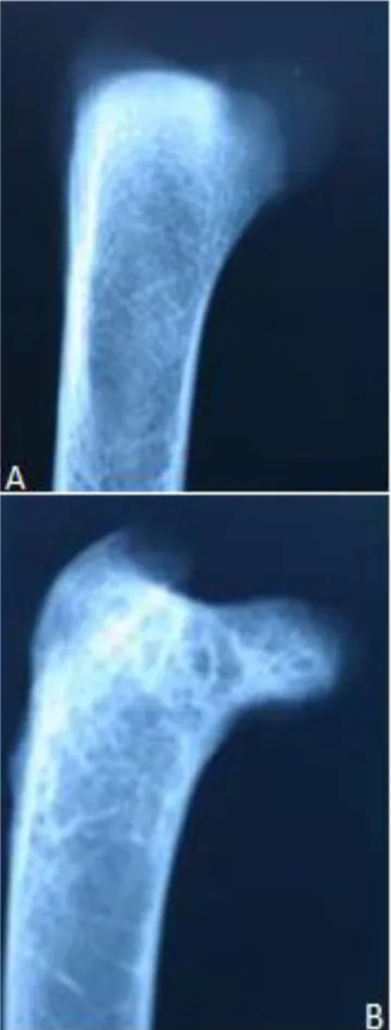
Figure 6. Lateromedial view of the proximal end of femur. A.
Figure 7. Craniocaudal view of the proximal growth plate (arrows) of the tibiotarsus. A.28th day. B. 35th day
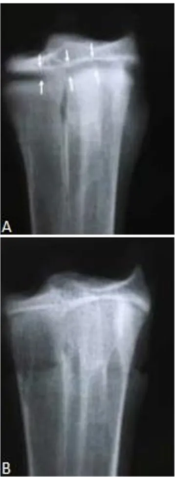
64 Figure 9. Dorsoplantar view of the proximal growth plate
(arrows) of the tarsometatarsus. A. 28th day. B. 35th day.
6. Conclusion
By comparison of leg development in pigeon and chicken, it is obvious that the skeleton elements in both birds are very similar in one sequence although the development in the chicken is longer than in the pigeon. In this study the existence of epiphyseal plate at the proximal extremity of tibia was doubtful that maybe causes the increase in the size of the diaphysis in all specimens or it was the secondary center of ossification that causes the traction epiphysis. In the pigeon, the ossification of the long bones of the leg begins at the end of the first week and all the elements in these bones are formed at the end of the fourth week. Hence the best time to say that all the hind limb elements are totally formed and ossified is the 35th day
after hatching.
Acknowledgment
This study was supported by Faculty of Veterinary Medicine, Urmia Branch, Islamic Azad University, Urmia, Iran.
References
1. Brachet A. Etudes sur la resorption du cartilage et le developpement des os longs chez lesoiseaux.
Internationale Monatsschrift ffr Anatomie and Physiologie, 1993; 10: 391-417.
2. Broom R. On the early development of the appendicular skeleton of the ostrich, with remarks on the origin of birds. Trans. S. Afr. phil. Soc, 1996; 16: 355-368.
3. Bellaairs A.D'A, Jenkin C.R. The skeleton of birds. In Biology and Comparative Physiology of Birds, vol. 1 (ed. A. J. Marshall). New York and London Academic Press, 1960; pp:241-300.
4. Breugelmans S., Muylle S., Cornillie, P., Saunders, J., Simoens, P. Age determination of poultry: a challenge for customs. Vlaams Diergeneeskundig Tijdschrift, 2007; 76: 423-429
5. Barnerr C.H., Lewis 0.J. The evolution of some traction epiphyses in birds and mammals. Journal of Anatomy, 1958; 92(4): 593-601.
6. Church L.E., Johnson L.C. Growth of long bones in the chicken – Rates of growth in length and diameter of the humerus, tibia, and metatarsus.
7. Chamberlain F.W. Atlas of Avian Anatomy. Michigan State College, 1943.
8. Fell H.B. The histogenesis of cartilage and bone in the long bones of the embryonic fowl. Journal of Morphology and Physiology, 1925; 40(3): 417-459. 9. Fell H.B., Canti R.G. Experiments on the development in vitro of the avian knee joint. Proc. R. Soc, B, 1934; 116: 316-349.
10.Franceschi M.P. On the appearance and evolution of secondary ossification centers in the tibia of Gallus gallus (Linn.). Acta Anatomica, 1967; 68: 169-188.
11.Fujioka T. Time and order of appearance of ossification centers in the chicken skeleton. Acta anatomica nipponica, 1955; 30: 140-150.
12.Hamilton H.L. Lillie's Development of the Chick, 3rd ed. New York: Henry Holt & Co. Herrick EH & Herrick EM. Rib abnormalities in fowls. Poultry Science, 1952; 35: 191-194.
13.Hines RW. Primitive form of epiphysis in long bones of tetrapods. Journal of Anatomy, 1938; 72: 323-343.
14.Hogg DA. A re-investigation of the centers of ossification in the avian skeleton after hatching.
Journal of Anatomy, 1980; 130(4): 725-743. 15.Hogg DA. The articulations of the neurocranium in
the postnatal skeleton of the domestic fowl (Gallua gallus domesticus). Journal of Anatomy, 1978; 127: 53-63.
16.Johnson A. On the development of the pelvic girdle and skeleton of the hind limb in the chick.
Quarterly Journal of Microscopical Science, 1883; 23: 399-411.
17.Kirkwood J.K., Duignan P.J., Kember N.F., Bennett P.M., Price D.J. The growth of the tarsometatarsus bone in birds. Journal of Zoology, 1989; 217, 403-416.
18.Lansdown A.B.G. A study of the normal development of the leg skeleton in the quail (Coturnix coturnix japonica). Journal of Anatomy, 1970; 106(1): 147-160.
19.Mitgutsch C., Wimmer C., Sánchez-Villagra M.R., Hahnloser R., Schneider R. A. Timing of ossification in duck, fowl, quail, and zebra finch: intraspecific variation, heterochronies, and life history evolution. Zoological Science, 2011; 28(7): 491.
20.MORSE E.S. On the tarsus and carpus of birds. Ann. Lyc. nat. Hist, N.Y.,1874;10:141-158.
21.Naldo J.L., Samour J.H., Bailey T.A. Radiographic monitoring of the ossification of long bones in kori
(Ardeotiskori) and white-bellied (Eupodotis senegalensis) bustards. Research in Veterinary Science, 1998; 65:161-163.
22.Naldo J.L., Bailey T.A., Samour J.H. Radiographic analysis of the growth rate of long bones in bustards. Research in Veterinary Science, 2000; 69: 233-240.
23.Nagaviri S.S., P.N. Dubey. Epiphysis at the lower end of chick tibia. Journal of Anatomical Society of India, 1975; 24: 35.
24.Nivnn J.S.F. The development in vivo and in vitro of the avian patella. Wilhelm Roux Arch. Entw Mech. Org, 1933; 8: 480-501.
25.O'Rahilly R., E. Gardener. The development of the knee joint of the chick and its correlation with embryonic staging. Journal of Morphology, 1956; 98: 49-88.
26.Parsons F.G. Observations on traction epiphyses.
Journal of Anatomy and Physiology, 1904; 38: 248-258.
27.Romanoff A.L. The Avian Embryo. New York: Macmillan, 1960.
28.Schryver H.F. A quantitative comparison of the growth of the embryonic chick tibiotarsus in vivo and in vitro. Journal of Experimental Zoology, 1967; 161: 81-88.
29.Sieglbaur F. Zur Entwicklung. der Vogelextremitait. Zeitschrift für Wissenschaftliche Zoologie, 1911;97:262-313.
30.Schepelmann K. Erythropoietic bone marrow in the pigeon: development of its distribution and volume during growth and pneumatization of bones.
Journal of Morphology, 1990; 203: 21–34.
31.Wetmore A. A revised classification of birds of the world. Smithson.misc. Collns, 1951;117 (4):1-22 32.Wolbach S.B., Hegsted D.M. Endochondral bone
growth in the chick. American Medical Association Archives o Pathology, 1952; 54(1): 1-12.
66
ناریا یکشزپماد یحارج هیرشن
لاس
2018
دلج ،
13
هرامش(
1
یپایپ هرامش ،)
28
هدیکچ
و یفارگویدار یسررب
تفاب
سانش
ی
زکارم
ناوختسا
زاس
ی
رتوبک رد چه زا سپ یفلخ یتکرح مادنا
ولقاجوا اضردمحم
1،
اضر یدهم
یئ
2 , *،
هدازیلع کمایس
2 1 شناد هتخومآ هفرح یارتکد ،یکشزپماد یا شناد هیمورا دحاو ،یکشزپماد هدک ،یملاسا دازآ هاگشناد ،
ناریا ،هیمورا
2
،هیمورا دحاو یملاسا دازآ هاگشناد ،یکشزپماد هدکشناد ،یهاگنامرد مولع هورگ مورا ی ه ناریا ، فده و یژولویدار یبایزرا و یمسج غولب نس نییعت هعلاطم نیا زا فده تفاب سانش ی زکارم ناوختسا زاس ی یتکرح مادنا رد یفلخ .دوب رتوبک حرط -.یداینب هعلاطم تاناویح -14 .رتوبک
راک شور ره و دنتفای شرورپ درادناتسا و ناسمه طیارش رد چه بقاعتم اهرتوبک نیا 7 زور ی ک راب ات 91 یگزور امزآ ی ش اه ی و یژولویدار تفاب سانش ی .دش ماجنا جیاتن و یژولویدار جیاتن ساسا رب تفاب
سانش ی زا یزکارم ندش رهاظ اب رتوبک رد یفلخ مادنا تلکسا زیامت فورضغ
اه ی زیفاید رد غلابان
ناوختسا رد دشر یلاوت .دش زاغآ موس هتفه نایاپ رد لاوبیف ناوختسا زین و لوا هتفه نایاپ رد سراتاتموسرات و سراتویبیت ،نار ناوختسا رد ناتشگنا و سراتاتموسرات ،لاوبیف ،سراتویبیت ،نار نیا دشر رثکادح و دش هدید یفلتخم لحارم یط
ناوختسا اه هب هرود اه ی رثکادح
ندش یناوختسا لحارم و یفورضغ تیلاعف نآ
اه هاگن تباث یفلخ مادنا تلکسا لوط اب ار دوخ دشر طابترا ،نار ناوختسا و دوب طوبرم م
ی
دراد جیاتن .تفای لیلقت نآ دشر نازیم مجنپ هتفه یاهتنا زا هچرگا ، تفاب
سانش ی ساسا رب ناوختسا یناقوف یاهتنا یتفاب عطاقم یسررب
.دوب نار هنومن اه ی رد یتفاب ی ک گزور ی ندش لیکشت هب عورش لوا هتفه یاهتنا زا ناوختسا زغم و هدوب ناوختسا زغم دقاف م
ی دنک دوجو .
یپا دشر هحفص رد یزیف
هنومن اه ساسا رب مجنپ هتفه لیاوا رد نآ ندش یناوختسا و .دش دییأت یژولویدار
تن ی هج گ ی ر ی ینیلاب دربراک و
-ساسا رب هک ار ینامز نیرتهب ،هعلاطم نیا م
ی ناوت هب ناونع دنور ندش لماک ناوختسا زاس ی لیکشت و همه تمسق اه ی ،دومن نایب رتوبک یفلخ مادنا تلکسا ًلاامتحا