O R I G I N A L R E S E A R C H
Open Access
Somatostatin receptor PET/CT in restaging
of typical and atypical lung carcinoids
Vikas Prasad
1*, Ingo G. Steffen
1, Marianne Pavel
2, Timm Denecke
3, Elisabeth Tischer
2, Konstantina Apostolopoulou
2,
Andreas Pascher
4, Ruza Arsenic
5and Winfried Brenner
1Abstract
Background:To assess the role of somatostatin receptor (SR) PET/CT using Ga-68 DOTATOC or DOTATATE in
staging and restaging of typical (TC) and atypical (AC) lung carcinoids.
Methods:Clinical and PET/CT data were retrospectively analyzed in 27 patients referred for staging (N= 5; TC,N= 4; AC,N= 1) or restaging (N= 22; TC,N= 8; AC,N= 14). Maximum standardized uptake value (SUVmax) of SR-positive lesions was normalized to the SUVmax of the liver to generate SUVratio; SR PET was compared to contrast-enhanced (ce) CT. The classification system proposed by Rindi et al. (Endocr Relat Cancer. 2014;21(1):1-16, 2014) was used for classification of patients in TC and AC groups.
Results:Only 18/27 patients were found to have metastases on PET/CT. Of the 186 lesions, 101 (54.3 %) were depicted on both PET and CT, 53 (28.5 %) lesions only on CT, and 32 (17.2 %) only on PET. SUVratio of lesions was significantly higher in AC as compared to TC (p< 0.001). In patients referred for restaging, additional findings on PET lead to upstaging with change in management strategy in 5/22 (22.7 %) patients (AC,N= 5; TC,N= 1). In four patients (all AC) referred for restaging and in one patient (TC) referred for staging, additional findings on CT missed on PET lead to correct staging.
Conclusions:Typical and atypical carcinoid patients have complex patterns of metastases which make it necessary to combine functional SR PET and contrast-enhanced CT for appropriate restaging. In patients referred for restaging SR, PET may have a relevant impact on treatment strategy in up to 22.7 of patients with typical and atypical lung carcinoids.
Keywords:Lung carcinoids; Atypical carcinoid; Typical carcinoid; Somatostatin receptor; PET/CT; Management; Restaging
Background
Neuroendocrine tumors (NET) of the lungs (LNET) rep-resent approximately 30 % of all NET [1, 2] and account for 1–2 % of all lung tumors. According to current WHO classification, LNET are sub-classified into typical carcin-oid (TC), atypical carcincarcin-oid (AC), and small cell and large cell neuroendocrine carcinoma (LCNEC). TCs are gener-ally low-grade tumors, and ACs are intermediate-grade tumors, whereas the other two entities, small cell lung cancer (SCLC) and LCNEC, are high-grade neoplasms by definition with usually poor prognosis [3]. Of special note
is the fact that up to 10 % of all lung tumors, especially, SCLC show neuroendocrine differentiation [4]. Diffuse idiopathic pulmonary neuroendocrine cell hyperplasia (DIPNECH) without any predisposing conditions has also been reported [5, 6]. DIPNECH is a disease with relatively indolent clinical course, usually remaining stable over sev-eral years but with the potential to metastasize in locore-gional lymph nodes and rarely to extra thoracic sites [7].
The wide range of histopathological variations of NET with distinct prognosis often poses a clinical challenge not only with respect to the choice of therapy but also to the selection of the appropriate imaging tool for sta-ging and restasta-ging. For small cell lung cancer, the clin-ical role of F-18-fluoro-deoxyglucose (FDG) PET is well documented for patient management [8]. However, for * Correspondence:vikas.prasad@charite.de
1
Department of Nuclear Medicine, Charité Universitätsmedizin, Berlin, Germany
Full list of author information is available at the end of the article
the other histological subtypes of lung neuroendocrine neoplasms, there is no general consensus regarding the relative value of CT, MRI (of the liver and spine), and functional imaging with radiolabelled somatostatin ana-logs for staging and restaging. In specialized centers, pa-tients with low- and intermediate-grade lung carcinoids like TC and AC [9] are usually imaged with somatostatin receptor (SR) scintigraphy or SR PET in addition to the conventional imaging procedures like CT and/or MRI. As yet, however, there has been only one prospective study examining the role of SR scintigraphy during the follow-up of patients after bronchial carcinoid resection [10].
Based on this background, we retrospectively analyzed all TC and AC patients referred to our ENETS Center of Excellence who had undergone both conventional contrast-enhanced CT imaging and SR PET/CT to evaluate if (a) SR PET and/or CT has an impact on the management of TC and AC, (b) to explore the correlation between SUVratio on tumor lesions and the histopathology, i.e., TC and AC, (c) compare SR PET and diagnostic CT in lesion detection, and (d) to look into the role of SR PET/CT in subset of DIPNECH patients.
Methods
Patient selection
Between 1.1.2008 and 13.2.2014, 36 patients with LNET were addressed for somatostatin receptor PET/CT; pa-tients with aggressive LNET (SCLC,N= 1; LCNEC,N= 2) and those with unknown histopathology (n = 6) were excluded. The remaining 27 patients with histologically proven AC (n= 15) and TC (n= 12) were included in this retrospective analyses after approval by our local ethics committee (Charité Universitätsmedizin Berlin). All pa-tients were followed up for a minimum of 6 months after the date of PET/CT.
PET/CT was performed in a total of 27 patients (18 females, 9 males) with TC + AC, for restaging after R0 (N= 20) and R1 resection (N= 2); in 5 patients, SR PET/ CT was performed for primary staging purposes. Median age of patients was 63.6 years (range, 33.5–84.1 years). Three patients had secondary tumor manifestations (one patient with ileum NET, one patient with MEN1 syn-drome, and one patient with prostate cancer). Patients’ characteristics are summarized in Table 1.
Histopathology of lung carcinoids
Internal and external written histopathological reports were reviewed by an experienced pathologist (RA). In unclear or discordant cases, the tumor specimens were re-reviewed by our pathologist (RA) to establish a final diagnosis.
Somatostatin receptor PET/CT
Ga-68 was eluted from Ge-68/Ga-68 generators and la-beled either with DOTATATE or DOTATOC according
to the respective standard labeling procedure already described elsewhere [11]. The selection of either DOTA-TATE or DOTATOC for imaging was purely based on the availability of the compound due to patent regula-tions. Ga-68-DOTATATE/DOTATOC PET/CT was per-formed according to the EANM Guidelines [12]. Mean radioactivity injected was 1.7 MBq/Kg of body weight, and the acquisition was performed 45–60 min after the injection of the radiotracer. Until June 2010, PET/CT was performed by a Biograph 16 PET/CT system (Siemens AG, Germany), five to six bed positions each with 3-min acquisition time. After June 2010, all PET scans were ac-quired in a 3-dimensional acquisition mode on a Gemini TF 16 PET/CT system (Philips Medical Systems) [13]. The standard 3D-LOR algorithm of the system software was used with default parameter settings to reconstruct trans-axial slices of 144 × 144 voxels with 4.0 × 4.0 × 4.0 mm3; 10–12 bed positions each with 1.5-min acquisition time; Table 1Patients’features: age is given as median/IQR and categorical variables are described by absolute and relative frequencies (%)
Parameter Patients (N= 27)
Age (years) 63.6/53.0–71.0
Gender
Female 18 (66.7 %)
Male 9 (33.3 %
Histopathology
TC 12 (44.4 %)
AC 15 (55.6 %)
Initial TNM staging (available for 19 patients)
T1 9 (47.4 %)
T2 9 (47.4 %)
T3 1 (5.3 %)
N0 15 (78.9 %)
N1 3 (15.8 %)
Nx 1 (5.3 %)
M0 12 (63.2 %)
M1 5 (26.3 %)
Mx 2 (10.5 %)
IASCL stage at initial diagnosis [27] (available for 19 patients)
Stage Ia 8 (42.1 %)
Stage Ib 5 (26.3 %)
Stage IIa 1 (5.3 %)
Stage IV 5 (26.3 %)
Resection status
R0 20 (74.1 %)
R1 2 (7.4 %)
CT was used for the attenuation correction for both the scanners. If contrast-enhanced multi-phase CT was performed at the time of PET/CT (N= 25), 70–100 ml Ultravist 370 (Bayer Schering Pharma, Berlin, Germany) with a delay of 30 s for the arterial phase, 50 s for the por-tovenous phase, and 70 s for venous phase was injected intravenously and images were acquired using bolus track-ing methodology, with a collimation of 0.75 mm and a slice thickness of 16 × 0.75 mm for arterial and portove-nous phase whereas for veportove-nous phase, slice thickness was 16 × 1.5 mm. In two patients, contrast-enhanced CT was performed within 4 weeks of PET/CT.
The somatostatin receptor expression in the tumor and normal liver tissue was semi-quantitatively assessed by calculating the maximum standardized uptake value (SUVmax). SUVmax for both the tumor region and the normal liver was determined by using a manual region of interest (ROI) in transaxial attenuation-corrected PET slices. The uptake in the liver was taken as reference value, and the SUVmax of the tumor lesions were normalized internally using SUVmax of the liver for normalization according to the formula normalized uptake in tumor (SUVratio) = SUVmax tumor / SUVmax liver.
SUV were measured only for those lesions which were definitely positive by visual assessment, i.e. the uptake of the lesion was higher than the uptake of the immediate normal surrounding tissue, and which had a size of more than 10 mm in diameter. For bone lesions, size was not taken into consideration according to RECIST criteria. The SUVmax values of Ga-68-DOTATATE/DOTATOC PET/CT can theoretically be influenced by several factors like difference in scanner type, acquisition and reconstruc-tion parameters, and differences in the peptide affinity to-wards somatostatin receptors among others. For these reasons the normalized values (SUVratio) were preferred over SUVmax for describing the characteristics in the degree of somatostatin receptor expression in both metas-tases and the primary tumors.
Image analyses
The PET/CT images were analyzed in an interdisciplin-ary tumor board by experienced and board-certified phy-sicians, primarily by a radiologist (TD), and a nuclear medicine physician (VP). For the image re-evaluation of this study, consensus of the two main readers, nuclear medicine physician (VP), and radiologist (TD) was con-sidered sufficient. In case of discrepancy between these two readers, a second nuclear medicine physician (WB) was involved for a final decision. Data were put in clinical perspective with the pathologist (RA), the attending gastro-enterologist (MP), and the surgeon (AP). Lesions seen on PET/CT were characterized as tumor tissue or metastases only if all the physicians achieved a common consensus; in case of any discrepancy between the panelists, lesions were
followed up with CT and/or MRI and by the clinical course. A tracer accumulation on PET images was defined as posi-tive tracer uptake by visual assessment by the two observers VP and TD. Lesions detected only by one modality (CT or PET) were termed positive or negative based on follow-up or complementary imaging modalities like MRI and/or CT. Those patients having both receptor-positive lesions as well as receptor-negative lesions appreciable on CT only were classified as having“mixed lesions”.
Statistical analyses
The R-software (version 3.1.3, R Foundation for Statistical Computing, Vienna, Austria) was used for statistical calcu-lations. Categorical variables were analyzed using contin-gency tables and chi-squared test. If the absolute frequency in contingency table cells was ≤5, Fisher’s exact test was used. According to histograms and quantile-quantile plots, a non-parametric distribution of metric variables (SUVmax, SUVratio) was assumed and descriptive parameters are given as median, interquartile range (IQR; 25th quantile-75th quantile), and range (minimum-maximum). Differ-ences between unpaired groups were analyzed using the non-parametric Kruskal-Wallis test (>2 groups) and the Mann-WhitneyUtest (2 groups), respectively. The associ-ation of a metric and a dichotomous variable was analyzed using receiver-operating characteristics (ROC) curves. The optimal cutoff value was defined by the point on the ROC curve with the minimal distance to the point with 100 % sensitivity and 100 % specificity. All tests were performed as two-sided tests, andpvalues of less than 0.05 were con-sidered as significant.
Results
Histopathology
Patient’s histopathology was classified according to the grading system proposed by Rindi et al. [14]. The major difference between the classification proposed by Rindi et al. and the WHO classification is the cutoff value of Ki67.
Based on the Rindi et al. classification, the patient series comprised 12 TC (44.4 %) and 15 AC (55.6 %) patients.
Assessment of Ki67 in tumor tissue (13 PT, 17 metas-tases) was available in 23 patients (8 TC, 15 AC). In six patients, Ki67 was available from different sites at differ-ent time points. The median proliferation rate (Ki67) in metastases (10.0; IQR, 5.0–15.0;N= 17) was significantly higher compared to primary tumors (5.0; IQR, 2.0–10.0;
Imaging
PET vs. CT—lesion-based analyses
Because of the retrospective nature of the study and eth-ical issues, none of the discordant lesions were histopatho-logically confirmed. The discrepant lesions between PET and CT were confirmed by clinical follow-up for at least 6 months and wherever needed also with correlative im-aging (CT, MRI, or PET).
Overall, 186 lesions were analyzed: 29 lesions in lungs suspected to be primary tumors (N= 6 patients, 3 with multiple lung nodules subclassified as DIPNECH), bone 52, LN 29, liver 49, and other metastases 27. One hun-dred one lesions (54.3 %) were concordant (both PET and CT visualized the lesions) whereas 53 (28.5 %) le-sions were only visible on CT and 32 (17.2 %) lele-sions were only positive in PET (Table 2). Lesions only positive in PET were significantly more frequent in AC patients (30/148 = 20.3 %) compared to TC patients (2/38 = 5.3 %,
p= 0.028).
PET failed to detect 21/29 lung lesions. PET detected 9/49 (18.4 %) additional liver metastases (Table 3), which were not visible on CT. In contrast, CT picked up 23/49 additional liver lesions (46.9 %) not seen on PET
(somatostatin receptor negative). One lesion seen on CT was later on classified as a liver cyst on biopsy. In this pa-tient, all the lesions seen on CT had the same characteris-tics as the lesion biopsied and therefore were considered as cysts. Two additional lymph nodes (6.9 %) were seen on PET while CT picked up 9/29 (31 %) pathologically en-larged lymph nodes confirmed as metastases by follow-up. CT missed 17/52 bone lesions (32.7 %) whereas PET depicted all 52 bone lesions (results are summarized in Table 2).
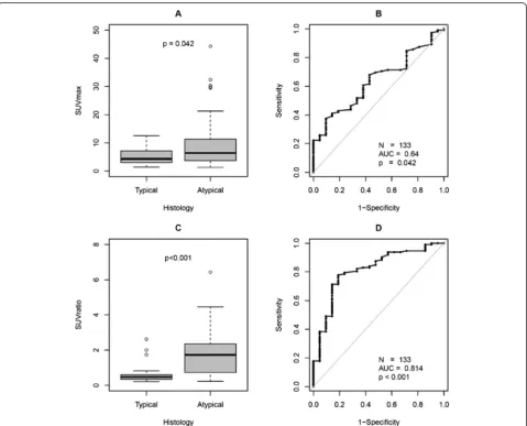
SUVmax of SR-positive tumor lesions (133/186) were normalized to the SUVmax of the liver to generate normal-ized SUV (SUVratio) values. SUVratio was significantly higher in AC (median/IQR/range, 1.7/0.7–2.4/ 0.2–6.4) as compared to TC (median/IQR/range, 0.5/0.3–0.6/0.2–2.6;
p< 0.001) with respect to all lesions (N= 133, PT 8, metas-tases 125; Fig. 2). AC metastatic lesions (median/IQR/ range, 1.7/0.8–2.4/0.2–6.4) also showed significantly higher SUVratio as compared to TC (median/IQR/range, 0.4/0.3– 0.6/0.2–2.0;p< 0.001).
PET vs. CT—patient-based analyses
Frequency and characteristics of metastases The fre-quency of metastases in patients with AC (13/15; 86.7 %) was higher compared to patients with TC with a trend towards significance (6/12; 50 %; p= 0.087). In pa-tients with AC, 4/15 had mixed lesions, 3/15 had somato-statin receptor-negative lesions, 2/15 had no detectable lesions on SR PET, whereas in the remaining 6/15, patients all the lesions were somatostatin receptor positive. In pa-tients with TC 1/12 had mixed lesions, 1/12 had PET-negative lesions, 7/12 had no detectable lesions on SR PET, whereas in the remaining three patients, all the lesions were somatostatin receptor positive (Table 4). Frequency of pa-tients with mixed lesions was not statistically significant between TC (1/12 = 8.3 %) and AC (4/15 = 26.7 %; p=
Fig. 1Ki67 of primary tumor (PT) and metastases depicted as boxplots and receiver operating curves (ROC). Proliferation rates in PT (N= 13) were significantly lower compared to metastases (N= 17)
Table 2Absolute and relative frequency of concordant and discordant lesions on PET/CT
Only positive on PET
Only positive on CT
Concordant positive on PET and CT
Total
PTa 0 (0 %) 21(72.4 %) 8 (27.6 %) 29 (15.6 %)
Liver 9 (18.4 %) 23 (46.9 %) 17 (34.7 %) 49 (26.3 %)
Bone 17 (32.7 %) 0 (0 %) 35 (67.3 %) 52 (28.0 %)
Lymph Node 2 (6.9 %) 9 (31.0 %) 18 (62.1 %) 29 (15.6 %)
Others 4 (14.8 %) 0 (0 %) 23 (85.2 %) 27 (14.5 %)
Total 32 (17.2 %) 53 (28.5 %) 101 (54.3 %) 186 (100 %) a
0.34). This was also true analyzing only patients with me-tastases (TC vs. AC, 1/6 = 16.7 % vs. 4/13 = 30.8 %;p= 1).
Bone metastases were present only in AC (N= 6) but not in TC patients, and all bone metastases were SR PET-positive lesions.
Effect of PET on management strategy Additional findings on PET missed on CT lead to upstaging in four
patients (AC N= 3; TC N= 1; all restaging) resulting in change in management strategy (Table 5). Two patients (1 AC, 1 TC) with liver metastases but no extrahepatic lesions were treated with transarterial embolization, and afterloading, in one patient (AC), salvage PRRT was ruled out because of stable disease in the bone, and in the fourth patient (AC), a wait-and-watch policy was ap-plied because of low tumor burden.
Table 3Patients’characteristics with confirmed liver metastases on CT or PET in follow-up
Patient 4 Patient 12 Patient 20 Patient 27 Patient 29 Patient 30 Patient 31 Patient 35
Ki67 15 % 10 % 5 % 10 % 1 10 % 20 % 7 %
Histo AC AC AC AC TC AC AC AC
Lesion size (mm) 7–32 14–40 20–150 – – 15–62 21–23 15–19
Somatostatin receptor-positive lung lesions 19/24 0/5 5/5 1/1 2/2 0/7 2/2 2/2
CT-positive lesions 21/24 5/5 4/5 0/1 0/2 7/7 2/2 2/2
SUVmax 7.4–17.4 – 5.2–10.5 16.2–44.4 16.5–17.5
In four patients referred for restaging (all, AC) and in one patient referred for staging (TC), additional findings on CT missed on PET lead to correct staging (Table 5). In patients referred for restaging, additional findings on PET lead to upstaging with change in management strat-egy in 4/22 (18.2 %) patients. In one patient (Table 5, patient #8), one of the liver lesions seen on CT was bi-opsied and was confirmed to be free of malignancy. All the lesions in this patient were found to be somatostatin receptor negative, and the disease was downstaged cor-rectly by PET.
Patients with multiple lung nodules Three of 27 pa-tients (11.1 %) had multiple lung nodules and were sub-classified into DIPNECH by the tumor board based on initial findings and the follow-up results. All the lung nodules diagnosed on CT were subclassified as primary tumor due to the absence of histopathological confirm-ation. One patient presented with nine lymph node me-tastases all positive on both PET and CT. However, only 6/26 (23.1 %) lung lesions range in size from 6 to 26 mm were found to be somatostatin receptor positive with very low SUVmax (Table 6) in these patients with DIPNECH.
Discussion
The incidence of LNET is increasing [2]. In the absence of evidence-based consensus guidelines on the manage-ment of LNET, the current standard of practice varies appreciably according to the availability of diagnostic tools: contrast-enhanced CT is standard in virtually all LNET patients often followed by somatostatin receptor scintigraphy or Ga-68 DOTATOC/DOTATATE PET/CT. There is only one study which prospectively examined the role of SR scintigraphy during the follow-up of patients after bronchial carcinoid resection [10]. Out of 16 patients enrolled, 15 had TC and 1 had AC. The authors compared CT and SR scintigraphy and found SR scintigraphy to be useful in 2/16 patients (12.5 %) whereas CT was found to be of additional benefit to SR scintigraphy in 1/16 patients; on the other hand, SR scintigraphy was found to be false positive due to co-existing sarcoidosis in one patient whereas CT was false positive for a lung nodule in another patient. Although prospective, this study comprised almost only TC patients, and there are no reliable data in AC patients available so far.
This difficulty in standardisation of imaging tools is partly attributable to the rarity as well as to the heterogen-eity of LNETs. Although our study presents the results of Table 4Absolute and relative frequency of somatostatin receptor-positive and somatostatin receptor-negative lesions in AC and TC patients
Histopathology All negative All positive Mixed lesions No metastases Total
TC 1 (8.3 %) 3 (25 %) 1 (8.3 %) 7 (58.3 %) 12
AC 3 (20.0 %) 6 (40.0 %) 4 (26.7 %) 2 (13.3 %) 15
Total 4 (14.8 %) 9 (33.3 %) 5 (18.5 %) 9 (39.3 %) 27
Table 5Patients with PET leading to correct and incorrect staging
Patient ID Sex Age Histo Ki67 Additional CT information Additional PET information Change in management due to PET
PET leading to correct staging
#27a M 67 AC 2 – 1 liver, 2 bone SD bone, no salvage PRRT indicated
#8a F 63 AC 3 Liver cysts Follow-up, without intervention
#28a F 34 AC 15 – 3 bone, 3 others, Low tumor burden, wait and watch, no PRRT
#29a F 68 TC 5 – 2 liver Afterloading of liver metastases
#31a F 53 AC 20 – 2 bone TAE of liver metastases seen on CT and SR PET
because of low tumor burden on bone
CT leading to correct staging
#1a F 74 AC 10 1 recurrent tumor in lung, 9 LN – –
#10b F 58 TC NA 8 PT – –
#12a M 59 AC 10 5 liver 1 LN –
#30a F 50 AC 10 7 liver – –
SDstable disease,LNlymph nodes,PTprimary tumor,TAEtransarterial embolization,PRRTpeptide receptor radionuclide therapy,SRsomatostatin receptor,
TCtypical carcinoid,ACatypical carcinoid,Mmale,Ffemale,NAnot available
a
Restaging
b
somatostatin receptor PET/CT in the largest patient series of low- and intermediate-grade neuroendocrine tumors of the lungs so far, collected over a time period of 6 years, it comprises still a low number of patients with however well-documented histopathology including proliferation rates.
An important aspect of tumor heterogeneity of LNET is the differential somatostatin receptor expression, partially depending on tumor grade. In our study, AC patients with intermediate-grade tumors, although not significant, were found to have a higher proportion of mixed lesions, i.e., both somatostatin receptor-positive and receptor-negative lesions as compared to TC patients which had more homogeneous somatostatin receptor expression. The lack of significance could be because of the relatively low num-ber of patients in the two subgroups as well as due to the lower frequency of metastases in TC as compared to AC. Moreover, in our patient population, proliferation rates of TC and AC metastases were significantly higher than those of the primaries which is partly due to differences between tumor clones in primary tumors and metastases [15] challenging the choice of the perfect tracer for these tumors, i.e., FDG as tracer for rather highly proliferative and high-grade tumors vs. Ga-68-labeled somatostatin receptor analogs, usually considered as tracer of choice for the well-differentiated and, thus, low- and intermediate-grade tumors. These complex inter- and intrapatient dif-ferences in the clonal behavior of the primary tumors and the metastases can theoretically be picked up only by combining different imaging tools. Indeed, in our study, only the combination of both functional SR PET aging and morphological contrast-enhanced CT im-aging yielded the maximum information necessary for appropriate staging and restaging because concordant results between SR PET and CT were observed in only 54 % of the lesions. This rather low concordance be-tween both imaging modalities clearly shows the need for combining both with each other to SR PET/con-trast-enhanced (ce) CT.
In general, CT was more sensitive for staging of liver and lung lesions whereas PET performed significantly
better in the detection of bone metastases. Lower sensi-tivity of PET in the detection of lung lesions as well as liver lesions as compared to CT is at least partly be at-tributable to the partial volume effect below 1 cm in diameter, normal physiological uptake of Ga-68 DOTA-TOC/DOTATATE in liver as well as to breathing move-ment artefacts [16]. In one patient, the disease in the liver was classified to be polycystic liver disease. In this patient, the hypodense lesions in the liver were all somatostatin receptor negative thereby making it essential to keep this as differential diagnosis in patients with neuroendocrine tumor and somatostatin receptor-negative lesions.
In contrast, additional lesions were detected by PET in 3 AC patients (patient 27, 1 liver lesion, 2 bone lesions; patient 28, 3 lymph node metastases, 3 bone lesions; patient 31, 2 bone lesions) and 1 TC patient (patient 29, 2 liver lesions). More importantly, in patients referred for restaging, additional findings on PET lead to upstaging with change in management strategy approximately every fifth patient.
Apart from allowing correct staging and restaging, com-bined SR-PET/CT allows selection of appropriate patients for PRRT by ruling out mixed lesions which is a contraindi-cation for performing PRRT and by allowing quantificontraindi-cation of somatostatin receptor expression and assessment of SR-positive tumor burden which is required for making a deci-sion on PRRT. In the absence of standardized systemic treatment option for AC and TC patients, PRRT appears to be a valuable therapeutic option. In our overall patient population (data not shown here), three AC patients SR-PET/CT showed very high somatostatin receptor expres-sion and no mismatch between PET and CT results. These patients were treated with PRRT, and an excellent therapy response could be shown in one patient (see Fig. 3).
The treatment strategy of LNETs also depends on their metastasizing potential. Our observation that TC metas-tasizes less frequently as compared to AC is in line with previous studies: this difference is related to their differ-ences in proliferative activity and, thus, aggressiveness, with AC having a higher frequency of nodal (50 %) and distant metastases (20 %) as compared to TC [17, 18]. However, typical carcinoids can also metastasize as shown in our retrospective analyses in which PET/CT revealed metastases in 50 % of the patients, making it mandatory to perform SR PET/CT in patients with TC at least once for staging/restaging to rule out distant metastases. In the presence of somatostatin receptor-negative lesions during initial staging with SR PET/CT, further follow-up exami-nations should then be based on clinical symptoms, CT, and serum tumor markers while SR PET/CT should be considered for follow-up examinations in patients with receptor-positive lesions.
For atypical carcinoids, especially in cases with high sus-picion of tumor recurrence after surgery and/or higher Table 6Characteristics of patients with diffuse pulmonary
neuroendocrine cell hyperplasia (DIPNECH)
Patient 3 Patient 10 Patient 25
Ki67 5 % NA 15 %
Transformation TC TC AC
Lesion size (mm) 2–18 2–26 2–26
Somatostatin receptor positive lung lesions
4/13 0/8 2/5
SUVmax 1.4–7.9 – 1.0–2.5
LN-Metastases on SR-PET and CT – – 9/9
grade tumors, alternatively FDG PET/CT could be per-formed in case of SR PET-negative lesions as is supported by the study of Kayani et al. [19] who found higher grade LNET to be more FDG avid as compared to Ga-68 DOTATATE. Of special note, they found FDG PET to be less useful in the differentiation of post-radiation changes from vital tumor tissue.
Surgery is generally offered with curative intent to all patients with operable well-differentiated metastases from NET regardless of the site of origin (foregut, mid-gut, or hindgut) [20]. The majority of patients will have recurrent disease at 5 years if distant metastases were present at initial diagnosis [20]. One of the patients in our retrospective analysis presented with local recur-rence 10 years after the first surgical resection (Fig. 4). Occurrence of late metastases in patients with carcin-oid lung tumors has been already previously reported and necessitates regular follow-up of such patients for at least 10 years [21] and probably even longer.
Tumor recurrence can either be hepatic and/or extrahe-patic. Liver is the most frequent site of distant metastases. Prior to liver surgery with curative intent, it is important to rule out extrahepatic metastases. High rates of detection of bone and lymph node metastases by SR PET suggest that at least one combined SR PET/CT should be performed in pa-tients with AC and TC prior to any liver surgery. However, if liver surgery is planned, MRI with liver-specific contrast
agent should always be performed in addition to CT and/or SR PET/CT because it has the highest sensitivity for detec-tion of liver metastases [22]. One of the inherent limitadetec-tions of SR PET in detection of liver metastases is the normal physiological uptake of the tracer in hepatocytes which lead to relatively low target non-target ratio, especially if the lesions are smaller than 1 cm or if the lesions have low somatostatin receptor expression.
On the other side of the spectrum of lung neuroendo-crine neoplasms, as far as receptor expression and mis-match between SR PET and CT results is concerned, are the DIPNECH. Management of patients with DIPNECH has always posed a major challenge because very little is known about their exact biological behavior and clinical course [5, 7]. In our analysis, we identified three patients with malignant transformation of initial DIPNECH into TC or AC. One of these patients also developed lymph node metastases and later on responded to chemotherapy underscoring the need of routine follow-up in this rare type of lung tumors (Fig. 5).
Our study is not the first one to look into the role of Ga-68-labeled somatostatin receptor PET/CT in LNET. Previous studies have compared Ga-68-DOTATATE and Ga-68-DOTATOC in comparison to FDG PET/CT in patients with AC and TC [19, 23, 24]. In these studies, the main purpose was to look at the different rates of som-atostatin receptor expression in TC and AC. We also
Fig. 5Patient with initially diffuse pulmonary neuroendocrine cell hyperplasia (DIPNECH) with transformation into an atypical carcinoid was referred for Ga-68 DOTATOC PET/CT. Based on weak somatostatin receptor expression, patients was treated with chemotherapy (folinic acid, 5 fluorouracil, and oxaliplatin) and showed a good response.a-eBefore chemotherapy.f-jAfter chemotherapy.a,fMaximum intensity projection PET images.b,c,g,hAxial PET images.d,e,i,jCorresponding axial CT images. Partial remission of the mildly receptor positive lesion in the right lung is well appreciated on CT (arrows). On MIP images, the previously receptor-positive hilar and mediastinal lymph node lesions also show response to treatment
looked at the degree of somatostatin receptor expression in TC and AC. Our findings, however, are contradictory to the previously reported results [19, 23, 24]. In our analysis, TC lesions were found to have a significantly lower tumor SUVmax and SUVratio than AC lesions (Fig. 2) whereas the previously published studies reported significantly higher SUVmax in TC as compared to AC [19, 23, 25]. This difference could be primarily due to the difference in the patient populations. While in our analysis, most of the patients (22/27; 81.5 %) underwent SR PET/CT for re-staging after primary tumor resection, in the study from Kayani et al. [19], 83 % (15/18) of the patients underwent SR PET/CT for staging, and the study of Venkitaraman et al. [23] considered only patients (N= 26) referred for staging. Furthermore, the ratio of TC (44 %) vs. AC (56 %) in our population is quite different in comparison to Kaya-ni’s group [19] with 72 % TC (N= 11) vs. 11 % AC (N= 2) or Venkitaraman et al. [23] (TC = 81 %, N= 21 vs. AC 19 %, N= 5). In their analyses of SUV in TC and higher grade LNET, Kayani et al. [19] categorized SCLC and NSCLC with NET differentiation in one group and LCNEC together with AC into another group of NEN which is not in accordance with the WHO classification [4] and is also distinct from the classification suggested by Rindi et al. [14]. Rindi et al. [14] included information on findings by SR scintigraphy in three patients with TC and five patients with AC and found a higher incidence of negative scinti-grams in TC as compared to AC (33 % vs. 20 %). Thus, our findings, based on a larger patient population, confirm these initial results by Rindi et al. showing that somato-statin receptor expression is also a valuable biomarker for tumor detection and (re) staging in patients with intermedi-ate grade AC tumors.
One of the major limitations of this analysis is its retro-spective nature. Although we included all patients with AC and TC which received SR PET/ceCT at our ENETS center in this analysis, there will probably be a selection bias due to the fact that rather patients with suspicion for relapse or metastatic disease will have been referred for SR PET/CT. Thus, our patient population may not repre-sent the full, i.e., unselected cohort of patients with AC and TC, and thus, the distribution of imaging characteris-tics of our patients might be biased to some extent. Apart from this, we used in our study both radiotracers, Ga-68 DOTATOC and Ga-68 DOTATATE, for SR PET imaging. The use of either tracer was solely based on its availability due to patent constraints but not on medical reasons. Although these tracers have slightly different binding af-finities to somatostatin receptor subtypes, there seems to be no clinically relevant difference in the diagnostic accur-acy for NET [20]. However, it would be interesting to also look into other somatostatin receptor analogs covering a broader spectrum of somatostatin receptor subtypes such as Ga-68 DOTANOC [26].
Conclusions
In conclusion, TC and AC patients have complex patterns of metastases which make it necessary to combine func-tional, i.e., Ga-68 SR PET and morphological imaging, i.e., contrast-enhanced CT for appropriate restaging because only 54 % of the lesions are concordantly detectable by both modalities. The major advantage of SR PET lies in the detection of additional bone lesions. Of similar importance, SR PET/CT allows correct discrimination of patients with heterogeneous (mixed lesions) and homogeneous (all lesions are either somatostatin receptor-positive or somatostatin receptor-negative) lesions which is an es-sential prerequisite for the selection of the appropriate therapy, especially with respect to PRRT. In patients referred for restaging SR, PET may have a relevant impact on treatment strategy in up to 18 % of patients with typical and atypical lung carcinoids.
Authors’contributions
VP designed, performed, analyzed, and wrote manuscript. IGS analyzed the data and wrote the manuscript. MP, TD, ET, KA, AP, RA, and WB revised the manuscript. MP and WB gave critical inputs in writing the manuscript. VP, TD, and WB evaluated all the imaging data. MP gave clinical input to the study whereas RA helped in performing the histopathological classification of the tumor specimens.
Acknowledgements
None.
Compliance with ethical standard
Ethic Commssion, Charité Universitätsmedizin Berlin
Funding
No funding was received for the study.
Conflict of interest
Marianne Pavel has received payments as a lecturer as well as a consultant for Novartis, Ipsen Pharma, Pfizer, and Lexicon Pharmaceuticals. Vikas Prasad has received payments as a lecturer as well as travel grants from Bayer Healthcare, Novartis, Ipsen Healthcare, Pfizer, and ITM Isotope Technologies Munich. In addition, he has received research funds from ITM Isotope Technologies.
Munich, Nordion and Affibody AG. Timm Denecke has received payments as a lecturer as well as travel grants from Bayer Healthcare, Novartis Pharma, and Ipsen Pharma. Winfried Brenner, Ingo G Steffen, Andreas Pascher, Ruza Arsenic, Konstantina Apostolopoulou, and Elisabeth Tischer declared that they have no competing interests.
Ethical approval
The retrospective analyses were performed in accordance with the ethical standards of the institutional ethics committee and with the 1964 Helsinki declaration and its later amendments.
Informed consent
Informed consent was obtained from all individual participants included in the study.
Author details
1Department of Nuclear Medicine, Charité Universitätsmedizin, Berlin,
Received: 3 August 2015 Accepted: 23 September 2015
References
1. Gustafsson BI, Kidd M, Modlin IM. Neuroendocrine tumors of the diffuse neuroendocrine system. Curr Opin Oncol. 2008;20(1):1–12.
2. Modlin IM, Lye KD, Kidd M. A 5-decade analysis of 13,715 carcinoid tumors. Cancer. 2003;97(4):934–59.
3. Travis WD, Brambilla E, Müller-Hermelink HK, Harris CC. Pathology andgenetics of tumours of the lung, pleura, thymus and heart. World Health Organization Classification of Tumours. Lyon, France: IARC Press; 2004. 4. Brambilla E, Travis WD, Colby TV, Corrin B, Shimosato Y. The new World Health
Organization classification of lung tumours. Eur Respir J. 2001;18(6):1059–68. 5. Gorshtein A, Gross DJ, Barak D, Strenov Y, Refaeli Y, Shimon I, et al. Diffuse
idiopathic pulmonary neuroendocrine cell hyperplasia and the associated lung neuroendocrine tumors: clinical experience with a rare entity. Cancer. 2012;118(3):612–9.
6. Modlin IM, Bodei L, Kidd M. A historical appreciation of bronchopulmonary neuroendocrine neoplasia: resolution of a carcinoid conundrum. Thorac Surg Clin. 2014;24(3):235–55.
7. Davies SJ, Gosney JR, Hansell DM, Wells AU, du Bois RM, Burke MM, et al. Diffuse idiopathic pulmonary neuroendocrine cell hyperplasia: an under-recognised spectrum of disease. Thorax. 2007;62(3):248–52.
8. Lu YY, Chen JH, Liang JA, Chu S, Lin WY, Kao CH. 18 F-FDG PET or PET/CT for detecting extensive disease in small-cell lung cancer: a systematic review and meta-analysis. Nucl Med Commun. 2014;35(7):697–703. 9. Krenning EP, Kwekkeboom DJ, Bakker WH, Breeman WA, Kooij PP, Oei HY, et
al. Somatostatin receptor scintigraphy with [111In-DTPA-D-Phe1]- and [123I-Tyr3]-octreotide: the Rotterdam experience with more than 1000 patients. Eur J Nucl Med. 1993;20(8):716–31.
10. Bini A, Grazia M, Stella F, Petrella F, Sellitri F, Fanti S, et al. The role of somatostatin receptor scintigraphy (Octreoscan) during follow-up of patients after bronchial carcinoid resection. A prospective study. J Cardiovasc Surg (Torino). 2005;46(3):318–9.
11. Zhernosekov KP, Filosofov DV, Baum RP, Aschoff P, Bihl H, Razbash AA, et al. Processing of generator-produced 68Ga for medical application. J Nucl Med. 2007;48(10):1741–8.
12. Virgolini I, Ambrosini V, Bomanji JB, Baum RP, Fanti S, Gabriel M, et al. Procedure guidelines for PET/CT tumour imaging with 68Ga-DOTA-conjugated peptides: 68Ga-DOTA-TOC, 68Ga-DOTA-NOC, 68Ga-DOTA-TATE. Eur J Nucl Med Mol Imaging. 2010;37(10):2004–10.
13. Surti S, Kuhn A, Werner ME, Perkins AE, Kolthammer J, Karp JS. Performance of Philips Gemini TF PET/CT scanner with special consideration for its time-of-flight imaging capabilities. J Nucl Med. 2007;48(3):471–80. 48/3/471. 14. Rindi G, Klersy C, Inzani F, Fellegara G, Ampollini L, Ardizzoni A, et al.
Grading the neuroendocrine tumors of the lung: an evidence-based proposal. Endocr Relat Cancer. 2014;21(1):1–16.
15. Yokota J. Tumor progression and metastasis. Carcinogenesis. 2000;21(3):497–503. 16. Kuehl H, Veit P, Rosenbaum SJ, Bockisch A, Antoch G. Can PET/CT replace
separate diagnostic CT for cancer imaging? Optimizing CT protocols for imaging cancers of the chest and abdomen. J Nucl Med. 2007;48 Suppl 1:45S–57S.
17. Fink G, Krelbaum T, Yellin A, Bendayan D, Saute M, Glazer M, et al. Pulmonary carcinoid: presentation, diagnosis, and outcome in 142 cases in Israel and review of 640 cases from the literature. Chest. 2001;119(6):1647–51. 18. Scott WJ. Surgical treatment of other bronchial tumors. Chest Surg Clin N
Am. 2003;13(1):111–28.
19. Kayani I, Conry BG, Groves AM, Win T, Dickson J, Caplin M, et al. A comparison of 68Ga-DOTATATE and 18F-FDG PET/CT in pulmonary neuroendocrine tumors. J Nucl Med. 2009;50(12):1927–32.
20. Pavel M, Baudin E, Couvelard A, Krenning E, Oberg K, Steinmuller T, et al. ENETS Consensus Guidelines for the management of patients with liver and other distant metastases from neuroendocrine neoplasms of foregut, midgut, hindgut, and unknown primary. Neuroendocrinology. 2012;95(2):157–76. 21. Ferolla P, Daddi N, Urbani M, Semeraro A, Ribacchi R, Giovenali P, et al.
Tumorlets, multicentric carcinoids, lymph-nodal metastases, and long-term behavior in bronchial carcinoids. J Thorac Oncol. 2009;4(3):383–7. 22. Schreiter NF, Nogami M, Steffen I, Pape UF, Hamm B, Brenner W, et al.
Evaluation of the potential of PET-MRI fusion for detection of liver metastases in patients with neuroendocrine tumours. Eur Radiol. 2012;22(2):458–67.
23. Venkitaraman B, Karunanithi S, Kumar A, Khilnani GC, Kumar R. Role of (68)Ga-DOTATOC PET/CT in initial evaluation of patients with suspected bronchopulmonary carcinoid. Eur J Nucl Med Mol Imaging.
2014;41(5):856–64.
24. Jindal T, Kumar A, Venkitaraman B, Meena M, Kumar R, Malhotra A, et al. Evaluation of the role of [18F]FDG-PET/CT and [68Ga]DOTATOC-PET/CT in differentiating typical and atypical pulmonary carcinoids. Cancer Imaging. 2011;11:70–5.
25. Poeppel TD, Binse I, Petersenn S, Lahner H, Schott M, Antoch G, et al. 68Ga-DOTATOC versus 68Ga-DOTATATE PET/CT in functional imaging of neuroendocrine tumors. J Nucl Med. 2011;52(12):1864–70.
26. Prasad V, Baum RP. Biodistribution of the Ga-68 labeled somatostatin analogue DOTA-NOC in patients with neuroendocrine tumors: characterization of uptake in normal organs and tumor lesions. Q J Nucl Med Mol Imaging.
2010;54(1):61–7.
27. Travis WD, Giroux DJ, Chansky K, Crowley J, Asamura H, Brambilla E, et al. The IASLC Lung Cancer Staging Project: proposals for the inclusion of broncho-pulmonary carcinoid tumors in the forthcoming (seventh) edition of the TNM Classification for Lung Cancer. J Thorac Oncol. 2008;3(11):1213–23.
Submit your manuscript to a
journal and benefi t from:
7Convenient online submission 7Rigorous peer review
7Immediate publication on acceptance 7Open access: articles freely available online 7High visibility within the fi eld
7Retaining the copyright to your article