M E T H O D O L O G Y
Open Access
An Initial Investigation of an Alternative
Model to Study rat Primordial Germ Cell
Epigenetic Reprogramming
Isabelle Hernandez Cantão
1,2, Renato Borges Tesser
1,2and Taiza Stumpp
1,2*Abstract
Background:Primordial germ cells (PGC) are the precursors of the gametes. During pre-natal development, PGC undergo an epigenetic reprogramming when bulk DNA demethylation occurs and is followed by sex-specific de novo methylation. The de novo methylation and the maintenance of the methylation patterns depend on DNA methyltransferases (DNMTs). PGC reprogramming has been widely studied in mice but not in rats. We have previously shown that the rat might be an interesting model to study germ cell development. In face of the difficulties of getting enough PGC for molecular studies, the aim of this study was to propose an alternative method to study rat PGC DNA methylation. Rat embryos were collected at 14, 15 and 19 days post-coitus (dpc) for the analysis of 5mC, 5hmC, DNMT1, DNMT3a and DNMT3b expression or at 16dpc for treatment 5-Aza-CdR, a DNMT inhibitor, in vitro.
Methods:Once collected, the gonads were placed in 24-well plates previously containing 45μm pore membrane and medium with or without 5-Aza-CdR. The culture was kept for five days and medium was changed daily. The gonads were either fixed or submitted to RNA extraction.
Results:5mC and DNMTs labelling suggests that PGC are undergoing epigenetic reprogramming around 14/15dpc. The in vitro treatment of rat embryonic gonads with 1μM of 5-Aza-CdR lead to a loss of 5mC labelling and to the activation ofPax6expression in PGC, but not in somatic cells, suggesting that 5-Aza-CdR promoted a PGC-specific global DNA hypomethylation.
Conclusions:This study suggests that the protocol used here can be a potential method to study the wide DNA demethylation that takes place during PGC reprogramming.
Keywords:Rat, Primordial germ cell, Epigenetic reprogramming
Background
Primordial germ cells (PGC) are peculiar cells that differentiate into gametes and transmit the genetic and epigenetic information to the next generations. In mice, PGC arise from a pool of epiblastic cells that retain the expression of pluripotent markers [1–3]. This pool of cells starts to express fragillisandBlimp1, which conduce to a germ cell fate [4–7]. PGC then migrates via the hindgut and its mesentery to reach the genital ridges, where they undergo male or female differentiation.
The switch from a somatic to a germ cell program depends initially on the maintenance of pluripotency markers and later on epigenetic reprogramming, when PGC DNA is widely hypomethylated [8–10] and then de novo hypermethylated in a sex-specific manner [11]. Mouse PGC reprogramming starts around 8dpc and ends around 16.5dpc in males, when DNA de novo methylation ends [4, 8, 10–15]. De novo DNA methyla-tion, as well as the maintenance of methylation patterns during cell division, occur via DNA methyltransferases (DNMT). DNMT3a and DNMT3b are described as de novo DNA methyltransferases capable of establishing methylation patterns whereas DNMT1 is considered the maintenance DNMT during DNA replication.
* Correspondence:[email protected]
1Laboratory of Developmental Biology, Department of Morphology and
Genetics, Federal University of Sao Paulo (UNIFESP), Sao Paulo, Brazil
2Rua Botucatu, 740. Ed. Leitão da Cunha, 2o andar. CEP: 04023-900, São
Paulo, Brazil
© The Author(s). 2017Open AccessThis article is distributed under the terms of the Creative Commons Attribution 4.0 International License (http://creativecommons.org/licenses/by/4.0/), which permits unrestricted use, distribution, and reproduction in any medium, provided you give appropriate credit to the original author(s) and the source, provide a link to the Creative Commons license, and indicate if changes were made. The Creative Commons Public Domain Dedication waiver (http://creativecommons.org/publicdomain/zero/1.0/) applies to the data made available in this article, unless otherwise stated.
Cantãoet al. Biological Procedures Online (2017) 19:9
It has been suggested that DNA demethylation in mouse PGC occurs through two mechanisms: oxidation of 5mC to 5hmC via TET enzymes and through replication-dependent dilution [16, 17]. These mecha-nisms operate in three steps: the first is a passive process of replicative-dilution of 5mC possibly due to a lack of DNMT1 function and occurs from 8.5dpc to 9.5dpc; the second is the active process where 5mC are oxidated to 5hmC via TET (Ten-eleven translocation) enzymes at 10.5dpc; and the third step is again a passive step that consists in the replicative-dilution of 5hmC from 10.5dpc to 13.5dpc [17].
Like in the early embryo, the de novo DNA methy-lation as well as the maintenance of DNA methyla-tion also seems to be mediated by the DNMTs, although the data are controversial. DNMT1 seems to be dispensable for the reacquisition of DNA methyla-tion in male germ cells [18, 19]. On the other hand, DNMT3a is responsible for de novo DNA methyla-tion [18, 20], whereas DNMT3b would funcmethyla-tion in the maintenance of de novo DNA methylation [20] or would not be required in prenatal germ cell develop-ment [18].
The natural epigenetic reprogramming by which germ cells go through is one of the most interesting events of their pre-natal development. Although the meaning of this event has not been clarified, it is likely to be related to the establishment of a toti-potent state that seems to be fundamental for germ cell differentiation [21] and maybe to confer the de-velopmental potential to the future gametes. Investi-gations about the role of DNA methylation in gene expression have made use of agents that impair the maintenance of DNA methylation, such as 5-Aza-deoxycitidine (5-Aza-CdR), to induce artificial DNA demethylation in vitro [12, 22]. 5-Aza-CdR is known to interact with DNMT1, inhibiting its activity. This compound acts in both proliferating and non-proliferating cells [23, 24] and is able to activate genes which expression is controlled by promoter methylation [12, 22].
Although the mouse has been the main model for studies on germ cell development, we have previously shown relevant differences in the detection of germ cell markers between mice and rats [25], suggesting that the rat also seems to be an important model to study germ cell development. Despite the relatively abundant studies about PGC reprogramming, espe-cially in the mouse, the methodologies involved are laborious due to the low number of PGC in the ini-tial phases of their development. When the rat is the model of choice, the difficulties are increased due to the fact that less information and tools are available for this species.
In face of the technical difficulties to study the control of gene expression by DNA methylation in PGC coupled to the impossibility to establish PGC cultures that maintain their in vivo characteristics, we aimed to find new and simpler methodologies to study the mecha-nisms involved in DNA methylation and demethylation in rat PGC. Here we describe DNMT expression in rat embryo gonads and a method that lead to the specific hypomethylation of PGC DNA by the DNMT inhibitor 5-Aza-CdR. This study may be useful for future investi-gations in the field.
Results
Dnmt Expression and Selection of the age for 5-Aza-CdR Treatment
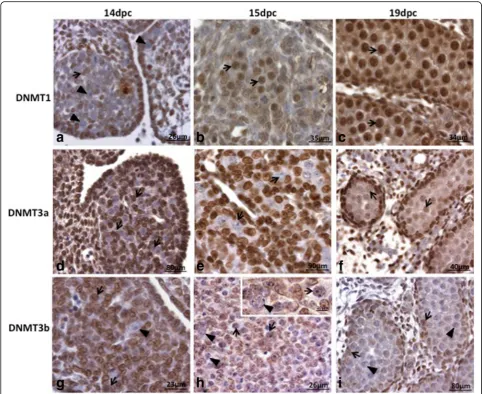
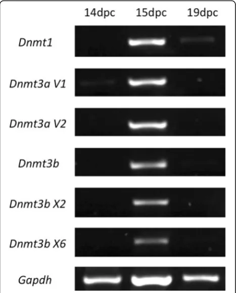
Considering that 5-Aza-CdR acts through the inhibition of DNMT1 activity [26], the presence of this enzyme was investigated in rat PGC (Figs. 1 and 2). DNMT1, which is responsible for maintaining DNA methylation (DNMT1), was not detected in PGC at 14dpc in both protein (Fig. 1a) and RNA (Fig. 2) levels, suggesting that DNMT1 activity is not necessary in PGC at this stage of rat germ cell development. On the other hand, at 15dpc (Fig. 1b) and at 19dpc (Fig. 1c) DNMT1 was detected in PGC at protein level, although at 19dpc its expression at RNA level could be barely detected (Fig. 2).
5mC and 5hmC Immunolabelling
Because 5-Aza-CdR is expected to promote DNA demeth-ylation through interaction with DNMT1, we investigated 5mC and 5hmC labelling in normal gonads as a basis for the analysis of 5-Aza-CdR effectiveness. 5mC was not de-tected at 14dpc (Fig. 3a) but appeared at 15dpc (Fig. 3b), when the presence of negative PGC was very rare. At 19dpc all PGC were positive for 5mC (Fig. 3c). These data are somewhat discordant with a recent study showing that rat PGC DNA is hypomethylated from 14.5–19.5dpc [29]. This discrepancy could be due to the different methodo-logical approaches used, such as fixative solutions, which are essential for the detection of this epigenetic mark.
5-hydroxymethylcytosine (5hmC) is the product of 5mC oxidation by TET (Ten-eleven translocation) enzymes. During mouse PGC reprogramming, DNA demethylation depends on 5mC oxidation to 5hmC [16, 17]. In the present study, 5hmC labelling was de-tected in a restricted area of PGC nucleus from at 14dpc and 15dpc (Figs. 3d and e). The labelling area showed a progressive reduction as the age increased, appearing very rarely at 15dpc until becoming un-detectable at 19dpc (Fig. 3f ). This agrees with recent data on rat PGC showing that 5hmC labelling is present between 14.5dpc and 16.5dpc but is absent at 19.5dpc [31].
Fig. 1DNMT1, DNMT3a and DNMT3b labelling in male rat embryonic gonads. DNMT1 was not detected in PGC (arrowheads) at 14dpc (a); the somatic cells were positive (arrows). At 15dpc (b) and 19dpc (c) all PGC/gonocytes were positive (arrows). These results coincides with 5mC labelling, indicating that PGC reprogramming ends around 15dpc. DNMT3a was not detected in PGC (arrows) at 14dpc (d) and 15dpc (e). At 19dpc (f) DNMT3a was detected (arrows) in all gonocytes, although the labelling intensity has varied among them. At 14dpc (g) and 15dpc (h) DNMT3b-positive (arrows) and DNMT3b-negative (arrowheads) PGC were observed. At 19dpc (i) DNMT3b was not detected (arrowheads) in the gonocytes and was detected in Sertoli cells (arrows)
In Vitro 5-Aza-CdR Administration
The DNMT1 and 5mC data obtained here were used to choose the embryonic age for the in vitro studies using 5-Aza-CdR. Furthermore, we have previously shown that rat PGC migration ends between 15dpc and 16dpc [25]. These findings led us to choose the age of 16dpc for 5-Aza-CdR treatment.
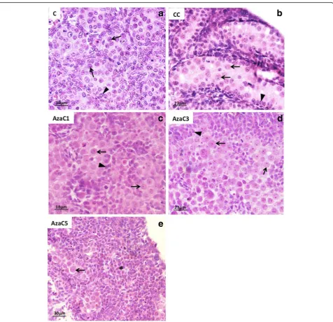
Morphological analysis of the gonads (Fig. 4) and 5mC labelling (Fig. 5) were used to select the adequate dose of 5-Aza-CdR for the aims of this study. Non-cultured (C) (Fig. 4a) and culture-control (CC) (Fig. 4b) were used as basis for comparison of the 5-Aza-CdR-treated gonads. Three concentrations of 5-Aza-CdR were used (1μM, 3μM and 5μM). The gonads treated with 1μM (Fig. 4c) showed well-preserved morphology when com-pared with C (Fig. 4a) and CC (Fig. 4b) gonads. The gonads treated with 3μM (Fig. 4d) and 5μM (Fig. 4e) of 5-Aza-CdR showed apparent reduction of its size and morphological alterations, such as cell degeneration (Fig. 4d). In the gonads treated with 5 μM of 5-Aza-CdR PGC were rare and the gonads contained basic-ally somatic cells (Fig. 4e).
5mC and 5hmC Labelling after 5-Aza-CdR Treatment The gonads treated with 1 μM and 3 μM of 5-Aza-CdR were submitted to 5mC immuno-labelling. The gonads treated with 5 μM of 5-Aza-CdR were excluded from this analysis due to the significant germ cell loss, as described above.
As observed for the 19dpc embryos, 5mC was detected in all PGC of the 16dpc embryos (Fig. 5a). After the in-cubation of embryonic male gonads without 5-Aza-CdR (CC), 5mC was also detected in all PGC and somatic cells (Figs. 5b). On the other hand, 5mC was barely detected in the PGC of the gonads incubated with 1μM of 5-Aza-CdR (Aza1), but was detected in the somatic cells (Fig. 5c), suggesting that the PGC are more sensi-tive to 5-Aza-CdR effect on DNA demethylation than the somatic cells. In the gonads treated with 3μM of 5-Aza-CdR (AzaC3) 5mC labelling was very reduced in both PCG and somatic cells (Fig. 5d), suggesting that this dose is able to induce the loss of 5mC not only in PGC DNA but also in somatic cell DNA. Therefore, we suggest that the dose of 1 μM represents an suitable concentration of 5-Aza-CdR to perform studies involv-ing 5mC detection in rat PGC in whole gonad culture systems.
In control embryo the gonads (C) 5hmC labelling was rarely observed and, when present, was restricted to a small area of PGC nucleus (Fig. 5e). Interestingly, both CC (Fig. 5f ) and in AzaC1 (Fig. 5g) gonads showed 5hmC labelling in PGC and Sertoli cells, although it was more intense in Sertoli cells than in PGC. This suggests that the protocol of gonad culture used here leads to an increase of 5hmC detection in PGC.
PGC Quantification and Proliferation
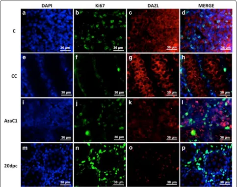
Because the morphological analysis of the AzaC1 gonads suggested an increase of PGC number, we decided to count these cells using a germ cell marker (DAZL) associated with a proliferation marker (Ki67). The Ki67/DAZL double-labelling indicated that PGC pro-liferation was active in the C gonads (Figs. 6a to d), but was inactive in both CC (Figs. 6e to h) and AzaC1 gonads (Figs. 6i to l). If germ cells continue to develop in vitro, it is indeed expected that their proliferation has ceased by the end of the culture period, when these cells would correspond to 20dpc germ cells. At this age rat male germ cells has entered mitotic arrest (Zogbi et al. [30]). The Ki67/Dazl double-labelling was then per-formed in 20dpc embryos to confirm this data. In-deed, no Ki67 labelling was observed in the 20dpc germ cells (Figs. 6m to p). Despite the fact that PGC were Ki67-negative by the end of the culture period, the number of PGC in the AzaC1 gonads was higher when compared with the control gonads (Table 1),
what lead to an increase of the numerical density (Nv) of these cells in the AzaC1 gonads (Fig. 7). This suggests that 5-Aza-CdR caused an alteration of germ cell proliferation, although their maturation, which can be indicated by mitotic arrest, which was not impaired.
Expression of Developmental Genes
The expression of developmental genes is a mark of the early phases of embryo development and of em-bryonic stem cells. In mouse PGC these genes are not expressed, although seems to be maintained in a bivalent state [31, 32]. Since DNMTs are fundamental for the control of developmental genes expression during embryo development [33], we investigated whether 5-Aza-CdR treatment induces the expression of the somatic developmental gene Pax6 in rat PGC. Pax6 expression was not detected in the control (C and CC gonads) PGC or in the somatic cells. On the other hand, in the in the AzaC1 gonads Pax6 expres-sion was detected in PGC but not in the somatic cells (Fig. 8), suggesting that the treatment with 5-Aza-CdR induced PGC-specific activation of this gene.
Discussion
Germ cell development depends on strictly controlled events that assure their normal differentiation and consequently the production of functional gametes and healthy embryos. In this context PGC epigenetic reprogramming is of particular importance, since it is required for PGC sex differentiation, imprinting es-tablishment and to provide adequate chromatin con-ditions for the future embryo development. Many important studies have been investigating and describ-ing PGC epigenetic reprogrammdescrib-ing, especially in the mouse, and have provided substantial data. However, studies about rat PGC epigenetics are very scarce, prob-ably due to the reduced information about molecular as-pects of the rat development when compared to mouse. This lead us to search for methodologies to study rat PGC epigenetics. Here we used 5-Aza-CdR, which is known to interact with DNMT1 and to block its activity, to promote male PGC DNA demethylation in vitro. To propose the protocol used here, we took into account two main aspects: the difficulties of getting enough num-ber of PGC during early reprogramming and the fact that DNA methylation represents a major form of gene ex-pression control.
Fig. 35mC and 5hmC immunolabelling in the embryo gonads at 14dpc, 15dpc and 19dpc. 5mC was not detected in PGC (arrows) at 14dpc (a). At 15dpc (b) 5mC was detected in most PGC (arrows), although very few PGC were still negative (arrowhead). At 19dpc 5mC was detected in all gonocytes (arrows) (c). The labelling of 5hmC was restricted to small nuclear areas at 14dpc (d) and 15dpc (e) PGC (arrowheads). 5hmC-negative PGC were also observed (arrows). The 5hmC-positive areas were more abundant at 14dpc (d) than at 15dpc (e). At 19dpc 5hmC was not detected in gonocytes (arrows) (f)
PGC reprogramming seems to involve the reduction of the activity of DNMT1 in the beginning of the repro-gramming period [13, 34] followed by the recruiting of DNMT3a and DNMT3b [18, 35, 36]. Since 5-Aza-CdR inhibits DNMT1 activity and also affects DNMT3a and DNMT3b [37, 38], it was necessary to investigate the ex-pression of these enzymes prior to establish the protocol of 5-Aza-CdR treatment in the rat gonad cultures. We detected DNMT1 in rat PGC at 15dpc and 19dpc, but not at 14dpc. Since DNMT1 is described as the main-tenance DNMT, it is expected that its presence
correlates with the moment of PGC de novo DNA methylation. However, a previous study showing epigen-etic marks in rat PGC indicated that 5mC reestablish-ment occurs around 19.5dpc [39], suggesting DNA de novo methylation occurs in late gestational period. On the other hand, in mouse PGC, DNMT1 was detected between 12.5dpc and 16.5dpc [19], when DNA is glo-bally demethylated [13], what agrees with our data. Conversely, [18] suggested that DNMT1 is not present in mouse PGC between 14.5dpc and 18.5dpc, a period that includes demethylated and de novo
methylated DNA [13]. DNMT1 expression agrees with 5mC labelling, which was not detected at 14dpc and was detected at 15dpc and 19dpc. These data suggest that rat PGC are hypomethylated at 14dpc and that de novo methylation may start between 14dpc and 15dpc.
The de novo methyltransferases DNMT3a and DNMT3b are fundamental for de novo methylation of the mouse embryo [29] and ES cells [40] although
seems to be dispensable for somatic cell nuclear reprogramming to a pluripotent state [41]. In the mouse germ cells DNMT3a, DNMT3b and DNMT3l are important for de novo methylation of imprinted regions during development and after birth and for retrotransposons silencing [42]. DNMT3a was re-ported to act in the acquisition of de novo methyla-tion in mouse PGC [18, 20], whereas DNMT3b seems to function in the maintenance of methylation [20]
Fig. 55mC and 5hmC labelling in the control (C), culture-control (CC) and 5-Aza-CdR-treated (Azac1 and AzaC3) gonads of 16dpc embryos. 5mC is detected in C (a) and CC (b) PGC (arrows) and somatic cells (arrowheads). In the gonads treated with 1μM of 5-Aza-CdR (c) very weak labelling was observed in PGC (arrows) whereas the somatic cells showed more intense labelling (arrowheads). The gonads treated with 3μM of 5-Aza-CdR (d) showed weak labelling in PGC (arrows) and somatic cells (arrowheads). 5hmC was detected in restricted regions of the germ cell nuclei in control (C) gonads (ar-rows) and in the whole nucleus of Sertoli cells (arrowheads) (e). In control-culture gonads (CC) germ cells (arrows) show weak labeling in the whole nu-cleus, whereas in the Sertoli cells (arrowheads) 5hmC labeling did not change when compared with C gonads (f). After 5-Aza-CdR (3μM) treatment (AzaC1), 5hmC labeling was more intense in germ cells (arrows) when compared with the CC gonads, whereas no alteration was observed in Sertoli cells (arrowheads) (g)
and to play a major role in the postnatal development of male germ cells [18]. Our data on DNMT3a expression suggest that this enzyme is not present in PGC at 14dpc, what agrees with the 5mC data. How-ever, at 15dpc and 19dpc the expression of this en-zyme at both protein and RNA level is controversial, indicating that further studies are needed. In contrast DNMT3b was detected at 14dpc and 15dpc but not at 20dpc, showing an opposite pattern to that
observed for DNMT3a. This suggests that these de novo DNA-methyltransferases seem to play distinct roles in rat germ cell development.
PGC reprogramming in mice seems to be associated with changes in the expression of the DNMTs and their partners. The downregulation ofUhrf1, which is essential for DNMT1 activity, was observed in mice PGC in the be-ginning of epigenetic reprogramming [43], suggesting that DNMT1 action needs to be annulled at this stage of germ cell development. Thus, the use of a DNMT1 inhibitor seems to be a reasonable way to mimic, at least in part, the global DNA demethylation that occurs during PGC reprogramming. Our analyses suggest that the incubation of rat embryonic gonads with the DNMT1 inhibitor 5-Aza-CdR was able to induce wide loss of 5mC in PGC DNA, without causing major global loss of this mark in somatic cells, as suggested by 5mC immno-labelling. The detection of Pax6 expression, which is controlled by
Fig. 6Ki67/DAZL immunolabelling the control (C) (atod), culture-control (CC) (etoh), 5-Aza-CdR-treated gonads (AzaC1) (itol) and 20dpc (m
top). Ki67 was detected in DAZL-positive cells in C gonads but not in CC, AzaC1 and 20dpc gonads. (a), (e), (i) and (m): DAPI; (b), (f), (j) and (n): Ki67 labelling; (c), (g), (k) and (o): DAZL labelling; (d), (h), (l) and (p): merge
Table 1Area and primordial germ cell count (N) averages in
CC and AzaC1 groups
CC AzaC1
Area 238,9 ± 7,0 256,8 ± 6,7
N(PGC) 149,8 ± 7,4 273,5 ± 16*
promoter methylation [44], observed in PGC but not in the somatic cells of the gonads treated with 5-Aza-CdR on in the control PGC corroborate to the hypothesis that 5-Aza-CdR treatment acted primarily on PGC. This sug-gests that in vitro treatment of whole-gonad with 5-Aza-CdR might be an interesting method to study the control of gene expression by DNA methylation in PGC.
Considering that 5-Aza-CdR is an inhibitor of DNMT1 activity, further studies investigating the pluripotency of PGC after 5-Aza-CdR treatment seems to be another point to be considered, since it has been shown that the absence of DNMTs leads to spontaneous pluripotency [45].
On the other hand, the alteration of 5hmC labelling even in the control culture gonads indicates that the cul-ture method by itself is able to induce epigenetic changes in PGC. This and other aspects of PGC epigen-etics, such as histone mark profile, need to be further
investigated to confirm whether this model would indeed be useful to study PGC epigenetics.
Conclusion
In conclusion, we describe the immuno-labelling of DNMTs in rat PGC and suggest that the administra-tion of 5-Aza-CdR to rat gonads in vitro leads to a wide demethylation of PGC DNA without major ef-fects on somatic cells. We finally suggest that this method might be a potential alternative method to study DNA methylation and demethylation, although additional studies are necessary to validate it.
Methods
Animals and Tissue Preparation
Male embryos were obtained from timed matings of adult Wistar rats (Rattus norvegicus albinus) from the Laboratory of Developmental Biology (EPM/UNIFESP, Sao Paulo–Brazil). The adult animals were kept in plas-tic cages under a 12–12 h light/dark cycle at 23–25 °C. Food and water were allowed ad libitum. The dams were anesthetized using the method of anaesthesia/analgesia (xylazin/ketamine, 10 mg/Kg and 100 mg/Kg, respect-ively) and euthanized by cardiac incision. The embryos were collected at 14, 15, 16 and 19dpc. At 14dpc the rat embryos do not show sexual dimorphism. At 15, 16 and 19dpc sexing was performed by morphological inspec-tion of the gonads and only male gonads were used. The gonads of 14, 15 and 19dpc embryos were fixed in Bouin’s solution for 6 h or in Carnoy’s solution for 48 h and processed for paraffin embedding. Cross sections (6 μm-thick) were obtained from embryos and testes and submitted to the labelling of DNMT1, DNMT3a
Fig. 7PGC quantification in 16dpc culture-control (CC) and Aza-treated (Aza) embryo gonads. An increase in the numerical density (Nv) of PGC is observed in the Aza-treated gonads
Fig. 8Expression of thePax6developmental gene in the PGC isolated from control (C), culture-control (CC) and Aza-treated (Aza) gonads. Pax6 expression was detected in Aza PGC but was absent in C and CC gonads. Somatic cells (SC) obtained from Aza gonads go-nads were used to check the PGC-specific activation ofPax6
by 5-Aza-CdR
and DNMT3b (see item 4.3). Five embryos from three different mothers were used for this analysis.
The male gonads of 16dpc embryos (20 embryos from 6 different mothers) were dissected and incubated with 5-Aza-2′-deoxicytidine (5-Aza-CdR) as described below (see item 4.2). The experiments were carried out under the rules of the local committee for animal care (CEUA Nr. 7,001,040,914).
5-Aza-CdR Experiment
The uterus of 18 pregnant females at gestation day 16 (GD16) were removed and taken to a culture room pre-sterilised with UV light. The gonad cultures were per-formed according to Livera et al. [26] and Habert et al. [27]. The experiment was performed three times, using 6 dams for each experiment (total of 18 dams). A total of 72 male embryo gonads (24 gonads per experiment) were dissected and placed on gridded cellulose/ester membrane (0.45 μm, HAWG047S0, Millipore, USA) in 24-well plates containing DMEM supplemented with GIBCO® GlutaMax (Life technologies), 0.5% BSA, 1% Penicillin/Streptomycin and 1 μM, 3μM or 5 μM of 5-Aza-CdR (InSolution™, Cat. 189,826 - Calbiochem). These gonads are from here on referred as AzaC. Con-trol culture (CC) gonads were performed using the same culture medium except for the addition of 5-Aza-CdR. The cultures were maintained for 5 days. 5-Aza-CdR was added on the 1st day of culture and replaced every day. The control and 5-Aza-CdR-treated gonad cultures were carried out concomitantly in every experiment, which was performed three times.
Six control and six 5-Aza-CdR-treated gonads (two from each experiment) were fixed in Carnoy’s solution and embedded in paraffin for 5mC and 5hmC labelling and Haematoxilyn and Eosin (H&E) staining. Other 30 control and 30 5-Aza-CdR-treated gonads (10 from each experiment) were used for PGC purification and RT-PCR (see item 4.4). The gonads were dissected from 20 male embryos from 6 different mothers.
Immunohistochemistry and Immunofluorescence
The sections were dewaxed in xylene, hydrated and sub-mitted to heat antigen retrieval using citrate buffer (pH 6.0) for 10 min (for DNMT1, DNMT3a, DNMT3b and H3K27me3) or to proteinase K (20μg/ml) for 5 min (for 5mC and 5hmC). Antigen blocking was performed using 5% BSA and the slides were incubated with primary antibodies: anti-5mC (1:50, Abcam, ab73938), anti-5hmC (1:50, Active Motif, 39,769), anti-DNMT1 (1:100, Santa Cruz, sc-20,701), anti-DNMT3a (1:100, Santa Cruz sc-20,703) anti-DNMT3b (1:100, Santa Cruz, sc-20,704), anti-Ki67 (Abcam, ab16667) and anti-DAZL (1:200, Serotec, MCA2336) overnight at 4 °C. For
immunohistochemistry the slides were washed in PBS (0.05 M, pH 7.2) and incubated with the LSAB system (DAKO Detection System - K0690) and then with streptavidin-peroxidase (LSAB, DAKO) for 30 min. The reactions were revealed with DAB (DAKO). For im-munofluorescence, after two PBS washes, the slides were incubated with the secondary antibodies Alexa (Invitro-gen, A10036) anti-mouse and FITC (Abcam, Ab6791) anti-rabbit. The nuclei were stained with DAPI.
Because of protocol and antibody incompatibilities the double labelling of the methylation marks and PGC markers was not possible. Thus, germ cells were identi-fied by their typical morphology (round nucleus and prominent nucleolus) and localization.
The slides were carefully analysed and the pattern of protein detection in germ cells was described using the Image Analysis System Leica QWin V3 (Cambridge, England) for immunohistochemistry and the NIS Element (Nikon) for immunofluorescence.
PGC Quantification and Proliferation
The Nv represents the number of cells in a given volume of tissue. PGC in the CC and AzaC gonads were counted and a numerical density (Nv) of these cells was obtained by the ratio between the number of PGC and the volume of the gonad tissue analysed (Zogbi et al. [46]). The quantification was performed in the Ki67/ DAZL double-stained gonads.
Two-Step PGC Sorting and RT-PCR
Acknowledgements
The authors thank Fundação de Amparo à Pesquisa do Estado de São Paulo (FAPESP) for the financial support: Isabelle H. Cantão - 2012/25087-9), Renato B. Tesser - 2011/07078-0 and Taiza Stumpp - Proc. 2012/01024-8.
Funding
Fundação de Amparo à Pesquisa do Estado de São Paulo (FAPESP) - (Proc. Nr. 2012/01024–8, 2011/07078–0 and 2012/25087–9).
Availability of Data and Materials
Not applicable.
Authors’Contributions
IHC: execution of the work and writing; RBT: contribution with
immunohistochemistry; TS: conception, design and writing. All authors read and approved the final manuscript.
Ethics Approval and Consent to Participate
The experiments were carried out under the rules of the local committee for animal care (CEUA Nr. 7,001,040,914).
Consent for Publication
Not applicable.
Competing Interests
The authors declare that they have no competing interests.
Publisher’s note
Springer Nature remains neutral with regard to jurisdictional claims in published maps and institutional affiliations.
Received: 11 May 2017 Accepted: 14 July 2017
References
1. Lawson KA, Hage WJ. Clonal analysis of the origin of primordial germ cells in the mouse. CIBA Found Symp. 1994;182:68–84. discussion 84-91 2. Pesce M, Klinger FG, De Felici M. Derivation in culture of primordial germ
cells from cells of the mouse epiblast: phenotypic induction and growth control by Bmp4 signalling. Mech Dev. 2002;112(1–2):15–24.
3. Sato M, Kimura T, Kurokawa K, Fujita Y, Abe K, Masuhara M, Yasunaga T, Ryo A, Yamamoto M, Nakano T. Identification of PGC7, a new gene expressed specifically in preimplantation embryos and germ cells. Mech Dev. 2002; 113(1):91–4.
4. Saitou M. Germ cell specification in mice. Curr Opin Genet Dev. 2009;19(4): 386–95.
5. Ohinata Y, Payer B, O'Carroll D, Ancelin K, Ono Y, Sano M, Barton SC, Obukhanych T, Nussenzweig M, Tarakhovsky A, Saitou M, Surani MA. Blimp1 is a critical determinant of the germ cell lineage in mice. Nature. 2005; 436(7048):207–13.
6. Vincent SD, Dunn NR, Sciammas R, Shapiro-Shalef M, Davis MM, Calame K, Bikoff EK, Robertson EJ. The zinc finger transcriptional repressor Blimp1/ Prdm1 is dispensable for early axis formation but is required for specification of primordial germ cells in the mouse. Development. 2005; 132(6):1315–25.
7. Lange UC, Saitou M, Western PS, Barton SC, Surani MA. The fragilis interferon-inducible gene family of transmembrane proteins is associated with germ cell specification in mice. BMC Dev Biol. 2003;3:1.
8. Guibert S, Forné T, Weber M. Global profiling of DNA methylation erasure in mouse primordial germ cells. Genome Res. 2012;22(4):633–41.
9. Seki Y, Hayashi K, Itoh K, Mizugaki M, Saitou M, Matsui Y. Extensive and orderly reprogramming of genome-wide chromatin modifications associated with specification andearly development of germ cells in mice. Dev Biol. 2005;278(2):440–58.
10. Hajkova P, Erhardt S, Lane N, Haaf T, El-Maarri O, Reik W, Walter J, Surani MA. Epigenetic reprogramming in mouse primordial germ cells. Mech Dev. 2002;117(1–2):15–23.
11. Kobayashi H, Sakurai T, Miura F, Imai M, Mochiduki K, Yanagisawa E, Sakashita A, Wakai T, Suzuki Y, Ito T, Matsui Y, Kono T. High-resolution DNA methylome analysis of primordial germ cells identifies gender-specific reprogramming in mice. Genome Res. 2013;23(4):616–27.
12. Hackett JA, Reddington JP, Nestor CE, Dunican DS, Branco MR, Reichmann J, Reik W, Surani MA, Adams IR, Meehan RR. Promoter DNA methylation couples genome-defence mechanisms to epigenetic reprogramming in the mouse germline. Development. 2012;139(19):3623–32.
13. Seisenberger S, Andrews S, Krueger F, et al. The dynamics of genome-wide DNA Methylation reprogramming in mouse primordial germ cells. Mol Cell. 2012;48(6):849–62.
14. Popp C, Dean W, Feng S, Cokus SJ, Andrews S, Pellegrini M, Jacobsen SE, Reik W. Genome-wide erasure of DNA methylation in mouse primordial germ cells is affected by AID deficiency. Nature. 2010;463(7284):1101–5. 15. Seki Y, Yamaji M, Yabuta Y, Sano M, Shigeta M, Matsui Y, Saga Y, Tachibana
M, Shinkai Y, Saitou M. Cellular dynamics associated with the genome-wide epigenetic reprogramming in migrating primordial germ cellsin mice. Development. 2007;134(14):2627–38.
16. Hackett JA, Surani MA. DNA methylation dynamics during the mammalian life cycle. Philos Trans R Soc Lond Ser B Biol Sci. 2013;368(1609):20110328. 17. Yamaguchi S, Hong K, Liu R, Inoue A, Shen L, Zhang K, Zhang Y. Dynamics
of 5-methylcytosine and 5-hydroxymethylcytosine during germ cell reprogramming. Cell Res. 2013;23(3):329–39.
18. La Salle S, Mertineit C, Taketo T, Moens PB, Bestor TH, Trasler JM. Windows for sex-specific methylation marked by DNAmethyltransferase expression profiles in mouse germ cells. Dev Biol. 2004;268(2):403–15.
19. Sakai Y, Suetake I, Itoh K, Mizugaki M, Tajima S, Yamashina S. Expression of DNA Methyltransferase (Dnmt1) in testicular germ cells during development of mouse embryo. Cell Struct Funct. 2001;26:685–91.
20. Kelly TL, Trasler JM. Reproductive epigenetics. Clin Genet. 2004;65(4):247–60. 21. Hackett JA, Zylicz JJ, Surani MA. Parallel mechanisms of epigenetic
reprogramming in the germline. Trends Genet. 2012;28(4):164–74. 22. Ramos MP, Wijetunga NA, AS ML, Suzuki M, Greally JM. DNA demethylation by
5-aza-2′-deoxycytidine is imprinted, targeted to euchromatin, and has limited transcriptional consequences. Epigenetics Chromatin. 2015 Mar 17;8:11. 23. Yamagata Y, Szabó P, Szüts D, Bacquet C, Arànyi T, Páldi A. Rapid turnover
of DNA methylation in human cells. Epigenetics. 2012 Feb;7(2):141–5. 24. Chik F, Szyf M. Effects of specific DNMT gene depletion on cancer cell
transformation and breast cancer cell invasion; toward selective DNMT inhibitors. Carcinogenesis. 2011 Feb;32(2):224–32.
25. Encinas G, Zogbi C, Stumpp T. Detection of four germ cell markers in rats during testis morphogenesis: differences and similarities with mice. Cells Tissues Organs. 2012;195(5):443–55.
26. Livera G, Delbes G, Pairault C, Rouiller-Fabre V, Habert R. Organotypic culture, a powerful model for studying rat and mouse fetal testis development. Cell Tissue Res. 2006 Jun;324(3):507–21.
27. Habert R, Devif I, Gangnerau MN, Lecerf L. Ontogenesis of the in vitro response of rat testis to gonadotropin-releasing hormone. Mol Cell Endocrinol. 1991 Dec;82(2–3):199–206.
28. Davidson S, Crowther P, Radley J, Woodcock D. Cytotoxicity of 5-aza-2′ -deoxycytidine in a mammalian cell system. Eur J Cancer. 1992;28(2–3):362–8. 29. Okano M, Bell DW, Haber DA, Li E. DNA methyltransferases Dnmt3a and
Dnmt3b are essential for de novo methylation and mammalian development. Cell. 1999 Oct 29;99(3):247–57.
30. Takeshima H, Suetake I, Shimahara H, Ura K, Tate S, Tajima S. Distinct DNA methylation activity of Dnmt3a and Dnmt3b towards naked and nucleosomal DNA. J Biochem. 2006 Mar;139(3):503–15.
31. Rose CM, van den Driesche S, Sharpe RM, Meehan RR, Drake AJ. Dynamic changes in DNA modification states during late gestation male germ line development in the rat. Epigenetics Chromatin. 2014;7:19.
32. Zogbi C, Tesser RB, Encinas G, Miraglia SM, Stumpp T. Gonocyte development in rats: proliferation, distribution and death revisited. Histochem Cell Biol. 2012 Aug;138(2):305–22.
33. Sachs M, Onodera C, Blaschke K, Ebata KT, Song JS, Ramalho-Santos M. Bivalent chromatin marks developmental regulatorygenes in the mouse embryonic germline in vivo. Cell Rep. 2013;3(6):1777–84.
34. Bernstein BE, Mikkelsen TS, Xie X, Kamal M, Huebert DJ, Cuff J, Fry B, Meissner A, Wernig M, Plath K, Jaenisch R, Wagschal A, Feil R, Schreiber SL, Lander ES. A bivalent chromatin structure marks key developmental genes in embryonic stem cells. Cell. 2006;125(2):315–26.
35. Lim DH, Maher ER. Genomic imprinting syndromes and cancer. Adv Genet. 2010;70:145–75.
36. Messerschmidt DM, Knowles BB, Solter D. DNA methylation dynamics during epigenetic reprogramming in the germline and preimplantation embryos. Genes Dev. 2014;28(8):812–28.
37. La Salle S, Trasler JM. Dynamic expression of DNMT3a and DNMT3b isoforms during male germ cell development in the mouse. Dev Biol. 2006; 296(1):71–82.
38. He X-J, Chen T, Zhu J-K. Regulation and function of DNA methylation in plants and animals. Cell Res. 2011;21(3):442–65.
39. Palii SS, Van Emburgh BO, Sankpal UT, Brown KD, Robertson KD. DNA methylation inhibitor 5-Aza-2′-deoxycytidine induces reversible genome-wide DNA damage that is distinctly influenced by DNA methyltransferases 1 and 3B. Mol Cell Biol. 2008 Jan;28(2):752–71.
40. Oka M, Meacham AM, Hamazaki T, RodićN, Chang LJ, Terada N. De novo DNA methyltransferases Dnmt3a and Dnmt3b primarily mediate the cytotoxic effect of 5-aza-2′-deoxycytidine. Oncogene. 2005 Apr 28;24(19):3091–9.
41. Chen T, Ueda Y, Dodge JE, Wang Z, Li E. Establishment and maintenance of genomic methylation patterns in mouse embryonic stem cells by Dnmt3a and Dnmt3b. Mol Cell Biol. 2003;23(16):5594–605.
42. Pawlak M, Jaenisch R. De novo DNA methylation by Dnmt3a and Dnmt3b is dispensable for nuclear reprogramming of somatic cells to a pluripotent state. Genes Dev. 2011;25(10):1035–40.
43. Kato Y, Kaneda M, Hata K, Kumaki K, Hisano M, Kohara Y, Okano M, Li E, Nozaki M. SasakiH.Role of the Dnmt3 family in de novo methylation of imprinted and repetitive sequences during male germ cell development in the mouse. Hum Mol Genet. 2007;16(19):2272–80.
44. Kurimoto K, Yabuta Y, Ohinata Y, Shigeta M, Yamanaka K, Saitou M. Complex genome-wide transcription dynamics orchestrated by Blimp1 for the specification of the germ celllineage in mice. Genes Dev. 2008;22(12): 1617–35.
45. Salem CE, Markl ID, Bender CM, Gonzales FA, Jones PA, Liang G. PAX6 methylation and ectopic expression in human tumor cells. Int J Cancer. 2000;87(2):179–85.
46. Schmidt CS, Bultmann S, Meilinger D, Zacher B, Tresch A, Maier KC, Peter C, Martin DE, Leonhardt H, Spada F. Global DNA hypomethylation prevents consolidation of differentiation programs and allows reversion to the embryonic stem cell state. PLoS One. 2012;7(12):e52629.
• We accept pre-submission inquiries
• Our selector tool helps you to find the most relevant journal • We provide round the clock customer support
• Convenient online submission • Thorough peer review
• Inclusion in PubMed and all major indexing services • Maximum visibility for your research
Submit your manuscript at www.biomedcentral.com/submit


![Effect of Ambient UV B on Stomatal Density, Conductance and Isotope Discrimination in Four Field Grown Soybean [Glycine max (L ) Merr ] Isolines](data:image/gif;base64,R0lGODlhAQABAIAAAP///wAAACH5BAEAAAAALAAAAAABAAEAAAICRAEAOw==)