Number 3 Fall 137S Medical Journal of the
Islamic Republic oflran November 1996
Basic
,
Science In Medicine
THE EFFECT OF HYPERTHERMIA ON THE
DIFFERENTIATION OF LEUKEMIC CELL LINES
ABDOL KHALEGH DEIZAJI AND BAHRAM GOLlAEI*
From the Institute of Biophysics and Biochemistry, University of Tehran, and *the National Research Center for Genetic Engineering and Biotechnology, Tehran, Islamic RepUblic of Iran.
ABSTRACT
Treatment of human promonocytic leukemic cell line U937 with mild hyperthermia in the temperature range of 40-43°C resulted in differentiation of these cells into monocyte/macrophage-like cells in a heat dose and time dependent manner. This process was accompanied by marked morphological, functional and
proliferational changes. U937 cells which normally grow in supension in the
logarithmic phase of growth showed marked inhibition in proliferation after treatment with heat in comparison with controls, without significant decrease in cell viability. The clonogenicity of these cells in semisolid agar cultures was also reduced upon heat treatment. Heat treatment increased the fraction of cells which could reduce nitro blue tetrazolium (NBT) and phagocytize latex particles. These data demonstrate that heat treatment can induce differentiation ofU937 cells into monocytes/macrophages and thus have possible applications in treatment of leukemia. Temperatures higher than 43°C or exposures of longer than 30 minutes at such high temperatures resulted in cytotoxic effects.
MJIRI, Vol.lD, No.3, 211-217,1996.
INTRODUCTION
It has been suggested that neoplasms are composed of malignant stem cells that have limited capacity for differentiation. This observation has provided the basis of the interesting possibility of treatment of various cancers and leukemia by induction of differentiation in malignant cells. I
Correspondence: Bahram Goliaei, Ph.D. National Research Center for
Genetic Engineering and Biotechnology P. O. Box 14155-6343
Tehran, Islami'c Republic of Iran.
Development of human myeloid/leukemic cell lines has provided useful means for studying the regulation of cell proliferation and differentiation as useful models in vitro.2 One such example is the human promonocytic cell line (U937) which was established by Christer Sundstrod and Kenneth Nilson in 1976.3 U937 cells have a potential to differentiate into monocyte/macrophage-like cells under the influence of several factors including phorbol ester tetradecanoiyl phorbol acetate (TPA), phorbol myristate acetate (PMA),4.5 dimethylsulfoxide (DMSO)6, vitamin D3 and it's metabolites/·8 formyl methionyl leucylphenylalanin (FMLP), chemqtactic peptides,9 gamma interferon1o and myosin light chain kinase inhibitor.1I
Monocytes and maciophages play a central role in the
J
Hyperthermia and Leukemic Cell Differentiation
primary defensive response of the body by their ability to present foreign antigens to T-cells, to phagocytize microorganisms invading the body, and to mediate various defense mechanisms against neoplasia.12,13 Many of these factors which induce differentiation in U937 cells are optimally active at concentrations which are cytotoxic or have many harmful side effects on normal cells. 14 Therefore exploring the factors which can induce differentiation in leukemic cells with fewer cytotoxic side effects and a higher rate of proliferation inhibition has been of primary importance and a subject of intensive investigation.
Hyperthermia (HT) or heating of cells at temperatures above 37°C results in toxicity to mammalian cells and can be directly lethal to malignant cells. 15 Prolonged exposure to mild HT or fractionated heating may lead to a heat resistance commonly known as thermotolerance.16 HT has also been reported to radiosensitize certain cell types and result in enhanced cytotoxicity and cell death. I?
In this work we have studied the effect of mild HT with limited cytotoxicity on the differentiation ofU93 7 leukemic cell lines using various criteria for differentiation.
MATERIALS AND METHODS
Cell culture
U937 leukemic cells were maintained in RPM! 1640 medium (Gibco) supplemented with 10% fetal calf serum (prepared freshly in our lab), 120 mg/L penicillin, ,md 200 mg/L streptomycin. The cells were incubated at 37°C, 7.5% CO2 and full humidity. The cells were maintained in logarithmic phase of growth by serial subculturing of 2 x 10⁵ - 106 cells/mL at 4 day intervals in 25 cm 2 culture flasks (Nunc). Cell viability was determined by trypan blue dye exclusion assay and was expressed as the percentage of cells excluding the dye.18
Hyperthermia application
1.2 mL of 2 x 10⁶ U937 cells/mL in RPM! 1640 culture medium without FCSI9 were exposed to immediate HT by immersing them in a thermostated water bath (Haake F3) with ± 0.1 °C precision. Heat was applied in the range of 40-45°C for different intervals of 15, 30,45, 60 and 90 minutes. After heat application the samples were allowed to sl<'Uld for 5 minutes at room temperature before further incubation.
After determination of cell viability 1 mL of the cells was transferred to 25 cm2 culture flasks (Nunc) conl<'lining 9 mL of the culture medium such that each flask ultimately contained 2 x 10⁵ cells/mL in RPM! 1640 supplemented with 10% FCS. The cells were incubated for a further 96 hours and then used for various assays.
NBT reduction assay
The NET solution was prepared as 1 mg/mL in 0.01 M
100 95 90 85 !!l Qi u 80 Q) :0 <1l ':;; 75 '0 c 0 70 U
�
L.L. 65 -e- 40°C ---- 41°C
60 ----A---- 42 °c
----y-- 43°C
55 ---+- 44°C
--+- 45°C
50
,-0 20 40 60 80 100
Time (min)
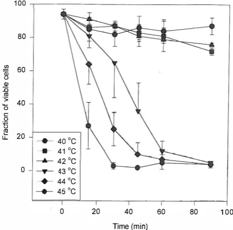
Fig.!. Fraction of viable U937 cells immediately after receiving hyperthermia at specified times and temperatures.
!!l Qi u Q) :0 <1l ':;; '0 c 0 U � L.L.
100 ,---________ �
80 60 40 20 0 -e- 40°C ____ 41°C ----A---- 42 °c ----y-- 43°C ---+- 44°C
--+- 45°C
o 20 40
Time (min)
60 80 100
Fig. 2. Fraction of viable U937 cells after 96 hrs of incubation at
37°C after receiving hyperthermia at ,specified times and doses,
of phosphate buffered saline, pH = 7.4. Then 0.1 mL of heat treated U937 cells-prepared as described above-containing 5x 1 ()4 cells in RPM! 1640 supplemented with 20% FCS was mixed with 0.1 mL of freshly prepared NBT solution ,md incubated for 45 min at 37°C. Cells were then maintained at 4°C. A minimum of 200 cells were counted using a
120
100
"I� 80 0 E .!!2 60
Qi
c.>
a
0 z 40
20
----T- 43 DC
---+- 44 DC -_*_ 45 DC
o �----r-�--,---'---'---'---�
o 20 40
Time (min)
60 80 100
Fig. 3. Number of U937 cells after 96 hrs of incubation at 37°C after receiving hyperthermia at specified times and doses.
35 .---.---�
30
25
;R e... 20
1)-c Q)
;g
15 illOJ c iii 10 0::
5
o ----T- 43 DC
---+- 44 DC
---+- 45 DC
o 20 40
Time (min)
60 80 100
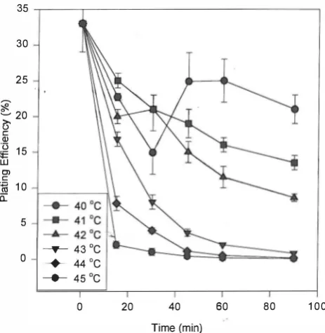
Fig. 4. The plating efficiency (PE) of U937 cells after treatment with hyperthermia at various temperatures and times.
hemocytometer and the fraction of differentiated cells capable of reducing NBT and producing a dark blue precipitate of nitro blue diformasan (NBD) was determined.2o,2!
Latex particles phagocytosis assay
Differentiated U937 cells were assayed for their ability to phagocytize protein coated latex particles according to a modification ofShaala et aIY A protein coated latex particle
Fig.S. Colonies produced by U937 cells in semi-solid agarcultures (xlOO).
suspension marketed commercially as a pregnancy test (Ortho Gravindex) was used for the assay. The commercial suspension of particles was diluted 1: 10 with PBS and 0.1 mL of the diluted suspension was mixed with 0.1 mL of RPMI 1640 supplemented with 20% FCS and containing 5xl()4 HT U937 cells and then incubated for a further 60 min. Finally cells were centrifuged at 1000 g for 20 min at 4°C. The cells were washed twice with PBS and resuspended in 0.2 mL of PBS. At least 200 cells were counted for latex particle phagocytosis positive cells (a cell containing a minimum of 10 particles was considered a positive cell).
Fluorescent antibody assessment test
The ability of differentiated U937 cells to bind to an antibody raised against mature alveolar macrophages of mice was used as a criterion of differentiation. The production and conjunction of this antibody to fluorescein isothiocyanate (FITC) has been described before.23Forthis assay 5xl04 HT treated U937 cells were washed 3 times with PBS and then incubated with 0.1 mL ofFITC-Ab for 45 min at 4°C. Then the cells were washed 3 times with PBS and counted for FTTC positive cells under a fluorescence microscope.
Cytological studies
Microscopic slides of U937 cells were prepared by cytocentrifuge (Cytospin, Shandow). The slides were stained with Wright-Geimsa (Merck) and observed under a light microscope.
Colony formation assay
Ixl(V U937 cells treated with HT were cultured in RPMI 1640 supplemented with 0.3% agar (Difco) and 20% FCS in 30 mm Petri dishes (Nunc) and incubated for 10 days in the above condition. Colonies were stained with Wright stain and counted. A minimum of 50 cells was considered as a colony. The plating efficiency was calculated as:
PE=(Number of colonies)j (Number of cells plated )x100
Hyperthermia and Leukemic Cell Differentiation
70 .---,---�
60
� � 50 !B.
fl
�
40 'iiio 0-
f-�
30'0 c o t5 20
� u..
10
_____ 40°C
___ 41°C ----A-- 42°C
----T-43°C
---+- 44°C
_____ 45°C
o �--_,----_,----_.----,_----,_--�
o 20 40
Time (min)
60 80 100
Fig. 6. Fraction of U937 cells which could reduce NBT after 96 hrs
of incubationat3rC following treatment with hyperthermia at various temperatures and exposure times.
Fig. 7. Photomicrograph ofNBT-positive U937 cells (x250).
All experiments described in various sections above were repeated 3 times.
RESULTS
HT caused both short and long term damage in U937 cells. Short term damage was expressed as a decrease in cell viability immediately after HT. As indicated in Figure 1 , viability decreased to 70% of the control after 9 0 min of heating at45°c. Long term damage was expressed when the cells were incubated at 37°C for 96 hrs after HT. As shown in Figure 2, 45 minutes of heating at 43°C dropped the viability to 30% of the control and 15 min at 45°C was
70 ,---r---�
60
.!!2 50
Qi
u (J
�
40o 0> !1l .c
0-'0 30 c: o t5
s:
2010
--*- 40 DC --*- 41 DC
----A-- 42 DC
---T- 43 DC
---+- 4 4 DC --*- 45 DC
O�----,_----._----,_----r_---,----�
o 20 40 60 80 100
Fig.S. Fraction of U9 37 cells which could phagocytize latex
particles after 96 hrs of incubation at 37°C following treatment with hyperthermia at various temperatures and exposure times.
enough to drop the viability to about 20% of the control. The actual counts of heat treated U937 cells after 96 hrs of , incubation are shown in Figure 3.
The alteration in the plating efficiency afterheattreatment is shown in Figure 4 as a function of the heating time. At temperatures below 43°C at which the cell count did not drop significantly there was a reduction in the plating efficiency of heat treated U937 cells. At 43°C and higher temperatures there was a sharp decrease in the plating efficiency similar to that observed for the total cell count. Figure 5 shows a photograph ofthe colonies produced by the HT U937 cells.
The result ofHT on U937 cells to produce 0-and reduce NBT is shown in Fig. 6. Two distinct effects ofHT on these cells are evident from the heating curves. The initial rise in the curves at low doses or exposure times indicated that HT has caused differentiation of U937 cells towards mature macrophages which were capable of reducing NBT. The decline in the final phase of the heating curves is an indication of the cytotoxic effect ofHT. It can be seen that 30-45 min of heat treatment at 43°C has induced the highest rate of differentiation in U937 cells. Figure 7 shows a photograph ofNBT positive cells.
Figure 8 shows some of the U937 cells which have been treated with hyperthermia and then allowed to phagocytize latex particles. Only 10% of untreated control U937 were able to phagocytize latex particles. However, heat treatment at 42°C for up to 90 min, 43°C for 15-45 min and 44°C for 15-30 min increased the fraction of cells which could
Deizaji and Goliaei
Fig. 9. Photomicrograph of U937 cells which have phagocytized latex particles (xl 000).
120
100
!!2 Qi
() 80 �
:;:l
"iii o c.
�60
u:::
c
240
() �
lJ..
20
---e-- 40°C --- 41 ° C �42°C ---.- 43°C -+- 44°C
---e-- 45°C
o �----,---,---,---.---,----�
o 20 40
Time (min)
60 80 100
Fig. 10. Fraction of U937 cells which showed positive reaction with FITC conjugated anti-macrophage antibody after 96 hr of incubation at 3rC following treatment with hyperthermia at various temperatures and exposure times.
phagocytize latex particles. Lower temperatures were not effective and 15 min at 45°C was slightly effective. Figure 9 shows a photograph of heat treated U937 cells which have phagocytized a large number of latex particles.
The interaction of FITC conjugated anti-macrophage antibody with U937 cells treated with hyperthermia is shown in Figure 10. Heat treatment at 42°C increased antibody binding by the cells in a dose dependent manner. Lower temperatures were almost ineffective. At higher temperatures there was a sharp increase up to 100% in the fraction ofFITC positive cells. A photomicrograph ofFITC
positi ve cells is shown in Figure 11.
Fig. 11. Photomicrograph of me positive U937 cells. Cells were treated with hyperthermia at various temperatures and exposure times, incubated at 37°C for 96 hrs, and treated with antibodies as described in the methods section (x400).
DISCUSSION
In the present work we have investigated the effects of hyperthermia on the proliferation and differentiation of U937 cells. Our results showed that mild hyperthermia could induce differentiation in U937 cells in adose dependent manner. Various experimental criteria were used to assess the differentiation ofU937 cells. For each criterion employed there appeared to exist a specific and distinct dose response curve and an optimum exposure time for the induction of differentiation. Such a phenomenon has been observed and reported for other compounds inducing differentiation in leukemic cells.24•25
Inducers of differentiation in leukemic cells have been classified as partial or strong inducers according to the degree of differentiation they induce in their target cells.26 Accordingly, hyperthermia treatment at temperatures of 42 and 43°C and exposure times of less than 45 minutes were considered as strong inducers. Lower temperatures or shorter exposure times could only partially induce differentiation, whereas at higher temperatures or longer exposure times the viability was reduced drastically and overshadowed the differentiating effect of hyperthermia in U937 cells.
Previous reports with HL60 cells had shown that hyperthermia treatment at temperatures between 42.5 and
43.SoC for 1 hr was most effective in induction of differentiation in those celisP However, in our studies this dose of heat reduced the viability ofU937 cells to less than 30%. The difference might be due to the difference in biological heat resistance and therefore the amount of thermal energy required to induce differentiation in the two cell lines. It might be considered that the partial induction of differentiation in these cells by mild hyperthermia is merely
Hyperthermia and Leukemic Cell Differentiation
a consequence of heat induced arrest of proliferation. However, the results of experiments with Acivin, an antagonist of glutamine, has shown that it could inhibit proliferation of U937 cells without triggering their differentiation.28
The three criteria used to evaluate the extent of differentiation, namely NBTpositivereaction, latex particle phagocytosis and FITC positive reaction, gave fairly similar and consistent results at low temperatures or short exposure times at temperatures above 43°C. However, at exposure
times longer than 30 min at high temperatures there was a
sharp increase in the fraction of FITC positive cells up to
100% (Fig. 10), while there was a decline of NBT positive
and phagocytic cells under similar conditions (Figs. 6 and
8).The difference can be partly described on the basis of the nature of the mechanisms of these reactions. At high temperatures the permeability of cell membranes increases; therefore there is influx ofFITC-Ab particles into the cells. These cells are considered as FITC positive cells. This is similar to results previously reported on the effect of hyperthermia on phagocytosis ofFITC- dextran particles by tumor cells.29 On the other hand, production of 02 and conversion of NBT to blue-black particles of nitro blue diformasan occurs only in viable cells.21
Hyperthermia has been widely used as an anti-cancer agent. There is some evidence that leukemic cells are more heat sensitive than their normal bone marrow progenitors or stem cells.3D The reason for this difference in heat sensitivity has been attributed to several parameters including the state of differentiation, chromosomal abnormalities, presence and function of several oncogenes, and the type of leuk emia.31 This increased heat sensi ti vity ofleukemic cells with respect to normals has been the rational basis of using hyperthermia to purge leukemic cells from bone marrow specimens.32 It has also been possible to use whole blood hyperthermia during bone marrow transplantation to destroy residual leukemic cells.33
The results we have presented here show that hyperthermia can be used as a differentiation inducing agent in leukemic cell lines. The extent of differentiation depends on the temperature and exposure time. This is a reasonable and promising alternative to cytotoxic properties of hyperthermia used in the purging experiments.
REFERENCES
1. Meeting Report: Differentiation therapy of cancer: laboratory and clinical investigation. Cancer Research. 53: 4109-4115, 1993.
2. Collins SJ, Gallo RC, Gallogher RC: Continuous growth and differentiation of myeloid leukaemic cells in suspension culture. Nature 270: 347-349, 1977
3. Sundstrom C, Nilsson K: Establishment and characterization of
a human histiOcytic lymphoma cell line (U937). Int J Cancer 17: 565-577, 1976.
4. Rovera G, O'Brien TG, Diamond L: Induction of differentiation in human promonocytic leukaemia cells by tumor promoters. Science 204: 868-870, 1979,
5. Hass R, Bartels H, TopleyN: TPA induced differentiation and adhesion ofU937 cells, changes in ultrastructure, cytoskeletal organization and expression of cell surface antigens. Europ J Cell Bioi 48: 282-293, 1989,
6. Collins SJ, Ruscetti FW: Terminal differentiation of human promyelomonocytic leukaemia cells induced by dimethyl sulfoxide and other polar compounds. Proc Nat! Acad Sci 75: 2458-2462, 1978.
7. Rigby WFC, Shen L, Edward D, Ball ED: Differentiation of a human monocytic cell line by 1,25 dihydroxy vitamin D3 (Calcitriol): a morphologic and functional analysis. Blood 64: 1110-1115,1984.
8. Dodd RC, C o hen MS: Vitamin D metabolites change the phenotype of monobla�tic U937 cells. Proc Nat! Acad Sci 80: 7538-7541, 1983.
9. Pollock K, Creba J: StimUlus-response coupling in
FMLP-stimulated U937 monocytic cells: effect of differentiation on Gi2 expression. Biochem et Biophys Acta 1051: 71-77,1990. 10. Kelsey SM, Allen PD, Razac K: Induction of surface TNF expression and possible facilitation of surface TNF release from U937 cells by GM-CSF or gamma interferon. Exp Hemat 21: 864-869, 1993.
11. Makishima M, Honma Y, Hozumi M: Differentiation of U937 cells induced by inhibitors of myosin light chain kinase and prevention byGM-CSF. Biochem et Biophys Acta 1176: 245-249, 1993.
12. Lasser A: The mononuclear phagocyte system. Human Pathology 14: 108-120, 1983.
13. Van de Loosdrecht AA, Ossenkoppele GJ: Apoptosis in tumor necrosis factor -dependent monocyte-mediated leukaemic cell death. Exp Hemat 21: 1628-1639, 1993.
14. Koeffler PH: Human acute myeloid leukaemic lines: models of leukaemogenesis. Seminars in Hematology 23: 223-236, 1986.
15. Carper SW, Duffy JJ: Heat shock proteins in thermotolerance and other cellular processes. Cancer Res 47: 5249-5255, 1987.
16. Kampinga HH: Thermotolerance in mammalian cells. J Cell Science·l 04: 11-17, 1993.
17. Harmon BV, Takana YS: Therole of apoptosis in the response of cells and tumours to mild hyperthermia. Int J Radiat BioI 59: 489-501, 1991.
18. Manford K, Patterson GR: Measurement of growth and viability. In: Jakoby WB (ed.), Methods in Enzymology. New York: Academic Press, 58: 150-152, 1979.
19. Sakagami H, Ikeda M: Dependence of cytokine-induced human myelogenic leukaemic cell differentiation on the type of serum in the medium. Anti Cancer Research 10: 385-390, 1990.
20. Absolom DR: Basic methods for the study of phagocytosis. In:
Jakoby WB (ed.), Methods in Enzymology. New York:
Academic Press, 132: 95-180, 1986.
21. Breiman TR: Growth and differentiation of human myeloid leukaemic cell line HL-60. In: Packer L (ed.), Methods in Enzymology. New York: Academic Press, 190: 118-130, 1990.
22. Shaala A Y, Dhaliwal HS: Ingestion of dyed opsonized yeasts as a simple way of detecting phagocytes. J ofImmun Methods 27: 175-187,1979.
23. Goliaei B, Deizadji A, Rabbani A: Heterogeneity of macrophage populations: antibodies detecting various populations of macrophages. Medical J IRI 5: 49-54, 1991.
24. Lindegaard JC: Thermosensitization induced by step down heating. Int J Hyperthermia 80: 561-586, 1992.
25. Langdon SP, HickmanJA: Correlation between the molecular weight and potency of polar compounds which induce the differentiation of HL-60 leukemic cells. Cancer Research 47: 140-144, 1 987.
26. Schwartz E, Ishigura L: Induction of differentiation by chemotherapeutic agents. Adv Enzyme Regulation 21:
3-20, 1983.
27. Richards FM, Watson A, Hickman JA: Investigation of the effects of heat shock and agents which induce a heat shock
response on the induction of differentiation of HL-60 cells. Cancer Research 48: 6715-6720,1988.
28. Nichols KE, Chithenin SR: Monocytoid differentiation of freshly isolated human myeloid leukemic cells and HL-60 cells induced by the glutamine antagonist acivicin. Blood 74: 1728-1733, 1989.
29. Vaupel P, et al: Pathophysiological mechanism ofhyperthermia in cancer therapy. Section 2.2.2, Tumor vascular permeability during hyperthem1ia. In: Streffer C, Vaupel P, Hahn GM, (eds.), Biological Basis of Oncological Thermotherapy. Berlin: Springer-Verlag, pp. 99-100,1990.
30. Mivechi NF: Heat sensitivity, thermotolerance and profile of heat shock proteins synthesis of human myelogenous leukemias. Cancer Research 49: 1954-1958,1989.
31. Mivechi NF, Rossi JJ: Use of polymerase chain reaction to detect the expression of the Mr. 70000 heat shock genes in control or heat shock leukemic cells correlated to their heat response. Cancer Research 50: 2877-2884, 1990.
32. Morryma Y, Narita M: Application of hyperthermia to the treatment of human acute leukemia: purging human leukemic progenitor cells by heat. Blood 67: 802-810, 1986.
33. Li GC, Mager JL: Heat induced protection of mice against thermal death. Cancer Research 43: 5758-5760, 1983.