R E S E A R C H
Open Access
Exercise intervention alters HDL subclass
distribution and function in obese women
Nicholas J Woudberg
1*, Amy E Mendham
2,4, Arieh A Katz
3, Julia H Goedecke
2,4and Sandrine Lecour
1Abstract
Background:Obesity is associated with a change in high-density lipoprotein (HDL) function and subclass. Exercise training reduces cardiovascular risk in obese patients. We aimed to explore the effect of an exercise training stimulus on HDL functionality and subclass in obese women.
Methods:Thirty-two obese black South African women were randomly assigned to exercise (combined aerobic
and resistance exercise) or control (no exercise) conditions for 12-weeks. Pre- and post-testing included venous blood sampling for analysis of lipid profile and HDL functionality, by measuring cellular cholesterol efflux capacity, reduction in endothelial vascular cell adhesion molecule (VCAM) expression (anti-inflammatory function),
paraoxonase (PON) (antioxidative function) and platelet activating factor acetylhydrolase (PAF-AH) activities (anti-thrombotic function). PON-1 and PAF-AH expression were determined in serum and in isolated HDL using Western blotting. Levels of large, intermediate and small HDL subclasses were measured using the Lipoprint® system. Results:Exercise training resulted in a decrease in body mass index (−1.0 ± 0.5% vs + 1.2 ± 0.6%,p= 0.010), PON activity (−8.7 ± 2.4% vs + 1.1 ± 3.0%,p= 0.021), PAF-AH serum expression (−22.1 ± 8.0% vs + 16.9 ± 9.8,p= 0.002), and the distribution of small HDL subclasses (−10.1 ± 5.4% vs + 15.7 ± 6.6%,p= 0.004) compared to controls. Exercise did not alter HDL cellular cholesterol efflux capacity and anti-inflammatory function.
Conclusions:These results demonstrate the potential for exercise training to modify HDL subclass distribution and HDL function in obese women.
Trial registration:Clinical trials number:PACTR201711002789113.
Keywords:Obesity, Exercise intervention, HDL structure, Cholesterol efflux, HDL subclass, Antioxidative, Anti-inflammatory
Background
Approximately 23% of the worldwide burden of ischemic heart disease can be attributed to obesity, with the preva-lence of obesity doubling since 1980 [1]. Exercise is a popular intervention designed to treat obesity and prevent the onset of associated non-communicable diseases [2]. Indeed, exercise training interventions (aerobic and/or re-sistance) reduce cardiovascular risk factors such as body fat mass, blood pressure, total cholesterol, low-density lipoprotein cholesterol (LDL-C) and raise high-density lipoprotein cholesterol (HDL-C) [2–7]. As little as 30 min of exercise per day can increase the concentration of
HDL-C in diabetic patients [8]. Although HDL-C concen-trations are inversely associated with risk for cardiovascu-lar disease [9], the recent outcomes of clinical trials aimed at reducing the risk of cardiovascular disease (CVD) by in-creasing HDL-C levels have been unsuccessful (reviewed by [10]). Accordingly, there has been a significant shift in focus from studying the quantity of HDL to studying the quality [11–15]. HDL quality refers to specific HDL func-tions and the distribution of HDL subclasses.
Although the principal function of HDL is reverse cholesterol efflux/transport (RCT), HDL displays add-itional physiological functions including antioxidative, anti-inflammatory and anti-thrombotic activities. These are mediated, at least in part, by HDL-associated enzymes such as paraoxonase (PON) (antioxidative) and platelet activating factor acetylhydrolase (PAF-AH) * Correspondence:nicholaswoudberg@gmail.com
1Hatter Institute for Cardiovascular Research in Africa, Department of
Medicine, Faculty of Health Sciences, University of Cape Town, Cape Town, South Africa
Full list of author information is available at the end of the article
(anti-thrombotic) [16, 17]. HDL can also be subdi-vided into several subclasses which display distinct functionalities [18].
The African black population has low prevalence of coronary artery and ischemic heart disease, which was originally attributed to higher serum HDL-C [19]. Interestingly, studies highlight that HDL-C is the same or even lower in black compared to white populations [19–22]. A more recent study has further shown that HDL functionality in African black women differed from their white counterparts, with black women dis-playing a higher level of HDL antioxidative function [22]. These data support the notion that HDL quality, rather than quantity may account for the difference in prevalence of CVD between the white and black popu-lations. Furthermore, exercise training may influence HDL function [23]; however, the impact of exercise training on HDL quality in a black population is still unclear [23]. The aim of the study was to examine the effects of exercise training on HDL functionality and subclass, in obese black South African women.
Methods
Full methodology regarding recruitment and testing is detailed by [24].
Participants
Forty-five women were recruited during 2015 and 2016 from the Western Cape, South Africa. Inclusion criteria were: 20–35 years in age, obese (BMI 30–40 kg/m2), weight stable for 6 months, black South African (both biological parents isiXhosa), sedentary (not participating in exercise training (< 1 session of < 20 min per week) for a minimum of 12 months, on injectable contraceptive (depot medroxyprogesterone acetate, 400 mg) for a mini-mum of 2 months, no known illness or chronic disease, not taking any medications, and had no surgical proce-dures within the last 6 months. This study was approved by the Human Research Ethics Committee at the Univer-sity of Cape Town (HREC REF: 054/2015), complies with Declaration of Helsinki principles and participants pro-vided written consent prior to testing. Clinical trials num-ber: PACTR201711002789113.
Study design
Participants were block randomized into either control (n= 22) or exercise (n= 23) conditions (Additional file1: Figure S1). Thirty-five women in control (n= 15) and exercise (n= 20) groups completed the study. The exer-cise intervention consisted of 12 weeks of supervised aerobic and resistance exercise training of moderate-vigorous intensity for 40 to 60 min, 4 days per week by a trained exercise physiologist. Exercises included car-diovascular exercises in the form of aerobic dance,
running, skipping, and stepping that were performed at a moderate-vigorous intensity (75–80% peak heart rate, HRpeak). Resistance exercises included the partici-pants using their own body weight and progressed to the use of equipment (eg bands and free weights). These exercises included squats, lunges, bicep curls, push-ups and shoulder press with a prescribed inten-sity of 60% to 70% HRpeak. Participants wore a heart rate monitor (Polar A300, Kempele, Finland) during all training sessions to ensure the prescribed intensity was maintained throughout the 12-week period. The control participants were instructed to continue their normal physical activity and dietary patterns. Partici-pants attended two pre- and two post-intervention testing sessions. The first session comprised of anthro-pometry and a graded exercise test for the assessment of peak oxygen consumption (VO2peak) and peak heart rate (HRpeak). After a minimum of 48-h recovery from the previous testing session, participants returned for fasting (10–12 h) venous blood collection for analysis of total cholesterol (total-C), low-density lipoprotein cholesterol (LDL-C), HDL-C concentration, HDL functionality and subclass distribution.
Nutritional and physical activity standardization
The exercise and control groups were instructed to maintain their usual dietary intake. The control group was also instructed to continue their habitual physical activity and to refrain from initiating any exercise pro-gram. Prior to the start of all testing sessions, partici-pants refrained from any physical activity for a minimum of 48 h, and from consumption of alcohol and caffeine for 24 h.
Anthropometry and graded exercise test
balance nitrogen; BOC Special Gas, Afrox Cape Town, South Africa).
Venous blood collection and lipid profile
Fasting (10–12 h) venous blood samples were col-lected in serum separating tubes (SST) and clotted for 15–30 min at room temperature. Samples were centri-fuged at 3000 rpm for 10 min at 4 °C. Serum was im-mediately stored at −80 °C until further analyses. Serum lipid profile (HDL-C, LDL-C and Total-C) was determined using a colorimetric assay (Randox (Pty) Ltd., Gauteng, South Africa).
HDL isolation
HDL was isolated from aliquots of serum using ultracen-trifugation, as previously described [26, 27]. Purity was confirmed using 12.5% reducing SDS-polyacrylamide gel electrophoresis (PAGE) stained with Coomassie Blue. The protein concentration of HDL was determined by the modified Lowry method [28] All samples were analysed in duplicate.
Quantification of HDL anti-inflammatory function
HDL anti-inflammatory function was measured using a cell culture model as previously described [27]. Briefly, human umbilical vein endothelial cells (HUVEC) were serum deprived prior to treatment with 10μg/ml of iso-lated HDL for 30 min. Eight participants per group were randomly selected (Additional file 1: Figure S1). Cells were then stimulated with 20 ng/ml murine tumour necrosis factor alpha (TNF-α) (PeproTech, 315-01A) for 5 h. Following RNA isolation and cDNA synthesis, cDNA was amplified for 25 cycles using the RT2 SYBR Green qPCR kit (Qiagen, 330,500) in the RotorGene6000 (Corbit Lifesciences) to quantify ex-pression levels of VCAM and GAPDH. Data is pre-sented as relative reduction in VCAM expression compared to an untreated control.
Quantification of HDL cellular cholesterol efflux capacity HDL induced cholesterol efflux was quantified using a modified method [29]. Briefly, RAW264.7 cells, generously donated by Prof Gil Dealtry (Nelson Mandela Metropol-itan University), were proliferated in RPMI-1640 media (Sigma, R8758) supplemented with 10% foetal calf serum and penicillin/streptomycin prior to seeding (100,000 cells/well) in 24-well culture plates for 16 h. Labelling medium was prepared by adding 4μCi/ml of [3H] choles-terol (Perkin Elmer, NET139001MC) to RPMI-1640 medium containing 2μg/ml of CoA cholesterol acyl-transferase (ACAT) inhibitor (Sandoz, Sigma, S9318) and supplemented with 5% foetal calf serum. Cells were then incubated in labelling medium for 24 h. Cells were washed with minimum essential eagle medium (MEM) in HEPES
buffer prior to addition of 25 μg/ml of isolated HDL in MEM-HEPES for 4 h. Cell culture media was extracted and added to Ultima Gold scintillant (Perkin Elmer, 6,013,327). Counts per minute (CPM) were enumerated using TriCarb® Liquid Scintillation Analyzer and QuantaS-martTM software with 2 Sigma terminator 0.5 and 30 min count time. Cellular cholesterol efflux capacity was calcu-lated as label present in the cell media relative to the un-treated control.
Paraoxonase (PON) activity
Serum paraoxonase activity was measured as previ-ously described [27]. Serum samples were diluted 1:10 in phosphate buffer containing 2 mM CaCl2 (pH 8). Diluted serum was added to 96-well plates in tripli-cate and paraoxon-ethyl substrate (Sigma, D9286) was added. Absorbance at A405 was measured at 30 s in-tervals over 20 min. One Unit of activity is defined as 1 nmol of substrate hydrolysed per min.
Platelet activating factor Acetylhydrolase (PAF-AH) activity
PAF-AH activity was measured in participant sera as previ-ously described [27], using the PAF Acetylhydrolase Assay Kit (Cayman Chemical, 760,901). Briefly, serum was added to an equal volume of 5, 5′-dithio-bis-(2-nitrobenzoic acid) (DTNB; Ellman’s Reagent) and assay buffer in triplicate into clear 96-well plates. All wells were incubated with 2-thio PAF substrate and absorbance at A412measured at 1 min time intervals for 20 min. One Unit of activity is defined as 1μmol of substrate hydrolysed per min.
PON-1 and PAF-AH expression
in the GeneGnome gel imager. Densitometry of PON-1 and PAF-AH blots was quantified using Quantity one soft-ware. PON-1 and PAF-AH relative expression data were corrected for control samples, repeated in each gel.
Quantification of HDL subclass distribution
Serum HDL subclass was determined using the Lipo-print® HDL system (Quantimetrix, Redondo Beach, CA) as previously described [27]. Briefly, serum (25 μl) was mixed with Lipoprint loading gel, containing Sudan black dye which binds proportionally to the cholesterol present in the sample. The mix was placed onto the upper part of the high resolution 3% polyacrylamide gel. Photopolymerisation was carried out for 30 min at room temperature and electrophoresis was performed for 50–60 min at 3 mA per gel tube. After a rest period of 30 min, gel tubes were scanned and analysed using the Lipoware software. For HDL subclass, the very low-density lipoprotein (VLDL) and LDL remained at the origin [Retention Factor (Rf ) = 0.0] and is shown as a grey peak to the left of the large HDL subclass distri-bution profile while albumin migrated as the leading front (Rf = 1.0). Between these, 10 HDL bands could be detected. HDL-1, HDL-2 and HDL-3 were defined as large HDL; HDL-4, HDL-5, HDL-6 and HDL-7 were defined as intermediate HDL and HDL-8, HDL-9 and HDL-10 were defined as small HDL. Each subclass was quantified and expressed as a percentage of total HDL.
Statistical analysis
Results are presented as mean or as percentage changes relative to baseline ± standard error of mean (SEM). Non-normally distributed data were log transformed prior to statistical analysis and included LDL-C, serum PON-1, serum and HDL PAF-AH expression. Non-normally
distributed data are presented as medians ± interquartile range (IQR). Sample size determination was based on pre-vious studies regarding exercise training interventions in obese individuals [10,30], using a significance level ofp < 0.05 and power of 80%, as described in detail previously [24]. Two-way repeated measures analysis of variance was used to compare changes in anthropometry, VO2peak, lipids, cholesterol efflux capacity, anti-inflammatory func-tion, paraoxonase activity, PAF-AH activity and HDL sub-class distribution between groups over the 12-week period, followed by Fischer post-hoc testing. Pearson cor-relation coefficients for the associations between anthro-pometry, VO2peak, HDL-C, HDL function and subclass were determined at baseline and changes in the combined sample. Where appropriate, statistical analysis were ad-justed for patient BMI.p< 0.05 was deemed statistically significant and statistical tests were performed using Sta-tistica (Version 13.2, Dell Inc., 2016).
Results
Changes in anthropometry, cardiorespiratory fitness and lipids
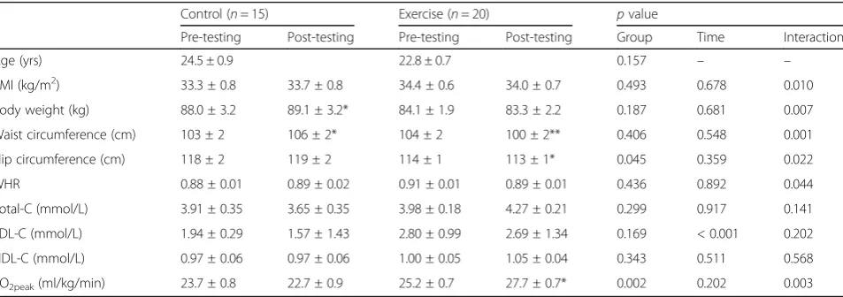
Adherence to the exercise training, expressed as the percentage attendance of total number of sessions, was 80.3 ± 3.0% (range: 60.4–100%). Exercise training re-sulted in a significant increase in cardiorespiratory fit-ness (VO2peak) compared to control (p= 0.003 for interaction, Table 1). BMI, body weight, waist and hip circumference and waist/hip ratio (WHR) decreased in response to the 12-week intervention in the exercise group compared to the controls (p< 0.05 for inter-action). Total-C, HDL-C and LDL-C concentrations did not vary between groups in response to the interven-tion (p= 0.141, p= 0.238 and p= 0.202 for interaction,
Table 1Changes in anthropometry, cardiorespiratory fitness and lipid profile in response to the 12-week intervention
Control (n= 15) Exercise (n= 20) pvalue
Pre-testing Post-testing Pre-testing Post-testing Group Time Interaction
Age (yrs) 24.5 ± 0.9 22.8 ± 0.7 0.157 – –
BMI (kg/m2) 33.3 ± 0.8 33.7 ± 0.8 34.4 ± 0.6 34.0 ± 0.7 0.493 0.678 0.010
Body weight (kg) 88.0 ± 3.2 89.1 ± 3.2* 84.1 ± 1.9 83.3 ± 2.2 0.187 0.681 0.007
Waist circumference (cm) 103 ± 2 106 ± 2* 104 ± 2 100 ± 2** 0.406 0.548 0.001
Hip circumference (cm) 118 ± 2 119 ± 2 114 ± 1 113 ± 1* 0.045 0.359 0.022
WHR 0.88 ± 0.01 0.89 ± 0.02 0.91 ± 0.01 0.89 ± 0.01 0.436 0.892 0.044
Total-C (mmol/L) 3.91 ± 0.35 3.65 ± 0.35 3.98 ± 0.18 4.27 ± 0.21 0.299 0.917 0.141
LDL-C (mmol/L) 1.94 ± 0.29 1.57 ± 1.43 2.80 ± 0.99 2.69 ± 1.34 0.169 < 0.001 0.202
HDL-C (mmol/L) 0.97 ± 0.06 0.97 ± 0.06 1.00 ± 0.05 1.05 ± 0.04 0.343 0.511 0.568
VO2peak(ml/kg/min) 23.7 ± 0.8 22.7 ± 0.9 25.2 ± 0.7 27.7 ± 0.7* 0.002 0.202 0.003
Results represent means ± SEM and as medians ± IQR for LDL-C. Unadjustedpvalues testing for significance of the grouping variable (Control vs Exercise), time (intervention duration) and the interaction (Group*Time). For Fischer post-hoc testing following interaction effect: *p< 0.05 **p< 0.005 pre vs post-testing.BMI
Body mass index,WHRWaist/hip ratio,Total-CTotal-cholesterol,LDL-CLow-density lipoprotein,HDL-CHigh-density lipoprotein and VO2peak, Peak
respectively), while there was a decrease in LDL-C over time in both groups (p< 0.001 for time).
Shift in HDL subclass distribution
The distribution and percent change of HDL subclasses in response to the exercise intervention are presented in representative scan sections of large, intermediate and small subclass distributions (Fig. 1). At baseline, the distribution of large, intermediate and small HDL subclasses were not different between the control and exercise groups (28.4 ± 2.2% vs 26.4 ± 1.8%, p= 0.803; 59.5 ± 1.4% vs 58.5 ± 1.0%, p= 0.701; and 11.8 ± 1.5% vs 15.0 ± 1.4%, p= 0.112, respectively). The distribution of large HDL subclasses did not change in response to the intervention (p= 0.105 for interaction), while the distribution of small HDL subclasses decreased in the exercise group compared to controls (p= 0.004 for interaction). When correcting for the change in BMI, this effect was maintained (p= 0.040 for interaction). The distribution of intermediate HDL subclasses was similar between groups in response to the intervention (p= 0.523 for interaction).
Changes in HDL function
At baseline, cholesterol efflux capacity did not differ between control and exercise groups (3.77 ± 0.22 vs 3.63 ± 0.20 AU,p= 0.808, respectively). HDL-mediated cholesterol efflux capacity did not change in response to a 12-week exercise intervention (p= 0.524 for inter-action, Fig. 2a). At baseline HDL anti-inflammatory function (expressed as relative reduction in VCAM ex-pression in HUVEC cells) did not differ between con-trol and exercise groups (0.47 ± 0.07 vs 0.50 ± 0.09 AU,
p= 0.504, respectively, Fig. 2b) and did not change in response to a 12-week exercise intervention (p= 0.516 for interaction).
At baseline, serum PON activity did not differ be-tween the control and the exercise groups (0.90 ± 0.07 vs 0.83 ± 0.05 U/L, p= 0.173, respectively). After 12 weeks, serum PON activity decreased in response to the exercise intervention only (p= 0.021 for inter-action, Fig. 3a), even after adjusting for the change in BMI (p= 0.006 for interaction). In contrast, serum and HDL PON-1 expression did not differ at baseline be-tween groups (p= 0.751 and p= 0.464, respectively,
A D G
B E H
C F I
Fig.3b-e) or in response to the intervention (p= 0.888 andp= 0.697 for interaction, respectively). The associ-ation between PON activity and expression was ex-plored at baseline in all participants and serum PON activity were positively correlated with serum and HDL PON-1 expression (r= 0.48, p= 0.016, andr= 0.57,
p= 0.001, respectively). However, percentage change in PON activity was not associated with change in serum and HDL PON-1 expression (r=−0.05, p= 0.817, and
r= 0.09,p= 0.633, respectively).
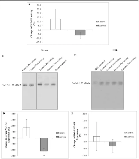
At baseline, serum PAF-AH activity did not differ be-tween the control and the exercise groups (12.7 ± 1.4 vs 15.2 ± 1.2 U/L,p= 0.311, respectively). There was no differ-ence in PAF-AH activity between groups in response to the intervention (p= 0.112 for interaction, Fig.4a). In contrast, serum PAF-AH expression decreased in response to the exercise intervention compared to controls (p= 0.002 for interaction, Fig.4d). This effect in the exercise group was maintained when correcting for the change in BMI (p = 0.003 for interaction). However, changes in HDL PAF-AH expression were not different between groups over time (p= 0.493 for interaction). No associations were found between PAF-AH activity and serum and HDL PAF-AH expression at baseline (r= 0.02,p= 921, and r = 0.09,p= 681, respectively) or between changes in activity and expression over the 12-week intervention (r= 0.38,
p= 0.055, andr= 0.17,p= 0.441, respectively).
Relationships between anthropomorphic measures, cardiorespiratory fitness and HDL function and subclass At baseline, higher BMI was associated with lower choles-terol efflux capacity (r=−0.42, p= 0.024) and less large
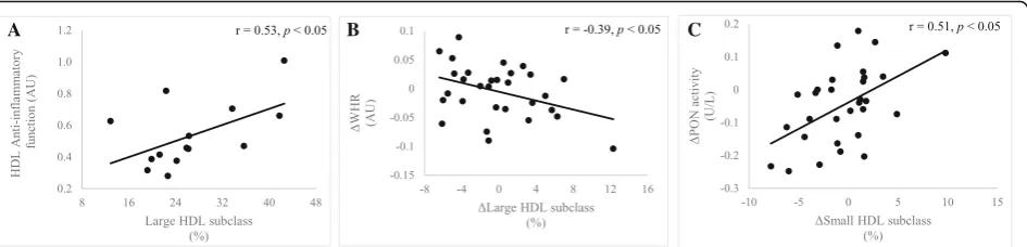
HDL subclasses (r=−0.37, p= 0.041, Additional file 1: Table S1). There were no associations between the changes in aerobic capacity, body composition, and HDL-C and changes in HDL function. However, a decrease in WHR in the combined sample over the twelve-week period, was as-sociated with an increase in the percentage of large HDL subclasses (r=−0.39,p= 0.035 Fig.5b).
Relationships between measures of HDL functionality with HDL subclass
At baseline, reverse cholesterol efflux was positively as-sociated with anti-inflammatory function (r= 0.68,p= 0.016, Additional file 1: Table S1). Similarly, higher HDL anti-in-flammatory function was associated with a greater percent-age of large HDL and lower percentpercent-age of intermediate HDL subclasses (r= 0.53,p= 0.049 andr=−0.54,p= 0.043, respectively, Fig.5a). In all participants, an increase in PON activity, over the twelve-week period, was associated with an increase in the percentage of small HDL subclasses (r= 0.51,
p= 0.004, Fig.5c).
Discussion
This study aimed to explore whether exercise training results in the modification of HDL functionality and HDL subclass distribution. Although HDL-C concentrations were unchanged, the 12-week exercise training interven-tion in obese black South African women resulted in a reduction in PON activity, serum PAF-AH expression and percentage of small HDL subclasses. For the first time, we provide evidence to suggest that exercise training may revert HDL subclass distribution back to a “non-obese” state in a black South African population.
A B
A
B C
D E
Fig. 3Changes in paraoxonase activity and protein expression in response to the intervention. Paraxonase activity of diluted sera was measured at A405over a 20 min time interval using the paraoxon-ethyl substrate. One unit of activity is defined as 1 nmol of substrate disintegrated per
A
B C
D E
Fig. 4Changes in PAF-AH activity and protein expression in response to the intervention. PAF-AH activity of diluted sera was measured at A412
Exercise interventions are routinely prescribed for obese individuals with the aim of reducing the risk of cardio-metabolic complications, and have been shown to pro-mote an increase in HDL-C concentration [8,31,32]. To our knowledge, this is the first study to examine the ef-fects of exercise training on HDL-C concentrations in a black African population, who present with lower HDL-C levels compared to other populations [19, 21]. However, the current study did not find any change in HDL-C fol-lowing the intervention. Despite no changes in HDL-C concentrations, the exercise intervention resulted in a sig-nificant decrease in small HDL subclasses.
A previous study in black South African women re-ported that obesity was associated with lower levels of large and high levels of smaller HDL subclasses, com-pared to normal-weight subjects [27]. In the current study, a statistically significant change in body compos-ition (BMI and WHR) following an exercise intervention was observed, however, these changes were minimal. Critically, when adjusting for the changes in BMI and WHR, the exercise training adaptations in HDL subclass were maintained. This suggests that changes in HDL subclass were potentially mediated by the stimulus of the exercise intervention and not necessarily through a change in body composition. These results are supported by other exercise intervention studies which have also shown that exercise training resulted in changes in HDL subclasses, favouring increases in larger HDL subclasses, that were accompanied by non-significant changes in BMI [33–36]. These results collectively suggest that ex-ercise training may stimulate additional mechanisms which may cause shifts in HDL subclass, independent of changes in body composition.
There is a significant debate in the literature regard-ing the contributions of each of the HDL subclasses to overall HDL functionality. It is also important to con-sider that nomenclature for HDL subclass depends on the methods used for quantification and separation [37]. This study designates HDL subclasses as large,
intermediate and small as quantified by the Lipoprint® System. Much of the existing literature describes two principal HDL subclasses, the larger, HDL2 and smaller HDL3 [38,39]. Whilst epidemiological studies describe lower HDL2 levels as an inverse predictor for cardio-vascular disease, pre-clinical studies describe the bene-fits of increased HDL3 owing to a higher association with cardioprotective proteins and lipids, as reviewed by [10]. Therefore, while the results from this study are consistent with other studies in the literature, it is diffi-cult at this stage to affirm whether the decrease in small HDL subclasses is of benefit to overall risk of CVD. This requires further investigation.
Common mechanisms stimulated by exercise training, in-cluding reductions in oxidative stress and inflammation [40, 41], may be contributing factors to the shift in HDL subclass, observed in the current study. The exercise inter-vention resulted in a decrease in the antioxidative activity of PON and the serum expression of PAF-AH, respectively. This is in contrast to other studies that found improve-ments in PON activity and overall HDL antioxidant func-tion after moderate aerobic exercise intervenfunc-tions (3 or 4 months) in metabolic syndrome and type 2 diabetic pa-tients, respectively [35, 36]. The unexpected PON activity results in the present study may be explained by the partici-pants being normolipidemic and nondiabetic prior to the intervention, and/or differences in the type and intensity of the exercise intervention. PON activity is largely modulated by genetic and environmental factors such as smoking and intake of antioxidants, and these factors may also contrib-ute to differences between studies [35, 36, 42]). Indeed, studies in overweight adolescents and type 2 diabetic pa-tients have reported that exercise training interventions may reduce oxidative stress and the potential risk of CVD [43]. This suggests that the beneficial aspects of exer-cise training may result in a compensatory reduction in PON activity owing to reduction in oxidative stress.
HDL not only controls oxidative stress but also per-forms several anti-inflammatory functions [17]. Here,
A B C
the exercise intervention did not improve HDL anti-in-flammatory function. In contrast, only one other study has explored how a 21-day dietary and exercise inter-vention in obese men (which resulted in a 3.2% de-crease in BMI), improved HDL anti-inflammatory function [35]. Despite a low sample size, our data do not support an association between changes in BMI or WHR with a change in anti-inflammatory function. In addition, the minimal changes in body composition ob-served in the current study may indeed suggest that sub-stantial changes in body composition may be required to influence changes in HDL anti-inflammatory function.
The current study showed that exercise training stimu-lated a decrease in the serum expression of PAF-AH. Previ-ous literature has reported increased PAF-AH activity in response to a short term (3 weeks) diet and exercise inter-vention in obese participants [44]. This suggests that diet and exercise-based interventions stimulate different mecha-nisms of PAF-AH activity, which may be specific to weight loss, changes in body composition and/or change in diet quality. In the current study, there were no associations be-tween changes in PAF-AH activity and expression with changes in BMI and WHR. Furthermore, participants did not display a substantial change in weight, thus the decrease in PAF-AH expression may relate to other mechanisms spe-cifically associated with an exercise training stimulus; how-ever, further research is required in this area. The lack of a significant correlation between HDL and serum PAF-AH expression may further explain this disparity.
Cholesterol efflux capacity is considered to be the pri-mary function of HDL in vivo and this did not change fol-lowing the 12-week intervention. These results are consistent with previous studies conducted in African American populations where a 6 month diet programme of reduced fat and energy, combined with low-intensity exercise, showed improvements in fitness and weight loss, but no changes in cholesterol efflux capacity [45]. Baseline results of the current study showed that cholesterol efflux capacity was associated with a lower WHR. Similarly, a study, examining the relationship between body compos-ition and HDL cholesterol efflux, indicated that an in-crease in waist circumference was an accurate predictor of impairment in cholesterol efflux capacity [46]. Previously, an association between increased BMI and lower choles-terol efflux capacity has been shown in obese subjects [47]. Commensurate with these findings, improvement in cholesterol efflux capacity in individuals undergoing exer-cise interventions were only significant in those individ-uals with significant weight loss [48]. Accordingly, the lack of clinically significant changes in body composition in our study may explain the lack of change in HDL choles-terol efflux capacity in response to the intervention.
The present study highlights that HDL function and subclass may be modified concurrently in response to
exercise training. Accordingly, it is recommended that both HDL function and subclass are considered when assessing changes in CVD risk in response to an exercise training intervention. We report an association between a decrease in small HDL subclasses and a decrease in PON activity. PON is preferentially associated with smaller HDL subclasses, therefore suggesting that a de-crease in PON activity is associated with a dede-crease in the distribution of small HDL subclasses [49]. HDL sub-classes have inherent functional differences; however, few studies have considered the associations between HDL size and function and have not done so using the Lipoprint® System. Therefore, this study presents novel findings that changes in traditional measures of HDL function can be linked to new measures of HDL size.
Notably, this study has limitations that must be consid-ered. In particular, this study was limited with a relatively small sample size. Whilst cholesterol efflux measurements were performed using biological triplicate measurements for each participant, due to technical constraints, the anti-inflammatory assay was only able to be performed on 8 participants. Furthermore, other clinical studies nor-mally employ J744 macrophage cells to test cholesterol ef-flux capacity. RAW264.7 cells were optimized for the conditions employed in this study and produced reprodu-cible results and therefore presented an applicable model. There was, however, good adherence to the exercise train-ing, thus allowing for adequate interpretation of its effects on HDL function and subclass distribution.
Conclusion
Despite no change in HDL-C concentrations, our study presents novel findings that exercise training may revert the HDL subclass distribution to a “non-obese” state in obese black women. Furthermore, exercise training al-tered HDL antioxidative and anti-thrombotic function, independent of changes in body composition. This study provides novel evidence on the association between HDL function and subclass distribution in a black Afri-can population, suggesting that studying HDL subclass and function may be a sensitive approach to assess CVD risk in this population compared to the measurement of HDL-C levels alone.
Additional file
Additional file 1:Figure S1.Breakdown of HDL analysis in control and exercise groups. The distribution of participants into exercise and control groups is presented. Further divisions for assessment of HDL function and subclass are expanded.Table S1.Associations between HDL functionality and subclass measures with body composition and HDL-C in all participants at baseline. Values are Pearson correlation coefficients. BMI, Body mass index; WHR, Waist/hip ratio;VO2peak, Peak oxygen consumption; HDL-C,
Abbreviations
ACAT:Acyl-CoA cholesterol acyltransferase; AU: Arbitary units; BMI: Body mass index; CPM: Counts per minute; CVD : Cardiovascular disease; HDL: High-density lipoprotein; HDL-C: High-density lipoprotein cholesterol; HRmax: Maximal heart rate; HUVEC : Human umbilical vein endothelial cells;
LDL: Low-density lipoprotein; LDL-C: Low-density lipoprotein cholesterol; MEM: Minimum essential eagle; PAF-AH : Platelet activating factor acetylhydrolase; PON : Paraoxonase; SDS-PAGE: Sodium dodecyl sulfate polyacrylamide gel electrophoresis; SEM: Standard error of mean;
TNF-α: Tumour necrosis factor alpha; VCAM : Vascular cell adhesion molecule; VEGF: Vascular endothelial growth factor; VLDL: Very low-density lipoprotein; VO2max: Maximal oxygen consumption; WHR: Waist/hip ratio
Acknowledgements
We thank Hendriena Victor, Keitumetse (Tumi) Smouse, Louise Clamp, Lindokuhle Phiri, Nandipha Sinyanya and Ntombekhaya Zoneleni from the Department of Human Biology at the University of Cape Town for their assistance in sample collection and recruitment. We thank Roshan Ebrahim and Kate Larmuth from the Division of Medical Biochemistry, Institute of Infectious Disease and Molecular Medicine at the University of Cape Town for their technical assistance with scintillation counting.
Funding
This study was funded by the National Research Foundation (Grant number 93577), the South African Medical Research Council and the Swiss South African Joint Research Programme (JRP16) to SL and RWJ and the University of Cape Town.
Availability of data and materials
The datasets used and/or analysed during the current study are available from the corresponding author on reasonable request.
Authors’contributions
AEM and JHD conceptualized the overall intervention design and coordinated sample collection. NJW assisted in sample collection, performed all analytical tests and analysis and compiled and edited the manuscript. SL provided the lab space and equipment for experiments. AK provided lab space and training for radioactive experiments. All authors read and approved the manuscript.
Ethics approval and consent to participate
This study was approved by the Human Research Ethics Committee at the University of Cape Town (HREC REF: 054/2015), complies with Declaration of Helsinki principles and participants provided written consent prior to testing. Clinical trials number: PACTR201711002789113.
Consent for publication
Strict confidentiality of study participants was maintained throughout and no individual data is presented in the study.
Competing interests
The authors declare that they have no competing interests.
Publisher’s Note
Springer Nature remains neutral with regard to jurisdictional claims in published maps and institutional affiliations.
Author details
1Hatter Institute for Cardiovascular Research in Africa, Department of
Medicine, Faculty of Health Sciences, University of Cape Town, Cape Town, South Africa.2Non-Communicable Diseases Research Unit, South African
Medical Research Council, Cape Town, South Africa.3UCT Research Unit for
Receptor biology, Department of Integrative Biomedical Sciences and Institute of Infectious Disease and Molecular Medicine, Faculty of Health Sciences, University of Cape Town, Cape Town, South Africa.4Division of
Exercise Science and Sports Medicine, Department of Human Biology, University of Cape Town, Cape Town, South Africa.
Received: 24 August 2018 Accepted: 27 September 2018
References
1. World Health Organization. World Health Organization: Fact Sheet: Obesity and Overweight [Internet]. 2014. Available from:www.who.int/mediacentre/ factsheets/fs311/en/. Accessed 2017
2. Curioni CC, Lourenco PM. Long-term weight loss after diet and exercise: a systematic review. Int J Obes. 2005;29:1168–74.
3. Hawley JA. Exercise as a therapeutic intervention for the prevention and treatment of insulin resistance. Diabetes Metab Res Rev. 2004;20:383–93. 4. Lavie CJ, Milani RV. Effects of cardiac rehabilitation, exercise training, and weight reduction on exercise capacity, coronary risk factors, behavioral characteristics, and quality of life in obese coronary patients. Am J Cardiol. 1997;79:397–401.
5. Ohta M, Nanri H, Matsushima Y, Sato Y, Ikeda M. Blood pressure-lowering effects of lifestyle modification: possible involvement of nitric oxide bioavailability. Hypertens Res. 2005;28:779–86.
6. Wood PD, Stefanick ML, Dreon DM, et al. Changes in plasma lipids and lipoproteins in overweight men during weight loss through dieting as compared with exercise. N Engl J Med. 1988;319:1173–9.
7. Slentz CA, Duscha BD, Johnson JL, et al. Effects of the amount of exercise on body weight, body composition and measures of central obesity. Arch Intern Med Med. 2004;164:31–9.
8. Argani N, Sharifi G, Golshahi J. Comparison of the effect of different intensity exercise on a bicycle ergometer on postprandial lipidemia in type II diabetic patients. Abstract Original Article. 2014;10:147–53.
9. Gordon T, Castelli WP, Hjortland MC, Kannel WB, Dawber TR. High density lipoprotein as a protective factor against coronary heart disease: the Framingham study. Am J Med. 1977;62:707–14.
10. Woudberg NJ, Pedretti S, Lecour S, et al. Pharmacological intervention to modulate HDL: what do we target? Front Pharmacol. 2018;8:1–16. 11. Woudberg NJ, Goedecke JH, Lecour S. Protection from Cardiovascular
Disease Due to Increased High-Density Lipoprotein Cholesterol in African Black Populations: Myth or Reality. Ethn Dis. 2016;26:553–60.
12. Kühnast S, Fiocco M, van der Hoorn JWA, Princen HMG, Jukema JW. Innovative pharmaceutical interventions in cardiovascular disease: focusing on the contribution of non-HDL-C/LDL-C-lowering versus HDL-C-raisingA systematic review and meta-analysis of relevant preclinical studies and clinical trials. Eur J Pharmacol. 2015;763:48–63.
13. Luscher TF, von Eckardstein A, Simic B. Therapeutic targets to raise HDL in patients at risk or with coronary artery disease. Curr Vasc Pharmacol. 2012; 10:720–4.
14. Chang TI, Streja E, Moradi H. Could high-density lipoprotein cholesterol predict increased cardiovascular risk? Curr Opin Endocrinol Diabetes Obes. 2017;24:140–7.
15. Rader DJ, Tall AR. The not-so-simple HDL story: Is it time to revise the HDL cholesterol hypothesis? Nat Med. 2012;18:1344–6.
16. Barter PJ, Rye KA. High density lipoproteins and coronary heart disease. Atherosclerosis. 1996;121:1–12.
17. Nofer J-R, Kehrel B, Fobker M, et al. HDL and arteriosclerosis: beyond reverse cholesterol transport. Atherosclerosis. 2002;161:1–16.
18. Camont L, Lhomme M, Rached F, et al. Small, dense high-density Lipoprotein-3 particles are enriched in negatively charged phospholipids relevance to cellular cholesterol efflux, Antioxidative, antithrombotic, anti-inflammatory, and Antiapoptotic functionalities. Arterioscler Thromb Vasc Biol. 2013;33:2715–23.
19. Sliwa K, Wilkinson D, Hansen C, et al. Spectrum of heart disease and risk factors in a black urban population in South Africa (the heart of Soweto study): a cohort study. Lancet. 2008;371:915–22.
20. Ellman N, Keswell D, Collins M, Tootla M, Goedecke JH. Ethnic differences in the association between lipid metabolism genes and lipid levels in black and white South African women. Atherosclerosis Elsevier Ltd. 2015;240:311–7.
21. Goedecke JH, Utzschneider K, Faulenbach MV, et al. Ethnic differences in serum lipoproteins and their determinants in South African women. Metabolism. 2010;59:1341–50.
23. Blazek A, Rutsky J, Osei K, Maiseyeu A, Rajagopalan S. Exercise-mediated changes in high-density lipoprotein: Impact on form and function. Am Heart J Mosby Inc. 2013;166:392–400.
24. Goedecke JH, Mendham AE, Clamp L, et al. An exercise intervention to unravel the mechanisms underlying insulin resistance in a cohort of black South African women: Protocol for a randomized controlled trial. JMIR Res Protoc. 2018;7(4):e75. 25. Norton K, Olds T, Norton KI. Anthropometrica: a textbook of body
measurement for sports and health courses. Sydney: UNSW Press; 1996. 26. Brulhart-Meynet MC, Braunersreuther V, Brinck J, et al. Improving
reconstituted HDL composition for efficient post-ischemic reduction of ischemia reperfusion injury. PLoS One. 2015;10:1–16.
27. Woudberg NJ, Goedecke JH, Blackhurst D, et al. Association between ethnicity and obesity with high-density lipoprotein (HDL) function and subclass distribution. Lipids Health Dis. 2016;15:92.
28. Markwell MAK, Haas SM, Bieber LL, Tolbert N. A modification of the Lowry procedure to simplify protein determination in membrane and lipoprotein samples. Anal Biochem. 1978;87:206–10.
29. Sankaranarayanan S, Kellner-Weibel G, de la Llera-Moya M, et al. A sensitive assay for ABCA1-mediated cholesterol efflux using BODIPY-cholesterol. J Lipid Res. 2011;52:2332–40.
30. Ortega JF, Hamouti N, Fernández-Elías VE, Mora-Rodriguez R. Comparison of glucose tolerance tests to detect the insulin sensitizing effects of a bout of continuous exercise. Appl Physiol Nutr Metab. 2014;39:787–92.
31. Kelley GA, Kelley KS. Aerobic exercise and lipids and lipoproteins in men: a meta-analysis of randomized controlled trials. J Mens Health Gend. 2006;3:61–70. 32. Kodama S, Tanaka S, Saito K, et al. Effect of aerobic exercise training on
serum levels of high-density lipoprotein cholesterol. Arch Intern Med. 2007; 167:999–1008.
33. Sarzynski MA, Burton J, Rankinen T, et al. The effects of exercise on the lipoprotein subclass profile: a meta-analysis of 10 interventions. Atherosclerosis. 2015;243:364–72.
34. Riedl I, Yoshioka M, Nishida Y, et al. Regulation of skeletal muscle transcriptome in elderly men after 6weeks of endurance training at lactate threshold intensity. Exp Gerontol. 2010;45:896–903.
35. Casella-Filho A, Chagas ACP, Maranho RC, et al. Effect of exercise training on plasma levels and functional properties of high-density lipoprotein cholesterol in the metabolic syndrome. Am J Cardiol Elsevier Inc. 2011;107:1168–72.
36. Ribeiro ICD, Iborra RT, Neves MQTS, et al. HDL Atheroprotection by aerobic exercise training in type 2 diabetes mellitus. Med Sci Sports Exerc. 2008;40:779–86.
37. Asztalos BF, Tani M, Schaefer EJ. Metabolic and functional relevance of HDL subspecies. Curr Opin Lipidol. 2011;22:176–85.
38. Rosenson RS, Brewer HB, Chapman MJ, et al. HDL measures, particle heterogeneity, proposed nomenclature, and relation to atherosclerotic cardiovascular events. Clin Chem. 2011;57:392–410.
39. Williams PT, Krauss RM, Vranizan KM, et al. Associations of lipoproteins and apolipoproteins with gradient gel electrophoresis estimates of high density lipoprotein subfractions in men and women. Arterioscler Thromb Vasc Biol. 1992;12:332–40.
40. Siti HN, Kamisah Y, Kamsiah J. The role of oxidative stress, antioxidants and vascular inflammation in cardiovascular disease (a review). Vascul Pharmacol Elsevier Inc. 2015;71:40–56.
41. Vinetti G, Mozzini C, Desenzani P, et al. Supervised exercise training reduces oxidative stress and cardiometabolic risk in adults with type 2 diabetes: a randomized controlled trial. Sci Rep. 2015;5:9238.
42. da Costa Vieira JL, Gomes ME, Almeida AB, Moriguchi EH. Changes in the profile of lipoprotein subfractions associated with hormone replacement therapy. Arq Bras Cardiol. 2001;76:177–88.
43. Li C, Feng F, Xiong X, Li R, Chen N. Exercise coupled with dietary restriction reduces oxidative stress in male adolescents with obesity. J Sports Sci. 2017;35:663–8. 44. Roberts CK, Ng C, Hama S, Eliseo AJ, Barnard RJ. Effect of a short-term diet
and exercise intervention on inflammatory/anti-inflammatory properties of HDL in overweight/obese men with cardiovascular risk factors. J Appl Physiol. 2006;101:1727–32.
45. Aicher BO, Haser EK, Freeman LA, et al. Diet-Induced Weight Loss in Overweight or Obese Women and Changes in High-Density Lipoprotein Levels and Function. Obesity. 2012;20:2057–62.
46. Attia N, Fournier N, Vedie B, et al. Impact of android overweight or obesity and insulin resistance on basal and postprandial SR-BI and ABCA1-mediated serum cholesterol efflux capacities. Atherosclerosis. 2010;209:422–9.
47. Sasahara T, Nestel P, Fidge N, Sviridov D. Cholesterol transport between cells and high density lipoprotein subfractions from obese and lean subjects. J Lipid Res. 1998;39:554.
48. Lesna IK, Suchanek P, Kovar J, Poledne R. Life style change and reverse cholesterol transport in obese women. Physiol Res. 2009;58:S33–8. 49. Sean Davidson W, Silva RAGD, Chantepie S, et al. Proteomic analysis of