R E S E A R C H
Open Access
Common sequence variants in CD36 gene and
the levels of triglyceride and high-density
lipoprotein cholesterol among ethnic Chinese
in Taiwan
Kuo-Liong Chien
1,2, Hsiu-Ching Hsu
2, Pi-Hua Liu
3, Hung-Ju Lin
2and Ming-Fong Chen
2*Abstract
Background:Evidence of the genetic association betweenCD36candidate gene and the risk of metabolic syndrome and its components has been inconsistent. This case–control study assessed the haplotype-tagged SNPs fromCD36on the risk of metabolic syndrome and components.
Methods and results:We recruited 1,000 cases and age, gender-matched controls were randomly selected from the participants with metabolic syndrome defined by International Diabetes Federation. Overall, the haplotype tagged SNPs of CD36 gene were not related to the risk of metabolic syndrome. For individuals with normal lipid levels, several SNPs were significantly associated with the triglycerides and HDL-cholesterol levels: Subjects with rs3211848 homozygote had a higher triglyceride level (99.16 ± 2.61 mg/dL), compared with non-carriers (89.27 ± 1.45 mg/dL,P= 0.001). In addition, compared with non-carriers, individuals with rs1054516 heterozygous and homozygous genotypes had a significantly lower HDL-cholesterol (46.6 ± 0.46 mg/dL for non-carrier, 44.6 ± 0.36 mg/dL for heterozygous, and 44.3 ± 0.56 mg/dL for homozygous,P= 0.0008).
Conclusion:TheCD36gene variants were significantly associated with triglycerides and HDL-cholesterol concentrations among ethnic Chinese in Taiwan.
Keywords:Metabolic syndrome, CD 36 gene polymorphism
Introduction
The CD36 is a glycoprotein located in membrane and plays various cellular processes such as lipid transport, immune regulation, coagulation and atherosclerosis [1], and the CD36 structure is related to scavenger receptor B1, and highly binds to oxidized LDL [2], which induced atherosclerosis process. CD36 gene variants regulated fatty acid metabolism and accumulation of fat and fat metabolites, and may influence the risk of metabolic syndrome and may be a target for further personalized medicine screening [3].
Emerging evidence indicating that variation in the
CD36 gene may play a role in the pathogenesis of
metabolic syndrome. Individuals with CD36 deficiency, which is prevalent among Asians, were likely to have the abnormal levels of triglycerides and HDL-cholesterol [4,5]. In addition, genetic variants of CD36 were related to acute myocardial infarction [6], type 2 diabetes [7,8], metabolic syndrome components [9,10], fatty acids [9] and adiponectin levels [11], and free fatty acids [12]. However, no special ethnic Chinese population was reported and only modest effects have been identified for variants of CD36 gene for metabolic syndrome, and the associations for metabolic syndrome itself have often been inconsistent. These inconsistent findings may be attributed to inadequate information from CD36 gene. In addition, the confounding effects by clinical and life-style factors should be considered concurrently [13]. Therefore, we examined the association of common variants of theCD36gene with the metabolic syndrome * Correspondence:mfchen@ntu.ntu.edu.tw
2
Department of Internal Medicine, National Taiwan University Hospital, Taipei 100, Taiwan
Full list of author information is available at the end of the article
and components using a case–control study among ethnic Chinese adults, controlling for clinical and life-style factors.
Methods
Study design and study participants
The study design was a case–control study design based on 8,911 adult participants who were recruited from the Health Management Center of one tertiary hospital from January 2004 to December 2007 and all provided the written informed consents with the study protocol being reported elsewhere [14,15]. In brief, details of socioeco-nomic status, along with medical and medication histor-ies were collected by questionnaires, and standardized clinical procedures were undertaken. We excluded the participants with concurrent severe medical diseases such as cancer and heart failure. The participants signed informed consent forms, and the protocol was approved by the Institutional Research Board of the National Taiwan University Hospital.
Details of subjects’ medical histories such as medica-tion, hospitalization and smoking status were included in the structural questionnaires. Standardized proce-dures for the physical examination, such as anthropo-metric measures and blood pressure, were performed [16,17]. Blood pressure was measured in a resting pos-ition by standard procedure. Body mass index (BMI) and standing height were measured with subjects dressed in light clothing and barefoot. BMI was calculated as weight (in kilograms)/square of height (in meters). Waist circumference was measured midline between the low costal margin and superior posterior iliac crest.
Laboratory measurements
Procedures for blood sampling and analytic methods were as previously described [18,19]. In brief, a blood sample was collected from each participant after at least a 12 hours fasting. Serum total cholesterol levels were measured using the CHOD-PAP method (Boehringer Mannheim, Germany). HDL-C was measured following precipitation of apolipoprotein B-containing lipopro-teins with phosphotungstic acid and magnesium ions (Boehringer Mannheim, Germany). Triglyceride concen-trations were measured by the GPO-DAOS method (Wako Co., Japan). All of the lipids mentioned above were measured using a Hitachi 7450 automated analyzer (Hitachi, Japan). LDL-C concentrations were calculated using the Friedewald formula. All the measures of both samples were carried out in a single hospital and the coefficients of variation were less than 5%.
Case ascertainment and matched control selection
We have recruited the participants from the matched case–control design during 2003 and 2007. We randomly
selecting from 8,911 participants recruited from the Health Management Center and all participants provided written informed consent. We used International Dia-betes Federation (IDF) criterion to define the metabolic syndrome cases. The IDF criterion included central obes-ity and other two components including high blood pres-sure, dyslipidemia and high glucose levels which were defined as: blood pressure of at least 130/85 mmHg or undergoing treatment for hypertension (39%); serum tri-glyceride of at least 150 mg/dL (25%); HDL cholesterol of <40 mg/dL in men and <50 mg/dL in women (53%); fasting glucose of 100 mg/dL or more (20%); and central obesity, waist circumference greater than 90 cm in men and 80 cm in women (38%). A total of 1000 cases of metabolic syndrome were randomly selected. In addition, the 1:1 matched control subjects were selected in the same samples, with frequency-matched according to age and gender.
DNA genotyping & polymorphism detection
Three primers were designed for each SNP detection with web-based software provided at Beckman Coulter Inc (www.autoprimer.com). A forward and areverse PCR primers were used to amplify a short stretch of DNA (90 bp) covering the SNP of interest, and a tagged SNP primer for single base primer extension to identify the SNP. The 5' portion of the tagged primer is complemen-tary to one of 12 unique single stranded DNA oligonu-cleotides that are spotted at a specific location within each well of a 384-well microplate. The 3' portion of the tagged primer is complementary and precisely adjacent to the SNP, which enables detection of the presence of the SNP through the incorporation of a fluorescent-labeled terminating nucleotide. The genotyping were performed in a 12-plex PCR reaction of a 384-well plate with the GenomeLab SNPstream genotyping platform (Beckman Coulter Inc. Fullerton, CA) and its accom-panying SNPstream software suite. The PCR reaction was carried out in a total volume of 5 μl containing
75 μM dNTPs (Applied Biosystem), 5 mM MgCl2,
and at 95°C for 10 min. The tagged extension reaction was carried out with‘cleaned’ PCR products as template and a mixture of 12 site-specific SNP primers. The multi-plex primer SNP extension reaction was performed in a total volume of 7 μl containing 3.76 μl SNP extension dilation buffer, 0.06 μl SNP primer mix (each 5 μM), 0.2 μl C/T (or C/G, T/C et al.) extension mix, 2.96 μl water, and 0.02μl SNPware DNA polymerase. The cycling conditions were 96°C for 3 minutes followed by 45 cycles of 94°C for 20 seconds, and 40°C for 11 seconds. The tagged extension primers were extended with single TAMRA- or fluorescein-labeled nucleotide and then spatially resolved by hybridization to the complementary oligonucleotides arrayed on the 384-well microplates (SNPware Tag array). The Tag array plates were imaged with a two-laser, two-color charged couple device-based imager (GenomeLab SNPstream array imager). The 12 individual SNPs were identified by their position and fluorescent color in each well according to the position of the tagged oligonucleotides. Sample genotype data were generated on the basis of the relative fluorescent inten-sities for each SNP. The image signals were then trans-ferred to genotyping software that translated the images of the arrays into genotype calls. The error rate on the basis of blind replicates was 0.1 to 1% for the SNPs exam-ined in the present study.
Using the common SNPs for Han Chinese were identi-fied from the International HapMap Project (www.hap-map.org), and we selected the tagged SNP for CD36 candidate gene, which contained 19 SNPs whose minor allele frequency was more than 5%. Tagger program was used to select haplotype-tagging SNPs (htSNPs) based on the pairwise linkage disequilibrium relation-ships (r2= 0.8). The common SNP subset represented the information from the remaining SNPs and was in linkage disequilibrium within each block.
Statistical analysis
All data were presented as mean and standard deviation for continuous variables and contingency tables for cat-egorical data and were listed by status of case patients and control subjects. The chi-square tests were used to access Hardy-Weinberg equilibrium among cases and controls and the allele frequency was compared between the cases of metabolic syndrome and control subjects. Subgroup analysis based on gender and age was per-formed for specific genotype. Relationships between metabolic component quantitative variables were ana-lyzed by multiple linear regression analysis adjusting for age, gender in specific subjects within the normal ranges of the metabolic syndrome cutoff values. False discovery rate [20] were used to correct for the multiple compari-son problems for the significant P value threshold as
0.05. In addition, we investigated the association between the genotypes of SNP rs1054516 and HDL cholesterol, after controlling clinical and lifestyle factors in the mul-tiple regression model.
The selected tagged SNPs spanning CD36 form one “block” whose algorithm was developed by Gabriel and colleagues[21], where blocks identified with the default settings in the Haploview program were merged if they have multiallelic D’greater than 0.8 and the cumulative frequency of common haplotypes (>5% frequency) in the merged block is >80% [22]. Haplotype frequencies were estimated by SAS/Genetics software using the expectation-maximization algorithm and the results were verified by the THESIAS program [23]. Pair-wise linkage disequilibrium coefficients, Lewontin's D' and correlation coefficients (r2) were estimated iteratively. Haplotype-specific analyses assessing the association be-tween haplotypes and the continuous lipid profiles, were performed by THESIAS program, which is based on the stochastic-estimation and maximization algorithm. The likelihood ratio test for haplotype-phenotype association was twice the difference between the log-likelihood under the null (one haplotype) and the alternative (add-itional haplotypes) model, and the chi-square with haplotype numbers as degree of freedom. We examined the global significance level in the multivariate adjusted linear models, and specifically tested the haplotype effects, compared with the most common haplotype for reference.
Previous literature showed that the relative risk be-tween CD36 SNP and the risk of metabolic syndrome was 1.3 ~ 1.4, and the allele frequencies of SNPs were 0.10 to 0.25 [9-11]. Under the additive mode of inherit-ance and the baseline risk for the metabolic syndrome as 0.10, we estimated the power for genetic effect was 90% with the sample size of 1000 cases & 1000 matched controls [24].
Results
Basic distribution of case–control subjects
Association ofCD 36gene variants and metabolic syndrome status
A total of 19 common htSNPs of CD36 gene were
selected from the HapMap website (http://www.hapmap. org) to further genotyping (Additional file 1: Table S1). We randomly selected 200 cases/200 controls from the study participants and genotyped these SNPs. From the pilot genotyping results, we found that several CD36 gene SNPs were associated with metabolic syn-drome status and metabolic trait, including HDL, tri-glycerides, body mass index, fasting glucose and triglyceride levels (Additional file 1: Table S2). Finally, we selected the 7 SNPs for completing genotyping work in all subjects.
Table 2 shows the minor allele frequency in the selected 7 SNPs forCD36gene in the study participants, call rates, and the Hardy-Weinberg equilibrium testing for cases and controls. We found that the call rates were satisfactory (>99%) and the Hardy-Weinberg equilibrium was not rejected in all SNPs, implying the genotyping quality was good. We cannot identify any SNP for a sig-nificant association with the status of metabolic syn-drome for all subjects and limited to < 55 years old,
as well as gender, in any mode of inheritance, including co-dominant, additive, dominant and recessive modes (data not shown).
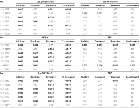
Our next strategy is to perform the genetic
asso-ciation between CD36 gene SNPs and various
meta-bolic syndrome components. Overall, the association between SNPs and metabolic components were found for triglycerides, HDL-cholesterol and blood pressure, as well as postprandial glucose, glycated hemoglobin and c-reactive protein. Among them, we found that triglycerides, cholesterol, triglycerides vs. HDL-cholesterol ratio, have the most significant level, and the significant level was between rs1054516 and HDL-cholesterol, with P< 0.001 in additive, dominant and co-dominant inheritance of models. Other SNPs, in-cluding rs3211849, rs17154258, rs3211869, rs3211958, and rs3211817, were also significantly associated with HDL-cholesterol (Table 3). The patterns were similar for log(triglycerides), triglycerides vs. HDL ratios, and the significance level was persistent after multiple
comparison adjustments, indicating the CD36 gene
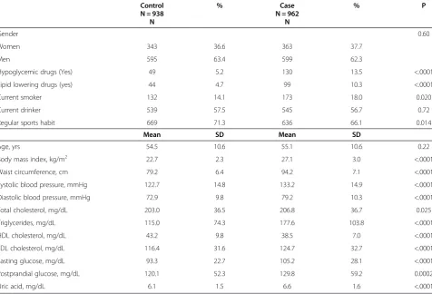
variants were significantly associated with triglycerides HDL-cholesterol, and systolic blood pressure. Figures 1, Table 1 Clinical characteristics of the study participants according to the metabolic syndrome status
Control % Case % P
N = 938 N = 962
N N
Gender 0.60
Women 343 36.6 363 37.7
Men 595 63.4 599 62.3
Hypoglycemic drugs (Yes) 49 5.2 130 13.5 <.0001
Lipid lowering drugs (yes) 44 4.7 99 10.3 <.0001
Current smoker 132 14.1 173 18.0 0.020
Current drinker 539 57.5 545 56.7 0.72
Regular sports habit 669 71.3 636 66.1 0.014
Mean SD Mean SD
Age, yrs 54.5 10.6 55.1 10.6 0.22
Body mass index, kg/m2 22.7 2.3 27.1 3.0 <.0001
Waist circumference, cm 79.2 6.4 94.2 7.1 <.0001
Systolic blood pressure, mmHg 122.7 14.8 133.2 14.9 <.0001
Diastolic blood pressure, mmHg 72.9 9.8 79.2 10.3 <.0001
Total cholesterol, mg/dL 203.0 36.5 206.8 36.7 0.025
Triglycerides, mg/dL 115.0 74.3 177.6 103.8 <.0001
HDL cholesterol, mg/dL 43.2 9.8 38.5 7.0 <.0001
LDL cholesterol, mg/dL 116.4 31.6 124.7 32.7 <.0001
Fasting glucose, mg/dL 93.3 22.7 105.2 28.1 <.0001
Postprandial glucose, mg/dL 120.1 52.3 129.8 59.2 0.0002
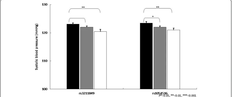
2, 3 and 4 showed the impact of CD36 gene variants on triglycerides, HDL-cholesterol and systolic blood pressure from pairwise genotype comparisons: subjects with rs3211848 homozygote had a higher triglycerides level (99.16 ± 2.61 mg/dL), compared with non-carriers
(89.27 ± 1.45 mg/dL, P= 0.001). In addition, com-pared with non-carriers, individuals with rs1054516 heterozygous and homozygous genotypes had a sig-nificant lower HDL-cholesterol (46.6 ± 0.46 mg/dL for non-carrier, 44.6 ± 0.36 mg/dL for heterozygous, and
Table 3 Association study forCD36gene variants and metabolic syndrome components in the“normal”subjects, adjusted for age and gender, the numbers in bold shows the significant level (<0.05); Lipid Profiles & blood pressure
#rs Log (Triglyceride) Total cholesterol
Additive Dominant Recessive Co-dominant Additive Dominant Recessive Co-dominant
rs3211849 0.013 0.21 0.001 0.005 0.52 0.40 0.92 0.69
rs17154258 0.20 0.34 0.06 0.14 0.029 0.022 0.93 0.07
rs3211869 0.026 0.09 0.034 0.06 0.46 0.51 0.58 0.76
rs3211958 0.019 0.040 0.08 0.06 0.75 0.51 0.83 0.72
rs3211817 0.019 0.17 0.007 0.021 0.62 0.84 0.48 0.78
rs1054516 0.11 0.41 0.06 0.17 0.95 0.73 0.79 0.87
rs17154233 0.73 0.74 0.85 0.94 0.34 0.52 0.14 0.31
#rs HDL-C SBP
Additive Dominant Recessive Co-dominant Additive Dominant Recessive Co-dominant
rs3211849 0.002 0.002 0.07 0.006 0.002 0.013 0.011 0.009
rs17154258 0.42 0.80 0.004 0.017 0.84 0.75 0.53 0.75
rs3211869 0.001 0.016 0.001 0.002 0.26 0.14 0.98 0.29
rs3211958 0.001 0.001 0.035 0.002 0.11 0.26 0.14 0.26
rs3211817 0.003 0.027 0.007 0.010 0.12 0.06 0.70 0.16
rs1054516 0.001 0.000 0.10 0.001 0.002 0.004 0.025 0.007
rs17154233 0.14 0.06 0.46 0.08 0.41 0.48 0.46 0.65
#rs log(TG/HDL-C) DBP
Additive Dominant Recessive Co-dominant Additive Dominant Recessive Co-dominant
rs3211849 0.002 0.033 0.001 0.002 0.08 0.45 0.017 0.06
rs17154258 0.18 0.37 0.013 0.041 0.22 0.20 1.00 0.43
rs3211869 0.003 0.026 0.004 0.006 0.55 0.32 0.72 0.46
rs3211958 0.002 0.003 0.032 0.007 0.64 0.79 0.60 0.87
rs3211817 0.003 0.05 0.002 0.004 0.51 0.37 0.99 0.64
rs1054516 0.011 0.042 0.033 0.039 0.05 0.20 0.06 0.13
rs17154233 0.39 0.32 0.88 0.56 0.77 0.88 0.57 0.85
Table 2 Allele frequency in the selected SNPs for theCD36gene (Chromosome #7) in the study participants and the Hardy-Weinberg equilibrium testing
rs# Allele Position MAF
Controls
Call rate HWE P value
Cases Cases Controls
rs3211849 A < G 80121259 0.350 0.368 99.6% 0.68 0.08
rs17154258 G < A 80134866 0.055 0.056 99.8% 0.76 0.53
rs3211869 A < T 80125088 0.288 0.287 99.7% 0.48 0.15
rs3211958 G < A 80142008 0.442 0.421 99.7% 0.51 1.00
rs3211817 G < T 80116043 0.351 0.348 99.6% 1.00 0.39
rs1054516 C < T 80122878 0.447 0.455 99.8% 0.70 0.23
Rs17154233 C < A 80131125 0.134 0.119 99.8% 0.42 0.64
44.3 ± 0.56 mg/dL for homozygous, P= 0.0008 in codo-minant mode).
The clinical distribution of total participants, accord-ing to the SNP rs1054516 genotype status was listed in Additional file 1: Table S3. The distributions among three genotypes were similar, except for HDL choles-terol: compared with those with CC and TC genotype, participants with TT genotypes had a higher HDL chol-esterol level (P = 0.009).
Table 4 showed the multiple regression models of SNP rs1054516 genotypes and various clinical variables, including age, gender, body mass index, metabolic syn-drome status, smoking, drinking and sports activity, as well as the hypoglycemic and lipid lowering drugs. The estimates for SNP rs1054516, after controlling for mul-tiple variables were significant: compared with TT geno-type, TC and CC genotypes had lower HDL cholesterol levels (−1.00±0.40 mg/dL between TC and TT, P = 0.012,
Figure 1Impact of CD36 gene on triglycerides levels (mg/dL ± SE) from pairwise genotype comparisons.Adjusted mean triglycerides for non-carriers (black bars), for heterozygous (gray bars) and subjects homozygous for the minor allele (white bars). Sample sizes (n for non-carriers, heterozygous, and homozygous subjects for SNP (rs3211849), n = 383, 408, 119, for SNP (rs3211958), n = 305, 432, 174, and for SNPs (rs3211817) n = 377, 416, 115, resulting co-dominant P-values as 0.004, 0.035, and 0.018.
-1.19±0.49 mg/dL between CC and TT, P = 0.012). Be-sides, we found that age, gender, body mass index, meta-bolic syndrome status, drinking, and sports activity status were related to HDL cholesterol levels.
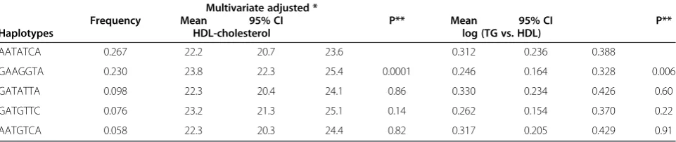
Haplotype analysis frequencies and association study with metabolic syndrome components
We performed the haplotypes analyses for the 7 SNPs from the CD36 gene to assess the association between
these haplotypes and various continuous metabolic syn-drome components. Table 5 lists the common haplotype (frequency > =5%) and the multivariate adjusted continu-ous traits, which showed significant reduction: individuals carrying the second common haplotype (GAAGGTA, fre-quency = 23.0%) had a significant level of HDL cholesterol (adjusted mean, 23.8, 95% confidence interval[CI], 22.3 to 25.4 mg/dL), as compared to those with the most fre-quent haplotype, AATATCA (26.7%, adjusted mean, 22.2,
Figure 3Impact ofCD36gene on HDL-cholesterol levels (mg/dL ± SE) from pairwise genotype comparisons.Adjusted mean HDL-cholesterol for non-carriers (black bars), for heterozygous (gray bars) and subjects homozygous for the minor allele (white bars). Sample sizes (nfor non-carriers, heterozygous, and homozygous subjects for SNP (rs3211958), n = 305, 432, 174, for SNP (rs3211817), n = 377, 416, 115, and for SNP (rs1054516), n = 281, 444, 187, resulting co-dominantP-values as 0.002, 0.010, and 0.0008.
95% CI, 20.7 to 23.6 mg/dL), P= 0.0001. The pattern was similar for triglycerides and triglycerides with regards to HDL-cholesterol ratio.
Discussion
Our study showed that the CD36 gene variants were associated with triglycerides and HDL cholesterol centrations among ethnic Chinese in Taiwan. We con-structed the haplotypes of CD36 gene and extensively checked the association of specific SNP rs1054516 and other clinical variables with HDL cholesterol level. Our study has provided the following clinical evidence. First, theCD36candidate gene is an important determinant of HDL-cholesterol and triglycerides among individuals with normal-range lipids. Second, the metabolic syn-drome itself, does not relate to the CD36 gene poly-morphism in our study.
The mechanism of theCD36gene for the pathogenesis of metabolic syndrome components has been proposed as the following: First, in the CD36 knockout mice, defective uptake and utilization of long-chain fatty acids
were found in muscle and adipose tissues [25]. Trans-genic mice over-expression CD36 protein was found to have reduced blood lipids [26]. The transgenic expres-sion of CD36 in the hypertensive rat model showed the decreased metabolic syndrome and insulin resistance burden. Cell culture study on monocyte CD36 expres-sion showed a 34% expresexpres-sion increase in type 2 diabetes as compared with control subjects [27]. Second, in human cells, the CD36 protein receptor has a highly af-finity to HDL; however, human LDL bound poorly to CD36 protein [28], indicating selective binding of CD36 protein to HDL, instead of to LDL. In addition, CD36 is a receptor for oxidized LDL and plays a role in scaven-ging LDL modified by oxidation and mediating the atherosclerosis process [1]. The expression patterns of
CD36 gene in human tissues were up-regulated in
response to oxidized LDL [29]. Furthermore, CD36 pro-tein facilitates a large fraction of fatty acid uptake and CD36 deficiency in humans was reported defect in myo-cardial cell fatty acid analog [25], and this effect on car-diac triglycerides storage was enhanced in obese status Table 4 The estimated parameters, standard errors and significant levels from the multiple linear regression models for HDL cholesterol levels in the study participants
Parameter Estimate Standard error P value
Intercept 55.3 1.9 <.0001
SNP rs1054516 CC vs. TT −1.19 0.49 0.015
TC vs. TT −1.00 0.40 0.012
age +1 yr 0.05 0.02 0.008
Sex Men vs. Women −6.76 0.41 <.0001
BMI +1 kg/m^2 −0.49 0.07 <.0001
Metabolic syndrome case vs. control −2.51 0.46 <.0001
Smoking Yes vs. No −0.51 0.51 0.31
Drinking habit Yes vs. No 1.07 0.37 0.004
Sports activity Frequent vs. No 1.45 0.39 0.0002
Hypoglycemic medication Yes vs. No −1.11 0.62 0.08
Lipid lowering medication Yes vs. No 0.99 0.68 0.15
Table 5 Common haplotypes (> = 5%) forCD36gene associations with continuous metabolic factors among the study participants
Haplotypes
Frequency
Multivariate adjusted *
Mean 95% CI P**
Mean 95% CI P**
HDL-cholesterol log (TG vs. HDL)
AATATCA 0.267 22.2 20.7 23.6 0.312 0.236 0.388
GAAGGTA 0.230 23.8 22.3 25.4 0.0001 0.246 0.164 0.328 0.006
GATATTA 0.098 22.3 20.4 24.1 0.86 0.330 0.234 0.426 0.60
GATGTTC 0.076 23.2 21.3 25.1 0.14 0.262 0.154 0.370 0.22
AATGTCA 0.058 22.3 20.3 24.4 0.82 0.317 0.205 0.429 0.91
The order of polymorphisms on the haplotype according toCD36SNPs. *: adjusted for gender and age.
[30]. In CD36-null mice, diet high in fructose induced markedly glucose intolerance and hyperinsulinemia [31]. In addition, consuming saturated fatty acid diets would increase LDL cholesterol level in the scavenger receptor class B type 1 gene variant, a similar effect asCD36gene [32]. Our data supportedCD36genetic effects on trigly-cerides and HDL cholesterol.
Evidence forCD36gene in dyslipidemia
Previous genetic studies showed various effects between
CD36 locus and metabolic syndrome components. A
genome-wide linkage scan among 418 individuals from 27 extended Mexican American families found that two loci in chromosome 7 were suggested as being linked to HDL cholesterol and triglycerides levels, besides the most linkage site in chromosome 15q [33]. In addition, among non-diabetic Mexican American families, quanti-tative trait locus study showed a strong linkage of one metabolic syndrome related factor, HDL and triglycer-ides, to chromosome 7 (LOD score up to 3.2) [34]; how-ever, other metabolic factors, including obesity and blood pressure, cannot be identified to linkage to chromosome 7, in which CD36 gene exists. Moreover, another large-scale genome-wide linkage scan among 8664 participants from multiple ethnicities showed that 7q36, a site for CD36, was associated with fasting glu-cose and insulin resistance [35]. To sum up, a meta-analysis based on genome-wide linkage studies on quan-titative lipid traits from the families ascertained from type 2 diabetes showed that CD36 gene locus (7p11-q21.11) was significantly linked to triglycerides and tri-glycerides/HDL cholesterol ratio, but not linked to LDL or total cholesterol [36]. Our findings also demonstrated significant association between CD36 gene polymorph-ism and triglycerides and HDL cholesterol, the traits predisposing to type 2 diabetes. In another study based on 1,375 patients with coronary heart disease, one SNP in CD36, rs3211956, was significantly associated with acute myocardial infarction as compared with stable cor-onary disease (allele frequency 11% vs. 8%, p = 0.04) [6]. However, the strength decreased modestly after adjust-ing covariates and multiple comparisons. Among a co-hort composed of 675 obese adults (age >40 yrs and body mass index >25) in the Netherlands, rs1527479, a C/T SNP in the upstream promoter region in the CD36 gene, homozygous carrier was associated with prevalent type 2 diabetes, and more so in women and high BMI (>27) group [8]. Furthermore, the homozygous carriers were more likely to have a high homeostasis model as-sessment index value [8]. Another case–control study on 61CD36-deficient patients and 25 controls showed that the patients were likely to have a higher type 2 diabetes prevalence, fasting glucose, glycated hemoglobin and
HDL-cholesterol, and likely to have a lower triglyceride value [4]. In addition, the study based on screening the coding sequence of the CD36 gene in 272 French individuals showed that one promoter variant allele (−178A/C) was associated with adiponectin levels (p= 0.036 after multiple testing correction) [11]. Among another French family study, a rare nonsense mutation (1079 T > G) inCD36locus showed linkage with familial type 2 diabetes risk [7]. In addition, genotyping 21 SNPs in the CD36 gene in 585 non-diabetic Caucasians, Ma and colleagues showed that 5 tagged SNPs for haplotype construction [9]. The 30294 G > C polymorphism was associated with free fatty acid level (p = 0.02) and the association was apparent among men. Compared with non-carriers, individuals carrying the haplotype AGGIG had a 31% higher free fatty acids (p = 0.0002) and 20% higher triglycerides (p = 0.025) [9]. Furthermore, this haplotype was associated with a higher risk of coronary heart disease among type 2 diabetic patients. However, a survey based on 831 adults from the health
screen-ing showed that one CD36 gene variant (Pro90Ser)
was associated with free fatty acids, but not related to HDL cholesterol nor triglycerides [12]. Extensive tagged
SNP study on CD36 gene among African-Americans
showed this gene was associated with metabolic syn-drome and HDL cholesterol [10]. A genome wide associ-ation study base on more than ten thousand individuals showed that biological lipoprotein metabolism related
genes, such as CYP7A1, NPC1L1 and SCARB1, were
related to lipid profiles [37]. Our study showed that the CD36 variants with differential effects on triglycerides and HDL cholesterol, consistent with previous findings.
Our study has several strengths. First, the study sam-ple size was moderate, which provided us with sufficient statistical power. In addition, the selection strategy on common htSNPs from public domain (HapMap website) could reduce genotyping costs, and provides an import-ant tool to explore the candidate gene effects. Second, this study population is relatively homogenous and thus may reduce the effect of population stratification. We recruited the participants from the health checkup cen-ter in a cen-tertiary university hospital, and these partici-pants had relatively high socioeconomic status and their health behavior and health promotion motivation were high. Third, due to the high prevalence of dyslipidemia and high blood pressure in the control subjects, we believe the heterogeneity in the metabolic syndrome reduced the association strength between gene and the metabolic syndrome. Further study on the pathogen-esis of CD36 gene expression and metabolic compo-nents, especially triglycerides and HDL cholesterol levels, is warranted.
and other environmental factors, rather than genetic effects, causing the traits to be the same. This was apparently due to the relatively middle and elderly parti-cipants in our study. These phenocopies and misclassifi-cation would reduce the power of the association and it may bias our results toward the null. Second, although our study had sufficient statistical power to detect large effects resulting from common alleles, the power to evaluate small effects due to rare alleles or the effect of interaction was limited. Further gene-environment inter-action study needs greater study numbers. Studying complex diseases has been shown to require very large sample sizes as multiple small effects may be expected. The study population in this study does exceed many very small studies, but may still lack power to detect small associations. Third, we did not include other gen-etic information, such asAPOE, APOA5,andLPL genes. Indeed, our previous studies have shown that the APOA5 and APOA1-C3-A4 genetic polymorphism was associated with dyslipidemia in Taiwanese population [15,38-40]. Finally, due to different components of the metabolic syndrome, multiple comparison issues may be a concern for inflating type I error.
In conclusion, our genetic association study demon-strated that CD36 gene variants were significantly associated with triglycerides and HDL-cholesterol levels. Further study on the pathogenesis of CD36 gene ex-pression and metabolic components, especially with regards to triglycerides and HDL cholesterol levels, is warranted.
Additional file
Additional file 1: Table S1.Minor Allele Frequency of the selected SNPs of CD36 gene in the study participants.Table S2.Selected SNPs for significant association between genetic polymorphisms and metabolic syndrome status as well as components in the study participants (200/ 200 cases-controls), according to adjusted status
(age, gender-adjusted) and the modes of inheritance, including the co-dominant, additive, dominant and recessive models.Table S3.Clinical characteristics of the study participants according to SNP rs1054516 genotype status.
Competing interests
The authors declare that they have no competing interests.
Authors’contributions
KLC, HCH and MFC proposed the study design. KLC, HJL, and BCL participated in data collection. KLC and PHL performed statistical and genetic analysis. HCH performed lipids and laboratory measurements and quality control. KLC and MFC conceived of the study, and participated in its design and coordination and helped to draft the manuscript. All authors read and approved the final manuscript.
Acknowledgements
The study was partly supported by grants from the National Taiwan University Hospital and National Science Council in Taiwan (DOH98-TD-I-111-TM009, NSC 97-2314-B-002 -130 -MY3, 97-3112-B-002-034-). The authors express thanks to the participants in this study.
Author details 1
Institute of Epidemiology & Preventive Medicine, National Taiwan University, Taipei, Taiwan.2Department of Internal Medicine, National Taiwan University
Hospital, Taipei 100, Taiwan.3Clinical Informatics and Medical Statistics Research Center, Chang Gung University, Tao-Yuan, Taiwan.
Received: 21 August 2012 Accepted: 4 December 2012 Published: 18 December 2012
References
1. Endemann G, Stanton LW, Madden KS, Bryant CM, White RT, Protter AA: CD36 is a receptor for oxidized low density lipoprotein.J Biol Chem1993, 268(16):11811–11816.
2. Calvo D, Gomez-Coronado D, Suarez Y, Lasuncion MA, Vega MA:Human CD36 is a high affinity receptor for the native lipoproteins HDL, LDL, and VLDL.J Lipid Res1998,39(4):777–788.
3. Pravenec M, Kurtz TW:Molecular genetics of experimental hypertension and the metabolic syndrome: from gene pathways to new therapies.
Hypertension2007,49(5):941–952.
4. Furuhashi M, Ura N, Nakata T, Shimamoto K:Insulin sensitivity and lipid metabolism in human CD36 deficiency.Diabetes Care2003,26(2):471–474. 5. Hirano K, Kuwasako T, Nakagawa-Toyama Y, Janabi M, Yamashita S,
Matsuzawa Y:Pathophysiology of human genetic CD36 deficiency.Trends Cardiovasc Med2003,13(4):136–141.
6. Knowles JW, Wang H, Itakura H, Southwick A, Myers RM, Iribarren C, Fortmann SP, Go AS, Quertermous T, Hlatky MA:Association of polymorphisms in platelet and hemostasis system genes with acute myocardial infarction.Am Heart J2007,154(6):1052–1058.
7. Lepretre F, Vasseur F, Vaxillaire M, Scherer PE, Ali S, Linton K, Aitman T, Froguel P:A CD36 nonsense mutation associated with insulin resistance and familial type 2 diabetes.Hum Mutat2004,24(1):104.
8. Corpeleijn E, van der Kallen CJ, Kruijshoop M, Magagnin MG, de Bruin TW, Feskens EJ, Saris WH, Blaak EE:Direct association of a promoter polymorphism in the CD36/FAT fatty acid transporter gene with Type 2 diabetes mellitus and insulin resistance.Diabet Med2006,23(8):907–911. 9. Ma X, Bacci S, Mlynarski W, Gottardo L, Soccio T, Menzaghi C, Iori E, Lager
RA, Shroff AR, Gervino EV,et al:A common haplotype at the CD36 locus is associated with high free fatty acid levels and increased cardiovascular risk in Caucasians.Hum Mol Genet2004,13(19):2197–2205.
10. Love-Gregory L, Sherva R, Sun L, Wasson J, Schappe T, Doria A, Rao DC, Hunt SC, Klein S, Neuman RJ,et al:Variants in the CD36 gene associate with the metabolic syndrome and high-density lipoprotein cholesterol.
Hum Mol Genet2008,17(11):1695–1704.
11. Lepretre F, Linton KJ, Lacquemant C, Vatin V, Samson C, Dina C, Chikri M, Ali S, Scherer P, Seron K,et al:Genetic study of the CD36 gene in a French diabetic population.Diabetes Metab2004,30(5):459–463.
12. Kajihara S, Hisatomi A, Ogawa Y, Yasutake T, Yoshimura T, Hara T, Mizuta T, Ozaki I, Iwamoto N, Yamamoto K:Association of the Pro90Ser CD36 mutation with elevated free fatty acid concentrations but not with insulin resistance syndrome in Japanese.Clin Chim Acta2001, 314(1–2):125–130.
13. Campbell H, Rudan I:Interpretation of genetic association studies in complex disease.Pharmacogenomics J2002,2(6):349–360. 14. Chien KL, Hsu HC, Lee YT, Chen MF:Renal function and metabolic
syndrome components on cardiovascular and all-cause mortality.
Atherosclerosis2008,197(2):860–867.
15. Chien KL, Chen MF, Hsu HC, Su TC, Chang WT, Lee CM, Lee YT:Genetic association study of APOA1/C3/A4/A5 gene cluster and haplotypes on triglyceride and HDL cholesterol in a community-based population.
Clin Chim Acta2008,388(1–2):78–83.
16. Chien KL, Chen WJ, Hsu HC, Su TC, Chen MF, Lee YT:Major gene effects on apolipoprotein B concentrations in families of adolescents-Results from a community-based study in Taiwan.ClinChimActa2006, 365(1–2):194–199.
17. Chien KL, Yang CY, Lee YT:Major gene effects in systolic and diastolic blood pressure in the families receiving health examination in Taiwan.
J Hypertens2003,21:1–7.
19. Chien K, Cai T, Hsu H, Su T, Chang W, Chen M, Lee Y, Hu FB:A prediction model for type 2 diabetes risk among Chinese people.Diabetologia2009, 52(3):443–450.
20. Benjamini Y, Hochberg Y:Controlling the false discover rate: a practical and powerful approach to multiple testing.J R Statist Soc B1995, 57:289–300.
21. Gabriel SB, Schaffner SF, Nguyen H, Moore JM, Roy J, Blumenstiel B, Higgins J, DeFelice M, Lochner A, Faggart M,et al:The structure of haplotype blocks in the human genome.Science2002, 296(5576):2225–2229.
22. Chen YC, Giovannucci E, Lazarus R, Kraft P, Ketkar S, Hunter DJ:Sequence variants of Toll-like receptor 4 and susceptibility to prostate cancer.
Cancer Res2005,65(24):11771–11778.
23. Tregouet DA, Escolano S, Tiret L, Mallet A, Golmard JL:A new algorithm for haplotype-based association analysis: the Stochastic-EM algorithm.
Ann Hum Genet2004,68(Pt 2):165–177.
24. Gauderman WJ:Sample size requirements for association studies of gene-gene interaction.Am J Epidemiol2002,155(5):478–484.
25. Coburn CT, Knapp FF Jr, Febbraio M, Beets AL, Silverstein RL, Abumrad NA: Defective uptake and utilization of long chain fatty acids in muscle and adipose tissues of CD36 knockout mice.J Biol Chem2000, 275(42):32523–32529.
26. Aitman TJ, Glazier AM, Wallace CA, Cooper LD, Norsworthy PJ, Wahid FN, Al-Majali KM, Trembling PM, Mann CJ, Shoulders CC,et al:Identification of Cd36 (Fat) as an insulin-resistance gene causing defective fatty acid and glucose metabolism in hypertensive rats.Nat Genet1999,21(1):76–83. 27. Sampson MJ, Davies IR, Braschi S, Ivory K, Hughes DA:Increased expression
of a scavenger receptor (CD36) in monocytes from subjects with Type 2 diabetes.Atherosclerosis2003,167(1):129–134.
28. de Villiers WJ, Cai L, Webb NR, de Beer MC, van der Westhuyzen DR, de Beer FC:CD36 does not play a direct role in HDL or LDL metabolism.
J Lipid Res2001,42(8):1231–1238.
29. Andersen M, Lenhard B, Whatling C, Eriksson P, Odeberg J:Alternative promoter usage of the membrane glycoprotein CD36.BMC Mol Biol2006, 7:8.
30. Coort SL, Hasselbaink DM, Koonen DP, Willems J, Coumans WA, Chabowski A, van der Vusse GJ, Bonen A, Glatz JF, Luiken JJ:Enhanced sarcolemmal FAT/CD36 content and triacylglycerol storage in cardiac myocytes from obese zucker rats.Diabetes2004,53(7):1655–1663. 31. Hajri T, Han XX, Bonen A, Abumrad NA:Defective fatty acid uptake
modulates insulin responsiveness and metabolic responses to diet in CD36-null mice.J Clin Invest2002,109(10):1381–1389.
32. Perez-Martinez P, Ordovas JM, Lopez-Miranda J, Gomez P, Marin C, Moreno J, Fuentes F, Fernandez de la Puebla RA, Perez-Jimenez F: Polymorphism exon 1 variant at the locus of the scavenger receptor class B type I gene: influence on plasma LDL cholesterol in healthy subjects during the consumption of diets with different fat contents.
Am J Clin Nutr2003,77(4):809–813.
33. Duggirala R, Blangero J, Almasy L, Dyer TD, Williams KL, Leach RJ, O'Connell P, Stern MP:A major susceptibility locus influencing plasma triglyceride concentrations is located on chromosome 15q in Mexican Americans.Am J Hum Genet2000,66(4):1237–1245.
34. Arya R, Blangero J, Williams K, Almasy L, Dyer TD, Leach RJ, O'Connell P, Stern MP, Duggirala R:Factors of insulin resistance syndrome–related phenotypes are linked to genetic locations on chromosomes 6 and 7 in nondiabetic mexican-americans.Diabetes2002,51(3):841–847.
35. An P, Freedman BI, Hanis CL, Chen YD, Weder AB, Schork NJ, Boerwinkle E, Province MA, Hsiung CA, Wu X,et al:Genome-wide linkage scans for fasting glucose, insulin, and insulin resistance in the National Heart, Lung, and Blood Institute Family Blood Pressure Program: evidence of linkages to chromosome 7q36 and 19q13 from meta-analysis.
Diabetes2005,54(3):909–914.
36. Malhotra A, Elbein SC, Ng MC, Duggirala R, Arya R, Imperatore G, Adeyemo A, Pollin TI, Hsueh WC, Chan JC,et al:Meta-analysis of genome-wide linkage studies of quantitative lipid traits in families ascertained for type 2 diabetes.Diabetes2007,56(3):890–896. 37. Teslovich TM, Musunuru K, Smith AV, Edmondson AC, Stylianou IM,
Koseki M, Pirruccello JP, Ripatti S, Chasman DI, Willer CJ,et al:Biological, clinical and population relevance of 95 loci for blood lipids.Nature2010, 466(7307):707–713.
38. Chien KL, Fang WH, Wen HC, Lin HP, Lin YL, Lin SW, Wu JH, Kao JT:APOA1/ C3/A5 haplotype and risk of hypertriglyceridemia in Taiwanese.
Clin Chim Acta2008,390(1):56–62.
39. Chien K-L, Hsu H-C, Chen Y-C, Su T-C, Lee Y-T, Chen M-F:Association between sequence variant of c.553 G > T in the apolipoprotein A5 gene and metabolic syndrome, insulin resistance, and carotid atherosclerosis.
Transl Res2009,154(3):133–141.
40. Kao JT, Wen HC, Chien KL, Hsu HC, Lin SW:A novel genetic variant in the apolipoprotein A5 gene is associated with hypertriglyceridemia.
HumMolGenet2003,12(19):2533–2539.
doi:10.1186/1476-511X-11-174
Cite this article as:Chienet al.:Common sequence variants in CD36 gene and the levels of triglyceride and high-density lipoprotein cholesterol among ethnic Chinese in Taiwan.Lipids in Health and Disease 201211:174.
Submit your next manuscript to BioMed Central and take full advantage of:
• Convenient online submission
• Thorough peer review
• No space constraints or color figure charges
• Immediate publication on acceptance
• Inclusion in PubMed, CAS, Scopus and Google Scholar
• Research which is freely available for redistribution