R E S E A R C H
Open Access
Human bone marrow mesenchymal stem
cell-derived exosomes stimulate cutaneous
wound healing mediates through TGF-
β
/
Smad signaling pathway
Tiechao Jiang
1,2*, Zhongyu Wang
2and Ji Sun
3Abstract
Background:Cutaneous wound healing represents a morphogenetic response to injury and is designed to restore anatomic and physiological function. Human bone marrow mesenchymal stem cell-derived exosomes (hBM-MSC-Ex) are a promising source for cell-free therapy and skin regeneration.
Methods:In this study, we investigated the cell regeneration effects and its underlying mechanism of hBM-MSC-Ex on cutaneous wound healing in rats. In vitro studies, we evaluated the role of hBM-MSC-Ex in the two types of skin cells: human keratinocytes (HaCaT) and human dermal fibroblasts (HDFs) for the proliferation. For in vivo studies, we used a full-thickness skin wound model to evaluate the effects of hBM-MSC-Ex on cutaneous wound healing in vivo.
Results:The results demonstrated that hBM-MSC-Ex promote both two types of skin cells’ growth effectively and accelerate the cutaneous wound healing. Interestingly, we found that hBM-MSC-Ex significantly
downregulated TGF-β1, Smad2, Smad3, and Smad4 expression, while upregulated TGF-β3 and Smad7 expression in the TGF-β/Smad signaling pathway.
Conclusions: Our findings indicated that hBM-MSC-Ex effectively promote the cutaneous wound healing through inhibiting the TGF-β/Smad signal pathway. The current results provided a new sight for the therapeutic strategy for the treatment of cutaneous wounds.
Keywords: Human bone marrow mesenchymal stem cells, Exosomes, Wound healing, TGF-β/Smad signaling
Introduction
Cutaneous wound healing is characterized by repairing damaged tissue, and the tissue regeneration was orches-trated by multiple cells to re-establish a protective barrier [1]. The primary goals of skin wound treatment consider rapid wound closure and scar-less healing process. Previous studies have shown that mesenchymal stem cells (MSCs)
are capable of self-renewal and multipotential differenti-ation capabilities, which shows promising therapeutic potential for tissue regeneration and skin function recovery [2,3]. However, MSC transplantation may induce immuno-reactivity to the host [4]. To avoid host-immunoreactivity, MSC-derived exosomes shows an alternative therapeutic attention for regenerative wound repair [5–7]. The current study has demonstrated that MSC-derived exo-somes accelerated the wound healing through increased re-epithelialization, angiogenesis, and tissue granulation [6]. In our previous publication, we have demonstrated that hBM-MSC-Ex treatment significantly reduced liver
© The Author(s). 2020Open AccessThis article is licensed under a Creative Commons Attribution 4.0 International License, which permits use, sharing, adaptation, distribution and reproduction in any medium or format, as long as you give appropriate credit to the original author(s) and the source, provide a link to the Creative Commons licence, and indicate if changes were made. The images or other third party material in this article are included in the article's Creative Commons licence, unless indicated otherwise in a credit line to the material. If material is not included in the article's Creative Commons licence and your intended use is not permitted by statutory regulation or exceeds the permitted use, you will need to obtain permission directly from the copyright holder. To view a copy of this licence, visithttp://creativecommons.org/licenses/by/4.0/. The Creative Commons Public Domain Dedication waiver (http://creativecommons.org/publicdomain/zero/1.0/) applies to the data made available in this article, unless otherwise stated in a credit line to the data.
* Correspondence:jiangtc@jlu.edu.cn 1
Department of Cardiology, The Third Hospital of Jilin University, 126 Xiantai St., Changchun 130033, Jilin, China
2Jilin Provincial Cardiovascular Research Institute, 126 Xiantai St., Changchun
130033, Jilin, China
fibrosis in rats [8]. In this current study, we used the promising exosomes type, i.e., hBM-MSC-Ex, to investi-gate the wound healing process and to investiinvesti-gate the associated signal pathway mechanism.
TGF-β/Smad signal pathway is an evolutionarily con-served pathway with numerous functions ascribed [9]. Transforming growth factor-beta (TGF-β) is considered as one of the essential growth factors in the natural wound healing process by TGF-β/Smad pathway [10]. Smad is well known as the major inducer of fibroblast differentiation, which is an essential factor for wound healing [11]. During this process, TGF-βactivates down-stream mediators such as Smad2 and Smad3, which result in the linage differentiation of fibroblasts into alpha-smooth muscle expressing (α-SMA) myofibroblasts [12]. The phosphorylated Smad2/Smad3 activates Smad7, which promoter to upregulate Smad7 expression in the differenti-ation process, and thereby, Smad7 inhibits TGF-β1 expres-sion for negative feedback regulation [13]. Recent studies have demonstrated that the TGF-β/Smad signal pathway plays an essential vital role in cutaneous wound healing via regulating the proliferation and migration of keratinocytes, dermal fibroblasts, and other skin cells in the affected areas to participate in the wound healing process [14, 15]. Furthermore, TGF-β/Smad signaling can regulate tissue fibrosis and scar formation [16]. In this current study, we hypothesized that hBM-MSC-Ex could promote the cuta-neous wound healing process via regulating the TGF-β/ Smad signaling pathway.
Materials and methods
Cell culture and exosome purification
HaCaT and HDFs were purchased from the Chinese Acad-emy of Medical Sciences, China. Human bone marrow mesenchymal stem cells (hBM-MSCs) were generously provided by Dr. Yi Wang (Jilin University, Changchun, China), P3-5 lines of hBM-MSCs were used in the experi-ments. Cells cultured in DMEM (Gibco, Grand Island, USA) supplemented with 10% FBS (Gibco, Grand island, USA) humidified 5% CO2atmosphere at 37 °C. The
purifi-cation of hBM-MSC-Ex involves several centrifugation steps, as described previously [17]. Briefly, hBM-MSCs were cultured in serum-free medium (SFM, Gibco, Grand Island, USA) for 2 days. Conditioned medium was first filtered using a 0.1-μm filtering unit. The supernatant was concentrated with a 100-kDa molecular weight cutoff (MWCO) hollow fiber membrane (Millipore, Billerica, MA, USA), at 1000g for 30 min. Then, the concentrated supernatant was loaded onto a 30% sucrose/D2O cushion
(5 ml, density 1.210 g/cm3), and ultra-centrifuged at 100, 000g for 3 h. After exosome-enriched fraction was col-lected, it was washed three times with fresh PBS, centri-fuged at 1500g(30 min each wash) with 100-KDa MWCO. Finally, purified exosomes were passed through a 0.22-μm
filter and stored at −80 °C until further use. The protein concentration of exosomes was measured by bicinchoninic acid (BCA) protein assay kit (Beyotime, Shanghai, China).
Cell proliferation assay
HaCaT and HDFs cells were cultured until 70~80% con-fluence, and trypsinized cells were plated in 96-well plates at a density of 4000 cells per well. Briefly, hBM-MSC-Ex purification and characterization were as per previously published methods [8]. The cells were treated with either hBM-MSC-Ex (25μg/mL) or PBS (control) (Invitrogen, Shanghai, China), then followed by incu-bated at 37 °C with 5% CO2for 5 days. The cell viability
was determined by 10% CCK-8 solution (Sigma, San Francisco, USA), the cultures were incubated for 30 min at 37 °C in 5% CO2, and corresponding OD value was
measured at the 490-nm wavelength.
Immunofluorescence staining (IF)
HaCaT and HDFs cultured by hBM-MSC-Ex (25μg/mL) or PBS were incubated in 24-well plate coated coverslip for 24 h. When cells reached 60~70% confluence, plate were washed with PBS and incubated with 4% parafor-maldehyde for 10 min (RT). The processed cells blocked with 1% bovine serum albumin (BSA; Biosharp, Hefei, China) for 30 min. The cells were incubated with primary antibodies against Rb anti-PCNA (1:100 dilution, BD Bio-sciences, Franklin Lakes, NJ, USA), and isotype-matched rabbit IgG/IgM (1:100 dilution, Abcam, Cambridge, UK) served as the negative controls. Anti-rb-FITC-488 second-ary antibody (1:500 dilution, Abcam, Cambridge, UK) for 2 h, and the nuclei were labeled with DAPI (Thermo Scien-tific, Waltham, USA) for 5 min. Images were acquired by fluorescence microscopy (EVOS, Thermo Scientific, Waltham, USA), and the PCNA positive cells were counted in ten random optical fields by using ImageJ software.
Animals and treatments
The wounded area was traced, and we circled the edge of wound on photograph, then calculate the circled area based on the pixels of that area. At the end of the study, the rats were euthanized on the 16th day to collect the healed and unhealed tissue area in the different treatment groups.
Histological examination
Skin tissue was collected from the mechanical injury region on 16 days, and samples were fixed in 10% formalin in PBS and embedded in paraffin. The skin tissue was sectioned at 4-μm thickness to perform hematoxylin and eosin (H&E) staining. The staining was performed following the manufacturer’s protocols (Sigma, San Francisco, USA). The sections were proc-essed for immunohistochemistry (IHC) using the Kit (Maixin KIT-9710, Fuzhou, China) following the manufacturer’s instructions. Briefly, the sections were deparaffinized, and antigen retrieval was performed by immersion slides in 0.01 M sodium citrate buffer solu-tion for 15 min. Endogenous peroxidase was quenched by processing the sections in 3% H2O2 for 15 min,
followed by blocked sections with 10% normal goat serum for 1 h at 37 °C. The sections were incubated with primary antibody anti-α-SMA or anti-VEGF with 1:500 dilution (Abcam, Cambridge, UK) for overnight at 4 °C. Next day, these sections were incubated with biotinylated goat-anti-rabbit IgG antibody for 2 h and incubated with avidin peroxidase reagent sequentially. Then, the sections were incubated with diaminobenzidine solution as the chromogenic agent at 37 °C for 5 min. Finally, we used hematoxylin staining for counterstaining the sections. These sections were photographed using a bright-field microscope (EVOS, Thermo Scientific, Waltham, USA). Cutaneous appendage separated by tissue on one microscope sphere was counted as one unit. The
α-SMA- and VEGF-positive area calculation is based on the ratio of positive area/total area of the observed field. We used 6 random fields per section and 6 sections in total (n= 8 rats) for the quantification of IHC images.
Western blot
Proteins were extracted from the skin healed tissue in the in SDS sample lysis buffer. The samples were heated to 95 °C for 10 min, and 40-μg protein samples were separated on SDS-polyacrylamide gels (5% stack-ing gel and 12% separation gel). Resolved proteins were then transferred onto nitrocellulose membranes, blocked in 5% nonfat powdered milk for 1 h, and probed with re-spective antibody against TGF-β1, TGF-β3, Smad2, Smad3, Smad4, Smad7, and glyceraldehyde-3-phosphate dehydrogenase (GAPDH) served as loading control (1: 1000 dilution, Abcam, Cambridge, UK) overnight at 4 °C. The blots were blocked in secondary HRP-conjugated goat anti-rabbit IgG antibodies (1:1000 dilution, Abcam,
Cambridge, UK) and visualized by chemiluminescent detection substrates (Immobilon western chemilumines-cent HRP substrate, Millipore). The densitometric quanti-fication were performed on the protein bands by using AlphaEaseFC software (Alpha Innotech).
Real-time PCR assay
Total RNA from skin healed tissue was extracted with Trizol (Invitrogen, Shanghai, China) according to the manufacturer’s protocol. Briefly, the first-strand cDNA was synthesized with 1μg of total RNA using Super-Script II (Invitrogen). SYBR Green I dye was used for reverse transcription in an ABI 7500 fluorescence quantitative PCR instrument, and the mRNA levels of TGF-β1, TGF-β3, Smad2, Smad3, Smad4, Smad7, and GAPDH were measured using the respective primers listed in Table (Supplementary Table S)1. The ther-mocycler conditions were as follow: initial step at 95 °C for 2 min, followed by 40 cycles at 95 °C for 15 s, and 60 °C for 1 min. Expression levels were re-corded as cycle threshold (Ct). Data were acquired using the 7500 Software (Applied Biosystems Life Technologies, Foster City, CA, USA). All reactions were performed in triplicate, and the data were ana-lyzed using the 2−ΔΔCt method.
Statistical analysis
Statistical analysis was performed using Prism 6 (Graph Pad software) and Image J. One-way ANOVA with post hoc Dunnett’s multiple comparison test was used to test for statistically significant differences between the groups. All quantitative data were given as the mean ± SD, and data were acquired by at least three independent experiments, and p< 0.05 was considered to be statisti-cally significant.
Results
In vitro studies: hBM-MSC-Ex improves skin cell proliferation
compared to the PBS group (62.1% vs 28.2% in HaCaT and 71.3% vs 34.8% in HDFs in the two types of skin cells (p< 0.01)). In vitro experiment, the results demon-strated that treatment of hBM-MSC-Ex can promote cell proliferation and maintain cell growth effectively in the both skin cells (HaCaT and HDFs).
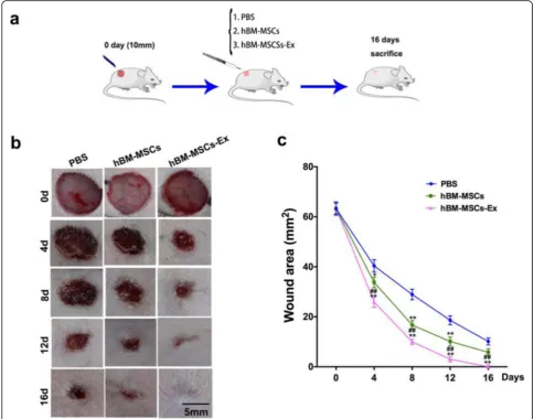
In vivo studies: hBM-MSC-Ex improves cutaneous wound healing
To investigate the roles of hBM-MSC-Ex in wound healing, we established a full-thickness skin wounds in-jury model in rats, as illustrated in the experimental de-sign in Fig.2a. The area measurements in the wounded region show a significant improvement in the wound closure after the treatment of MSC-Ex and hBM-MSCs treatment group (Fig. 2b, c). We observed a sig-nificant reduction in the wound area in the hBM-MSCs treatment group starting from day 4 after the surgery and constantly improved in reduction until the day 16
compared with PBS. The wound in hBM-MSC-Ex group completely healed on the 16 ± 2.3 days after sur-gery (p< 0.05, p< 0.01). These results indicates that hBM-MSCs’isolated exosomes can more effectively ac-celerated the cutaneous wound healing process as dem-onstrated in the animal model.
hBM-MSC-Ex restore the normal skin morphology
To understand the process of restoring the skin morphology regenerated during the healing process, we assessed the effects of the hBM-MSC-Ex on the wound healing quality. To study the general morph-ology of skin regeneration, we performed H&E stain-ing. The observation of H&E staining images shows an indication that regeneration of cutaneous append-ages in the affected area, including restoring hair folli-cles and sebaceous glands in hBM-MSC-Ex group (21.3 ± 5.4 /filed), was compared to other treatments such as the PBS group (1.2 ± 2.8/filed, p< 0.001) the
and hBM-MSC group (15.4 ± 4.1 /filed, p< 0.001) (Fig. 3a, b). α-SMA and VEGF are both important in-dicators of angiogenesis. We performed IHC in the wound area samples from the different treatment groups. The α-SMA-positive area quantification results showed that the percentage of α-SMA positive area were signifi-cantly increased in the hBM-MSC-Ex group (8.1% ± 1.2), when compared with the PBS group (1.3% ± 0.5/HPF,p< 0.001) as well as the hBM-MSC group (5.6% ± 0.9, p< 0.01) (Fig. 3a, c). Consistent with the above results, the percentage of VEGF-positive area was significantly in-creased in the hBM-MSC-Ex group (12.3% ± 2.4), com-pared to the PBS group (1.6% ± 1.5/HPF, p< 0.001) and the hBM-MSC group (5.1% ± 1.7, p< 0.001) (Fig. 3a, d). The above results indicated that hBM-MSC-Ex not only enhanced the wound process but also restored skin func-tion and angiogenesis in the affected areas.
hBM-MSC-Ex regulate the TGF-β/Smad signal pathway
Finally, we investigated the underlying mechanism of hBM-MSC-Ex-induced skin healing process in the af-fected tissue. The expression level of TGF-β1, TGF-β3, Smad2, Smad3, Smad4, and Smad7 (components of the TGF-β/Smad signaling pathway) were analyzed by Western blot and qPCR. Both Western blot and RT-qPCR results showed the significantly decreased (re-spective protein and RNA levels) expression of TGF-β1, Smad2, Smad3, and Smad4 in hBM-MSC-Ex treatment group, compared with the other two control groups (p< 0.05, p< 0.01). The alteration of signaling molecule dur-ing the healdur-ing the process illustrated that hBM-MSC-Ex treatment plays a major mechanism in inhibiting TGF-β/Smad signaling pathway (Fig.4a, b).
In the current study, we aimed to study how the
TGF-β/Smad signal modulated during wound healing. In our
result, we detected that Smad7 levels were significantly increased in the hBM-MSC-Ex-treated group compared to other treatment groups (Fig. 4a, b;p< 0.05, p< 0.01). These results suggested that altering the levels of Smad7 could be beneficial in the wound regeneration process. Inhibitory Smad7 is activated by the binding of the TGF-β super family to the cell surface receptors.
TGF-β1 is fibrotic isoform, while TGF-β3 is the anti-fibrotic isoform. The hBM-MSC-Ex treatment group signifi-cantly upregulated the expression of TGF-β3, compared with the other two control groups (Fig. 4a, b; p< 0.05,
p< 0.01). These results suggest that hBM-MSC-Ex ef-fectively promote the cutaneous wound healing through inhibiting the TGF-β/Smad signal pathway (Fig.5).
Discussion
In this study, our results indicate that hBM-MSC-Ex stimulates cutaneous wound healing both in vitro and in vivo.In vitro, hBM-MSC-Ex promotes both two types of skin cell (HaCaT and HDFs) proliferation effectively. In vivo, hBM-MSC-Ex accelerates cutaneous wound healing, via inhibiting the TGF-β/Smad signal pathway.
Recently, studies have shown that the prospective ap-plication of MSC-derived exosomes promoted cutaneous wound healing [5, 7, 19]. The possible roles of MSC-derived exosomes in wound healing are promotion of cell proliferation, migration, differentiation, angiogenesis, and matrix reconstruction [6]. HaCaT and HDFs are the two major skin cells types which participate in cutane-ous wound healing [20]. In our study, we found that hBM-MSC-Ex promote HaCaT and HDFs growth effect-ively (Fig. 1). It indicated that hBM-MSC-Ex promote HaCaT and HDF cell proliferation to participate in the process of wound healing. In addition, fibroblasts are critical players for exosomes in wound healing and are the main cell types that synthesize, secrete, and deposit ECM collagen and elastic fibers [21]. Studies have shown that MSCs exert their therapeutic effect via secretion of soluble factors, including the exosomes [5, 6]. There is substantial evidence that exosomes have the ability to promote skin regeneration when applied topically or
injected systemically [22, 23]. In this study, we have ad-ministrated subcutaneous injection of exosomes at multi-directional to delay its clearance in the body. In addition, hBM-MSCs appear to be limited because of poor cell retention at the wound site [24], To overcomes this issue, we adopted intravenous injection routes to maximize the effect of treating wounds in our treatment groups.
MSC-derived exosomes accelerate wound healing process through promoting angiogenesis and restoration of skin function [25]. Previous studies have proved that stem cell-conditioned medium may contain exosomes that contain pro-angiogenesis factors to promote wound healing in skin injury [26, 27]. Our results demonstrate that hBM-MSC-Ex significantly accelerate wound heal-ing (Fig. 2b, c) and modulate α-SMA expression which is an important indicator of angiogenesis (Fig. 3a, c). MSC-derived exosome contains various growth factors which play an important role in cutaneous regeneration
and repair [6, 19]. Interestingly, we also found there are many cutaneous appendages regeneration, such as hair follicles and sebaceous glands (Fig. 3a, b). It provides fa-vorable conditions for hBM-MSC-Ex to restore skin func-tion during wound healing.
Recent studies indicated that different types of MSC therapy or combined therapies with some unique bio-technology factors can stimulate cutaneous wound healing [28–31]. Current studies have report that a new combination therapeutical approach shows effective combinational treatment composed of adipose-derived mesenchymal stem cells (AD-MSCs) platelet-rich plasma (PRP) and hyaluronic acid (HA) dressing which shown to stimulate the wounds healing and regener-ation process [30–32]. The complete closure of wound is inducing a new neodermis and stimulating regener-ation and a protected environment in a humid environ-ment [33–35]. Many supporting evidence indicates that adipose-derived stem cells (ASCs) and adipocyte-secreted exosomal microRNA promote wound repair [36]. Another study has demonstrated hBMSCs on gel-atin scaffold with poly N-isopropylacrylamide
(pNIPAAm) as transplanted grafts for improving skin regeneration [37]. More recent clinical trials have shown to improve the hair density by administration human follicle stem cells (HFSCs) [38–40]. In addition, autologous fat grafting is a better approach to consider for the correction of wound scars in the affected re-gions [41, 42]. However, in our current study, we ad-ministrated directly hBM-MSC-Ex on mechanical damaged skin area, which is more clinically transfer-able, more safe, and effective cell-free reagents com-pared with their parents’ cell skin regeneration therapies. Moreover, we have demonstrated that MSC-Ex treatment is more effective than of hBM-MSCs in wound healing, such as some indicators of wound area and cutaneous appendages.
TGF-β1/Smad pathway is an important pathogenic mechanism in wound healing [9]. TGF-β1 is considered to be a key mediator in tissues scarring and mostly by acti-vating its downstream against decapentaplegic (Smad) sig-naling [13]. It has proven that TGF-β1 exerts its biological effects by activating downstream mediators, including Smad2 and Smad3 [15]. The phosphorylated cytoplasmic
Fig. 5Illustration of hBM-MSC-Ex stimulates cutaneous wound healing by regulating the TGF-β/Smad signal pathway. hBM-MSC-Ex inhibited TGF-β1 and activated TGF-β3 expression; TGF-βisoforms and activins stimulate intracellular signaling via Smad-2/3 transcription factors;
mediators, Smad2 and/or Smad3, and a heterotrimeric complex are formed with Smad4 that translocate into the nucleus, bind a consensus sequence, and regulate gene transcription [14], while these activities are negatively reg-ulated by Smad7 expression [16]. In our study, we found hBM-MSC-Ex significantly downregulate TGF-β1, Smad2, Smad3, and Smad4 expression and upregulate of Smad7 expression (Fig. 4). It demonstrated that hBM-MSC-Ex might accelerate cutaneous wound healing through inhi-biting the TGF-β/Smad signal pathway (Fig. 5). TGF-β1 are associated with fibrosis, while TGF-β3 has been associ-ated with anti-fibrotic or scar-less wound healing activity, and they have been observed to play an essential role in regulating epidermal and dermal cell movement during wound repair [10]. In our study, we found hBM-MSC-Ex decreased of TGF-β1 expression and increased TGF-β3 expression (Fig.4). This may be one of the crucial reasons to promote the skin scar-less wound healing.
In conclusion, we successfully investigated the role of hBM-MSC-Ex on cutaneous wound healing. Our results demonstrated that hBM-MSC-Ex could exert promoting effect of cutaneous wound healing via inhibiting the TGF-β/Smad signal pathway. The current approach pro-vides a better knowledge in the wound healing process and new therapeutic strategy for the treatment of cuta-neous wounds.
Supplementary information
Supplementary informationaccompanies this paper athttps://doi.org/10. 1186/s13287-020-01723-6.
Additional file 1: Table S1.Primers used for qRT-PCR.
Abbreviations
hBM-MSCs:Human bone marrow-derived mesenchymal stem cells; hBM-MSC-Ex: Human bone marrow mesenchymal stem cell-derived exosomes; ALP: Alkaline phosphatase; ALT: Alanine aminotransferase; AST: Aspartate aminotransferase; HSCs: Hepatic stellate cells; Hyp: Hydroxyproline; MDA: Malonaldehyde; qRT-PCR: Quantitative real-time PCR; HaCaT: Human keratinocytes; HDFs: Human dermal fibroblasts; TBIL: Total bilirubin; TP: Total protein;α-SMA: Alpha-smooth muscle actin; VEGF: Vascular endothelial growth factor;γ- GT: Gamma glutamyl transpeptidase; AD-MSCs: Adipose derived mesenchymal stem cells; PRP: Combined treatment composed of platelet-rich plasma; HA: Hyaluronic acid; pNIPAAm:N-Isopropylacrylamide; HFSCs: Human follicle stem cells
Acknowledgements
The authors thank Professor Wang, Jilin University, for his assistance in histology.
Authors’contributions
T.J. carried out the molecular genetics and animal studies, conceived of the study, and participated in its design and coordination and helped to draft the manuscript. Z.W. carried out the WB and animal studies. J.S. participated in the design of the study and performed the statistical analysis. All authors read and approved the final manuscript.
Funding
This work was supported by the National Natural Science Foundation of China (No. 81570360).
Availability of data and materials
The datasets used and/or analyzed during the present study are available from the corresponding author on reasonable request.
Ethics approval and consent to participate
All the protocols and procedures were approved by the Animal Experiment Ethics Committee of the Jilin University, China (approval No. YXA2019-0136).
Consent for publication Not applicable.
Competing interests
The authors declared no potential conflicts of interest with respect to the research, authorship, and/or publication of this article.
Author details
1Department of Cardiology, The Third Hospital of Jilin University, 126 Xiantai
St., Changchun 130033, Jilin, China.2Jilin Provincial Cardiovascular Research
Institute, 126 Xiantai St., Changchun 130033, Jilin, China.3Department of
Pediatric Neurology, The First Hospital of Jilin University, 71 Xinmin St., Changchun 130021, Jilin, China.
Received: 22 February 2020 Revised: 6 May 2020 Accepted: 11 May 2020
References
1. Singer AJ, Clark RA. Cutaneous wound healing. N Engl J Med. 1999;341(10): 738–46.
2. McFarlin K, Gao X, Liu YB, Dulchavsky DS, Kwon D, Arbab AS, Bansal M, Li Y, Chopp M, Dulchavsky SA, et al. Bone marrow-derived
mesenchymal stromal cells accelerate wound healing in the rat. Wound Repair Regen. 2006;14(4):471–8.
3. Kim WS, Park BS, Sung JH, Yang JM, Park SB, Kwak SJ, Park JS. Wound healing effect of adipose-derived stem cells: a critical role of secretory factors on human dermal fibroblasts. J Dermatol Sci. 2007;48(1):15–24. 4. Francois S, Mouiseddine M, Mathieu N, Semont A, Monti P, Dudoignon N,
Sache A, Boutarfa A, Thierry D, Gourmelon P, et al. Human mesenchymal stem cells favour healing of the cutaneous radiation syndrome in a xenogenic transplant model. Ann Hematol. 2007;86(1):1–8.
5. Wang X, Jiao Y, Pan Y, Zhang L, Gong H, Qi Y, Wang M, Gong H, Shao M, Wang X, et al. Fetal dermal mesenchymal stem cell-derived exosomes accelerate cutaneous wound healing by activating notch signaling. Stem Cells Int. 2019;2019:2402916.
6. Kim S, Lee SK, Kim H, Kim TM. Exosomes secreted from induced pluripotent stem cell-derived mesenchymal stem cells accelerate skin cell proliferation. Int J Mol Sci. 2018;19(10):3119.
7. Qiu H, Liu S, Wu K, Zhao R, Cao L, Wang H. Prospective application of exosomes derived from adipose-derived stem cells in skin wound healing: a review. J Cosmet Dermatol. 2019(3):574-81.
8. Rong X, Liu J, Yao X, Jiang T, Wang Y, Xie F. Human bone marrow mesenchymal stem cells-derived exosomes alleviate liver fibrosis through the Wnt/beta-catenin pathway. Stem Cell Res Ther. 2019;10(1):98. 9. Hu HH, Chen DQ, Wang YN, Feng YL, Cao G, Vaziri ND, Zhao YY. New
insights into TGF-beta/Smad signaling in tissue fibrosis. Chem Biol Interact. 2018;292:76–83.
10. Lichtman MK, Otero-Vinas M, Falanga V. Transforming growth factor beta (TGF-beta) isoforms in wound healing and fibrosis. Wound Repair Regen. 2016;24(2):215–22.
11. Walraven M, Gouverneur M, Middelkoop E, Beelen RH, Ulrich MM. Altered TGF-beta signaling in fetal fibroblasts: what is known about the underlying mechanisms? Wound Repair Regen. 2014;22(1):3–13.
12. Lindert S, Wickert L, Sawitza I, Wiercinska E, Gressner AM, Dooley S, Breitkopf K. Transdifferentiation-dependent expression of alpha-SMA in hepatic stellate cells does not involve TGF-beta pathways leading to coinduction of collagen type I and thrombospondin-2. Matrix Biol. 2005;24(3):198–207. 13. Mokoena D, Dhilip Kumar SS, Houreld NN, Abrahamse H. Role of
photobiomodulation on the activation of the Smad pathway via TGF-beta in wound healing. J Photochem Photobiol B. 2018;189:138–44. 14. Guo J, Lin Q, Shao Y, Rong L, Zhang D. miR-29b promotes skin wound
TGF-beta1/Smad/CTGF signaling pathway. Can J Physiol Pharmacol. 2017;95(4): 437–42.
15. Walton KL, Johnson KE, Harrison CA. Targeting TGF-beta mediated SMAD signaling for the prevention of fibrosis. Front Pharmacol. 2017;8:461. 16. Jun EK, Zhang Q, Yoon BS, Moon JH, Lee G, Park G, Kang PJ, Lee JH, Kim A,
You S. Hypoxic conditioned medium from human amniotic fluid-derived mesenchymal stem cells accelerates skin wound healing through TGF-beta/ SMAD2 and PI3K/Akt pathways. Int J Mol Sci. 2014;15(1):605–28.
17. Busser H, Najar M, Raicevic G, Pieters K, Velez Pombo R, Philippart P, Meuleman N, Bron D, Lagneaux L. Isolation and characterization of human mesenchymal stromal cell subpopulations: comparison of bone marrow and adipose tissue. Stem Cells Dev. 2015;24(18):2142–57.
18. Rong X, Chu W, Zhang H, Wang Y, Qi X, Zhang G, Wang Y, Li C. Antler stem cell-conditioned medium stimulates regenerative wound healing in rats. Stem Cell Res Ther. 2019;10(1):326.
19. Zhao B, Zhang Y, Han S, Zhang W, Zhou Q, Guan H, Liu J, Shi J, Su L, Hu D. Exosomes derived from human amniotic epithelial cells accelerate wound healing and inhibit scar formation. J Mol Histol. 2017;48(2):121–32. 20. Gabbott CM, Sun T. Comparison of human dermal fibroblasts and HaCat
cells cultured in medium with or without serum via a generic tissue engineering research platform. Int J Mol Sci. 2018;19(2):388.
21. Darby IA, Laverdet B, Bonte F, Desmouliere A. Fibroblasts and myofibroblasts in wound healing. Clin Cosmet Investig Dermatol. 2014;7:301–11.
22. Riau AK, Ong HS, Yam GHF, Mehta JS. Sustained delivery system for stem cell-derived exosomes. Front Pharmacol. 2019;10:1368.
23. Colombo M, Raposo G, Thery C. Biogenesis, secretion, and intercellular interactions of exosomes and other extracellular vesicles. Annu Rev Cell Dev Biol. 2014;30:255–89.
24. Lee DE, Ayoub N, Agrawal DK. Mesenchymal stem cells and cutaneous wound healing: novel methods to increase cell delivery and therapeutic efficacy. Stem Cell Res Ther. 2016;7:37.
25. McBride JD, Rodriguez-Menocal L, Guzman W, Candanedo A, Garcia-Contreras M, Badiavas EV. Bone marrow mesenchymal stem cell-derived CD63(+) exosomes transport Wnt3a exteriorly and enhance dermal fibroblast proliferation, migration, and angiogenesis in vitro. Stem Cells Dev. 2017;26(19):1384–98.
26. Lee CY, Kang JY, Lim S, Ham O, Chang W, Jang DH. Hypoxic conditioned medium from mesenchymal stem cells promotes lymphangiogenesis by regulation of mitochondrial-related proteins. Stem Cell Res Ther. 2016;7:38. 27. An Y, Liu WJ, Xue P, Ma Y, Zhang LQ, Zhu B, Qi M, Li LY, Zhang YJ, Wang
QT, et al. Autophagy promotes MSC-mediated vascularization in cutaneous wound healing via regulation of VEGF secretion. Cell Death Dis. 2018;9(2):58. 28. Gentile P, Scioli MG, Bielli A, Orlandi A, Cervelli V. Comparing different
nanofat procedures on scars: role of the stromal vascular fraction and its clinical implications. Regen Med. 2017;12(8):939–52.
29. Gentile P, Colicchia GM, Nicoli F, Cervelli G, Curcio CB, Brinci L, Cervelli V. Complex abdominal wall repair using a porcine dermal matrix. Surg Innov. 2013;20(6):Np12–5.
30. Gentile P, Scioli MG, Bielli A, Orlandi A, Cervelli V. Concise Review: The use of adipose-derived stromal vascular fraction cells and platelet rich plasma in regenerative plastic surgery. Stem Cells (Dayton). 2017;35(1):117–34. 31. Cervelli V, Lucarini L, Spallone D, Palla L, Colicchia GM, Gentile P, De Angelis
B. Use of platelet-rich plasma and hyaluronic acid in the loss of substance with bone exposure. Adv Skin Wound Care. 2011;24(4):176–81.
32. Scioli MG, Bielli A, Gentile P, Cervelli V, Orlandi A. Combined treatment with platelet-rich plasma and insulin favours chondrogenic and osteogenic differentiation of human adipose-derived stem cells in three-dimensional collagen scaffolds. J Tissue Eng Regen Med. 2017;11(8):2398–410. 33. Nicoli F, Balzani A, Lazzeri D, Gentile P, Chilgar RM, Di Pasquali C, Nicoli
M, Bocchini I, Agovino A, Cervelli V. Severe hidradenitis suppurativa treatment using platelet-rich plasma gel and Hyalomatrix. Int Wound J. 2015;12(3):338–43.
34. Gentile P, Bottini DJ, Spallone D, Curcio BC, Cervelli V. Application of platelet-rich plasma in maxillofacial surgery: clinical evaluation. J Craniofac Surg. 2010;21(3):900–4.
35. Gentile P, Scioli MG, Orlandi A, Cervelli V. Breast reconstruction with enhanced stromal vascular fraction fat grafting: what is the best method? Plastic Reconstr Surg Glob Open. 2015;3(6):e406.
36. Gentile P, Garcovich S. Concise Review: Adipose-derived stem cells (ASCs) and adipocyte-secreted exosomal microRNA (A-SE-miR) modulate cancer growth and promote wound repair. J Clin Med. 2019;8(6):855.
37. Perng CK, Kao CL, Yang YP, Lin HT, Lin WB, Chu YR, Wang HJ, Ma H, Ku HH, Chiou SH. Culturing adult human bone marrow stem cells on gelatin scaffold with pNIPAAm as transplanted grafts for skin regeneration. J Biomed Mater Res A. 2008;84(3):622–30.
38. Gentile P, Scioli MG, Bielli A, Orlandi A, Cervelli V. Stem cells from human hair follicles: first mechanical isolation for immediate autologous clinical use in androgenetic alopecia and hair loss. Stem Cell Invest. 2017;4:58. 39. Gentile P. Autologous cellular method using micrografts of human
adipose tissue derived follicle stem cells in androgenic alopecia. Int J Mol Sci. 2019;20(14):3446.
40. Gentile P, Garcovich S. Advances in regenerative stem cell therapy in androgenic alopecia and hair loss: Wnt pathway, growth-factor, and mesenchymal stem cell signaling impact analysis on cell growth and hair follicle development. Cells. 2019;8(5):466.
41. Gentile P, De Angelis B, Pasin M, Cervelli G, Curcio CB, Floris M, Di Pasquali C, Bocchini I, Balzani A, Nicoli F, et al. Adipose-derived stromal vascular fraction cells and platelet-rich plasma: basic and clinical evaluation for cell-based therapies in patients with scars on the face. J Craniofac Surg. 2014; 25(1):267–72.
42. Gentile P, Casella D, Palma E, Calabrese C. Engineered fat graft enhanced with adipose-derived stromal vascular fraction cells for regenerative medicine: clinical, histological and instrumental evaluation in breast reconstruction. J Clin Med. 2019;8(4):504.
Publisher’s Note