R E V I E W
Open Access
Cardiomyocyte differentiation of
mesenchymal stem cells from bone
marrow: new regulators and its
implications
Xiaofei Guo
1, Yan Bai
1, Li Zhang
1, Bo Zhang
1, Naufal Zagidullin
2, Katherine Carvalho
3, Zhimin Du
1and Benzhi Cai
1*Abstract
In the past years, cardiac mortality has decreased,
but cardiac diseases are still responsible for millions
of deaths every year worldwide. Bone-marrow
mesenchymal stem cells (BMSCs) transplantation may
be a promising therapeutic strategy because of its
capacity to differentiate into cardiac cells. Current
research indicates that chemical substances,
microRNAs, and cytokines have biological functions
that regulate the cardiomyocytes differentiation of
BMSCs. In this review, we chiefly summarize the
regulatory factors that induce BMSCs to differentiate
into cardiomyocytes.
Keywords:
Bone marrow-derived mesenchymal stem
cells, Cardiomyocytes, Differentiation, microRNAs,
Cytokines, Microenvironment
Background
Cardiac diseases remain the leading cause of death
worldwide, both in developed and developing countries.
Cardiac diseases can progress rapidly, such as acute
myocardial infarction (AMI), or progress slowly, such as
cardiac remodeling, which is characterized by cardiac
hypertrophy and myocardial fibrosis that can eventually
lead to heart failure. Although, a variety of measures
have been put into clinical practice and achieved certain
curative effects, the poor prognosis and irreversible
* Correspondence:caibz@ems.hrbmu.edu.cn
1Department of Pharmacy, the Second Affiliated Hospital of Harbin Medical
University, No. 246 Xuefu Road, Harbin, Heilongjiang Province 150081, People’s Republic of China
Full list of author information is available at the end of the article
pathology of cardiac remodeling still limit their
thera-peutic effect for cardiac diseases.
Nowadays, significant advances have been made in the
field of cardiac diseases and stem cell transplantation-based
therapies have emerged as a promising therapeutic tool for
improving cardiac regeneration and function [1
–
3]. In
addition, pluripotent stem cells, including embryonic stem
(ES) cells, induced pluripotent stem (iPS) cells, and
multi-potent/unipotent stem cells like bone marrow-derived
mesenchymal stem cells (BMSCs) can be differentiated into
cardiomyocytes in vitro [4
–
7]. However, the ideal source of
stem cells remains elusive, with the drawbacks of limited
engraftments and differentiation potential, ethical issues,
and immunologic incompatibility. Of these stem cell types,
BMSCs have several advantages of easy availability,
powerful capacity of proliferation, immune modulatory
properties, and migration to damaged tissues [8]. BMSC
transplantation is considered a promising cardiac disease
strategy due to differentiation [9
–
11]. Many efforts have
been proved to facilitate differentiation of BMSCs into
car-diomyocytes, such as chemical substances, microRNAs,
and cytokines, or alter culture intermediaries [12
–
14].
Chemicals
5-Azacytidine (5-aza) is an important chemical inducer
that can induce BMSCs to differentiate into
cardiomyo-cytes in murine samples [15]. Antonitsis et al. also found
that 5-aza could stimulate BMSCs to differentiate into
cardiomyocytes via random demethylation of DNA in
the human body [16,
17]. Although 5-aza has been
ex-tensively used in stem cell differentiation, the
carcino-genicity of 5-aza still blocks therapeutic applications [18]
so other alternative options for BMSCs to differentiate
into cardiomyocytes are imperative.
MicroRNAs
MicroRNAs (miRNAs) are a class of noncoding RNAs
about 22 nucleotides long that can act as negative
regu-lators of gene expression by binding to the 3
′
UTR of
mRNAs [19]. Previous studies have confirmed that
miR-NAs can play a significant role in cell development,
dif-ferentiation, proliferation, and apoptosis [20]. Studies
have also demonstrated that miRNAs can regulate
differ-entiation of BMSCs [21,
22]. However, whether miRNAs
could regulate cardiomyocyte differentiation of BMSCs
is still little known.
Zhao et al.
’
s [23] study reveals that miR-1a could
pro-mote the differentiation of BMSCs into myocardial cells.
Their results show that BMSCs could differentiate into
myocardial cells in special conditioned medium, but will
be efficient when overexpressing miR-1a. As an in-depth
study, they demonstrate that Delta-like 1 (Dll-1) is the key
inhibitor of myocardium gene expression during
myocar-dium differentiation and that miR-1a can reduce the
expression of Dll-1 by targeting the 3
′
UTR, leading to
the dramatic upregulation of myocardium gene protein.
Cai et al. [24] show that BMSCs are transformed into
cardiomyocytes by coculture with cardiomyocytes, and
cardiac-specific markers such as atrial natriuretic peptide
(ANP), cardiac troponin T (cTnT), and
α
-myosin heavy
chain (
α
-MHC) are detected. miRNA assay indicates that
the level of miR-124 is significantly downregulated
dur-ing cardiomyocte differentiation of BMSCs. The authors
then performed functional experiments on the
acquisi-tion or loss of miR-124 and find that overexpression of
miR-124 would inhibit cardiomyocyte differentiation of
BMSCs. By further study into the molecular mechanism
of this progress, the authors demonstrate that miR-124
exerts a negative effect on myogenic differentiation of
BMSCs via targeting signal transducers and activators of
transcription 3 (STAT3) [24].
Shen et al. [25] find the expression of miR1-2 is
signifi-cantly increased after 5-aza treatment. In order to clear
the role of miR1-2 in modulating cardiomyocyte
differen-tiation, miR1-2 mimics are transferred into BMSCs, and
these cells are induced to differentiate into cardiomyocytes
by the expression of cardiac-specific genes GATA binding
protein 4 (GATA4), cardiac troponin I (cTnI), and
Homeobox protein 2.5 (Nkx2.5). Further study shows that
miR1-2 could activate the Wnt/
β
-catenin signaling
path-way, whereas BMSCs pretreated with Wnt/
β
-catenin
sig-naling inhibitor LGK-974 can weaken the differentiation
of BMSCs into cardiomyocytes. To sum up, miR1-2 could
regulate the differentiation of BMSCs into cardiomyocytes
via the Wnt/
β
-catenin signaling pathway [25].
miRNAs could regulate gene expression and the
cardiomyocyte development and differentiation of stem
cells [26], including BMSCs. For instance, miR-23b
in-hibits the osteogenic differentiation of BMSCs via
targeting Runx2 during treatment with TNF-
α
[27]. The
miR-1/133 family has a high level in the heart, but they
have opposing effects: miR-1 promotes and miR-133
blocks differentiation into cardiac cells. A previous study
has shown that Jagged 1 protein could activate Notch
sig-nal and promote the differentiation of BMSCs into
cardio-myocytes in vitro and in vivo [28], and miR-1 could
promote myocardial differentiation in stem cells via
tar-geting Dll-1, a Notch ligand expressed in ES cells [29].
The Wnt signaling pathway has an essential role in
cardio-myocyte development and
β
-catenin could promote the
occurrence of the heart in Drosophila [30]. The Wnt
sig-naling pathway also regulates the proliferation and
differ-entiation of BMSCs [31]. miR-29c-3p is significantly
upregulated and could regulate the osteoblast
differenti-ation of rat BMSCs by targeting Dishevelled 2, a key
mediator of the Wnt/
β
-catenin signaling pathway, in a
hyperlipidemia environment [32]. Therefore, the increasing
in miR-124 level is considered to be an important trigger of
the transition from proliferation to neural differentiation
[33]. STAT3 has a significant role in self-renewal,
differenti-ation, and paracrine activation of BMSCs [7,
34]. Activation
of STAT3 has been reported that could enhance the
differ-entiation of transplanted BMSCs and produce better
func-tion of infarcted myocardium. miR-124 regulates the
activation of STAT3 and in turn affects myogenic
differenti-ation of BMSCs.
The miRNAs mimics group have a lower apopotic rate
than the 5-aza group, indicating miRNAs are less
cyto-toxic. BMSCs treated with miRNAs express
cardiac-specific genes but these cells are still short of the
morphology of cardiomyocytes, indicating that further
investigation needs to be done. Long noncoding RNAs
have been shown to play important roles in multiple
physiological processes. Nowadays, lncRNA H19 could
mediate osteogenesis differentiation of BMSCs by
spon-ging miR-138 [35]. This might be a new strategy to
in-duce cardiomyocyte differentiation of BMSCs through
miRNAs.
Cytokines
Growth factor
Growth factors such as epidermal growth factor,
platelet-derived growth factor, fibroblast growth factor, nerve
growth factor, and insulin-like growth factor are cytokines
that can affect cell growth and differentiation. Whether it
can regulate the differentiation of cardiomyocytes in
BMSCs needs to be further explored.
Insulin-like growth factor-1
cardiomyocyte-like cells (CLCs) which have the capacity
for contractility and can express the cardiac-specific gene
[39
–
41]. Whether IGF-1 could participate in
cardiomyo-cytes differentiation of BMSCS, more experiments need to
be done.
Transplanted BMSCs need a feasible
microenviron-ment to differentiate into cardiomyocytes in the
ische-mic area [42]. In-vitro experiments suggest that HGF
has a significant role in promoting myocardial
differenti-ation of BMSCs, but lacking the ability to proliferate and
inhibit apotpsis. However, IGF-1 could significantly
sup-ply the gap of HGF. Research has tried to investigate the
effect of combination of two factors in AMI therapy
[43], and the combination of IGF-1 and HGF could
pro-mote the cardioprotective effects of adipose-derived
stem cells [44]. Zhang et al. [45] find that a combination
of HGF and IGF-1 could inhibit BMSC apoptosis and
in-crease angiogenesis. To further investigate whether HGF
and IGF-1 could induce the cardiomyocyte
differenti-ation of BMSCs, immunofluorescence staining,
qRT-PCR, and western blot analysis are executed. After
treat-ment of BMSCs with HGF and IGF-1, the level of
cardiac-specific markers like cardiac troponin T (cTnT),
GATA4, NKx2.5, and Connexin 43 (CX43) are all
in-creased, suggesting that the combination of HGF and
IGF-1 could achieve the dual purpose of not only
pro-moting differentiation of BMSCs into cardiomyocytes
but also inhibiting apoptosis induced by hypoxia [45].
However, the mechanism is not clear.
IGF-1 can affect cell proliferation, apoptosis,
angiogen-esis, and cardiac protection, especially the differentiation
of stem cells, and a previous study shows that
insulin-like growth factor could promote differentiation after
transplanting ES cells for myocardial renovation [46].
IGF-1R is the significant individual component, and
IGF/IGF-1R can be activated to enhance proliferation
and survival of many cells [47]. Gong et al. [48] notice
the increased expression of myocardium markers cTnT,
cTnI, and phosphorylation IGF-1 receptor (pIGF-1R) after
exposing BMSCs to IGF-1. To confirm whether the
differ-entiation of BMSCs into CLCs is induced by IGF-1,
BMSCs are treated with IGF-1 and I-OMe AG538, an
IGF-1R kinase inhibitor that can block its
autophosphoryl-ation. The results show that the expression of cTnT and
cTnI is decreased through the MAPK and PI3K pathways,
which are the two major IGF-1R-related intracellular
sig-naling pathways. In conclusion, IGF-1 induces the BMSCs
to differentiate into CLCs via IGF-1R [48]. PI3K/Akt is the
downstream signaling pathway of IGF-1, and plays a role
in cell survival and migration [49], and the MAPK/ERK
pathway could modulate the expression of proteins
in-volved in differentiation. Whether the MAPK/ERK
path-way participates in the processes of differentiating BMSCs
into CLCs needs further research.
Insulin gene enhancer binding protein ISL-1
Insulin gene enhancer binding protein ISL-1 (Islet-1) is a
significant regulator of cardiac development and
cardio-myocyte differentiation [50,
51], and is also a marker of
undifferentiated cardiac progenitors [52]. A previous
study verifies that overexpressed Islet-1 in BMSCs could
play a critical role in cardiomyocyte differentiation [52].
This is discussed in the context of how Islet-1 carries
the differentiation function. Yi et al. [53] find that
in-creasing expression of Islet-1 by lentiviral vector could
promote the differentiation of MSCs into CLCs, and the
level of GATA4 is also elevated. Further studies
demon-strate that Islet-1 alters the histone acetylation levels of
GATA4 and the DNA methylation levels of GATA4
pro-moter region through Gcn5 and DNMT-1 [53].
Epigenetic modifications, including histone acetylation
and DNA methylation, have been demonstrated to serve
an important role in cardiomyocyte differentiation of
MSCs [54]. Histone acetylation changes the transcriptional
activity of chromatin [55], while DNA methylation alters
the function of DNA by methylation modifications [56].
Gcn5, the first discovered histone acetyltransferase, mainly
modifies nucleosomal histones and free histones [57].
Dur-ing the process of Islet-1-induced BMSC differentiation
into CLCs, the expression of Gcn5 and binding to GATA4
promoter regions are both increased, subsequently
enhan-cing the expression of GATA4 to promote the
cardimoy-cyte differentiation of BMSCs. The main role of DNMT1 is
to form DNA methylation, the expression of DNMT1 and
binding to GATA4 promoter regions are both decreased in
the process of Islet-1 induction. The function of two
epi-genetic modifications presents a crosscurrent and may
regulate the expression of GATA4 reciprocally.
Basic fibroblast growth factor
of bFGF and hydrocortisone can not only induce BMSCs
to differentiate into cardiomyocytes, but is more efficient
than treatment with 5-aza.
A previous study shows that bFGF could increase
migra-tory activity, engraftment, and therapeutic potency [61].
bFGF could promote new arteriolar formation and LV
functional improvements, and is essential in MSC
angio-genesis and improving the ischemic surroundings [62,
63].
It also enhances differentiation of BMSCs for cardiac
re-pair after myocardial infarction [64], suggesting that bFGF
is important in cardiac diseases. Hydrocortisone has an
important role in the regulation of cardiomyocyte
prolifer-ation and differentiprolifer-ation [60]. In the role of maintenance
and differentiation of mesodermal cells, bFGF and
hydro-cortisone may be a good selection to induce
cardiomyo-cyte differentiation. Experiments such as Hafe et al also
prove this character [60].
Interleukin
Cytokines could induce MSCs differentiating into cardiac
lineage in the microenvironment. These factors can be
de-rived by autocrine and paracrine signaling. Interleukin, a
cytokine, is also vital for regulating cardiac function,
de-velopment, and pathogenesis. The interleukin-1 family is a
member of proinflammatory cytokines and has been
divided into two types: IL-1
α
and IL-1
β
, which have
bio-logical function in a variety of cells, especially
cardiomyo-cytes [65]. Recent studies have shown that IL-1
β
plays a
critical role in pathogenesis, development, and function of
cardiomyocytes in impaired heart [66]. A previous study
demonstrates that IL-1
β
could mediate the
neovasculari-zation after myocardial ischemia [67] and cardiac
develop-ment processes.
Khajeniazi [68] finds that IL-1
β
has a positive effect on
cardiac differentiation as well as 5-aza in vitro. On
pretreat-ment of BMSCs with IL-1
β
, myocardial marker proteins
cTnI, cTnT, CX43, and
α
-cardiac actin are expressed in the
process of differentiation. Then, BMSCs are treated with
IL-1
β
and 5-aza, and the data show that the combination of
IL-1
β
and 5-aza is more effective than using IL-1
β
or 5-aza
respectively.
These results show that IL-1
β
has a potential role in
pro-moting cardiomyocyte differentiation of BMSCs. The
ex-pression of Notch ligand Jagged1 can be induced by IL-1
β
on human dystrophic myogenic cells, which could promote
cardiomyocyte differentiation [69]. Based on its pleiotropic
features and these data, further studies are needed to
delin-eate whether IL-1
β
could affect the differentiation process
of MSCs into cardiomyocytes. Khajeniazi
’
s [68] result
shows that IL-1
β
could induce differentiation of BMSCs
into cardiomyocytes, similar to 5-aza, but when IL-1
β
is
applied in combination with 5-aza they exert a synergistic
impact on cardiomyocyte differentiation. However, the
mechanism of IL-1
β
-induced cardiomyocyte differentiation
is not clear. Wnt signaling pathway is essential in
cardiomyocyte development and it has also been noted
that IL-1
β
has the ability to induce osteogenic
differenti-ation of hMSCs via Wnt-5a/receptor tyrosine kinase-like
orphan receptor 2 pathways [70]. It can be suspected that
IL-1
β
might come into play via the Wnt signaling pathway.
TGF-
β
family
Transforming growth factor
β
1
TGF-
β
1 belongs to the TGF-
β
family, which can regulate
a load of biological processes, including proliferation,
survival, differentiation, and migration of various cells
[71,
72]. After treatment with TGF-
β
1, murine BMSCs
increase the expression of cardiac-specific markers, such
as cTnI, cTnT,
α
-MHC, and
α
-sarcomeric actin,
suggest-ing that TGF-
β
1 may promote differentiation of BMSCs
into cardiomocytes [73]. Further studies report that
BMSCs treated with autologous serum and TGF-
β
1 are
significantly more sensitive than BMSCs treated with
medium supplement containing 10% FBS and
serum-free medium by detecting the expression of cTnT and
GATA4, exhibiting a higher rate of proliferation and the
capacity to differentiate into cardiomyocytes [74].
Elec-trical stimulation also affects the cardiomyocyte
micro-environment [75], and would regulate the cariogenic
differentiation of stem cells. Previous studies have found
that electrical stimulation promotes the mRNA expression
of GATA4 and Nkx2.5 in BMSCs [76], indicating that
electrical stimulation could induce cardiomyocyte
differ-entiation of BMSCs. The current study shows TGF-
β
1
may be involved in this process [77]. The expression of
TGF-
β
1 is increased during electrical stimulation. To
in-vestigate whether electrical stimulation induces the
car-diomyocyte differentiation of BMSCs through TGF-
β
1,
BMSCs are treated with PFD (a TGF-
β
1 inhibitor); protein
levels of the cardiac markers CX43 and
α
-actinin 2 are
higher in the electrical stimulation group than in the
elec-trical stimulation + PFD group, indicating that elecelec-trical
stimulation induces the cardiomyocyte differentiation of
BMSCs through TGF-
β
1.
were cultured in autologous serum medium into LV
in-farcted patients to repair the jeopardized myocardium.
The electrical microenvironment is a key regulating
factor of cardiomyocytes in vivo, and could trigger the
cardiac-specific marker expression of various types of
cells including fibroblasts [83], human mesenchymal
stem cells [84], and ES cells [85]. Because of cardiac
de-velopment of TGF-
β
1 during embryogenesis [86] and
various cardiac pathologies [87], a hypothesis that
whether electrical stimulation could increase the
cardio-myocytes differentiation of BMSCs with TGF-
β
1
supple-ment is provided. The experisupple-ment demonstrates that
electrostimulation would induce the cardiomyocyte
dif-ferentiation of BMSCs via TGF-
β
1 with a higher
effi-ciency. However, the exact molecular mechanism and
the signaling pathway mediating this process remain
un-known, making it clear this could be useful in promoting
the therapeutic efficacy of BMSCs for clinical use.
Bone morphogenetic protein-2
Bone morphogenetic proteins (BMPs) belonging to the
TGF-
β
family play roles in bone formation and cardiac
dis-eases [88
–
90]. BMP-2 is a member of BMPs that has
dem-onstrated therapeutic potential in MI by improving the
contractility of cardiomyocytes and preventing cell death
[91]. Whether it could regulate the cardiac differentiation
of BMSCs requires further experiments. After treatment of
BMSCs with BMP-2, Lv et al. [92] find that the
differenti-ation of BMSCs into cardiomyocytes is enhanced by
de-tecting the ultrastructural characterization,
cardiomyocyte-specific protein expression, and mRNA expression of
transcription factors. Furthermore, BMP-2 combined with
Salvianolic acid B extracted from
Salvia miltiorrhiza
could
produce better efficiency. To data, BMP-2 plays a role in
inducing the cardiomyocyte differentiation of BMSCs and
can play a synergy role with Salvianolic acid B [92].
BMP-4 could induce cardiomyogenic differentiation of
human amniotic epithelial cells [93] and promote cardiac
differentiation of mouse ES cells with autologous serum
supplement [94]. BMP-10 comes to be critical in
embryo-genesis of the heart. As a member of the BMP family,
BMP-2 is known to induce various types of stem cells into
osteoblasts, chondrocytes, or adipocytes [95]. The BMP
signaling pathway also plays key roles in regulating
prolif-eration, differentiation, and survival of cardiac progenitor
cells [96]. The expression of BMP-2 is increased after
myocardial infarction, not only anti-apoptosis, but also
regulating the cardiomyocyte differentiation of cardiac
progenitors [97]. By controlling the expression of BMP-2,
ES cells could differentiate into cardiomyocytes [98]. A
previous study also shows that BMP-2 might differentiate
BMSCs into a myocardial cell line. Salvianolic acid B
could play a cardioprotective role in ES cell-derived
cardi-omyocytes in a hypoxia condition. Salvianolic acid B also
could regulate the differentiation of various types of cells.
For example, Salvianolic acid B promotes osteogenesis of
human mesenchymal stem cells [99] and enhances BMSC
differentiation into type I alveolar epithelial cells [100].
Salvianolie acid B could be used to induce myocardial
dif-ferentiation of BMSCs due to its function of
cardioprotec-tive and regulationg differentiation.
Microenvironment
Many research studies show that the cell-culture
microenvironment may influence cell proliferation
and differentiation. Recently, in-vitro studies have shown
that culturing cells with specific medium could alter the
cardiac-specific gene expression and differentiation of
stem cells.
Wu et al. [101] utilize a high-voltage electrostatic field
system to form nanosized collagen particles from collagen
I solution. To further investigate whether collagen I
nano-molecules could affect BMSC differentiation, BMSCs are
cultured in medium with or without collagen I
nanoparti-cles. After 24 h, 5-aza is added to induce the
cardiomyo-cyte differentiation of BMSCs. The expression of two
transcription factors (GATA4 and Nkx2.5) and four
cardiac-specific markers (cTnI,
β
-MHC, CX43, and cardiac
α
-actin) are evaluated in BMSCs pretreatment with
colla-gen I nanomolecules compared with BMSCs which not
ex-posed to
collagen I nanomolecules. These
results
demonstrate that collagen I nanomolecules can synergize
with 5-aza to induce the cardiomyocyte differentiation of
BMSCs, but the mechanism remains to be further
explored.
Recently, in-vitro studies have shown that culturing
substrates could modulate MSC differentiation [102].
Due to its physical and chemical properties and its effect
on differentiation of MSCs [103], graphene has attracted
much attention as a new type of MSC culture dish. To
determine whether graphene could regulate the
cardio-myocyte differentiation of human bone marrow-derived
MSCs, Park et al. [104] conduct a series of studies. After
cell seeding, cardiac-specific markers, including GATA4,
cardiac actin,
β
-MHC, and cTnT, are all higher in MSCs
cultured on graphene than in MSCs cultured on
coverslips. Furthermore, the level of cardiomyogenic
differentiation-associated extracellular matrix proteins
(collagen I, collagen III, collagen IV, fibronectin, and
laminin) in MSCs cultured with graphene supplement is
increased. Taken together, these data suggest that
gra-phene could promote cardiomyocyte differentiation of
MSCs through differentiation-associated ECM proteins
and related signaling pathways.
secrete cardiotrophic factors [106]. Extracellular matrix is
an essential property of the microenvironment cells
inter-act with, and has a key role in influencing cell behavior
and determining cell fate. Furthermore, MSCs cultured in
collagen patches provide not only structural support to
damaged myocardium but also promote tissue repair and
enhance regenerative potential of MSCs [107
–
109].
Previous studies have shown that stem cell
–
extracellular
matrix (ECM) interactions may take part in the
cardiomyo-genic differentiation of stem cells [110
–
112], whereas
cardiomyogenic differentiation-associated ECM proteins
can induce cardiac differentiation of ES cells [113].
Graphene-based materials have emerged with various
functions in multiple biomedical applications, such as
gene and drug delivery, cancer therapy, and tissue
regen-eration [114
–
116], due to their electrical and chemical
properties. Moreover, they have been used to culture
and differentiate stem cells [117,
118]. Ahadian et al.
[119] find that graphene could induce spontaneous
car-diac differentiation in embryoid bodies. Phosphorylated
focal adhesion kinase (FAK) and ERK play an important
role in regulating cardiomyogenic differentiation of stem
cells [120]. Graphene could enhance stem cell adhesion,
as the expression of FAK is increased [35], and it also
in-creases the phosphorylation level of ERK. Park et al
’
s
[104] results are also consistent with this. Western blot
analysis results show that these differentiation-related
pathways, FAK and ERK, are both activated in
graphene-cultured MSCs.
Others
Caveolin-1
Caveolin-1 is an important part of caveolae, a specialized
membrane invagination. Previous reports indicate that
Caveolin-1 could play an important role in proliferation
and differentiation of BMSCs [121
–
123], but the role of
Caveolin-1 in cardiomyocyte differentiation of BMSCs
remains unknown. Chen et al. [124] find that both
mRNA and protein levels of Caveolin-1 are increased in
5-aza-treated BMSCs, suggesting that Caveolin-1 may be
involved in the differentiation of BMSCs into
cardio-myocytes. To further explore the role of Caveolin-1 in
BMSC differentiation, Caveolin-1 siRNA is used. In the
presence of siRNA, qRT-PCR and western blot analysis
are performed to detect cardiac markers both at mRNA
and protein levels, suggesting that knockdown of
Caveolin-1 could enhance the cardiomyocyte
differenti-ation of BMSCs [124].
The major finding of this study is that the
downregula-tion of Caveolin-1 can promote the cardiomyocyte
dif-ferentiation of BMSCs by regulating the activation of
STAT3 signaling. Previous studies show that the
expres-sion of Caveolin-1 is increased in terminally
differenti-ated mesenchymal lineage cells [125,
126], indicating
that Caveolin-1 might prevent continued growth and
dif-ferentiation. Guimaraes et al. [127] find that BMSCs
iso-lated
from
the
Caveolin-1
null
mouse
have
an
osteogenic differentiation potential, suggesting that
Caveolin-1 could inhibit osteogenesis. Caveolin-1 also
could block the neuronal differentiation and adipogenesis
[128,
129]. The cardiomyocyte differentiation of BMSCs is
a complex and multisignal process. As stated, STAT3
could enhance the differentiation of transplanted BMSCs,
and inhibition of its activation could suppress the
differen-tiation of mouse ES cells into cardiomyocytes induced by
the cooperation of leukemia inhibitory factor and BMP-2
[130]. But this may be contradictory. Natarajan et al.
’
s
[131] research shows that inhibition of STAT3 could
pro-mote neuron differentiation at the expense of
astroglio-genesis. In this study, the activation of STAT3 is
drastically decreased during inhibition of the expression of
Caveolin-1 in BMSCs with or without 5-aza induction.
Al-though the study demonstrates that Caveolin-1 plays an
important role in cardiomyocyte differentiation of BMSCs
through the STAT3 signaling pathway, the change of
STAT3 activation is not consistent with previous reports
and requires further investigation.
Vanilloid receptor 1
Vanilloid receptor 1 (VR-1) is a Ca
2+-permeable cationic
channel belonging to the family of transient receptor
po-tential ion channels [132]. The transient receptor popo-tential
ion channel family regulates neurons and osteocytes
differ-entiation [133,
134]. VR-1 could promote osteoclast and
osteoblast differentiation [135,
136]. Previous studies have
indicated that VR-1 is expressed in cardiomyocytes and
has roles in cardiac remodeling and differentiation from
mouse ES cells to cardiomyocytes [137,
138]. Whether
VR-1 could function in differentiation from BMSCs into
cardi-omyocytes is still not clear cut. After treatment with 5-aza,
Ren et al. [139] find that the levels of VR-1 are evaluated in
BMSCs, suggesting that VR-1 may play a role in the
differ-entiation of BMSCs into cardiomyocytes. In order to
fur-ther explore the potential role of VR-1 in cardiomyocyte
differentiation of BMSCs, VR-1 is knocked down using
siRNA. The mRNA and protein expressions of
α
-MHC,
α
-actin, and Nkx2.5 in the 5-aza group are significantly lower
than those in the negative control group. These data show
that VR-1 knockdown would inhibit the cardiomyocyte
dif-ferentiation of BMSCs induced by 5-aza.
knocked down in BMSCs. These results suggest that VR-1
could participate in the cardiomyocyte differentiation of
BMSCs via the Wnt/
β
-catenin signaling pathway [139].
Histone deacetylase 1
Currently, epigenetic modifications play an important
role in cardiomyocyte differentiation processes of stem
cells, especially histone acetylation [142,
143]. Previous
studies have reported that HDAC1 knockdown could
in-duce myocardial differentiation in P19CL6 cells and ES
cells [144,
145]. In a preliminary experiment, the
de-crease of HDAC1 expression in BMSCs is identified
dur-ing cardiomyocytes differentiation [146]. But whether
HDAC1 is involved in the process is unclear. To further
investigate this hypothesis, Lu et al. construct the
opti-mal HDAC1-RNAi lentiviral vector to reduce the
ex-pression of HDAC1 in BMSCs. After treatment of
BMSCs with the HDAC1-RNAi lentiviral vector, mRNA
levels of cardiac-specific genes such as Nkx2.5, GATA-4,
MHC, CX43, and cTnT are detected by qRT-PCR. The
results show that the lentivirus-infected BMSC
expres-sion of these five genes is significantly higher than the
vector or NC vector, indicating that inhibition of
HDAC1 expression could promote the directional
differ-entiation of BMSCs into cardiomyocytes [146].
Epigenetic modification of histone acetylation has been
demonstrated to play significant roles in differentiation
process of stem cells. Histone deacetylase (HDAC) has
many subtypes that participate in this process, such as
CUDC-907 regulating adipocytic differentiation of bone
marrow stromal cells via HDAC [147], lncRNA H19
inhibiting adipocyte differentiation of BMSCs through
HDAC4
–
6 [148], and HDAC8 suppressing osteogenic
differentiation of bone marrow stromal cells [149]. On
treatment of BMSCs with nonspecific inhibitors of HDAC
such as suberoylanilide hydroxamic acid and trichostatin
A which could suppress multiple HDAC subtypes except
HDAC1, the expression of cardiomyocyte-specific genes is
increased [142,
150]. In this study, cardiomyocytes
differ-entiation is enhanced upon treatment of BMSCs with
HDAC1-RNAi lentiviral vector, suggesting that HDAC1
also plays a role in cardiomyocytes differentiation of
BMSCs.
Prospects
In recent years, a series of research studies have provided
different approaches to induce the cardiomyocytes
differ-entiation of BMSCs, including chemical inducers,
cyto-kines, microRNAs, culture intermediaries, and so on.
Recent studies have also shown that hMSCs cocultured
with primary cardiomyocytes could promote nuclear
modification of hMSCs for cardiomyogenic-like cell
differ-entiation [151]. But there are also series of problems, such
as the carcinogenicity of 5-aza and the low differentiation
efficiency of many inducers, all of which hinder the
clin-ical application of BMSC transplantation. However, these
research studies supply a train of thought for inducing
BMSCs to differentiate into cardiomyocytes as well.
Transplantation of BMSCs will become a new promising
therapeutic strategy for clinical application in cardiac
dis-eases in the future.
Conclusions
Many researches have been done to explore the efficient
therapy of cardiac diseases, and stem cells transplanting
has been a promising therapeutic strategy. Stem cells
could be induced into cardiomyocytes, and then
mi-grated to damaged location to play the therapeutic
ef-fect. BMSCs would be the ideal source of stem cells due
to easy availability, powerful capacity of proliferation and
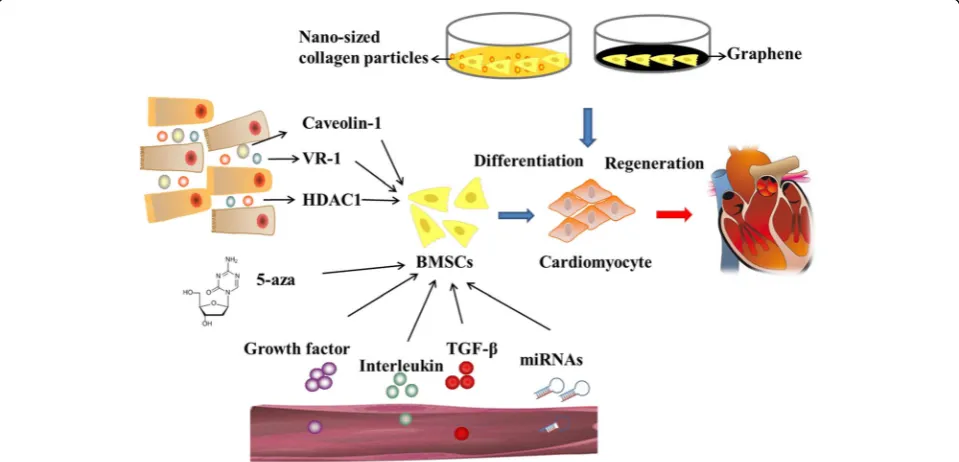
immune modulatory properties. In this article, some
regulatory factors which could induce the cardiomyoctes
differentiation have been summarized, including 5-aza,
miRNAs, cytokines, microenvironment, Caveolin-1,
VR-1 and HDACVR-1 (Fig.
1). More exploration are needed to
elucidate the mechanism of BMSCs differentiate into
cardiomyocytes and accelerate the clinical application.
Abbreviations
AMI:Acute myocardial infarction; ANP: Atrial natriuretic peptide; 5-aza: 5-Azacytidine; bFGF: Basic fibroblast growth factor; BMP: Bone morphogenetic protein; BMSC: Bone marrow-derived mesenchymal stem cell; CLC: Cardiomyocyte-like cell; cTnC: Cardiac troponin C; cTnI: Cardiac troponin I; cTnT: Cardiac troponin T; CX43: Connexin 43; Dll-1: Delta-like 1; ES: Embryonic stem; FAK: Focal adhesion kinase; GATA4: GATA binding protein 4;
HGF: Hepatocyte growth factor; IGF-1: Insulin-like growth factor-1; iPS: Induced pluripotent stem; Islet-1: Insulin gene enhancer binding protein ISL-1; α-MHC: Alpha myosin heavy chain; Nkx2.5: Homeobox protein 2.5; pIGF-1R: Phosphorylation IGF-1 receptor; STAT3: Targeting signal transducers and activators of transcription 3; VR-1: Vanilloid receptor 1
Acknowledgements Not applicable.
Funding
This work was supported by the National Natural Science Fund of China (81170096/81573434).
Availability of data and materials
Data sharing is not applicable to this article as no datasets were generated or analyzed during the current study.
Authors’contributions
ZmD and BzC planned the review. XfG, YB, LZ, and BZ wrote the manuscript. NZ and KC proposed amendments and new directions of discussion. BzC was responsible for final approval of the manuscript. All authors read and approved the final manuscript.
Ethics approval and consent to participate Not applicable.
Consent for publication Not applicable.
Competing interests
The authors declare that they have no competing interests.
Publisher
’
s Note
Springer Nature remains neutral with regard to jurisdictional claims in published maps and institutional affiliations.
Author details
1Department of Pharmacy, the Second Affiliated Hospital of Harbin Medical
University, No. 246 Xuefu Road, Harbin, Heilongjiang Province 150081,
People’s Republic of China.2Department of Internal Diseases, Bashkir State Medical University, Ufa, Russia.3Cell Therapy and Biotechnology in Regenerative Medicine Research Group, Pequeno Príncipe Faculty, Pelé Pequeno Príncipe Institute, Curitiba, Brazil.
References
1. Dowell JD, Rubart M, Pasumarthi KB, Soonpaa MH, Field LJ. Myocyte and myogenic stem cell transplantation in the heart. Cardiovasc Res. 2003;58:336–50.
2. Murry CE, Field LJ, Menasche P. Cell-based cardiac repair: reflections at the 10-year point. Circulation. 2005;112:3174–83.
3. Wang H, Hao J, Hong CC. Cardiac induction of embryonic stem cells by a small molecule inhibitor of Wnt/beta-catenin signaling. ACS Chem Biol. 2011;6:192–7.
4. Mummery C, Ward-van Oostwaard D, Doevendans P, Spijker R, van den Brink S, Hassink R, van der Heyden M, Opthof T, Pera M, de la Riviere AB, Passier R, Tertoolen L. Differentiation of human embryonic stem cells to cardiomyocytes: role of coculture with visceral endoderm-like cells. Circulation. 2003;107:2733–40.
5. Kattman SJ, Witty AD, Gagliardi M, Dubois NC, Niapour M, Hotta A, Ellis J, Keller G. Stage-specific optimization of activin/nodal and BMP signaling promotes cardiac differentiation of mouse and human pluripotent stem cell lines. Cell Stem Cell. 2011;8:228–40.
6. Martin-Rendon E, Brunskill S, Doree C, Hyde C, Watt S, Mathur A, Stanworth S. Stem cell treatment for acute myocardial infarction. Cochrane Database Syst Rev. 2008;(4):CD006536.
7. Xu H, Yang YJ, Qian HY, Tang YD, Wang H, Zhang Q. Rosuvastatin treatment activates JAK-STAT pathway and increases efficacy of allogeneic mesenchymal stem cell transplantation in infarcted hearts. Circ J. 2011;75:1476–85. 8. Ryan JM, Barry FP, Murphy JM, Mahon BP. Mesenchymal stem cells avoid
allogeneic rejection. J Inflamm (Lond). 2005;2(1):8.
9. Chang SA, Lee EJ, Kang HJ, Zhang SY, Kim JH, Li L, Youn SW, Lee CS, Kim KH, Won JY, Sohn JW, Park KW, Cho HJ, Yang SE, Oh WI, Yang YS, Ho WK, Park YB, Kim HS. Impact of myocardial infarct proteins and oscillating pressure on the differentiation of mesenchymal stem cells: effect of acute myocardial infarction on stem cell differentiation. Stem Cells. 2008;26:1901–12.
10. Quevedo HC, Hatzistergos KE, Oskouei BN, Feigenbaum GS, Rodriguez JE, Valdes D, Pattany PM, Zambrano JP, Hu Q, McNiece I, Heldman AW, Hare JM. Allogeneic mesenchymal stem cells restore cardiac function in chronic ischemic cardiomyopathy via trilineage differentiating capacity. Proc Natl Acad Sci U S A. 2009;106:14022–7.
11. Hahn JY, Cho HJ, Kang HJ, Kim TS, Kim MH, Chung JH, Bae JW, Oh BH, Park YB, Kim HS. Pre-treatment of mesenchymal stem cells with a combination of growth factors enhances gap junction formation, cytoprotective effect on cardiomyocytes, and therapeutic efficacy for myocardial infarction. J Am Coll Cardiol. 2008;51:933–43.
12. Collino F, Bruno S, Deregibus MC, Tetta C, Camussi G. MicroRNAs and mesenchymal stem cells. Vitam Horm. 2011;87:291–320.
13. Martin-Rendon E, Sweeney D, Lu F, Girdlestone J, Navarrete C, Watt SM. 5-Azacytidine-treated human mesenchymal stem/progenitor cells derived from umbilical cord, cord blood and bone marrow do not generate cardiomyocytes in vitro at high frequencies. Vox Sang. 2008;95:137–48. 14. Yoon BS, Yoo SJ, Lee JE, You S, Lee HT, Yoon HS. Enhanced differentiation of
human embryonic stem cells into cardiomyocytes by combining hanging drop culture and 5-azacytidine treatment. Differentiation. 2006;74:149–59. 15. Makino S, Fukuda K, Miyoshi S, Konishi F, Kodama H, Pan J, Sano M,
Takahashi T, Hori S, Abe H, Hata J, Umezawa A, Ogawa S. Cardiomyocytes can be generated from marrow stromal cells in vitro. J Clin Invest. 1999;103:697–705.
16. Xu W, Zhang X, Qian H, Zhu W, Sun X, Hu J, Zhou H, Chen Y. Mesenchymal stem cells from adult human bone marrow differentiate into a cardiomyocyte phenotype in vitro. Exp Biol Med (Maywood). 2004;229:623–31.
17. Antonitsis P, Ioannidou-Papagiannaki E, Kaidoglou A, Papakonstantinou C. In vitro cardiomyogenic differentiation of adult human bone marrow mesenchymal stem cells. The role of 5-azacytidine. Interact Cardiovasc Thorac Surg. 2007;6:593–7.
19. Gu S, Jin L, Zhang F, Sarnow P, Kay MA. Biological basis for restriction of microRNA targets to the 3′untranslated region in mammalian mRNAs. Nat Struct Mol Biol. 2009;16:144–50.
20. Wang K, Sun T, Li N, Wang Y, Wang JX, Zhou LY, Long B, Liu CY, Liu F, Li PF. MDRL lncRNA regulates the processing of miR-484 primary transcript by targeting miR-361. PLoS Genet. 2014;10:e1004467.
21. Kim YJ, Hwang SJ, Bae YC, Jung JS. MiR-21 regulates adipogenic differentiation through the modulation of TGF-beta signaling in mesenchymal stem cells derived from human adipose tissue. Stem Cells. 2009;27:3093–102.
22. Wu T, Zhou H, Hong Y, Li J, Jiang X, Huang H. miR-30 family members negatively regulate osteoblast differentiation. J Biol Chem. 2012;287:7503–11.
23. Zhao XL, Yang B, Ma LN, Dong YH. MicroRNA-1 effectively induces differentiation of myocardial cells from mouse bone marrow mesenchymal stem cells. Artif Cells Nanomed Biotechnol. 2016;44:1665–70.
24. Cai B, Li J, Wang J, Luo X, Ai J, Liu Y, Wang N, Liang H, Zhang M, Chen N, Wang G, Xing S, Zhou X, Yang B, Wang X, Lu Y. microRNA-124 regulates cardiomyocyte differentiation of bone marrow-derived mesenchymal stem cells via targeting STAT3 signaling. Stem Cells. 2012;30:1746–55.
25. Shen X, Pan B, Zhou H, Liu L, Lv T, Zhu J, Huang X, Tian J. Differentiation of mesenchymal stem cells into cardiomyocytes is regulated by miRNA-1-2 via WNT signaling pathway. J Biomed Sci. 2017;24:29.
26. Huang F, Tang L, Fang ZF, Hu XQ, Pan JY, Zhou SH. Corrigendum to “miR-1-Mediated Induction of Cardiogenesis in Mesenchymal Stem Cells via Downregulation of Hes-1”. Biomed Res Int. 2016;2016:8510747.
27. Deng L, Hu G, Jin L, Wang C, and Niu H. Involvement of microRNA-23b in TNF-alpha-reduced BMSC osteogenic differentiation via targeting runx2. J Bone Miner Metab. 2017.
28. Li H, Yu B, Zhang Y, Pan Z, Xu W. Jagged1 protein enhances the differentiation of mesenchymal stem cells into cardiomyocytes. Biochem Biophys Res Commun. 2006;341:320–5.
29. Ivey KN, Muth A, Arnold J, King FW, Yeh RF, Fish JE, Hsiao EC, Schwartz RJ, Conklin BR, Bernstein HS, Srivastava D. MicroRNA regulation of cell lineages in mouse and human embryonic stem cells. Cell Stem Cell. 2008;2:219–29. 30. Kim YS, Kim MJ, Koo TH, Kim JD, Koun S, Ham HJ, Lee YM, Rhee M, Yeo SY,
Huh TL. Histone deacetylase is required for the activation of Wnt/beta-catenin signaling crucial for heart valve formation in zebrafish embryos. Biochem Biophys Res Commun. 2012;423:140–6.
31. Gwak J, Hwang SG, Park HS, Choi SR, Park SH, Kim H, Ha NC, Bae SJ, Han JK, Kim DE, Cho JW, Oh S. Small molecule-based disruption of the Axin/beta-catenin protein complex regulates mesenchymal stem cell differentiation. Cell Res. 2012;22:237–47.
32. Huang X, Li D, Wang Z, Huang Z, Dong X, Li C, Lan J. Study of microRNAs targeted Dvl2 on the osteoblasts differentiation of rat BMSCs in hyperlipidemia environment. J Cell Physiol. 2017.https://doi.org/10.1002/jcp.26392. 33. Kapsimali M, Kloosterman WP, de Bruijn E, Rosa F, Plasterk RH, Wilson SW.
MicroRNAs show a wide diversity of expression profiles in the developing and mature central nervous system. Genome Biol. 2007;8:R173. 34. Arminan A, Gandia C, Bartual M, Garcia-Verdugo JM, Lledo E, Mirabet V,
Llop M, Barea J, Montero JA, Sepulveda P. Cardiac differentiation is driven by NKX2.5 and GATA4 nuclear translocation in tissue-specific mesenchymal stem cells. Stem Cells Dev. 2009;18:907–18.
35. Wu J, Zhao J, Sun L, Pan Y, Wang H, Zhang WB. Long non-coding RNA H19 mediates mechanical tension-induced osteogenesis of bone marrow mesenchymal stem cells via FAK by sponging miR-138. Bone. 2017;108:62–70. 36. Haylor J, Hickling H, El Eter E, Moir A, Oldroyd S, Hardisty C, El Nahas AM.
JB3, an IGF-I receptor antagonist, inhibits early renal growth in diabetic and uninephrectomized rats. J Am Soc Nephrol. 2000;11:2027–35.
37. Hu C, Wu Y, Wan Y, Wang Q, Song J. Introduction of hIGF-1 gene into bone marrow stromal cells and its effects on the cell’s biological behaviors. Cell Transplant. 2008;17:1067–81.
38. Schnabel LV, Lynch ME, van der Meulen MC, Yeager AE, Kornatowski MA, Nixon AJ. Mesenchymal stem cells and insulin-like growth factor-I gene-enhanced mesenchymal stem cells improve structural aspects of healing in equine flexor digitorum superficialis tendons. J Orthop Res. 2009;27:1392–8.
39. Liechty KW, MacKenzie TC, Shaaban AF, Radu A, Moseley AM, Deans R, Marshak DR, Flake AW. Human mesenchymal stem cells engraft and demonstrate site-specific differentiation after in utero transplantation in sheep. Nat Med. 2000;6:1282–6.
40. Orlic D, Kajstura J, Chimenti S, Limana F, Jakoniuk I, Quaini F, Nadal-Ginard B, Bodine DM, Leri A, Anversa P. Mobilized bone marrow cells repair the infarcted heart, improving function and survival. Proc Natl Acad Sci U S A. 2001;98:10344–9.
41. Kudo M, Wang Y, Wani MA, Xu M, Ayub A, Ashraf M. Implantation of bone marrow stem cells reduces the infarction and fibrosis in ischemic mouse heart. J Mol Cell Cardiol. 2003;35:1113–9.
42. Miyahara Y, Nagaya N, Kataoka M, Yanagawa B, Tanaka K, Hao H, Ishino K, Ishida H, Shimizu T, Kangawa K, Sano S, Okano T, Kitamura S, Mori H. Monolayered mesenchymal stem cells repair scarred myocardium after myocardial infarction. Nat Med. 2006;12:459–65.
43. Ellison GM, Torella D, Dellegrottaglie S, Perez-Martinez C, Perez de Prado A, Vicinanza C, Purushothaman S, Galuppo V, Iaconetti C, Waring CD, Smith A, Torella M, Cuellas Ramon C, Gonzalo-Orden JM, Agosti V, Indolfi C, Galinanes M, Fernandez-Vazquez F, Nadal-Ginard B. Endogenous cardiac stem cell activation by insulin-like growth factor-1/hepatocyte growth factor intracoronary injection fosters survival and regeneration of the infarcted pig heart. J Am Coll Cardiol. 2011;58:977–86.
44. Savi M, Bocchi L, Fiumana E, Karam JP, Frati C, Bonafe F, Cavalli S, Morselli PG, Guarnieri C, Caldarera CM, Muscari C, Montero-Menei CN, Stilli D, Quaini F, Musso E. Enhanced engraftment and repairing ability of human adipose-derived stem cells, conveyed by pharmacologically active microcarriers continuously releasing HGF and IGF-1, in healing myocardial infarction in rats. J Biomed Mater Res A. 2015;103:3012–25.
45. Zhang GW, Gu TX, Guan XY, Sun XJ, Qi X, Li XY, Wang XB, Lv F, Yu L, Jiang DQ, Tang R. HGF and IGF-1 promote protective effects of allogeneic BMSC transplantation in rabbit model of acute myocardial infarction. Cell Prolif. 2015;48:661–70.
46. Kofidis T, de Bruin JL, Yamane T, Balsam LB, Lebl DR, Swijnenburg RJ, Tanaka M, Weissman IL, Robbins RC. Insulin-like growth factor promotes engraftment, differentiation, and functional improvement after transfer of embryonic stem cells for myocardial restoration. Stem Cells. 2004;22:1239–45.
47. Shuang T, Fu M, Yang G, Wu L, Wang R. The interaction of IGF-1/IGF-1R and hydrogen sulfide on the proliferation of mouse primary vascular smooth muscle cells. Biochem Pharmacol. 2017.
48. Gong H, Wang X, Wang L, Liu Y, Wang J, Lv Q, Pang H, Zhang Q, Wang Z. Inhibition of IGF-1 receptor kinase blocks the differentiation into cardiomyocyte-like cells of BMSCs induced by IGF-1. Mol Med Rep. 2017;16:787–93.
49. McMahon LA, Prendergast PJ, Campbell VA. A comparison of the involvement of p38, ERK1/2 and PI3K in growth factor-induced chondrogenic differentiation of mesenchymal stem cells. Biochem Biophys Res Commun. 2008;368:990–5. 50. Laugwitz KL, Moretti A, Caron L, Nakano A, Chien KR. Islet1 cardiovascular
progenitors: a single source for heart lineages? Development. 2008;135:193–205. 51. Nakano A, Nakano H, Chien KR. Multipotent islet-1 cardiovascular
progenitors in development and disease. Cold Spring Harb Symp Quant Biol. 2008;73:297–306.
52. Cai CL, Liang X, Shi Y, Chu PH, Pfaff SL, Chen J, Evans S. Isl1 identifies a cardiac progenitor population that proliferates prior to differentiation and contributes a majority of cells to the heart. Dev Cell. 2003;5:877–89. 53. Yi Q, Xu H, Yang K, Wang Y, Tan B, Tian J, Zhu J. Islet-1 induces the
differentiation of mesenchymal stem cells into cardiomyocyte-like cells through the regulation of Gcn5 and DNMT-1. Mol Med Rep. 2017;15:2511–20.
54. Bhuvanalakshmi G, Arfuso F, Kumar AP, Dharmarajan A, Warrier S. Epigenetic reprogramming converts human Wharton’s jelly mesenchymal stem cells into functional cardiomyocytes by differential regulation of Wnt mediators. Stem Cell Res Ther. 2017;8:185.
55. Zhang XL, Wu J, Wang J, Shen T, Li H, Lu J, Gu Y, Kang Y, Wong CH, Ngan CY, Shao Z, Zhao X. Integrative epigenomic analysis reveals unique epigenetic signatures involved in unipotency of mouse female germline stem cells. Genome Biol. 2016;17:162.
56. Hawkins RD, Hon GC, Lee LK, Ngo Q, Lister R, Pelizzola M, Edsall LE, Kuan S, Luu Y, Klugman S, Antosiewicz-Bourget J, Ye Z, Espinoza C, Agarwahl S, Shen L, Ruotti V, Wang W, Stewart R, Thomson JA, Ecker JR, Ren B. Distinct epigenomic landscapes of pluripotent and lineage-committed human cells. Cell Stem Cell. 2010;6:479–91.
58. Rosenblatt-Velin N, Lepore MG, Cartoni C, Beermann F, Pedrazzini T. FGF-2 controls the differentiation of resident cardiac precursors into functional cardiomyocytes. J Clin Invest. 2005;115:1724–33.
59. Khezri S, Valojerdi MR, Sepehri H, Baharvand H. Effect of basic fibroblast growth factor on cardiomyocyte differentiation from mouse embryonic stem cells. Saudi Med J. 2007;28:181–6.
60. Hafez P, Jose S, Chowdhury SR, Ng MH, Ruszymah BH. Abdul Rahman Mohd R. Cardiomyogenic differentiation of human sternal bone marrow mesenchymal stem cells using a combination of basic fibroblast growth factor and hydrocortisone. Cell Biol Int. 2016;40:55–64.
61. Yang X, Hao J, Mao Y, Jin ZQ, Cao R, Zhu CH, Liu XH, Liu C, Ding XL, Wang XD, Chen D, Wu XZ. bFGF promotes migration and induces cancer-associated fibroblast differentiation of mouse bone mesenchymal stem cells to promote tumor growth. Stem Cells Dev. 2016;25(21):1629–39.
62. Rodrigues M, Griffith LG, Wells A. Growth factor regulation of proliferation and survival of multipotential stromal cells. Stem Cell Res Ther. 2010;1:32. 63. Khanabdali R, Rosdah AA, Dusting GJ, Lim SY. Harnessing the secretome
of cardiac stem cells as therapy for ischemic heart disease. Biochem Pharmacol. 2016;113:1–11.
64. Wang X, Zhen L, Miao H, Sun Q, Yang Y, Que B, Lopes Lao EP, Wu X, Ren H, Shi S, Lau WB, Ma X, Ma C, Nie S. Concomitant retrograde coronary venous infusion of basic fibroblast growth factor enhances engraftment and differentiation of bone marrow mesenchymal stem cells for cardiac repair after myocardial infarction. Theranostics. 2015;5:995–1006.
65. Bagchi AK, Sharma A, Dhingra S, Lehenbauer Ludke AR, Al-Shudiefat AA, Singal PK. Interleukin-10 activates Toll-like receptor 4 and requires MyD88 for cardiomyocyte survival. Cytokine. 2013;61:304–14.
66. Van Tassell BW, Toldo S, Mezzaroma E, Abbate A. Targeting interleukin-1 in heart disease. Circulation. 2013;128:1910–23.
67. Mohr T, Haudek-Prinz V, Slany A, Grillari J, Micksche M, Gerner C. Proteome profiling in IL-1beta and VEGF-activated human umbilical vein endothelial cells delineates the interlink between inflammation and angiogenesis. PLoS One. 2017;12:e0179065.
68. Khajeniazi S, Solati M, Yazdani Y, Soleimani M, Kianmehr A. Synergistic induction of cardiomyocyte differentiation from human bone marrow mesenchymal stem cells by interleukin 1beta and 5-azacytidine. Biol Chem. 2016;397:1355–64.
69. Nagata Y, Kiyono T, Okamura K, Goto YI, Matsuo M, Ikemoto-Uezumi M, Hashimoto N. Interleukin-1beta (IL-1beta)-induced Notch ligand Jagged1 suppresses mitogenic action of IL-1beta on human dystrophic myogenic cells. PLoS One. 2017;12:e0188821.
70. Sonomoto K, Yamaoka K, Oshita K, Fukuyo S, Zhang X, Nakano K, Okada Y, Tanaka Y. Interleukin-1beta induces differentiation of human mesenchymal stem cells into osteoblasts via the Wnt-5a/receptor tyrosine kinase-like orphan receptor 2 pathway. Arthritis Rheum. 2012;64:3355–63. 71. Massague J, Blain SW, Lo RS. TGFbeta signaling in growth control, cancer,
and heritable disorders. Cell. 2000;103:295–309.
72. Siegeland PM, Massague J. Cytostatic and apoptotic actions of TGF-beta in homeostasis and cancer. Nat Rev Cancer. 2003;3:807–21.
73. Gwak SJ, Bhang SH, Yang HS, Kim SS, Lee DH, Lee SH, Kim BS. In vitro cardiomyogenic differentiation of adipose-derived stromal cells using transforming growth factor-beta1. Cell Biochem Funct. 2009;27:148–54. 74. Rouhi L, Kajbafzadeh AM, Modaresi M, Shariati M, Hamrahi D. Autologous
serum enhances cardiomyocyte differentiation of rat bone marrow mesenchymal stem cells in the presence of transforming growth factor-beta1 (TGF-beta1). In Vitro Cell Dev Biol Anim. 2013;49:287–94. 75. Ingber DE. Mechanical signaling and the cellular response to extracellular
matrix in angiogenesis and cardiovascular physiology. Circ Res. 2002;91:877–87. 76. Yan Z, Yang G, Cui L, He X, Kuang W, Wu W, Liu X, Li L. Effects of electrical
stimulation on the differentiation of mesenchymal stem cells into cardiomyocyte-like cells. Sheng Wu Yi Xue Gong Cheng Xue Za Zhi. 2013;30:556–61.
77. He X, Li L, Tang M, Zeng Y, Li H, Yu X. Biomimetic electrical stimulation induces rat bone marrow mesenchymal stem cells to differentiate into cardiomyocyte-like cells via TGF-beta 1 in vitro. Prog Biophys Mol Biol. 2017. 78. Ramesh S, Singh A, Cibi DM, Hausenloy DJ, Singh MK. In vitro culture of
epicardial cells from mouse embryonic heart. J Vis Exp. 2016;(110). 79. Shikatani EA, Chandy M, Besla R, Li CC, Momen A, El-Mounayri O, Robbins CS,
Husain M. c-Myb regulates proliferation and differentiation of adventitial Sca1+ vascular smooth muscle cell progenitors by transactivation of myocardin. Arterioscler Thromb Vasc Biol. 2016;36:1367–76.
80. Nimura A, Muneta T, Koga H, Mochizuki T, Suzuki K, Makino H, Umezawa A, Sekiya I. Increased proliferation of human synovial mesenchymal stem cells with autologous human serum: comparisons with bone marrow mesenchymal stem cells and with fetal bovine serum. Arthritis Rheum. 2008;58:501–10.
81. Mizuno M, Katano H, Otabe K, Komori K, Kohno Y, Fujii S, Ozeki N, Horie M, Tsuji K, Koga H, Muneta T, Sekiya I. Complete human serum maintains viability and chondrogenic potential of human synovial stem cells: suitable conditions for transplantation. Stem Cell Res Ther. 2017;8:144.
82. Chachques JC, Herreros J, Trainini J, Juffe A, Rendal E, Prosper F, Genovese J. Autologous human serum for cell culture avoids the implantation of cardioverter-defibrillators in cellular cardiomyoplasty. Int J Cardiol. 2004;95 Suppl 1:S29–33.
83. Guerrero-Robles CI, Vazquez-Zapien GJ, Mata-Miranda MM, Noriega-Gonzalez JE, Gonzalez-Diaz CA. Electrical bioimpedance spectroscopy as biosensor technique to identify cells lineages and cell differentiation process. Conf Proc IEEE Eng Med Biol Soc. 2017;2017:3568–71.
84. Thrivikraman G, Madras G, Basu B. Electrically driven intracellular and extracellular nanomanipulators evoke neurogenic/cardiomyogenic differentiation in human mesenchymal stem cells. Biomaterials. 2016;77:26–43.
85. Koga T, Shiraki N, Yano S, Suico MA, Morino-Koga S, Sato T, Shuto T, Kume S, Kai H. Mild electrical stimulation with heat shock guides differentiation of embryonic stem cells into Pdx1-expressing cells within the definitive endoderm. BMC Biotechnol. 2017;17:14.
86. Valdimarsdottirand G, Mummery C. Functions of the TGFbeta superfamily in human embryonic stem cells. APMIS. 2005;113:773–89.
87. Al-Tamari HM, Dabral S, Schmall A, Sarvari P, Ruppert C, Paik J, DePinho RA, Grimminger F, Eickelberg O, Guenther A, Seeger W, Savai R, Pullamsetti SS. FoxO3 an important player in fibrogenesis and therapeutic target for idiopathic pulmonary fibrosis. EMBO Mol Med. 2017.
88. Salazar VS, Gamer LW, Rosen V. BMP signalling in skeletal development, disease and repair. Nat Rev Endocrinol. 2016;12:203–21.
89. Lu J, Sun B, Huo R, Wang YC, Yang D, Xing Y, Xiao XL, Xie X, Dong DL. Bone morphogenetic protein-2 antagonizes bone morphogenetic protein-4 induced cardiomyocyte hypertrophy and apoptosis. J Cell Physiol. 2014;229:1503–10.
90. Sun B, Huo R, Sheng Y, Li Y, Xie X, Chen C, Liu HB, Li N, Li CB, Guo WT, Zhu JX, Yang BF, Dong DL. Bone morphogenetic protein-4 mediates cardiac hypertrophy, apoptosis, and fibrosis in experimentally pathological cardiac hypertrophy. Hypertension. 2013;61:352–60.
91. Ebelt H, Hillebrand I, Arlt S, Zhang Y, Kostin S, Neuhaus H, Muller-Werdan U, Schwarz E, Werdan K, Braun T. Treatment with bone morphogenetic protein 2 limits infarct size after myocardial infarction in mice. Shock. 2013;39:353–60. 92. Lv Y, Gao CW, Liu B, Wang HY, Wang HP. BMP-2 combined with salvianolic
acid B promotes cardiomyocyte differentiation of rat bone marrow mesenchymal stem cells. Kaohsiung J Med Sci. 2017;33:477–85. 93. Han X, Wan Q, Wu W, Zheng A, Li L, Liu X. Activin A and BMP-4 induce
cardiomyocyte-like cells differentiation of human amniotic epithelial cells. Sheng Wu Yi Xue Gong Cheng Xue Za Zhi. 2011;28:1217–22.
94. Tahaand MF, Valojerdi MR. Effect of bone morphogenetic protein-4 on cardiac differentiation from mouse embryonic stem cells in serum-free and low-serum media. Int J Cardiol. 2008;127:78–87.
95. Zhou N, Li Q, Lin X, Hu N, Liao JY, Lin LB, Zhao C, Hu ZM, Liang X, Xu W, Chen H, Huang W. BMP2 induces chondrogenic differentiation, osteogenic differentiation and endochondral ossification in stem cells. Cell Tissue Res. 2016;366:101–11. 96. Liu C, Chen L, Zeng J, Cui J, Ning JN, Wang GS, Belguise K, Wang X,
Qian GS, Lu KZ, Yi B. Bone morphogenic protein-2 regulates the myogenic differentiation of PMVECs in CBDL rat serum-induced pulmonary microvascular remodeling. Exp Cell Res. 2015;336:109–18.
97. Zhang Z, Li H, Ma Z, Feng J, Gao P, Dong H. Efficient cardiomyogenic differentiation of bone marrow mesenchymal stromal cells by combination of Wnt11 and bone morphogenetic protein 2. Exp Biol Med (Maywood). 2012;237:768–76.
98. Hoxha E, Lambers E, Wasserstrom JA, Mackie A, Ramirez V, Abramova T, Verma SK, Krishnamurthy P, Kishore R. Elucidation of a novel pathway through which HDAC1 controls cardiomyocyte differentiation through expression of SOX-17 and BMP2. PLoS One. 2012;7:e45046.
100. Gao P, Yang J, Gao X, Xu D, Niu D, Li J, Wen Q. Salvianolic acid B improves bone marrow-derived mesenchymal stem cell differentiation into alveolar epithelial cells type I via Wnt signaling. Mol Med Rep. 2015;12:1971–6. 101. Wu YJ, Chen SY, Chang SJ, Kuo SM. Enhanced differentiation of rat MSCs
into cardiomyocytes with 5-azacytidine/collagen I nano-molecules. Conf Proc IEEE Eng Med Biol Soc. 2013;2013:322–5.
102. Kim HN, Jiao A, Hwang NS, Kim MS, Kang DH, Kim DH, Suh KY. Nanotopography-guided tissue engineering and regenerative medicine. Adv Drug Deliv Rev. 2013;65:536–58.
103. Lee WC, Lim CH, Shi H, Tang LA, Wang Y, Lim CT, Loh KP. Origin of enhanced stem cell growth and differentiation on graphene and graphene oxide. ACS Nano. 2011;5:7334–41.
104. Park J, Park S, Ryu S, Bhang SH, Kim J, Yoon JK, Park YH, Cho SP, Lee S, Hong BH, Kim BS. Graphene-regulated cardiomyogenic differentiation process of mesenchymal stem cells by enhancing the expression of extracellular matrix proteins and cell signaling molecules. Adv Healthc Mater. 2014;3:176–81.
105. Nart J, de Tapia B, Pujol A, Pascual A, Valles C. Vancomycin and tobramycin impregnated mineralized allograft for the surgical regenerative treatment of peri-implantitis: a 1-year follow-up case series. Clin Oral Investig. 2017. https://doi.org/10.1007/s00784-017-2310-0.
106. Rashedi I, Talele N, Wang XH, Hinz B, Radisic M, Keating A. Collagen scaffold enhances the regenerative properties of mesenchymal stromal cells. PLoS One. 2017;12:e0187348.
107. Dayan V, Yannarelli G, Billia F, Filomeno P, Wang XH, Davies JE, Keating A. Mesenchymal stromal cells mediate a switch to alternatively activated monocytes/macrophages after acute myocardial infarction. Basic Res Cardiol. 2011;106:1299–310.
108. Nemeth K, Leelahavanichkul A, Yuen PS, Mayer B, Parmelee A, Doi K, Robey PG, Leelahavanichkul K, Koller BH, Brown JM, Hu X, Jelinek I, Star RA, Mezey E. Bone marrow stromal cells attenuate sepsis via prostaglandin E(2)-dependent reprogramming of host macrophages to increase their interleukin-10 production. Nat Med. 2009;15:42–9.
109. Waterman RS, Tomchuck SL, Henkle SL, Betancourt AM. A new mesenchymal stem cell (MSC) paradigm: polarization into a pro-inflammatory MSC1 or an immunosuppressive MSC2 phenotype. PLoS One. 2010;5:e10088.
110. Engler AJ, Sen S, Sweeney HL, Discher DE. Matrix elasticity directs stem cell lineage specification. Cell. 2006;126:677–89.
111. Martino MM, Mochizuki M, Rothenfluh DA, Rempel SA, Hubbell JA, Barker TH. Controlling integrin specificity and stem cell differentiation in 2D and 3D environments through regulation of fibronectin domain stability. Biomaterials. 2009;30:1089–97.
112. Ogura N, Kawada M, Chang WJ, Zhang Q, Lee SY, Kondoh T, Abiko Y. Differentiation of the human mesenchymal stem cells derived from bone marrow and enhancement of cell attachment by fibronectin. J Oral Sci. 2004;46:207–13.
113. Sato H, Takahashi M, Ise H, Yamada A, Hirose S, Tagawa Y, Morimoto H, Izawa A, Ikeda U. Collagen synthesis is required for ascorbic acid-enhanced differentiation of mouse embryonic stem cells into cardiomyocytes. Biochem Biophys Res Commun. 2006;342:107–12.
114. Bayer IS. Thermomechanical properties of polylactic acid-graphene composites: a state-of-the-art review for biomedical applications. Materials (Basel). 2017;10(7).
115. Wang Y, Wang H, Liu D, Song S, Wang X, Zhang H. Graphene oxide covalently grafted upconversion nanoparticles for combined NIR mediated imaging and photothermal/photodynamic cancer therapy. Biomaterials. 2013;34:7715–24.
116. Wang L, Wu Y, Hu T, Guo B, Ma PX. Electrospun conductive nanofibrous scaffolds for engineering cardiac tissue and 3D bioactuators. Acta Biomater. 2017;59:68–81. 117. Shah S, Yin PT, Uehara TM, Chueng ST, Yang L, Lee KB. Guiding stem cell
differentiation into oligodendrocytes using graphene-nanofiber hybrid scaffolds. Adv Mater. 2014;26:3673–80.
118. Kim TH, Shah S, Yang L, Yin PT, Hossain MK, Conley B, Choi JW, Lee KB. Controlling differentiation of adipose-derived stem cells using combinatorial graphene hybrid-pattern arrays. ACS Nano. 2015;9:3780–90.
119. Ahadian S, Zhou Y, Yamada S, Estili M, Liang X, Nakajima K, Shiku H, Matsue T. Graphene induces spontaneous cardiac differentiation in embryoid bodies. Nanoscale. 2016;8:7075–84.
120. Choi SH, Jung SY, Yoo SM, Asahara T, Suh W, Kwon SM, Baek SH. Amine-enriched surface modification facilitates expansion, attachment, and maintenance of human cardiac-derived c-kit positive progenitor cells. Int J Cardiol. 2013;168:100–7.
121. Baker N, Zhang G, You Y, Tuan RS. Caveolin-1 regulates proliferation and osteogenic differentiation of human mesenchymal stem cells. J Cell Biochem. 2012;113:3773–87.
122. Lee MY, Ryu JM, Lee SH, Park JH, Han HJ. Lipid rafts play an important role for maintenance of embryonic stem cell self-renewal. J Lipid Res.
2010;51:2082–9.
123. Wang S, Kan Q, Sun Y, Han R, Zhang G, Peng T, Jia Y. Caveolin-1 regulates neural differentiation of rat bone mesenchymal stem cells into neurons by modulating Notch signaling. Int J Dev Neurosci. 2013;31:30–5.
124. Chen Y, Wang C, Huang Q, Wu D, Cao J, Xu X, Yang C, Li X. Caveolin-1 plays an important role in the differentiation of bone marrow-derived
mesenchymal stem cells into cardiomyocytes. Cardiology. 2017;136:40–8. 125. Bandara N, Gurusinghe S, Lim SY, Chen H, Chen S, Wang D, Hilbert B,
Wang LX, Strappe P. Molecular control of nitric oxide synthesis through eNOS and caveolin-1 interaction regulates osteogenic differentiation of adipose-derived stem cells by modulation of Wnt/beta-catenin signaling. Stem Cell Res Ther. 2016;7:182.
126. Guan X, Wang N, Cui F, Liu Y, Liu P, Zhao J, Han C, Li X, Leng Z, Li Y, Ji X, Zou W, Liu J. Caveolin-1 is essential in the differentiation of human adipose-derived stem cells into hepatocyte-like cells via an MAPK pathway-dependent mechanism. Mol Med Rep. 2016;13:1487–94. 127. Guimaraes SE, Rothschild MF, Ciobanu D, Stahl CH, Lonergan SM. SNP
discovery, expression and association analysis for the SDHD gene in pigs. J Anim Breed Genet. 2007;124:139–43.
128. Volpicelli F, Caiazzo M, Moncharmont B, di Porzio U, Colucci-D'Amato L. Neuronal differentiation dictates estrogen-dependent survival and ERK1/2 kinetic by means of caveolin-1. PLoS One. 2014;9:e109671.
129. Chang CC, Chen CY, Wen HC, Huang CY, Hung MS, Lu HC, Chen WL, Chang CH. Caveolin-1 secreted from adipose tissues and adipocytes functions as an adipogenesis enhancer. Obesity (Silver Spring). 2017;25:1932–40. 130. Xie X, Chan KS, Cao F, Huang M, Li Z, Lee A, Weissman IL, Wu JC. Imaging
of STAT3 signaling pathway during mouse embryonic stem cell differentiation. Stem Cells Dev. 2009;18:205–14.
131. Natarajan R, Singal V, Benes R, Gao J, Chan H, Chen H, Yu Y, Zhou J, Wu P. STAT3 modulation to enhance motor neuron differentiation in human neural stem cells. PLoS One. 2014;9:e100405.
132. Xia R, Dekermendjian K, Lullau E, Dekker N. TRPV1: a therapy target that attracts the pharmaceutical interests. Adv Exp Med Biol. 2011;704:637–65. 133. Leuner K, Kraus M, Woelfle U, Beschmann H, Harteneck C, Boehncke WH, Schempp CM, Muller WE. Reduced TRPC channel expression in psoriatic keratinocytes is associated with impaired differentiation and enhanced proliferation. PLoS One. 2011;6:e14716.
134. Shin HY, Hong YH, Jang SS, Chae HG, Paek SL, Moon HE, Kim DG, Kim J, Paek SH, Kim SJ. A role of canonical transient receptor potential 5 channel in neuronal differentiation from A2B5 neural progenitor cells. PLoS One. 2010;5:e10359.
135. Bellini G, Torella M, Manzo I, Tortora C, Luongo L, Punzo F, Colacurci N, Nobili B, Maione S, Rossi F. PKCbetaII-mediated cross-talk of TRPV1/CB2 modulates the glucocorticoid-induced osteoclast overactivity. Pharmacol Res. 2017;115:267–74.
136. Mikami R, Mizutani K, Aoki A, Tamura Y, Aoki K, Izumi Y. Low-level ultrahigh-frequency and ultrashort-pulse blue laser irradiation enhances osteoblast extracellular calcification by upregulating proliferation and differentiation via transient receptor potential vanilloid 1. Lasers Surg Med. 2017.https://doi. org/10.1002/lsm.22775.
137. Zhang Y, Li L, Hua Y, Nunn JM, Dong F, Yanagisawa M, Ren J. Cardiac-specific knockout of ET(A) receptor mitigates low ambient temperature-induced cardiac hypertrophy and contractile dysfunction. J Mol Cell Biol. 2012;4:97–107. 138. Qi Y, Qi Z, Li Z, Wong CK, So C, Lo IC, Huang Y, Yao X, Tsang SY. Role of
TRPV1 in the differentiation of mouse embryonic stem cells into cardiomyocytes. PLoS One. 2015;10:e0133211.
139. Ren M, Wang T, Huang L, Ye X, Xv Z, Ouyang C, Han Z. Role of VR1 in the differentiation of bone marrow-derived mesenchymal stem cells into cardiomyocytes associated with Wnt/beta-catenin signaling. Cardiovasc Ther. 2016;34:482–8.
140. Eisenbergand LM, Eisenberg CA. Wnt signal transduction and the formation of the myocardium. Dev Biol. 2006;293:305–15.
142. Feng C, Zhu J, Zhao L, Lu T, Zhang W, Liu Z, Tian J. Suberoylanilide hydroxamic acid promotes cardiomyocyte differentiation of rat mesenchymal stem cells. Exp Cell Res. 2009;315:3044–51.
143. Li L, Zhu J, Tian J, Liu X, Feng C. A role for Gcn5 in cardiomyocyte differentiation of rat mesenchymal stem cells. Mol Cell Biochem. 2010;345:309–16.
144. Liu Z, Li T, Liu Y, Jia Z, Li Y, Zhang C, Chen P, Ma K, Affara N, Zhou C. WNT signaling promotes Nkx2.5 expression and early cardiomyogenesis via downregulation of Hdac1. Biochim Biophys Acta. 2009;1793:300–11. 145. Dovey OM, Foster CT, Cowley SM. Histone deacetylase 1 (HDAC1), but not
HDAC2, controls embryonic stem cell differentiation. Proc Natl Acad Sci U S A. 2010;107:8242–7.
146. Lu DF, Wang Y, Su ZZ, Zeng ZH, Xing XW, He ZY, Zhang C. Knockdown of the HDAC1 promotes the directed differentiation of bone mesenchymal stem cells into cardiomyocytes. PLoS One. 2014;9:e92179.
147. Ali D, Alshammari H, Vishnubalaji R, Chalisserry EP, Hamam R, Alfayez M, Kassem M, Aldahmash A, Alajez NM. CUDC-907 promotes bone marrow adipocytic differentiation through inhibition of histone deacetylase and regulation of cell cycle. Stem Cells Dev. 2017;26:353–62.
148. Huang Y, Zheng Y, Jin C, Li X, Jia L, Li W. Long non-coding RNA H19 inhibits adipocyte differentiation of bone marrow mesenchymal stem cells through epigenetic modulation of histone deacetylases. Sci Rep. 2016;6:28897. 149. Fu Y, Zhang P, Ge J, Cheng J, Dong W, Yuan H, Du Y, Yang M, Sun R, Jiang H.
Histone deacetylase 8 suppresses osteogenic differentiation of bone marrow stromal cells by inhibiting histone H3K9 acetylation and RUNX2 activity. Int J Biochem Cell Biol. 2014;54:68–77.
150. Yang G, Tian J, Feng C, Zhao LL, Liu Z, Zhu J. Trichostatin a promotes cardiomyocyte differentiation of rat mesenchymal stem cells after 5-azacytidine induction or during coculture with neonatal cardiomyocytes via a mechanism independent of histone deacetylase inhibition. Cell Transplant. 2012;21:985–96. 151. Labovsky V, Hofer EL, Feldman L, Fernandez Vallone V, Garcia Rivello H,