R E S E A R C H
Open Access
Detection, identification and quantification of
Campylobacter jejuni,
coli
and
lari
in food matrices
all at once using multiplex qPCR
Lucie Vondrakova
*, Jarmila Pazlarova and Katerina Demnerova
Abstract
Background:ThermotolerantCampylobacter jejuni,coliandlariare recognized as leading food-borne pathogens causing an acute bacterial enteritis worldwide. Due to narrow spectrum of their biochemical activity, it is very complicated to distinguish between individual species. For reliable risk assessment, proper incidence evaluation or swift sample analysis regarding individual species, a demand for simple and rapid method for their distinguishing is reasonable. In this study, we evaluated a reliable and simple approach for their simultaneous detection, species identification and quantification using multiplex qPCR.
Results:Species specific primers and hydrolysis probes are directed to hippuricase gene ofC. jejuni, serine hydroxymethyltransferase gene ofC. coliand peptidase T gene ofC. lari. Efficiencies of reactions were 90.85% for
C. jejuni, 96.97% forC. coliand 92.89% forC. lari. At 95.00% confidence level and when cut off is set to 38 cycles, limits of detection are in all cases under 10 genome copies per reaction which is very appreciated since it is known that infectious doses are very low.
Conclusions:Proposed assay was positively validated on different food matrices (chicken wing rinses, chicken juice and homogenized fried chicken strips). No inhibition of PCR reaction occurred. Assay was evaluated in accordance with MIQE handbook.
Keywords:ThermotolerantCampylobacterspp, Multiplex qPCR, Quantification, MIQE
Background
Alimentary infections caused by various food-borne path-ogens generally pose a threat to public health which has to be determined and, if possible, prevented or eliminated.
Thermotolerant bacteria belonging to Campylobacter genus (especiallyC. jejuni,C. coli,C. lariand marginally
C. upsaliensis) are recognised as leading human
food-borne pathogens causing an acute gastrointestinal dis-ease called campylobacteriosis. Since 2007, the incidence of campylobacteriosis in Czech Republic is significantly higher than incidence of similar well known disease sal-monellosis [1]. However, this trend is also apparent in all developed countries worldwide [2].
Digestive tracts of domesticated animals farmed for meat (especially poultry, pigs, cattle and sheep) and wild
warm-blooded animals are significant reservoirs of ther-motolerantCampylobacters. However, many other sources are also known (e.g. sewage, both drinking and environ-mental water, raw milk, pets, various kinds of seafood, in-sects etc.). From all these sources they are able to spread into a food chain or to an immediate proximity of human beings and can cause the infection [3-6]. Leaving aside typical clinical symptoms [3-5,7], another significant prob-lem is the possibility of developing various post-infectious complications e.g. reactive arthritis, urticaria or erythema nodosum. The most serious sequel is the Guillain-Barré syndrome (GBS) which manifests as an acute polyneurop-athy affecting peripheral nervous system leading to a typ-ical ascending paralysis. Especially infection caused byC. jejuni is a common trigger of this disease and it is esti-mated that its infection proceeded to about 30% of all GBS cases [3,4,8].
Although normative methods for detection and enu-meration of Campylobacterspp. e.g. [9] show relatively * Correspondence:[email protected]
Institute of Chemical Technology, Prague, Faculty of Food and Biochemical Technology, Department of Biochemistry and Microbiology, Technicka 5, Prague 166 28, Czech Republic
high degree of specificity, their main disadvantages are ap-parent. These procedures are based on selective enrich-ment of desired bacteria, which consequently does not enable their quantification in original sample, followed by their isolation from background microflora, biochemical characterization and phenotypic or serotype identification. Considering Campylobacters’ special requirements for optimal growth, the detection according to standardised methods may take up to 7–10 days. Another problem ap-pears when identification of individual species is required. This is caused mainly because of their relatively narrow spectrum of biochemical reactivity. However, also increas-ing numbers of nalidixic acid resistantC. jejuniandC. coli strains, orC. jejunistrains which are not able to hydrolyse hippuric acid under laboratory conditions (some of the biochemical tests used for species differentiation) make this situation more complicated as well [3,10-14]. Another issue linked with classical microbiological methods, which should be mentioned, is their inability to detect viable but non-culturable bacteria (VBNC). Especially C. jejuni is known for its capability to enter this state when stressed, starved or physically damaged. Since this phenomenon has not yet been properly explored, it is assumed that such bacteria may be able to regenerate and therefore become infectious again [15,16].
With a regard to already mentioned problems it is evi-dent that an availability of reliable alternatives for rapid detection, identification and quantification, especially in the food and agricultural industry, is undoubtedly an issue of the day. Over the last several years, various campylobacter-focused studies implementing real-time PCR approach, which seems to be suitable to provide appropriate solution meeting all of the above require-ments, have been proposed. However, only very few of them were carried out in a platform of multiplex quan-titative real-time PCR (qPCR), which enables complex analysis of a given sample. Main drawbacks of proposed studies are either that they are not focused on all above-mentioned main thermotolerant Campylobacters, and/ or enable detection without species identification or quantification e.g. [17-25], or include undesired pre-enrichment step [26-28]. Also one of the most interest-ing studies [28] dealinterest-ing with this issue suffers from several shortcomings. Briefly, C. coli identification is based on amplification of a gene encoding one of the subunits of a cytolethal distending toxin (cdtA), which is one of its viru-lence factors. SincecdtAis not housekeeping gene, is not essential and its predisposition to mutate is higher, some publications reporting data concerning its mutations and deletions in campylobacter genome have been already published and alsocdtAnegative strains are known as well [29-33]. Also the achieved detection sensitivity of the assay (about 38 genome copies per reaction), should be higher and improved by further optimization.
Moreover, during our extensive research on other assays concerned real-time PCR released after 2009, we surpris-ingly found no publication which would be written in accordance with so called MIQE handbook (Minimum in-formation for publication of quantitative real-time PCR experiments), which we consider unfortunate. MIQE is very detailed set of guidelines describing the minimum in-formation which are necessary and should be provided every time when qPCR experiments are evaluated [34-37]. Although MIQE is not obligatory, there is no doubt that such checklist helps to assure the quality of obtained re-sults and for this reason the present study is written in accordance with it.
Methods
Bacterial strains and culture conditions
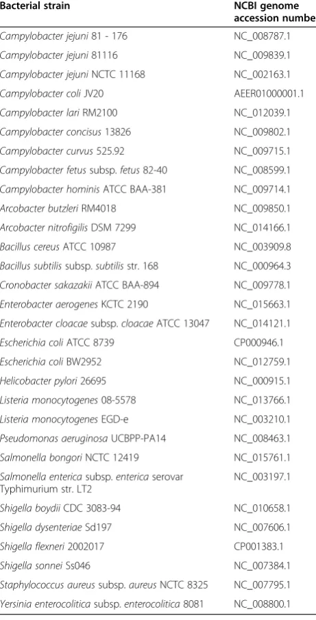
Bacterial strains used for experimental analyses in this study are listed in Table 1. Before further handling, all
Campylobacterstrains were incubated in Park and
San-ders enrichment broth (HiMedia, India) for 24–48 h at 42°C under microaerobic atmosphere (5% O2, 10% CO2
and 85% N2; O2/CO2incubator MCO-18, Sanyo, USA).
Non-campylobacter bacterial strains were aerobically grown in BHI broth (brain heart infusion; Merck, Germany) for 24 h at 37°C.
Design of qPCR assay
Material and methods section is only a brief extract from the very complex MIQE checklist in Additional file 1 where are provided all available details about experimen-tal design and procedures.
DNA extraction
Genomic DNA from pure bacterial cultures (Table 1) was extracted by thermal lysis. Concentration and purity of the DNA were determined spectrophotometrically using
Nano-Photometer™ (Implen, Germany). Only samples whose
A260/A280ratio ranged from 1.7 to 2.1 were used for further
analyses. Real genome copy number was determined using the formula:Genome copies/µl = (C × NA× 10−9)/
(genome length(bp) × Mw), where C is a measured
con-centration of extracted DNA (ng/μl), NA is Avogadro’s
constant (6.02 × 10²³molecule/mole) and Mwis molecular
weight of 1 bp which is 660 Da [28].
In silico analyses
for biotechnology information) and FastPCR molecular biology software [38]. Additional more comprehensive analysis for primer pair specificity checking was con-ducted using Primer-BLAST tool at NCBI. As a database
query “Genome (chromosome of all organisms)” was
selected and as an organism query was selected “ bac-teria (taxid: 2)”. In order to enable proper optimization of qPCR protocol for a multiplex platform, necessary de-termination of primers and probe chemical characteris-tics was also carried out using FastPCR molecular biology software [38].
Empirical primers’specificity screen
In addition to in silico analyses experimental primers’ specificity verification was implemented. Horizontal agarose-gel electrophoresis and melt curve analysis util-izing SYBR Green fluorescent dye (2×Power SYBR Green PCR Master Mix, Applied Biosystems, USA) were
performed. All target and non-target Campylobacter
species listed in Table 1 were used for these analyses. Table 1 List of experimentally includedCampylobacters
and other bacterial species
Species Strain Source Real-time PCR
specificity
C. jejunisubsp.jejuni CCM 6212 Human blood +
C. jejunisubsp.jejuni NCTC 11168
Human faeces +
C. jejunisubsp.jejuni 81-176 Human faeces +
C. jejuni 1 K Chicken meat +
C. jejuni 667/C4 Human clinical isolate
+
C. jejuni 681/C5 Human clinical isolate
+
C. jejuni 2517 Human clinical isolate
+
C. jejuni 3316 Human clinical isolate
+
C. jejuni 13 Wastewater treatment plant
+
C. jejuni 53G Turkey breasts +
C. coli CCM 6211 Pig +
C. coli 549/6 Wastewater treatment plant
+
C. coli 490/3 Wastewater treatment plant
+
C. coli C253 Human clinical isolate
+
C. coli C254 Human clinical isolate
+
C. coli C226 Human clinical isolate
+
C. coli 2463 Human clinical isolate
+
C. coli 2521 Human clinical isolate
+
C. coli 2530 Human clinical isolate
+
C. coli 52 Chicken breasts +
C. lari CCM 4897 Herring gull, cloacal swab
+
C. fetussubsp.fetus CCM 6213 Human blood
-C. upsaliensis ATCC 43954
Animal faeces
-Arcobacter butzleri 2013/43 Chicken thigh
-Arcobacter cryaerophilus
2012/1 Wastewater treatment plant
-Arcobacter skirrowii 2013/34 Cow teat
-Bacillus cereus DBM 3035 Not found
-Bacillus megaterium CCM 2007 Not found
-Bacillus subtilis
subsp.spizizenii
CCM 1999 Not found
-Cronobacter sakazakii
ATCC 29544
Child’s throat
-Table 1 List of experimentally includedCampylobacters and other bacterial species(Continued)
Enterobacter cloacae CCM 1903 Plasma
-Enterococcus faecalis CCM 4224 Urine
-Escherichia coli CCM 4517 Human faeces
-Escherichia coli 485 Raw milk
-Listeria innocua CCM 4030 Cow brain
-Listeria ivanovii
subsp. ivanovii
CCM 5884 Sheep
-Listeria monocytogenes
CCM 5576 Guinea pig mesenteric lymph node
-Listeria
monocytogenes EGD-e
ATCC BAA-679
Animal tissue
-Pseudomonas aeruginosa
CCM 1968 Not found
-Pseudomonas aeruginosa
49 Turkey meat
-Rhodococcus equi CCM 3429 Lung abscess of foal
-Salmonella enterica
subsp.enterica
CCM 7205 Animal tissue
-Staphylococcus aureussubsp.aureus
CCM 3953 Clinical isolate
-Yersinia enterocolytica
CNCTC 7252 Y41/ 73
Human faeces
-Yersinia ruckeri CCM 6093 Rainbow trout
-ATCC–American Type Culture Collection. CCM–Czech Collection of Microorganisms.
Singleplex qPCR
Choice of probes and primers concentrations for the first experiments was based on our research on previ-ously published assays where the most common final concentrations were 0.40 μM and 0.20 μM for primers and probes respectively e.g. [22,24,39-42]. Before stand-ard curves were generated, one experiment was carried out with all target and non-targetCampylobacterspecies as well as with other bacterial strains (Table 1) in order to experimentally verify also specificity of probes. After the positive verification only three reference target
Cam-pylobacter species (C. jejuni CCM 6212, C. coli CCM
6211 and C. lari CCM 4897) were used for further ex-periments. Standard curves for singleplex qPCR plat-form were generated as described in Additional file 1. All samples were run in duplicates and non-template (NTC) and positive controls were included.
Multiplex PCR
Multiplex qPCR was evaluated in two phases. In the first phase, reaction was performed with all components (primers and probes) with DNA from one strain in a tube in order to find out whether undesirable inhibition caused by interaction between components occurs or not. Stand-ard curves were generated and quantification cycles (Cq), y-intercepts, slopes, efficiencies (E), standard deviations (SD), correlation coefficients (R2) and linear ranges were determined using 7500 Software (Applied Biosystems, USA, version 2.0.5), Microsoft Office Excel 2007 (Micro-soft, USA, version 2007) and GenEx software (MultiD Analyses AB, Sweden, version GenEx 5 Enterprise).
In the second phase, reaction mixture contained all chemical components as well as mixed DNA sample from all target Campylobacter strains. Standard curves
were generated and abovementioned parameters deter-mined using the same approach. In accordance with ob-tained results the concentrations of individual components and reaction conditions were optimized until satisfying values were obtained (Additional file 1).
Data analysis
Output data were analysed with instrument compliant 7500 Software (Applied Biosystems, USA, version 2.0.5), Microsoft Office Excel 2007 (Microsoft, USA, version 2007) and GenEx software (MultiD Analyses AB, Sweden, version GenEx 5 Enterprise).
Assay validation on food samples
Sample collection and processing
Three different types of food matrices were chosen for empirical assay validation – raw chicken wings (local butchery, Prague, Czech Republic), whole frozen chicken without giblets (local hypermarket, Prague, Czech Repub-lic) and fried chicken strips (fast food restaurant, Prague, Czech Republic). All samples were processed immediately after the purchase as described in Additional file 1, and obtained chicken wing rinses, chicken juice and homogen-ate from fried chicken strips were further examined.
Artificial contamination of food samples
Each sample was divided into four aliquots. One remained unspiked and the rest was artificially contaminated with pure culture of individual reference Campylobacter strain and then serially diluted in order to achieve the range of approximately 101-105CFU/ml. The number of Campylo-bactercells used for the spiking was determined on Karmali agar with Campylobacter selective supplement (sodium pyruvate, vancomycin, cefoperazone and cycloheximide; Table 2 PCR primers and probes in this assay
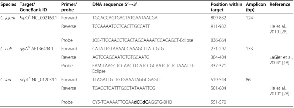
Species Target/ GeneBank ID
Primer/ probe
DNA sequence 5’→3’ Position within
target
Amplicon (bp)
Reference
C. jejuni hipOaNC_002163.1 Forward TGCACCAGTGACTATGAATAACGA 809-832 124
Reverse TCCAAAATCCTCACTTGCCATT 911-932 He et al.,
2010 [28]
Probe JOE-TTGCAACCTCACTAGCAAAATCCACAGCT-Eclipse 836-864
C. coli glyAbAF136494.1 Forward CATATTGTAAAACCAAAGCTTATCGTG 271-297 133
Reverse AGTCCAGCAATGTGTGCAATG 384-404 LaGier et al.,
2004* [18] Probe
FAM-TAAGCTCCAACTTCATCCGCAATCTCTCTAAATTT-Eclipse
337-371
C. lari pepTcNC_012039.1 Forward TTAGATTGTTGTGAAATAGGCGAGTT 519-544 86
Reverse TGAGCTGATTTGCCTATAAATTCG 581-604 He et al.,
2010* [28]
Probe CY5-TGAAAATTGGAAdCGdCAGGTG-BHQ 551-570 a
hippurate hydrolase. b
serine hydroxymethyltransferase. c
peptidase T.
Oxoid, UK) using a drop plate method [43], by our qPCR as well as via genome copy number determination in pure cultures used for spiking [28].
DNA extraction and qPCR
DNA was isolated from 750 μl of each food sample
using commercial PrepSEQ® Spin Sample Preparation Kit with Protocol (Applied Biosystems, USA) in accord-ance with manufacturer’s recommendations.
qPCR reaction was performed as described in Additional file 1. Briefly, standard curves were constructed using 5μl of mixed DNA extracted from target Campylobacters. Used DNA concentrations ranged approximately from 100
to 106 genome copies (CFU equivalent) per well for C. jejuniand from 100to 105forC. colias well as forC. lari. In the case of food samples 10 μl of DNA were added into reaction. Simultaneously, all food samples
were examined for presence of Campylobacters using
drop plate method [43].
Results and discussion
qPCR assay
For implementation of multiplex qPCR assay three species specific target genes (Table 2) were selected based on re-search of previously published studies e.g. [23,27,28,44-49]. Considering the very short length and high similarity [50] of campylobacters genomes (from 1.5 to 1.7 Mbp), it was necessary to carefully choose such primers and probes which interact exclusively with its target gene, do not form any secondary structures and also have similar chemical characteristics in order to allow the co-amplification of multiple targets in one tube without any competition or inhibition. Regarding obtained results, C. lari specific probe, previously designed by another research group [28], was additionally internally modified with propynyl at two cytosines (Eastport, Czech Republic) because of its shorter length and therefore lower melting temperature in com-parison with the others. Our modification increased its melting temperature by 5°C and therefore its utilization in multiplex platform was possible.
Primers’ specificity was experimentally verified with simple horizontal agarose-gel electrophoresis and melt curve analysis performed with SYBR Green fluorescent
dye. DNA from target and non-target Campylobacter
species (Table 1) was used. As expected, only specific melting peaks of amplified products were obtained. Nonspecific amplicons of different lengths or primer-dimers did not form. Amplification of non-target DNA
(C. fetussubspfetusCCM 6213 andC. upsaliensisATCC
43954) did not occur as well.
Singleplex qPCR
First singleplex qPCR served for specificity screen of hy-drolysis probes. As a sample DNA isolated from all bacteria listed in Table 1 was used. Three different combinations of primers and probes final concentrations in reaction were tested as follows: 0.40×0.20, 0.30×0.10 and 0.20×0.05μM respectively. There were no significant differences between Cq values, therefore for further experiments in singleplex platform the combination of concentrations 0.40×0.20μM were used. No fluorescent signal was detected when non-target DNA was used as a sample. When specificity of probes was positively verified standard curves for target Campylobacterswere generated. Quantification cycles and efficiencies were 18.54 and 84.56% forC. jejuni, 25.27 and 87.11% for C. coli, 16.19 and 77.93% for C. lari (Table 4) when 106 genome copies per well used. Because of very Table 3 Bacterial strains forin silicospecificity screen of
primers and probes
Bacterial strain NCBI genome
accession number
Campylobacter jejuni81 - 176 NC_008787.1
Campylobacter jejuni81116 NC_009839.1
Campylobacter jejuniNCTC 11168 NC_002163.1
Campylobacter coliJV20 AEER01000001.1
Campylobacter lariRM2100 NC_012039.1
Campylobacter concisus13826 NC_009802.1
Campylobacter curvus525.92 NC_009715.1
Campylobacter fetussubsp.fetus82-40 NC_008599.1
Campylobacter hominisATCC BAA-381 NC_009714.1
Arcobacter butzleriRM4018 NC_009850.1
Arcobacter nitrofigilisDSM 7299 NC_014166.1
Bacillus cereusATCC 10987 NC_003909.8
Bacillus subtilissubsp.subtilisstr. 168 NC_000964.3
Cronobacter sakazakiiATCC BAA-894 NC_009778.1
Enterobacter aerogenesKCTC 2190 NC_015663.1
Enterobacter cloacaesubsp.cloacaeATCC 13047 NC_014121.1
Escherichia coliATCC 8739 CP000946.1
Escherichia coliBW2952 NC_012759.1
Helicobacter pylori26695 NC_000915.1
Listeria monocytogenes08-5578 NC_013766.1
Listeria monocytogenesEGD-e NC_003210.1
Pseudomonas aeruginosaUCBPP-PA14 NC_008463.1
Salmonella bongoriNCTC 12419 NC_015761.1
Salmonella entericasubsp.entericaserovar Typhimurium str. LT2
NC_003197.1
Shigella boydiiCDC 3083-94 NC_010658.1
Shigella dysenteriaeSd197 NC_007606.1
Shigella flexneri2002017 CP001383.1
Shigella sonneiSs046 NC_007384.1
Staphylococcus aureussubsp.aureusNCTC 8325 NC_007795.1
high Cq value forC. coliwhen compared with the others, another combinations of primer and probe concentrations were tested as follows: 0.40×0.20 to 0.50, 0.50×0.20 to 0.50 and 0.80×0.80 μM. However, no significant differences were observed. Anotherin silicoanalysis showed one non-complementary base at 3’end of the forward primer, which was not issue when used in original study [18] where only duplex qPCR was evaluated and quantification cycles ranged between 18.80-23.00 (when 106genome copies per well used). Therefore 3’ end of original primer was amended by adding a two bases which increased a stability and specificity of annealing step (Additional file 1). Our adjustment caused significant decrease in Cq value to 15.72 (when 106genome copies per well used) and slight increase in efficiency as well (89.24%) when concentrations of primers and probes in reaction were 0.40×0.20 μM (Table 4). Due to the fact that singleplex platform was mainly performed in order to verify the functionality of the reaction, we proceeded directly to the multiplex without further optimization in order to improve obtained values.
Multiplex qPCR
In the first experiment, all components were present in reaction mixture at the same concentration as in the sin-gleplex, but DNA sample in each reaction originated only from individual species (not mixed sample). Con-sidering greater number of components when multiplex-ing, reaction volume was increased from 25μl to 30 μl. Results showed that there is no inhibition of the reaction caused by interaction between components. Serial dilu-tions of DNA were in the range of 100-107genome cop-ies (CFU equivalents) per well. Quantification cycles and efficiencies were 23.80 and 91.35% for C. jejuni, 23.41 and 95.23% for C. coliand 21.53 and 92.12% for C. lari when 105genome copies per well used (more details in Additional file 1).
In second phase multiplex with mixed DNA sample was evaluated. First experiment was carried out under the same conditions as the singleplex. Serial dilutions of mixed DNA were in the range of 100-107genome copies of each strain per well. Although Cq values and efficien-cies forC. coli andC. lari were comparable with previ-ous multiplex results (DNA from single strain), it was unambiguous that strong inhibition of amplification oc-curred in the case of C. jejuni because of a complete
disappearance of its PCR product. Therefore conven-tional multiplex PCR (all three pairs of primers and three probes) with end point horizontal agarose-gel elec-trophoresis was conducted with two possible combina-tions of DNA present in sample (C. jejuni×C. coli; C. jejuni×C. lari) in order to determine in which case the problem occurs. Based on results it was found that when all components are present in reaction with DNA sam-ple mixed ofC. jejuniandC. larithe amplification ofC. jejunitarget is affected and the typical PCR product does not form. Having regard to the fact that there was no in-hibition due to competition for other reaction compo-nents when multiplex with DNA sample from each strain individually was performed, this indicated that there was some interaction betweenC. jejuniandC. lari
DNA even though the trend of C. lari reaction was
not affected at all. Considering this fact, another op-timization was necessary and various concentrations of
C. jejuni and C. lari specific primers and probes were
tested (results not shown).
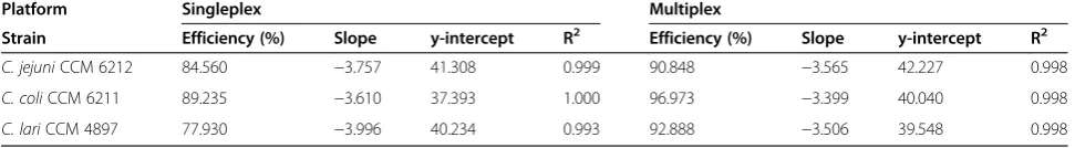
Fully optimized reaction mixture consisted of 0.80μM C. jejuni, 0.40 μM C. coli and 0.05 μMC. lari primers and 0.20μM of each probe. Eight points of ten-fold ser-ial dilutions in the range of 100-107 genome copies of each strain per well were used to generate standard curves. Values of quantification cycles and efficiencies are 22.99 and 90.85% forC. jejuni, 20.77 and 96.97% for C. coli, 20.04 and 91.05% for C. lari, when 105 genome copies per well used (Table 4). All reactions were linear over seven orders of magnitude in the range 101-107 with potential to cover wider range in higher orders. Detection limits of this assay were determined to be between 6.62-16.10 genome copies/well for C. jejuni, 5.13-6.30 genome copies/well for C. coli and 4.87-5.23 genome copies/well for C. lari. All other parameters are provided in Additional file 1.
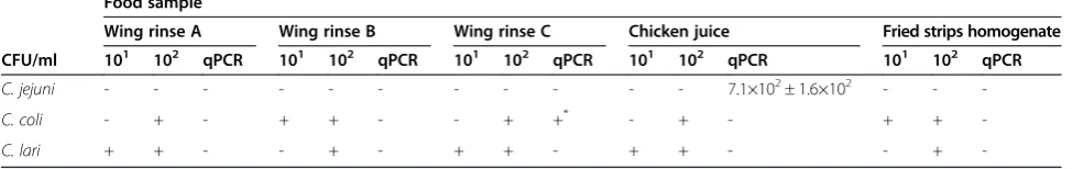
Food sample analyses
For empirical assay evaluation on food samples, three different food matrices which were likely to be naturally
contaminated with Campylobacter species were
exam-ined (chicken wing rinses, chicken juice and homogen-ate prepared from fried chicken strips). Sample aliquots were artificially contaminated with individual target Campylobacters. Unspiked samples were tested for natural Table 4 Comparison of results for singleplex qPCR and optimized multiplex qPCR with pure cultures
Platform Singleplex Multiplex
Strain Efficiency (%) Slope y-intercept R2 Efficiency (%) Slope y-intercept R2
C. jejuniCCM 6212 84.560 −3.757 41.308 0.999 90.848 −3.565 42.227 0.998
C. coliCCM 6211 89.235 −3.610 37.393 1.000 96.973 −3.399 40.040 0.998
C. lariCCM 4897 77.930 −3.996 40.234 0.993 92.888 −3.506 39.548 0.998
R2
contamination as well. Plate counting method [43] and proposed qPCR assay were simultaneously compared.
Using qPCR, quantification of target Campylobacters was possible in all tested food samples even when the highest dilutions were used for spiking. Quantification by plate counting was always possible in the range of 103 -105 CFU/ml however, for some target Campylobacters failed when higher dilutions were used for spiking (Table 5). Quantification ofC. jejuniby plate counting, re-gardless of the sample analysed, always failed with the di-lution corresponding to 102CFU/ml and higher in which case no growth on the plates was observed even after pro-longed incubation for 72 hours. Therefore this concentra-tion seems to be the detecconcentra-tion limit for this specie when the plate counting is used. All the unspiked food samples were determined to beCampylobacterfree by plate count-ing. On the contrary, the unspiked chicken juice was de-termined to be naturally contaminated by C. jejuni using qPCR (Table 5). Also one of the unspiked chicken rinses was reliably determined to be naturally contaminated by
C. coli however, its numbers were below quantification
limit. As mentioned above, there is a possibility that food samples orCampylobactercultures used for spiking con-tained certain numbers of dead or VBNC cells, which were detected and quantified with qPCR but did not grow on plates. However, considering fact that cultures were fresh and under no stress, it is highly unlikely that number of such cells would be significant.
Conclusions
In conclusion, we provided a reliable method for detec-tion, identification and quantification of three most abun-dant thermotolerantCampylobacters. The main advantage of this approach over normative methods for their charac-terization is a possibility to exclude the pre-enrichment step. Exclusion of this part dramatically reduces the time required for analysis. Also the possibility to identify all three species at once is appreciated, since cases of co-contamination and co-infection with more than one Cam-pylobacterspecie are relatively common [51-53]. Also this publication is written in accordance with the MIQE hand-book [35,36] which introduces a very good way to estab-lish a consensus on how best to perform and interpret
qPCR experiments in order to facilitate cooperation be-tween laboratories, comparability and reproducibility of obtained results, and generally serves for higher stan-dardization of real-time PCR experiments.
Additional file
Additional file 1:Supplementary text 1.
Competing interests
Authors declare that they have no competing interests.
Authors’contributions
LV, JP and KD participated in the design of the study. LV performed experiments, collected and analysed data. All authors provided ideas, comments and prepared a draft manuscript and approved the final manuscript.
Acknowledgements
Financial support provided from specific university research MSMT No 21/ 2012 and MSMT No 21/2013.
Received: 3 February 2014 Accepted: 4 May 2014 Published: 9 May 2014
References
1. NIPH:Infectious diseases in Czech Republic.Nat Int Pub Health2013, [http://www.szu.cz/publikace/data/infekce-v-cr]
2. EFSA:EFSA explains zoonotic diseases.Campylobacter European Food Safety Authority2013, [www.efsa.europa.eu/]
3. Allos BM:Campylobacter jejuniinfections: Update on emerging issues and trends.Clin Infect Dis2001,32:1201–1206.
4. Humphrey T, O’Brien S, Madsen M:Campylobacters as zoonotic pathogens: A food production perspective.Int J Food Microbiol2007,
117:237–257.
5. Moore JE, Corcoran D, Dooley JSG, Fanning S, Lucey B, Matsuda M, McDowell DA, Mégraud F, Millar BC, O’Mahony R, O’Riordan L, O’Rourke M, Rao JR, Rooney PJ, Sails A, Whyte P:Campylobacter.Vet Res2005,
36:351–382.
6. Sabatkova Z, Pazlarova J, Demnerova K:Sample processing effect on polymerase chain reaction used for identification ofCampylobacter jejuni.Folia Microbiol2004,49:693–697.
7. Butzler JP:Campylobacter, from obscurity to celebrity.Clin Microbiol Infect
2004,10:868–876.
8. Drenthen J, Yuki N, Meulstee J, Maathuis EM, van Doorn PA, Visser GH, Blok JH, Jacobs BC:Guillain-Barre syndrome subtypes related to
Campylobacterinfection.J Neurol Neurosurg Psychiatry2011,82:300–305. 9. Anonymous:Microbiology of food and animal feeding stuffs - horizontal
method for detection and enumeration ofCampylobacterspp. Part 1: Detection methods.International organization for Standardization2006,
ISO 10272–1:2006.
10. Caner V, Cokal Y, Cetin C, Sen A, Karagenc N:The detection ofhipOgene by real-time PCR in thermophilicCampylobacterspp. with very weak and Table 5 Comparison of food sample analyses results obtained by plate counting and qPCR
Food sample
Wing rinse A Wing rinse B Wing rinse C Chicken juice Fried strips homogenate
CFU/ml 101 102 qPCR 101 102 qPCR 101 102 qPCR 101 102 qPCR 101 102 qPCR
C. jejuni - - - 7.1×10²± 1.6×10² - -
-C. coli - + - + + - - + +* - + - + +
-C. lari + + - - + - + + - + + - - +
-- negative quantification. + positive quantification.
negative reaction of hippurate hydrolysis.Antonie Van Leeuwenhoek2008,
94:527–532.
11. Rautelin H, Jusufovic J, Hänninen M-L:Identification of hippurate-negative thermophilic campylobacters.Diagn Microbiol Infect Dis1999,35:9–12. 12. Totten PA, Patton CM, Tenover FC, Barrett TJ, Stamm WE, Steigerwalt AG,
Lin JY, Holmes KK, Brenner DJ:Prevalence and characterization of hippurate-negativeCampylobacter jejuniin King County, Washington.
J Clin Microbiol1987,25:1747–1752.
13. Endtz HP, Ruijs GJ, Vanklingeren B, Jansen WH, Vanderreyden T, Mouton RP:
Quinolone resistance inCampylobacterisolated from man and poultry following the introduction of fluoroquinolones in veterinary medicine.
J Antimicrob Chemother1991,27:199–208.
14. Aarestrup FM, Engberg J:Antimicrobial resistance of thermophilic
Campylobacter.Vet Res2001,32:311–321.
15. Rollins DM, Colwell RR:Viable but nonculturable stage ofCampylobacter jejuniand its role in survival in the natural aquatic environment.
Appl Environ Microbiol1986,52:531–538.
16. Silva J, Leite D, Fernandes M, Mena C, Gibbs PA, Teixeira P:Campylobacter
spp. as a foodborne pathogen: a review.Front Microbiol2011,2:200. eCollection 2011.
17. Hong J, Jung WK, Kim JM, Kim SH, Koo HC, Ser J, Park YH:Quantification and differentiation ofCampylobacter jejuniandCampylobacter coliin raw chicken meats using a real-time PCR method.J Food Prot2007,
70:2015–2022.
18. LaGier MJ, Joseph LA, Passaretti TV, Musser KA, Cirino NA:A real-time multiplexed PCR assay for rapid detection and differentiation of
Campylobacter jejuniandCampylobacter coli.Mol Cell Probes2004,
18:275–282.
19. Leblanc-Maridor M, Beaudeau F, Seegers H, Denis M, Belloc C:Rapid identification and quantification ofCampylobacter coliand
Campylobacter jejuniby real-time PCR in pure cultures and in complex samples.BMC Microbiol2011,11:113.
20. Toplak N, Kovac M, Piskernik S, Mozina SS, Jersek B:Detection and quantification ofCampylobacter jejuniandCampylobacter coliusing real-time multiplex PCR.J Appl Microbiol2012,112:752–764.
21. Bonjoch X, Calvo L, Soler M, Ruiz-Rueda O, Garcia-Gil LJ:A new multiplexed real-time PCR assay to detectCampylobacter jejuni,C. coli,C. lari, andC. upsaliensis.Food Anal Methods2010,3:40–46.
22. Debretsion A, Habtemariam T, Wilson S, Nganwa D, Yehualaeshet T: Real-time PCR assay for rapid detection and quantification ofCampylobacter jejunion chicken rinses from poultry processing plant.Mol Cell Probes
2007,21:177–181.
23. Nogva HK, Bergh A, Holck A, Rudi K:Application of the 5‘-nuclease PCR assay in evaluation and development of methods for quantitative detection of
Campylobacter jejuni.Appl Environ Microbiol2000,66:4029–4036. 24. Perelle S, Josefsen M, Hoorfar J, Dilasser F, Grout J, Fach P:A LightCycler
real-time PCR hybridization probe assay for detecting food-borne thermophilicCampylobacter.Mol Cell Probes2004,18:321–327. 25. Persson S, Petersen HM, Jespersgaard C, Olsen KEP:Real-time TaqMan
polymerase chain reaction-based genus-identification and
pyrosequencing-based species identification ofCampylobacter jejuni,C. coli,C. lari,C. upsaliensis, andC. fetusdirectly on stool samples.
Diagn Microbiol Infect Dis2012,74:6–10.
26. Josefsen MH, Jacobsen NR, Hoorfar J:Enrichment followed by quantitative PCR both for rapid detection and as a tool for quantitative risk assessment of food-borne thermotolerant campylobacters.
Appl Environ Microbiol2004,70:3588–3592.
27. Sails AD, Fox AJ, Bolton FJ, Wareing DRA, Greenway DLA:A real-time PCR assay for the detection ofCampylobacter jejuniin foods after enrichment culture.Appl Environ Microbiol2003,69:1383–1390.
28. He YP, Yao XM, Gunther NW, Xie YP, Tu SI, Shi XM:Simultaneous detection and differentiation ofCampylobacter jejuni,C. coli, andC. lariin chickens using a multiplex real-time PCR assay.Food Anal Methods2010,3:321–329. 29. AbuOun M, Manning G, Cawthraw SA, Ridley A, Ahmed IH, Wassenaar TM,
Newell DG:Cytolethal distending toxin (CDT)-negativeCampylobacter jejunistrains and anti-CDT neutralizing antibodies are induced during human infection but not during colonization in chickens.Infect Immun
2005,73:3053–3062.
30. Bang DD, Nielsen EM, Scheutz F, Pedersen K, Handberg K, Madsen M:PCR detection of seven virulence and toxin genes ofCampylobacter jejuni
andCampylobacter coliisolates from Danish pigs and cattle and
cytolethal distending toxin production of the isolates.J Appl Microbiol
2003,94:1003–1014.
31. Martinez I, Mateo E, Churruca E, Girbau C, Alonso R, Fernandez-Astorga A:
Detection ofcdtA,cdtB, andcdtCgenes inCampylobacter jejuniby multiplex PCR.Int J Med Microbiol2006,296:45–48.
32. Samosornsuk W, Asakura M, Yoshida E, Taguchi T, Nishimura K, Eampokalap B, Phongsisay V, Chaicumpa W, Yamasaki S:Evaluation of a cytolethal distending toxin (cdt) gene-based species-specific multiplex PCR assay for the identification ofCampylobacterstrains isolated from poultry in Thailand.Microbiol Immunol2007,51:909–917.
33. Fernandes M, Mena C, Silva J, Teixeira P:Study of cytolethal distending toxin (cdt) inCampylobacter coliusing a multiplex polymerase chain reaction assay and its distribution among clinical and food strains.
Foodborne Pathog Dis2010,7:103–106.
34. Bustin SA:Why the need for qPCR publication guidelines?-The case for MIQE.Methods2010,50:217–226.
35. Bustin SA, Beaulieu JF, Huggett J, Jaggi R, Kibenge FSB, Olsvik PA, Penning LC, Toegel S:MIQE precis: Practical implementation of minimum standard guidelines for fluorescence-based quantitative real-time PCR experiments.BMC Mol Biol2010,11:74.
36. Bustin SA, Benes V, Garson JA, Hellemans J, Huggett J, Kubista M, Mueller R, Nolan T, Pfaffl MW, Shipley GL, Vandesompele J, Wittwer CT:The MIQE guidelines: minimum information for publication of quantitative real-time PCR experiments.Clin Chem2009,55:611–622.
37. Taylor S, Wakem M, Dijkman G, Alsarraj M, Nguyen M:A practical approach to RT-qPCR-Publishing data that conform to the MIQE guidelines.
Methods2010,50:S1–S5.
38. Kalendar R, Lee D, Schulman AH:FastPCR software for PCR primer and probe design and repeat search.Genes, Genomes and Genomics2009,
3:1–14 [www.biocenter.helsinki.fi/bi/programs/fastpcr.htm] 39. Banihashemi A, Van Dyke MI, Huck PM:Long-amplicon propidium
monoazide-PCR enumeration assay to detect viableCampylobacterand
Salmonella.J Appl Microbiol2012,113:863–873.
40. Bui XT, Wolff A, Madsen M, Bang DD:Reverse transcriptase real-time PCR for detection and quantification of viableCampylobacter jejunidirectly from poultry faecal samples.Res Microbiol2012,163:64–72.
41. Flekna G, Schneeweiss W, Smulders FJM, Wagner M, Hein I:Real-time PCR method with statistical analysis to compare the potential of DNA isolation methods to remove PCR inhibitors from samples for diagnostic PCR.Mol Cell Probes2007,21:282–287.
42. Rothrock MJ, Cook KL, Bolster CH:Comparative quantification of
Campylobacter jejunifrom environmental samples using traditional and molecular biological techniques.Can J Microbiol2009,55:633–641. 43. Chen CY, Nace GW, Irwin PL:A 6 × 6 drop plate method for simultaneous
colony counting and MPN enumeration ofCampylobacter jejuni,Listeria monocytogenes, andEscherichia coli.J Microbiol Methods2003,55:475–479. 44. Englen MD, Fedorka-Cray PJ:Evaluation of a commercial diagnostic PCR
for the identification ofCampylobacter jejuniandCampylobacter coli.Lett Appl Microbiol2002,35:353–356.
45. Flekna G, Stefanic P, Wagner M, Smulders FJM, Mozina SS, Hein I:
Insufficient differentiation of live and deadCampylobacter jejuniand
Listeria monocytogenescells by ethidium monoazide (EMA) compromises EMA/real-time PCR.Res Microbiol2007,158:405–412.
46. Linton D, Lawson AJ, Owen RJ, Stanley J:PCR detection, identification to species level, and fingerprinting ofCampylobacter jejuniandCampylobacter colidirect from diarrheic samples.J Clin Microbiol1997,35:2568–2572. 47. Wang H, Farber JM, Malik N, Sanders G:Improved PCR detection of
Campylobacter jejunifrom chicken rinses by a simple sample preparation procedure.Int J Food Microbiol1999,52:39–45.
48. Yamazaki-Matsune W, Taguchi M, Seto K, Kawahara R, Kawatsu K, Kumeda Y, Kitazato M, Nukina M, Misawa N, Tsukamoto T:Development of a multiplex PCR assay for identification ofCampylobacter coli,Campylobacter fetus,
Campylobacter hyointestinalissubsphyointestinalis,Campylobacter jejuni,
Campylobacter lariandCampylobacter upsaliensis.J Med Microbiol2007,
56:1467–1473.
49. Yang CB, Jiang Y, Huang KH, Zhu CQ, Yin YL:Application of real-time PCR for quantitative detection ofCampylobacter jejuniin poultry, milk and environmental water.FEMS Immunol Med Microbiol2003,38:265–271. 50. Chang N, Taylor DE:Use of pulsed-field agarose-gel electrophoresis to
size genomes ofCampylobacterspecies and to construct aSalI map of
51. Koene MGJ, Houwers DJ, Dijkstra JR, Duim B, Wagenaar JA:Simultaneous presence of multipleCampylobacterspecies in dogs.J Clin Microbiol2004,
42:819–821.
52. Lawson AJ, Logan JMJ, O’Neill GL, Desai M, Stanley J:Large-scale survey of
Campylobacterspecies in human gastroenteritis by PCR and PCR-enzyme-linked immunosorbent assay.J Clin Microbiol1999,37:3860–3864. 53. Richardson JF, Frost JA, Kramer JM, Thwaites RT, Bolton FJ, Wareing DRA,
Gordon JA:Coinfection withCampylobacterspecies: an epidemiological problem?J Appl Microbiol2001,91:206–211.
doi:10.1186/1757-4749-6-12
Cite this article as:Vondrakovaet al.:Detection, identification and quantification ofCampylobacter jejuni,coliandlariin food matrices all
at once using multiplex qPCR.Gut Pathogens20146:12.
Submit your next manuscript to BioMed Central and take full advantage of:
• Convenient online submission
• Thorough peer review
• No space constraints or color figure charges
• Immediate publication on acceptance
• Inclusion in PubMed, CAS, Scopus and Google Scholar
• Research which is freely available for redistribution