Basic and Clinical
July 2016. Volume 7. Number 3
Hossein Zeinali1, Homa Manaheji2*, Jalal Zaringhalam3, Zahra Bahari1, Samad Nazemi4, Mehdi Sadeghi5
Age-Related Differences in Neuropathic Pain Behavior and
Spinal Microglial Activity after L5 Spinal Nerve Ligation in
Male Rats
Introduction: Several studies have reported the involvement of age-related changes in the
development of neuropathic pain behaviors. However, limited data are available on the role of age in establishing and maintaining chronic neuropathic pain after peripheral nerve injury.
Methods: In the present study, we examined age-related neuropathic behavior among rats in 4 age
groups: pups (4 weeks old; weight, 60–80 g), juvenile rats (6 weeks old; weight, 120–140 g), and mature rats (10–12 weeks old; weight, 200–250 g). Because the exact contribution of spinal microglia and its association with the development of neuropathic pain remains unknown, we also evaluated the expression of spinal Iba1, a microglial marker, by using western blotting before and 5 days after spinal nerve ligation (SNL) as well as after the daily IP administration of minocycline (30 mg/kg).
Results: Our results showed that SNL-induced mechanical allodynia but not thermal hyperalgesia
in mature rats but not in pups (P<0.05 and P<0.01, respectively). The expression of spinal Iba1 in the juvenile rats was significantly lower than that in pups and mature rats (P<0.01). Moreover, administration of minocycline decreased the expression of spinal Iba1 in the pup rats more than in juvenile rats (P<0.001) and in the juvenile rats more than in the mature rats (P<0.05).
Conclusion: These data suggest that the development of neuropathic behaviors and microglial
activation after SNL could be age dependent.
A B S T R A C T
Key Words: Age- related,
Hyperalgesia, Allodynia, Iba1
1. Introduction
europathic pain is characterized by spontaneous pain, hyperalgesia, and al-lodynia. To date, no effective treatment is available for neuropathic pain. The
mechanisms underlying this debilitating disorder are not completely understood. It has been suggested that development of chronic pain may be influenced by age (Resnick, Levy, & Jannetta, 1998; Rokyta et al., 2008) i.e. changes in neurochemical and anatomical
organiza-tion occur with advanced age (Mills, Hains, Johnson,
N
Article info:Received: 16 March 2015 First Revision: 03 April 2015
Accepted: 06 September 2015
1. Department of Physiology, School of Medicine, Shahid Beheshti University of Medical Sciences, Tehran, Iran. 2. Neurophysiology Research Center, Shahid Beheshti University of Medical Sciences, Tehran, Iran.
3. Neuroscience Research Center, Shahid Beheshti University of Medical Sciences, Tehran, Iran. 4. Department of Physiology, School of Medicine, Sabzevar University of Medical Sciences, Sabzevar, Iran. 5. Department of Physiology, Faculty of Medicine, Boushehr University of Medical Sciences, Boushehr, Iran.
* Corresponding Author: Homa Manaheji, PhD
Address: Neurophysiology Research Center, Shahid Beheshti University of Medical Sciences, Tehran, Iran. Tel: +98 (21) 22439971
E-mail: manahejih@sbmu.ac.ir hshardimanaheji@yahoo.com
Citation: Zeinali, H., Manaheji, H., Zaringhalam, J., Bahari, Z., Nazemi, S., & Sadeghi, S. (2016). Age-related differences in neuropathic pain behavior and spinal microglial activity after L5 spinal nerve ligation in male rats. Basic and Clinical Neuroscience, 7(3), 203-212.
http://dx.doi.org/10.15412/J.BCN.03070305
:
: http://dx.doi.org/10.15412/J.BCN.03070305
Hulsebosch, 2002; Mogil, 2006), and the exact effect of age differences on the development of neuropathic pain is unclear. Some studies have reported that the lower incidence of neuropathic pain in children compared to
adults (Drew, Siddall, & Duggan, 2001; Gwak, Hains,
Johnson, & Hulsebosch, 2003).
Postnatal development of nociceptive pathways and central mechanisms of neuropathic pain in the first weeks of life are completely functional in juvenile
ani-mals (Nakamura & Bregman, 2001; Hains, Everhart,
Fullwood, 2003). A recent study indicated that periph-eral nerve injury does not induce mechanical allodynia in rats aged less than 3 weeks. Interestingly, pain symp-toms are not observed even when these rats (nerve-in-jured) reach the age at which an injury normally evokes
mechanical allodynia (Stuesse, Cruce, Lovell,
McBur-ney, & Crisp, 2000; Gwak, et al., 2003).
Microglial cells play a pivotal role in the development of neuropathic pain (Tsuda, Inoue, & Salter, 2005). Spi-nal cord microglia is strongly activated after nerve injury
in the adult rats (Nakamura & Bregman, 2001). Spared
nerve injury (SNI) evokes dorsal horn microglial activa-tion 5 days after operaactiva-tion. Unlike a feeble microglia response in young animals at day 1 of SNI, there was strong astrocytes response, something that was not seen in adults. These results reveals interesting points about the microglia response to nerve injury which may show
the lack neuropathic allodynia in young animals (
Vega-Avelaira, Moss, & Fitzgerald, 2007; Guasti et al., 2009;
Padi & Kulkarni, 2008).
Some studies suggest that activated microglia exhibit the increased the expression of microglial marker Iba1 (Mitchell & Boss, 2002; Mogil, Chesler, Wilson, Juras-ka, & Sternberg, 2000). Glial cell proliferation is rarely detected under normal conditions. However, robust mi-croglial proliferation has been observed in different
neu-ropathic pain models (Mogil et al., 1999; Shir, Ratner,
Raja, Campbell, & Seltzer, 1998). Some studies have shown that preemptive, intraperitoneal, or the intrathe-cal application of minocycline induces antiallodynic and antihyperalgesic effects in neuropathic pain (Kar & Quirion, 1995; Magnone, Rossolini, Piantanelli, Migani,
2000). Therefore, the first aim of the present study was
to evaluate the effect of age differences on the initiation and development of neuropathic pain behavior and the
2. Methods
2.1. AnimalsMale Wistar rats belonging to 3 age groups (pup rats: 4 weeks old, 60–80 g; juvenile rats: 6 weeks, 120–140g; and mature rats: 10–12 weeks, 200–250 g) were caged with 12:12 h light/ dark cycles and free access to food and water ad libitum. Animal experiments were per-formed according to the National Institute of Health (NIH) guidelines for the Care and Use of Laboratory Animals and were approved by the Animal Ethics Com-mittee of Shahid Beheshti University of Medical Sci-ences, Tehran, Iran (194/-90/3/18; 2011).
2.2. Surgical preparation
Neuropathic pain was induced according to the
method described by Kim and Chung (1992). Animals
were anesthetized using pentobarbital sodium (60 mg/ kg, intraperitoneal). The left paraspinal muscles were
separated at the L4–S2 levels, and the left transverse
process of the L6 vertebra was removed. The L5 spinal
nerve was isolated and tightly ligated using a silk thread (6/0), and the distal part of the ligature was transected. The wound was closed later with 3/0 silk threads. In the control sham group, the same surgical procedure was
performed, except that the left L5 spinal nerve was not
ligated and transected instead. Only those animals that showed no sign of motor deficiency (not limping) were considered for further experiments. To study the age– related role of microglia, minocycline (10 and 30 mg/ kg) (Sigma, USA) dissolved in saline was immediately injected intraperitoneally after the operation and then daily up to day 5 after the surgery in all the rats.
2.3. Behavioral study
Von Frey and Hargreaves tests were used to confirm the successful induction of neuropathy. Sham-operated (n=8) and nerve-injured rats (n=8) were examined for the development of mechanical allodynia and thermal hyper-algesia 1 day before and 2, 5, and 7 days after neuropathy.
2.3.1. Mechanical allodynia
To measure paw withdrawal threshold to mechanical stimuli, each animal was placed in a Plexiglas cham-ber with a metal mesh floor. Thresholds determined by
Basic and Clinical
July 2016. Volume 7. Number 3
Yaksh, 1994). Mechanical stimuli were applied perpen-dicular to the plantar surface at the proximal half of the third and fourth toes of the injured hindpaw by using a set of Von Frey monofilaments (Stoelting Co, Wood Dale, IL, USA). The evoked hindpaw (left hind paw) was stimulated by using 1 of the 7 Von Frey filaments, with logarithmically incremental stiffness (2, 4, 6, 8, 15, 26, and 60 (force) g). Each monofilament was applied to the left hindpaw for approximately 2-3 seconds with sufficient force to buckle the filament.
Each filament was applied 3 times at approximately 5 minutes intervals. Each application started using an 8 (force) g stimulus, that increased or decreased in in-tensity until the obvious paw withdrawal was observed. Quick withdrawal or licking of the paw in response to stimulation by a particular filament was considered as a positive response. In the presence of a response, the next filament was applied with a feeble force. In the absence of a response, the next filament was applied with an
in-Figure 1. Comparison of mechanical (A) and thermal (B) threshold ratios (SNL/preSNL), 1 day before and 2, 5, 7 and 21 days after SNL in pup, juvenile, and mature rats. SNL decreased mechanical threshold ratio (postSNL/preSNL) in mature rats than
pup and juvenile ones. This ratio was significantly lower in mature rats than pup ones on day 5 (F(1, 14)=29.95, P<0.05) until day 21 (F(4, 56)=37.36, P<0.01) postinjury. (2-way ANOVA repeated measures, n=6, *P<0.05 and **P<0.01). No significant
differences were observed between pup and juvenile rats, juvenile and mature rats. Moreover, the thermal threshold ratio
showed no significant difference between the 3 different age groups. Day
-1 2 5 7 14 21 -1 2 5 7 14 21 Day
Mature Mechanical threshold
SN
L/Pr
e-SNL r
atio %
SN
L/Pr
e-SNL r
atio %
Mechanical threshold
Mature
Juveniles Juveniles
Pups Pups
150
100
50
0
100
50
0
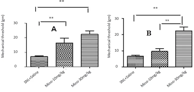
Figure 2. The effect of different doses of minocycline 5 days after SNL plus intraperitoneal administration of minocycline im
-mediately and daily after SNL up to 5th day after SNL on threshold of mechanical allodynia in juvenile (A) and mature (B)
rats (Unpaired t-test, n=6, **P<0.01). Administration of minocycline (10, 30 mg/kg) significantly increased the threshold of mechanical allodynia in juvenile rats. However, only minocycline (30 mg/kg) significantly increased the threshold of mechani-cal allodynia in mature rats compared to the sham group. Since the pup rats showed no significant changes in mechanimechani-cal
allodynia, they omitted from this experiment.
30
20
10
0
30
20
10
0
Me
chanic
al thr
eshold (
gm)
Me
chanic
al thr
eshold (
gm)
SNL+Saline SNL+Saline
Mino-10mg /kg
Mino-10mg /kg
Mino-30mg /kg
Thermal hyperalgesia was assessed as paw withdrawal latency (PWL) by using Hargreaves test. All tests were performed between 8 and 12 AM. The rats were placed in a clear plastic container on an elevated floor made of clear, heat-tempered glass (Plantar Test, Ugo Basile, Italy). After 15 minutes of habituation, a radiant heat source (50 W halogen reflector bulbs with intensity controlled by a constant voltage source) was focused on the plantar surface of the ipsilateral and contralateral
hindpaws (Nazemi, Manaheji, Zaringhalam, Sadeghi, &
Haghparast, 2012 ). Each paw was tested 5 times with 5 minutes intervals, and the average value of PWL for 5 consecutive tests was recorded. The cut-off time in the absence of a response was 33 seconds to avoid tissue damage. The Hargreaves test confirmed the successful induction of neuropathy before the administration of drugs or a vehicle.
2.4. Western blotting
To study the spinal microglial marker Iba1 expression, rats were killed under isoflurane anesthesia, and the lum-bar (L5–L6) region of the spinal cord was removed. Tis-sue samples were homogenized (Brinkmann Polytron Homogenizer, 20000 rpm, 30 s) in RIPA buffer (50 mM Tris-HCl (pH 7.5), 150 mM NaCl, 1 mM ethylenedi-aminetetraacetic acid, 1% NP40, 0.5% sodium dodecyl sulfate (SDS), 1 mM sodium orthovanadate, 2.5 µg/ml aprotinin, 2 µg/mL leupeptin, and 2 µg/mL pepstatin A) and were cleared by centrifugation (10000×g at 15°C for 10 min). Protein concentration in the supernatant was
determined using Bradford assay (Bradford, 1976).
Samples containing 60 µg of protein were heated for 8 minutes at 99°C in loading buffer (4% SDS, 25 mM Tris-HCl (pH 6.8), 5% glycerol, 0.5% 2-mercaptoetha-nol, and 0.01% bromophenol blue) and resolved by SDS polyacrylamide gel electrophoresis on 10% poly-acrylamide gels (120 V for 60 min). After electrophore-sis, the proteins were electrophoretically transferred to PVDF membranes (Millipore, Bedford, MA) by using miniprotein II (Bio-Rad) at 100 V for 85 minutes. Non-specific binding sites were blocked using 2% blocking buffer (0.2% Aurora Blocking Reagent) in Tris-buffered saline (TBS) with 0.1% Tween 20 (TBST) for 90 min-utes at 24°C. Mouse monoclonal anti-Iba1 (1:2000 dilu-tion; Abcam, Cambridge, UK) was used to detect the level of microglial cell activation.
in blocking buffer (1:10000 dilution) for 1 hour at room temperature. After another three 10-minute washes with TBST and another wash in TBS, the immunoreactivity of the proteins on the membrane was visualized using chemiluminescence detection system (ECL Advance; Amersham). The blots were stripped (stripping buffer: 100 µM 2-mercaptoethanol, 2% SDS, and 62.5 mM Tris, pH 6.7) at 50°C for 30 minutes and reprobed using a mouse polyclonal primary antibody against β-actin (1:5000 dilution; cell signaling) as a loading control.
The results were quantified by performing densitomet-ric scanning of the films. Data analysis was performed using ImageJ (V1.41, NIH, USA) after background sub-traction. Each experiment was replicated 3 times with new groups of rats.
2.5. Statistical analysis
All data are presented as mean±standard error of the mean (SEM). In the behavioral study, the data were ana-lyzed using 1- and 2-way repeated measures ANOVA, followed by Bonferroni post hoc test. Graphical and sta-tistical analyses were performed using GraphPad Prism Version 5.0 (GraphPad Prism Software, San Diego, CA, USA).
3. Results
3.1. Time profile of mechanical allodynia and thermal hyperalgesia among rats in 3 age groups
SNL decreased mechanical threshold ratio (postSNL/ preSNL) in mature rats compared to pup and juve-nile ones. This ratio was significantly lower in mature rats than pup ones on day 5 (F(1, 14)=29.95, P<0.05) and continued until 21 days (F(4, 56)=37.36, P<0.01) postinjury (Figure 1-A). No significant differences were observed between pup rats and juvenile ones, or between juvenile rats and mature ones. Moreover, the thermal threshold ratio showed no significant difference among 3 age groups of rats (Figure 1-B).
3.2. Effect of minocycline on SNL-induced allo-dynia
mino-Basic and Clinical
July 2016. Volume 7. Number 3
cycline (30 mg/kg) increased significantly threshold of mechanical allodynia in mature rats compared to sham groups (Figure 2-B). Since the pup rats showed no sig-nificant changes in mechanical allodynia, they omitted from this experiment.
3.3. Effect of SNL and minocycline on Iba1 ex-pression
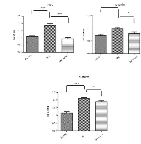
SNL induced the overexpression of spinal Iba1 in all group of the rats. In contrast, administration of minocy-cline significantly decreased the expression of Iba1 in all different age groups, especially juvenile group (Fig-ures 3, 4).
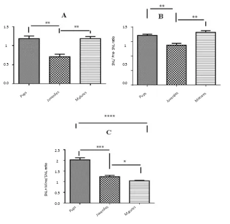
3.4. Comparison of Iba1 expression ratio among rats belonging to different age groups
Expression of Iba1 was significantly lower in juvenile rats than pup and mature ones (Figure 5-A). The ratio of Iba1 expression (postSNL/preSNL) significantly in-creased after SNL; However, it was lower in juvenile group compared to pup and mature rats (Figure 5-B). Moreover, after minocycline administration, the expres-sion ratio was significantly lower in mature rats than pup and juvenile ones (Figure 5-C).
4. Discussion
In this study, SNL resulted in higher induction of mechanical allodynia in juvenile and mature rats
com-pared to pup ones. Comparison of PWL ratio of thermal stimuli showed no significant differences among rats in these 3 age groups. Our results are consistent with those of Fitzgerald study, which showed that brachial plexus injury induced devastating chronic neuropathic pain in
adults but minimal chronic pain in neonates (Fitzgerald,
2005). Facial neuralgia is rare in children (Fitzgerald & MacDermott, 2005; Grazzi, Usai, & Rigamonti, 2005) and adults; its early symptoms manifest during
child-hood, which do not need any therapy (Howard, Walker,
Mota, & Fitzgerald, 2005). It is generally accepted that perinatal and early postnatal periods play a crucial role
in ontogenetic development (Rokyta et al., 2008; Wei et
al., 2010). Postnatal development of nociceptive path-ways and central mechanisms of neuropathic pain in the first few weeks of life are completely functional in
juvenile animals (Hains, Everhart, Fullwood, 2003;
Na-kamura & Bregman, 2001).
Prenatal or neonatal stress may disturb the develop-ment of the nociceptive system and induce long-term behavioral changes that persist during adulthood. How-ever, some type of neuropathic pain cannot be induced in the first two weeks of postnatal period. Therefore, a mature nervous system is required to observe the
devel-opment of pathological behaviors (Rokyta et al., 2008;
Howard, 2003). Apparently, different mechanisms exist for allodynia and hyperalgesia development and these
behaviors are age dependent (Wei et al., 2010). These
events play a pivotal role in age-related neuropathic
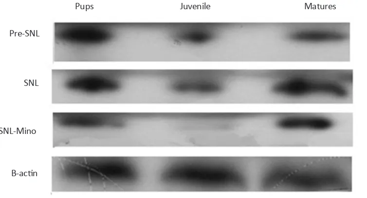
Figure 3. Evaluation of Iba1 expression by western blot among 3 different age groups before SNL (Pre SNL), on the 5th day after
SNL (post SNL), and on the 5th day after SNL plus administration of minocycline (30 mg/kg) immediately and daily after SNL
up to 5th day after SNL. SNL induced the overexpression of spinal Iba1 in all different age groups. In contrast, administration
of minocycline decreased significantly the expression of Iba1 in all age groups, especially in juvenile rats.
Pre-SNL
SNL
SNL-Mino
B-actin
pain after peripheral nerve injury (Wen, Tan, Cheng, Liu, & Ji, 2011; Echeverry, Shi, & Zhang, 2008).
Previous studies have indicated that microglial activa-tion in the spinal cord and higher centers is one of the main causes of increasing sensitivity to pain transmis-sion after a nerve injury (Ledeboer et al., 2005; Suter, Wen, Decosterd, & Ji, 2007). Also, a relationship ex-ists between nerve damage, microglial activation, and neuropathic pain (Anand & Birch, 2002; Grazzi, Usai, & Rigamonti, 2005). In the present study, SNL
signifi-cantly increased spinal Iba1 expression in all rats. How-ever, allodynia and hyperalgesia were not significant in pups after SNL.
Several studies have also confirmed the association of hyperalgesic state and prolife ratio with activation of microglia in the spinal cord and higher centers. It has been shown that lack of allodynia in nerve-injured pups
may arise from immature microglial response (
Fitzger-ald, 2005). Furthermore, CCI and SNL neuropathy acti-vate spinal microglia in rats belonging to all age groups
Figure 4. Spinal Iba1 expression before SNL (preSNL), 5 days after SNL (postSNL) and on the 5th day after SNL plus intraperi
-toneal administration of minocycline (30mg/kg) immediately and daily after SNL in 3 different age groups (Unpaired t-test, n=6, *P<0.05, **P<0.01, and ***P<0.001). Iba1 ratio: Iba1/β-actin. SNL induced overexpression of spinal Iba1 in all age groups of the rats. In contrast, administration of minocycline significantly decreased the expression of Iba1 in all different age groups.
Matures Pre-SN
L
Pre-SN L
Pre-SN L
SNL+Mino SNL+Mino
SNL+Mino
SNL SNL
SNL
Iba-1 Ra
tio
Iba- 1 Ra
tio
Iba- 1 Ra
tio
2
1.5
1
0.5
0.0
2.5
2
1.5
1
0.5
0.0
1
0.5
Basic and Clinical
July 2016. Volume 7. Number 3
(Guasti et al., 2009; Padi & Kulkarni, 2008). Moreover, a study has shown microglial activation after bilateral damage to the spinal nerve with PLS in young male
Wi-star rats (weight, 120–150 g) (Ledeboer et al., 2005).
Several studies have also confirmed association of hy-peralgesic state and proliferation with activation of mi-croglia in the spinal cord and higher centers. Glial cells such as microglia are also critical for the development of neuropathic pain. These studies also showed that
increasing neuronal release of mediators such as ATP, glutamate, and neuropeptides after nerve injury induces glial cell activity (Grazzi, Usai, & Rigamonti, 2005;
Zhuo, Wu, & Wu, 2011). However, the potential factors that may contribute to microglial activation after nerve injury are still unclear. It has been shown that daily ad-ministration of minocycline (10, 20, or 40 mg/kg intra-peritoneally) beginning 1 hour before nerve transection decreases mechanical hyperalgesia and allodynia, with a maximum inhibitory effect at the dose of 20 and 40
Figure 5. Comparison of Iba1 expression ratios among different age groups before SNL (preSNL), 5 days after SNL (postSNL)
and after SNL plus administration of minocycline (30 mg/kg) (Unpaired t-test, n=6, *P<0.05, **P<0.01, and ***P<0.001). Iba1 ratio: Iba1/β-actin. The expression of Iba1 was significantly lower in juvenile rats than pup and mature ones (A). The ratio of Iba1 expression (post SNL/pre SNL) significantly increased after SNL; However, it was lower in juvenile rats than pup and mature rats (B). Moreover, after minocycline administration, the expression ratio was significantly lower in mature rats than
pup and juvenile rats (C).
Pups Pups
Pups Juvenile
s
Juvenile s
Juvenile s Matur
es
Matur es
Matur es
2.5
2
1.5
1
0.5
0.0 1.5
1
0.5
0.0
1.5
1
0.5
0.0
Iba- 1 Ra
tio
SN
L/ Pr
e- SN
L r
atio
SN
L+ Mino/ SN
L r
that inhibition of microglial activation may prevent the occurrence of neuronal hypersensitivity in neuropathic pain (Raghavendra, Tanga, & DeLeo, 2003).
In our study, after-SNL/before-SNL ratio of spinal Iba1 expression was different among three age groups, i.e. higher in pup and mature rats. It seems that SNL-induced expression of spinal Iba1 is age dependent, but the relationship between age and the level of expression is different. The effects of age on Iba1 expression and microglial activity before nerve injury are unknown. It has indicated that in adult rats, SNI provokes strong microglia response at 5 and astrocyte at 7 days after surgery. Unlike adults, in young animals, SNI lead to poor response in the microglia and strong in astrocytes at day 1. These results provide interesting facts about the microglia response to nerve injury which may show the lack neuropathic allodynia in young animals, some-thing that was not seen in adults. Also SNI-induced Iba1 overexpression is age dependent but the relationship be-tween age and Iba1 expression is different to some ex-tent (Vega-Avelaira, Moss, & Fitzgerald, 2007; Guasti et al., 2009; Padi & Kulkarni, 2008).
In our study, daily administration of minocycline (mi-croglial inhibitor) decreased allodynia in juvenile and mature rats. However, the decrease was higher in juve-nile rats than in mature rats and the effect of minocy-cline decreased with an increase in age. It seems that age is a pivotal factor for the activation, induction, and maintenance of neuropathic pain after SNL. Consistent
with the present study, Mika, Osikowicz, Makuch, &
Przewlocka (2007) showed that minocycline (20 and 30 mg/kg) prevented the activation of microglia and symp-toms of neuropathic pain in rats after SNI.
The present study showed that mechanical allodynia (not thermal hyperalgesia) develops in parallel to the expression of spinal Iba1, which is age dependent. Moreover, the effect of minocycline on allodynia and spinal Iba1 expression is age dependent too.
Acknowledgements
This project was supported by the Neuroscience Re-search Center of Shahid Beheshti University of Medical Sciences, Tehran, Iran.
Bennett, A. D., Everhart, A. W., & Hulsebosch, C. E. (2000). Intrathecal administration of an NMDA or a non-NMDA
receptor antagonist reduces mechanical but not thermal al
-lodynia in a rodent model of chronic central pain after spinal cord injury. BrainResearch, 859(1), 72-82.
Bradford, M. M. (1976). A dye binding assay for protein. Ana-lytical Biochemistry, 72, 248-254.
Chaplan, S., Bach, F., Progel, J., Chung, J., & Yaksh, T. (1994).
Quantitative assessment of tactile allodynia in the rat paw. JournalofNeuroscienceMethods, 53(1), 55-63.
Drew, G. M., Siddall, P. J., & Duggan, A. W. (2001). Responses
of spinal neurones to cutaneous and dorsal root stimuli in rats with mechanical allodynia after contusive spinal cord injury. BrainResearch,893(1), 59-69.
Echeverry, S., Shi, X. Q., & Zhang, J. ( 2008). Characterization
of cell proliferation in rat spinal cord following peripheral nerve injury and the relationship with neuropathic pain.
Pain, 135(1-2), 37-47.
Fitzgerald, M. (2005). The development of nociceptive circuits.
NatureReviewNeuroscience,6(7), 507-520.
Fitzgerald, M., & MacDermott, A. (2005). The development of
pain systems. In L. Ede (Ed.), NeurobiologyofPain:Molecular
andCellularNeurobiology (pp. 207-238). UK: Oxford
Univer-sity Press.
Grazzi, L., Usai, S., & Rigamonti, A. (2005). Facial pain in
chil-dren and adolescents. NeurologicalSciences, 26(2), 101-103.
Guasti, L., Richardson, D., Jhaveri, M., Eldeeb, K., Barrett, D., Elphick, M. R., et al. (2009). Minocycline treatment inhibits
microglial activation and alters spinal levels of endocan
-nabinoids in a rat model of neuropathic pain. MolecularPain,
5(35), 1-10.
Gwak, Y. S., Hains, B. C., Johnson, K. M., & Hulsebosch, C. E. ( 2004). Effect of age at time of spinal cord injury on behavioral
outcomes in rat. JournalofNeurotroma, 21(8), 983-93.
Gwak, Y. S., Nam, T. S., Paik, K. S., Hulsebosch, C. E., & Leem, J. W. (2003). Attenuation of mechanical hyperalgesia
fol-lowing spinal cord injury by administration of antibodies to nerve growth factor in the rat. NeuroscienceLetter, 336(2),
117-120.
Hains, B. C., Willis,W. D., & Hulsebosch C. E. (2003). Seroto-nin receptors 5-HT1A and 5-HT3 reduce hyperexcitability
of dorsal horn neurons after chronic spinal cord hemisection injury in rat. ExperimentalBrainResearch, 149(2), 174-186.
Hains, B. C., Everhart, A. W., Fullwood, S. D., & Hulsebosch, C. E. (2002). Changes in serotonin, serotonin transporter expression and serotonin denervation supersensitivity:
in-volvement in chronic central pain after spinal hemisection in the rat. ExperimentalNeurology, 175(2), 347-362.
Howard, R. F. (2003). Current status of pain management in
Basic and Clinical
July 2016. Volume 7. Number 3
Howard, R. F., Walker, S. M., Mota, P. M., & Fitzgerald, M. (2005). The ontogeny of neuropathic pain: Postnatal onset of
mechanical allodynia in rat spared nerve injury (SNI) and chronic constriction injury (CCI) models. Pain, 115(3), 382-389.
Kar, S., & Quirion, R. (1995). Neuropeptide receptors in de-veloping and adult rat spinal cord: An in vitro quantitative autoradiography study of calcitonin gene-related peptide, neurokinins, mu-opioid, galanin, somatostatin, neurotensin
and vasoactive intestinal polypeptide receptors. Journal of
ComparativeNeurology, 354(2), 253-281.
Kim, S. H., & Chung, J. M. (1992). An experimental model for
peripheral neuropathy produced by segmental spinal nerve ligation in the rat. Pain,50(3), 355-363.
Ledeboer, A., Sloane, E. M., Milligan, E. D., Frank, M. G., Ma-hony, J. H., & Maier, S. F. (2005). Minocycline attenuates mechanical allodynia and proinflammatory cytokine
expres-sion in rat models of pain facilitation. Pain, 115(1-2), 71-83.
Magnone, M. C., Rossolini, G., Piantanelli, L. & Migani, P. (2000). Neurochemical parameters of the main
neurotrans-mission systems in aging mice. ArchivesofGerontologyand Geriatric, 30(3), 269-279.
Mika, J., Osikowicz, M., Makuch, W., & Przewlocka, B. (2007).
Minocycline and pentoxifylline attenuate allodynia and hy
-peralgesia and potentiate the effects of morphine in rat and mouse models of neuropathic pain. EuropeanJournalof
Phar-macology,560(2-3), 142-149.
Mills, C. D., Hains, B. C., Johnson, K. M., & Hulsebosch, C. E. (2001). Strain and model differences in behavioral outcomes
after spinal cord injury in rat. Journalof Neurotrauma, 18(8), 743-756.
Mills, C. D., & Hulsebosch, C. E. (2002). Increased expression
of metabotropic glutamate receptor subtype 1 on spinotha
-lamic tract neurons following spinal cord injury in the rat.
NeuroscienceLetter, 319(2), 59-62.
Mitchell, A. & Boss, B. J. (2002). Adverse effects of pain on the nervous systems of newborns and young children: A review
of the literature. JournalofNeuroscienceNursing, 34(5), 228-236.
Mogil, J. S. (2006). Sex, gender and pain. In F. Cervero & T. S. Jensen (Eds.), Pain:Handbookofclinicalneurology (Vol. 81, pp. 325-343). Amstredam: Elsevier, B.V.
Mogil, J. S., Chesler, E.J., Wilson, S. G., Juraska, J. M., & Stern-berg, W. F. (2000). Sex differences in thermal nociception and
morphine antinociception in rodents depend on genotype.
NeuroscienceBiobehavoralReview, 24(3), 375-89.
Mogil, J. S., Wilson, S. G., Bon, K., Lee, S. E., Chung, K., & Raber, P., et al. (1999). Heritability of nociception II.Types’
of nociception revealed by genetic correlation analysis. Pain,
80(1), 83-93.
Nakamura, M., & Bregman, B. S. (2001). Differences in neu-rotrophic factor gene expression profiles between neonate
and adult rat spinal cord after injury. Experimental Neurology, 169(2), 407-415.
Nazemi, S., Manaheji, H., Zaringhalam, J., Sadeghi, M., & Haghparast, A. (2012). Post-injury repeated administrations
of minocycline improve the antinociceptive effect of mor
-phine in chronic constriction injury model of neuropathic
pain in rat. Pharmacology,BiochemistryandBehavior, 102,
520-525.
Padi, S. S., & Kulkarni, S. K. (2008). Minocycline prevents the development of neuropathic pain, but not acute pain: Pos-sible anti-inflammatory and antioxidant mechanisms. Euro-peanJournalof Pharmacology, 601(1-3), 79-87.
Raghavendra, V., Tanga. F., & DeLeo, J. A. (2003). Inhibition of
microglial activation attenuates the development but not ex
-isting hypersensitivity in a rat model of neuropathy. Journal
ofPharmacologyandExperimentalTheraputhic, 306(2), 624-630.
Resnick, D. K., Levy, E. I., & Jannetta, P. J. (1998).
Microvascu-lar decompression for pediatric onset trigeminal neuralgia.
Neurosurgery, 43(4), 804-807.
Rokyta, R., Yamamotova, A., Slamberova, R., Frankek, M., Va-culin, S., & Hruba, L., et al. (2008). Prenatal and perinatal fac-tors influencing nociception, addiction and behavior during
ontogenetic development. PhysiologicalResearch, 57(3), 79-88.
Shir, Y., Ratner, A., Raja, S. N., Campbell, J. N., & Seltzer, Z. (1998). Neuropathic pain following partial nerve injury in
rats is suppressed by dietary soy. NeuroscienceLetter, 240(2),
73-76.
Stuesse, S. L., Cruce, W. L., Lovell, J. A, McBurney, D. L., & Crisp, T. (2000). Microglial proliferation in the spinal cord
of aged rats with a sciatic nerve injury. NeuroscienceLetter,
287(2), 121-124.
Suter, M. R., Wen, Y. R., Decosterd, I., & Ji, R. R. (2007). Do glial
cells control pain? NeuronGliaBiology, 3(3), 255-268.
Tsuda, M., Inoue, K., & Salter, M. W.( 2005). Neuropathic pain and spinal microglia: a big problem from molecules in “small’’ glia. TrendsinNeurosciences,28(2), 101-107.
Vega-Avelaira, D., Moss, A., & Fitzgerald, M. (2007).
Age-re-lated changes in the spinal cord microglial and astrocytic re -sponse profile to nerve injury. Brain, Behavior,andImmunity,
21(5), 617-623.
Wei, H., Hao, B., Huang, J. L., Ma, A. N., Li, X. Y., & Wang, Y. X. (2010). Intrathecal administration of a gap junction
decou-pler, an inhibitor of Na+ K+ 2Cl- cotransporter 1, or a GABA
A
receptor agonist attenuates mechanical pain hypersensitivity
induced by REM sleep deprivation in the rat. Pharmacology
BiochemistryandBehavior,97(2), 377-383.
Wen, Y. R., Tan, P. H., Cheng, J. K., Liu, Y. C., & Ji, R. R. (2011). Microglia: a promising target for treating neuropathic and
postoperative pain, and morphine tolerance. Journal of the
Formosan Medical Association, 110(8), 487-494.
Zhuo, M., Wu, G., & Wu, L. J. (2011). Neuronal and microglial