Open Access
Primary research
Human mast cells decrease SLPI levels in type II – like alveolar cell
model,
in vitro
Camilla Hollander
1, Max Nyström
2,3, Sabina Janciauskiene*
4and
Ulla Westin
1,2Address: 1Department of Otolaryngology and Head and Neck Surgery, University of Lund, SE-205 02 Malmö, Sweden, 2Department of Surgical
Pathophysiology, University of Lund, SE-205 02 Malmö, Sweden, 3Department of Surgery, University of Lund, SE-205 02 Malmö, Sweden and 4Department of Medicine, University of Lund, SE-205 02 Malmö, Sweden
Email: Camilla Hollander - [email protected]; Max Nyström - [email protected]; Sabina Janciauskiene* - [email protected]; Ulla Westin - [email protected] * Corresponding author
SLPImast cellsinflammationmigration
Abstract
Background: Mast cells are known to accumulate at sites of inflammation and upon activation to release their granule content, e.g. histamine, cytokines and proteases. The secretory leukocyte protease inhibitor (SLPI) is produced in the respiratory mucous and plays a role in regulating the activity of the proteases.
Result: We have used the HMC-1 cell line as a model for human mast cells to investigate their effect on SLPI expression and its levels in cell co-culture experiments, in vitro. In comparison with controls, we found a significant reduction in SLPI levels (by 2.35-fold, p < 0.01) in a SLPI-producing, type II-like alveolar cell line, (A549) when co-cultured with HMC-1 cells, but not in an HMC-1-conditioned medium, for 96 hours. By contrast, increased SLPI mRNA expression (by 1.58-fold, p < 0.05) was found under the same experimental conditions. Immunohistochemical analysis revealed mast cell transmigration in co-culture with SLPI-producing A549 cells for 72 and 96 hours.
Conclusion: These results indicate that SLPI-producing cells may assist mast cell migration and that the regulation of SLPI release and/or consumption by mast cells requires interaction between these cell types. Therefore, a "local relationship" between mast cells and airway epithelial cells might be an important step in the inflammatory response.
Background
The inflammatory process in the respiratory airways includes the release of several mediators such as chemoat-tractants, cytokines and proteinases that regulate the adhesion of molecules, and the processes of cell migra-tion, activation and degranulation. The characteristic destruction of tissue in inflammatory diseases is to a large
extent mediated by an excess of neutral serine proteinases and matrix metalloproteinases (MMP) [1–4]. The serine proteinases also contribute to the activation of MMPs, which are typically released in a latent form [5]. Therefore, the regulation of proteolytic enzyme activity in the respi-ratory airways by endogeneous inhibitors is a prerequisite for the maintenance of tissue integrity, and for the repair Published: 20 August 2003
Cancer Cell International 2003, 3:14
Received: 10 March 2003 Accepted: 20 August 2003
This article is available from: http://www.cancerci.com/content/3/1/14
of tissue damage. Proteinase inhibitors that provide pro-tection against the extracellular activity of serine protein-ases include alpha1-antitrypsin (AAT), secretory leukocyte proteinase inhibitor (SLPI) and elafin/skin–derived anti-leukoproteinase (SKALP). Whereas AAT is produced mainly by the liver and reaches the tissues via passive dif-fusion [6], SLPI and elafin/SKALP are produced locally [7–12]
SLPI is found in considerable amounts in nasal, bronchial and cervical mucous, in saliva, and in seminal fluid [7,9,13–16]. There is increasing evidence that SLPI has numerous functions that are not related to its protease-inhibitory activity. SLPI is a non-glycosylated, hydropho-bic, cationic 12 kDa protein, consisting of two homolo-gous cystein-rich domains of 53 and 54 amino acids [17]. The carboxyl-terminal domain of SLPI manifests inhibi-tory activities against chymotrypsin, trypsin, granulocyte and pancreatic elastase, cathepsin G and mass cell chy-mase [18–22], whereas anti-inflammatory, anti-bacterial and anti-fungal activities appear to reside in its amino-ter-minal domain [23,24]. SLPI is shown to reduce LPS-induced TNFα production in the macrophage cell line [25,26], to suppress the production of prostaglandin E2 and metalloproteinase in monocytes [27], and also to antagonize up-regulation of nuclear transcription factor (NF-κB) activation [28]. Lentsch and co-workers have demonstrated that SLPI attenuates the acute inflamma-tory response caused by the deposition of IgG immune complexes in the lungs [29]. In addition, Ashcroft and associates found that SLPI might play a crucial role in wound healing [30]. Recently SLPI has also been shown to inhibit HIV-1 replication in cultured human monocytes [23]. The up-regulation of SLPI by bacterial lipopolysac-charides, and cytokines such as TNFα and IL-1β, com-bined with a broad spectrum antibiotic activity against gram-positive and gram-negative bacteria, suggest it to be a potent anti-microbial "defensin-like" peptide produced by the lungs. States of impaired healing are characterized by excessive proteolysis and often bacterial infection, leading to the hypothesis that SLPI may also have a role in this process.
Historically, SLPI was first purified from secretions of patients with chronic, obstructive pulmonary disease and cystic fibrosis [18], and it was suggested that SLPI being a major anti-elastase inhibitor of the bronchi, is an impor-tant molecule for protecting the respiratory epithelium [13,15]. In contrast to α1-antitrypsin, SLPI blocks elastin-bound elastase in the alveolar walls, which might also protect against the development of emphysema [31]. The interaction between SLPI and elastase is reversible, proba-bly facilitating the transfer of neutrophil elastase to α 1-antitrypsin [32]. It is important to point out that neu-trophil elastase has been found to increase SLPI mRNA
expression in lung epithelial cells in vitro, but this increase in SLPI expression was accompanied by a decrease in SLPI protein release [33]. The local induction of SLPI might be important to break the cycle of inflammation. However the mechanisms involved in the regulation of SLPI expres-sion and release still remain to be elucidated. It has been shown that SLPI is up-regulated by pro-inflammatory stimuli including LPS, TNFα, IL-6 and IL-1β, in vitro [25,26,34,35]. Corticosteroids have also been found to enhance SLPI mRNA levels in airway epithelial cells lead-ing to the suggestion that antiinflammatory effects of cor-ticosteroids may be related to the stimulated SLPI levels [36]. The demonstration that neutrophil defensins increase SLPI release from the bronchial epithelial cells supports the idea that leukocytes play a prominent role in the regulation of SLPI production [37].
Contradictory results have been presented concerning the levels of SLPI during allergic rhinitis in antigen-challenged atopic subjects. For example, lower SLPI levels were found in the bronchial secretions of asthma patients [38]. Other studies indicated that levels of SLPI are also lower in nasal secretion of allergic rhinitis patients compared to healthy controls. After allergen challenge SLPI seems to decrease in the nasal secretions in atopic subjects, which probably indicates mucosal damage [39]. However, the question why SLPI levels is decreased during certain allergic reac-tions, still remains to be answered.
Histamine release from HMC-1 cells
Figure 1
Histamine release from HMC-1 cells. Histamine release was measured at different time points: 0, 3, 6 and 24 h and calcu-lated as a percent of the total cellular histamine content. Each point represents mean ± S.D. of five or six separate experiments. Histamine release increases from 4 % to 25 % over time (p < 0.05).
Cell incubation time (h)
0 5 10 15 20 25 30
Histamine release
(%
of total cellular
his
tamine levels)
Allergic inflammation, including rhinitis, asthma, anaph-ylaxis and urticaria are all disorders associated with mast cell activation [40]. Mast cells are multifunctional cells capable of secreting a wide variety of cytokines, chemok-ines and growth factors [41,42]. The mediators released by mast cells can independently, and in synergy with mac-rophage- and T-cell-derived cytokines, induce much of the inflammatory pathology and serve to stimulate a complex immune response [43,44]. Mast cells are the primary ini-tiating cell of IgE-mediated hypersensitivity. Allergen binding to, or the cross-linking of IgE on the surface of mast cells, which is bound to the high affinity IgE-recep-tor, leads to the rapid release of inflammatory mediators that further provoke a profound immunological and inflammatory process. There are indications that SLPI can inhibit IgE-mediated histamine release from rodent and human nasal mucosa mast cells [45,46]. SLPI may also counterbalance the proteolytic activities caused by pro-tease leakage from the cells [47]. Mast cell and leukocyte serine proteinases are shown to be elevated in the airways of asthmatic patients [40,48]. Individuals with reduced anti-proteinase activity as a result of AAT deficiency, have an increased propensity to develop asthma [49,50]. Together, these findings indicate that proteinase-antipro-teinase imbalance in the airways contributes to the patho-physiological responses in the airways. Because SLPI provides a potent, broad-spectrum inhibitory activity against mast cell and leukocyte serine proteinases, this protein is suggested to be an effective protector against antigen-induced inflammatory responses in the airways. The purpose of this study is to further elucidate how local SLPI levels may be influenced during mast cell interaction with epithelial cells. A co-culture model was used, in which we studied mast cells HMC-1 affect on SLPI levels released from the type II alveolar cell line (A549) derived from human lung carcinoma.
Results
Mast cell characterisation
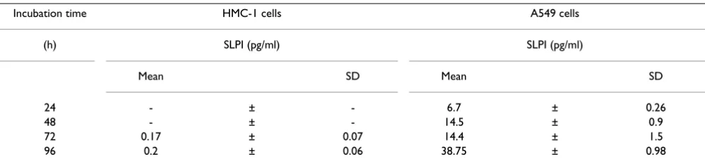
Most studies performed on the mechanisms of the mast cell degranulation are based on the release of histamine. Histamine is electrostatically linked to the protein heparin complex in a manner, which allows it to be released very easily. As shown in figure 1, a spontaneous histamine release from HMC-1 cells consistently increases with incu-bation time (up to 24 h) from 4% to 25% relative to the total cellular histamine content. Continues cell incuba-tion (up to 96 h) showed no further changes in histamine release (data not shown). In addition, the supernatants from the HMC-1 cells alone did not contain SLPI after cell culture for 24 and 48 h, and only trace amounts of SLPI are detected after 72 and 96 h of incubation (Table 1). The ability of HMC-1 cells to express SLPI was also confirmed by immunohistochemical analysis (Fig. 2A and 2B).
Cell co-culture experiments
Epithelium cells, A549, cultured alone for 24, 48, 72 and 96 h increased the SLPI release from 6.7 ± 0.26 to 38.8 ± 0.98 pg/ml (Table 1). Next, we studied the effect of mast cells on SLPI levels by using a transmigration model in which mast cells HMC-1 migrated across the topside of the transwell filter towards the A549 cells. The results obtained with this model show that, during the first 48 h of cell co-culture, mast cells did not migrate into the lung epithelium cell culture (Fig. 3A). However, as shown in figure 3B, cell co-culture for 72 h resulted in a slight mast cell transmigration as indicated by the presence of immu-noreactive HMC-1 cells among the A549 cells. This was, furthermore, confirmed by the co-culture of these cells for 96 h after which a large number of immunoreactive mast cells was detected among the A549 cells (Fig. 3C). It should be pointed out that HMC-1 cells showed no ability to transmigrate through a "blank" transwells. Moreover, cells which had not migrated into the polyester mem-brane inserts displayed immunoreactivity to tryptase for all incubation times, from 24 to 96 h, showing that under the chosen experimental conditions mast cells preserved
Table 1: Time dependent SLPI release from the mast cells, HMC-1, and type II epithelium cells, A549, cultured alone
Incubation time HMC-1 cells A549 cells
(h) SLPI (pg/ml) SLPI (pg/ml)
Mean SD Mean SD
24 - ± - 6.7 ± 0.26
48 - ± - 14.5 ± 0.9
72 0.17 ± 0.07 14.4 ± 1.5
96 0.2 ± 0.06 38.75 ± 0.98
their capacity to differentiate (not shown). We further examined the effect of mast cell trans-migration on the amount of SLPI released by the A549 cells into cell culture media. Only a slight decrease in SLPI levels was observed during 72 h of cell co-culture compared to the SLPI-pro-ducing cells alone (Fig. 4). However, when cells were co-cultured for 96 h a significant decrease in SLPI levels was found. As shown in figure 3, SLPI levels decline by 2.35-fold (p < 0.01) in the presence of mast cells relative to the A549 cells alone.
Effects of conditioned media on SLPI levels
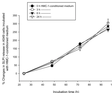
In the next series of experiments we aimed to investigate if concentration of SLPI can be influenced by the incuba-tion of A549 cells with HMC-1-condiincuba-tioned medium for various time periods (from 24 to 96 h). In this case, a medium collected from mast cells at different time points was added to the A549 cells and allowed to act for 24, 48, 72 and 96 h. As shown in Figure 5, the culture of epithelium cells in the mast cell-media caused no changes in SLPI levels.
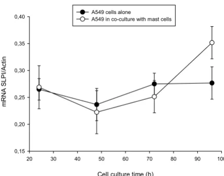
SLPI mRNA expression by A549 cells
To determine whether the decrease of SLPI levels in co-culture with mast cells involves changes in SLPI mRNA expression in A549 cells, we monitored the SLPI mRNA/
β-Actin ratio in A549 cells alone and in co-culture over the period of 24 to 96 h. As shown in figure 6, the levels of SLPI mRNA expression were relatively unchanged when A549 cells were cultured alone. By contrast, increased SLPI
mRNA levels were observed in co-culture experiments after 96 h (1.58-fold, p < 0.05) compared to 24 or 48 h. Under the same experimental conditions, we also exam-ined whether addition of conditioned medium from HMC-1 cells effects SLPI mRNA expression in same way. Consistent with our earlier observations (Fig. 5) showing that A549 cells incubated with conditioned medium from mast cells did not change SLPI protein levels, we found that under these experimental conditions SLPI mRNA lev-els are not changed either (data not shown).
Discussion
Mast cells are widely distributed within the connective tis-sue, with a preferential localization adjacent to the small blood vessels. They play a central role in inflammatory and allergic reactions, and are involved in tissue remodelling during wound healing [40]. The mast cell responses involve the ingestion and killing of adherent substances, unlike that of traditional phagocytic cells. Concomitant with this endocytic activity, inflammatory mediators are released by the mast cells.
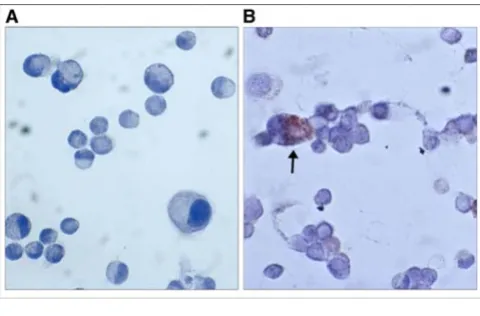
Mast cells constitute a heterogeneous group of cells con-taining several proteases, i.e. tryptase, carboxypeptidase, cathepsin G and chymase [51]. Mainly there are two kinds of mast cells, those in the connective tissue (MCTC) and those in the mucosa (MCT). They differ in protease con-tent, MCTC cells contain tryptase and chymase, whereas MCT cells contain only tryptase. The MCTC cells are pre-dominant in the nasal mucosa while MCT cells are more prevalent in the peripheral lung tissue. Mast cells have also been found to interact with different types of cells, including fibroblasts, endothelial cells, lymphocytes, macrophages, neutrophils, eosinophils, nerve cells and cancer cells [40,43,52,53]. However, the adhesive Localisation of SLPI in HMC-1-mast cells. A, the control
slides were incubated with specific antiserum previously adsorbed with SLPI (1/500) and no positive staining for HMC-1 cells was found (original magnification × 500)
Figure 2
Localisation of SLPI in HMC-1-mast cells. A, the control slides were incubated with specific antiserum previously adsorbed with SLPI (1/500) and no positive staining for HMC-1 cells was found (original magnification × 500). B, HMC-1 cells stained with polyclonal goat-anti-SLPI antibody (1/500) show immunoreactivity for SLPI (original magnifica-tion × 500).
Mast cell HMC-1 migration towards the lung type II epithe-lium cells, A549
Figure 3
mechanisms initiating cell-cell interaction and the conse-quences of this are not well understood.
The present study was designed to investigate the adher-ence of mast cells, HMC-1, to the cell line A549, which represents epithelial cells within the respiratory tract, and to investigate the effects of this cell-cell interaction on SLPI levels.
The mast cell line HMC-1 is known to express a number of β1- and α-integrins as well as other receptors which per-mit the binding of these cells to the extracellular matrix compounds [53–55]. The factors that stimulate mast cell migration still remain largely undefined, although recent reports have implicated the transforming growth factor-β family (TGFβ) as the potential candidate for acting as mast cell chemotaxin, recruiting mast cells into inflamma-tory reactions [56]. In accordance with other studies, we found that HMC-1 mast cells growing in suspension adhere efficiently and spread on top of cell monolayers, in our case on top of A549 cells. After 72 h of HMC-1/A549 co-culture, solitary HMC-1 cells were detected among the A549 cells, while after 96 h a large number of mast cells was found to be adhering to the A549 cells. The mainte-nance of HMC-1 cell maturity was verified by the tryptase immunoreactivity each of the periods of cell culture.
The role of mast cells as primary effector cells in IgE-dependent, immediate hypersensitivity is well established [57]. The discovery that mast cells can release a wide variety of immune mediators, including proteases, cytokines, chemokines and growth factors, suggests an additional role of mast cells in modulating late-phase reactions and other chronic inflammatory processes [40,42]. Here we also demonstrate by immunocytochem-istry that HMC-1 mast cells are SLPI-positive, and trace amounts of SLPI were found in the cell culture superna-tants collected after 72 and 96 h of culture. Although the amount of SLPI released by few activated mast cells had no importance for the present study, this observation still extends earlier findings showing that SLPI and other chy-mase inhibitors, i.e. α1-antichymotrypsin and α 1-antit-rypsin are present in stimulated mast cells and protect the microenvironment against chymase activity [47]. Previ-ously it has been shown that chymase degranulates mast cells, induces histamine release and an increase in SLPI concentration [58]. Neutrophil elastase was also found to increase SLPI transcript levels in primary and transformed human airway epithelial cells in a time- and dose-depend-ent manner [34,36]. These observations suggest that the sensitive regulation of anti-proteases, such as SLPI, in rela-tion to local levels of proteases, may play an important role in minimising tissue destruction.
Effects of mast cells HMC-1 on the ability of the A549 cells to release SLPI
Figure 4
Effects of mast cells HMC-1 on the ability of the A549 cells to release SLPI. The SLPI release from the A549 cells was decreased in co-cultured with HMC-1 cells, compared to the A549 cells alone (p < 0.006). The most pronounced fall in SLPI production was observed after 96 hours of cell co-cul-ture (**p < 0.01). Each point represents mean of six or five separate experiments.
Incubation time (h)
20 30 40 50 60 70 80 90 100
Changes i
n
S
L
P
I r
e
le
ase (
pg/m
l)
0 10 20 30 40 50
A549 cells
Co-culture A549/HMC-1cells
Effects of HMC-1-conditioned medium on SLPI levels in A549 cells
Figure 5
Effects of HMC-1-conditioned medium on SLPI levels in A549 cells. Medium from HMC-1 cells cultured for 0, 3, 6, 8 and 24 h was used to incubate A549 cells for various time periods (from 24 to 96 h). The mast cell-media caused no changes in SLPI levels. Each point represents mean of three independent experiments
Incubation time (h)
20 30 40 50 60 70 80 90 100
%
C
h
a
n
g
e
s
in
S
L
P
I r
e
le
a
s
e
in
A
5
4
9
c
e
lls
in
c
u
b
a
te
d
w
ith HM
C-1-condi
ti
oned m
e
di
um
0 50 100 150 200 250 300 350
0 h HMC-1-conditioned medium 3 h
---With these results as a background we have further inves-tigated the effects of mast cells on the capacity of epithe-lial-presenting cells, A549, to express and release SLPI. In our experimental model, SLPI-producing epithelial cells were cultured for determined time points in conditioned media obtained from mast cells or in co-culture with mast cells. By using this approach, we were able to show that under cell co-culture conditions SLPI levels in cell culture media are gradually decreased, although the expression of mRNA SLPI increases. A significant diminition in SLPI lev-els (by 2.35-fold) was observed after 96 h of cell co-cul-ture, although the SLPI mRNA was up-regulated (by 1.58-fold). In contrast, the A549 cell culture in the mast cell conditioned medium for the time periods chosen had no influence either on SLPI protein levels or on SLPI mRNA expression. By measuring histamine release and tryptase activity in media from mast cells cultured alone for up to 96 h, we were able to show that these cells are not acti-vated. Histamine levels plateaued after 24 h of cell culture which explains why the conditioned media collected from mast cells and added to the SLPI producing cells (A549) had no effect on SLPI levels.
Based on these findings one can conclude that mast cells exert an inhibitory effect on SLPI levels only when they are in close contact with SLPI-producing, A549, cells. Since our primary goal was to find out if mast cell interaction with epithelial cells by itself can induce expression of SLPI and reduce its levels in media, we specifically did not investigate mast cell exogenous activation under these
conditions. This indirectly shows that SLPI is either con-sumed (for example degraded or in complex with enzymes) or its release is inhibited. Studies on this point are in progress in our laboratory.
Recently, van Wetering and co-workers have shown that neutrophil elastase increases SLPI mRNA expression, while it decreases SLPI protein release, in vitro [37]. On the other hand, studies by Hill and co-workers, have indicated that SLPI concentration does not decrease until the elastase activity of the samples is in excess of 50 nM [48]. The relationship between SLPI and elastase is therefore found not to be a simple linear, indicating that a certain amount of elastase and/or other enzymes is needed in order to induce epithelial damage or interfere with epithe-lial cell metabolism resulting in a decreased SLPI secretion [12,32,34].
SLPI levels were found to be decreased in nasal secretion after antigen challenge in vivo [39] as well as in bronchial alveolar lavage obtained from asthmatics compared to healthy subjects [38]. In the airways of allergic patients, mast cells are found in the close proximity of airway thelial cells, which may indicate that mast cells and epi-thelial cells influence each other's properties. Together, previous findings, that SLPI levels are lower in inflamma-tory loci in airways and data from our experimental model, that co-culture of mast cells with SLPI-producing epithelial-like, A549, cells, results in decreased SLPI levels in cell co-culture media suggest that decrease in local anti-protease activity might sustain mucosal damage in reac-tions in which mast cells are participating.
Conclusion
Airway inflammation, present in asthma, bronchitis, and bronchiectasis, is characterised by the presence of acti-vated inflammatory cells. The proteinases, presumably leaking from the cells during their migration from the blood into the extracellular space, can be detrimental to the connective tissue. Proteinase inhibitors, such as SLPI, produced locally in the airway epithelium, are thought to be important in minimising proteolytic damage during inflammation. On the other hand, proteinases may also play a role in the regulation of the antiproteinase profile at the epithelial site. Thus, the auto-regulatory loop observed, namely that the up-regulation of SLPI mRNA parallels apparent down-regulation of the levels of the SLPI in the mast/epithelial like cell, A549, co-culture model, will need further investigations. An understanding of the mechanisms involved in SLPI protein expression, release and consumption may provide important knowl-edge regarding the dynamics of the regulation of the pro-tease/antiprotease systems.
SLPI mRNA expression by lung epithelium, A549, cells. Increased SLPI mRNA levels in co-culture experiments were observed after 96 h (* p < 0.05), compared to 24 and 48 hours
Figure 6
SLPI mRNA expression by lung epithelium, A549, cells. Increased SLPI mRNA levels in co-culture experiments were observed after 96 h (* p < 0.05), compared to 24 and 48 hours. No change in SLPI mRNA levels was found when lung carcinoma cells were cultured alone.
Cell culture time (h)
20 30 40 50 60 70 80 90 100
m
RNA SL
PI
/A
ct
in
0,15 0,20 0,25 0,30 0,35
Materials and Methods
Cell cultures
The human mast cells HMC-1, established from a patient with mast cell leukaemia, were obtained from Dr J.H. But-terfield, Mayo Clinic, Rochester, MN, USA [59] The cells were cultured in 75 cm2 flasks in Iscove's modified Dul-becco's medium (Gibco BRL, Paisly, UK) supplemented with 10% (v/v) iron-supplemented fetal calf serum (FCS) (Gibco BRL), 1.2 mM α-thioglycerol (Sigma, St Louis, MO, USA), 100 IU/ml penicillin, 100 µg/ml streptomycin and 2.5 µg/ml amphotericin B and fungizone (Gibco BRL), in humidified air with 5% CO2, at 37°C. At conflu-ence, the cells were centrifuged at 2000 g for 5 min, washed in PBS, re-suspended in cell culture medium and counted in a Bürker chamber. The viability of the HMC-1 cells was ≥ 95%.
SLPI producing a type-II alveolar cell line (A549) derived from human lung carcinoma, was cultured in RMPI-1640 medium supplemented with 10% FCS, 100 IU/ml penicil-lin, 100 µg/ml streptomycin and 2.5 µg/ml amphotericin B and fungizone at 37°C in an atmosphere of humidified air saturated with 5% CO2. The cells were subcultured every 4–5 days by tripsinization and were used for experi-ments after reaching confluence.
Human type II lung epithelium cells (A549) and HMC-1 co-culture models
Epithelium cells (A549) were cultured in the lower com-partment of the cell co-culture plates (Nunc, Wiesbaden, Germany) to obtain confluent monolayers. A constant amount of HMC-1 cells (106 cells/ml) was seeded on fibronectin-coated polycarbonate transwell filters (pore size 3 µm and 12 mm in diametre). The cells were co-cul-tured in an Iscove's modified Dulbecco's medium for 24, 48, 72 and 96 h. The cell viability was analysed after 96 h of cell co-culture by a trypan blue staining, and was found to be 90 % and 95% for the mast cells and for the lung epithelium cells, respectively. For the controls, A549 cells and mast cells were cultured separately in co-culture plates under the same experimental conditions as described above. The experiments were repeated six times.
SLPI expression by A549 cells alone and in co-culture models
Total RNA was isolated and quantified from A549 cells cultured alone and in co-culture. The cells were lysed in 1.5 ml lyses buffer (4 M guanidinum thiocyanate, 25 mM sodium citrate (pH 7), containing 0.5% antifoam A and 100 mM 2-mercaptoethanol). Total RNA was extracted using a single-step method based on acid-guanidinum thiocyanate-phenol-chloroform extraction described by Chomczynski and Sacchi [60]. The total RNA yield was quantified at 260 and 280 nm. The transcript levels of SLPI and β-actin were evaluated by slot blot, using a
Magnagraph (MSI) nylon membrane according to the manufacturer's instructions. Briefly, 5 µg of total RNA was mixed with 100 µl of dilution buffer containing 7.4% for-maldehyde-7 and SSPE (150 mM sodium chloride, 10 nM sodium phosphate and 1 mM EDTA), denatured for 5 min in boiling water, cooled very fast (0°C) for 2 min and loaded onto the nylon membrane. SLPI mRNA was detected with a cocktail of an equimolar mixture of three single stranded oligonucleotide probes (British Biotech-nology Products LTD, Oxon, UK). The probes were based on the antisense sequence and modified at the 5' end with digoxigenin. The digoxigenin-labelled β-actin probe was purchased from Roche, catalogue number 1498045. The blots were exposed to Kodak XAR-5 X-ray film (Sigma Chemical, St. Louise, MO). Autoradiographs were ana-lysed using the Fuij film LAS-100, Luminescent Image analyser and Image reader macintosh version 1.0 was used to determine the densitometric units for both SLPI and β-actin. The data represent the mean SLPI/β-actin ratio.
SLPI and histamine quantification assays
SLPI was quantified in the cell supernatants obtained from each experimental condition. Analyses of SLPI were performed by using the quantitative sandwich ELISA kit according to the manufacture recommendations (R&D systems, Inc, USA). The minimum detectable dose of SLPI was less than 25 pg/ml. The mast cell degranulation was verified by the amount of histamine released. Histamine was quantified in the supernatants obtained from the mast cells cultured alone after the various incubation time points. Histamine was measured by a sensitive radioen-zyme assay based on the conversion of histamine to [3H] methylhistamine in the presence of the enzyme histamine – N-methyltransferase using S-adenosyl-L-[methyl-3H] methionine as the methyl donor, using a commercial radioimmunoassay (RIA)-kit (Immunotech, KEMILA, Sol-lentuna, Sweden). Histamine secretion is expressed as a percentage of total cellular content (cell lysate plus spon-taneous release) and is corrected for sponspon-taneous release.
Immunohistochemistry
To monitor the differentiation of the mast cells we stained cells for tryptase after the various incubation times of 24, 48, 72 and 96 h. The mast cells were also stained for SLPI in order to eliminate the possibility that these cells them-selves produce significant amounts of this protein. To confirm mast cell migration, the lung carcinoma cells, co-cultured with mast cells for various time periods, were immunohistochemically stained with a monoclonal anti-mast cell antibody.
(produced at our laboratory) was used as primary anti-body at dilutions of 1/500 and 1/1000, a monoclonal mouse anit-tryptase antibody, Mab 1254 (DAKO, Glos-trup, Denmark) diluted 1/1000 was added and allowed to react for 90 min at room temperature. To identify migrated mast cells, a mouse anti-human mast cell 229; kidney juxtaglomerular CE (Swant, Switzerland) diluted 1/5000 was applied under the conditions described above. Control slides were incubated with the buffer or non-immunised mouse IgG (negative control), instead of primary antibody. The slides were washed and the second labelled antibody was applied and left for 30 min at room temperature. The slides were incubated with biotinylated rabbit anti-goat IgG antibody (5 mg/ml buffer) and then incubated with avidin DH biotinylated horseradish per-oxidase (ABC) complexes. After this, the slides were washed again and stained with 0.06% 3,3'-diamino-ben-zidine-tetrahydrochloride (DAB) for 20 min and mounted. In addition, the control slides were incubated with specific antiserum previously adsorbed with SLPI.
Human type II lung epithelium cells, A549, cultured in HMC-1-conditioned medium
Lung epithelium, A549, cells and mast cells were cultured and prepared as described above. A549 cells were re-seeded into the six-well plates and grown till confluence. The supernatants collected from the mast cells after differ-ent periods of time (0, 3, 9 and 24 h) were added to the A549 cells for a further incubation of 24, 48, 72 and 96 h. A549 cell controls were cultured in a cell growth medium for the same length of time. SLPI levels were measured in the supernatants collected from the HMC-1 cells before and after addition to the lung carcinoma cells. These experiments were repeated four times.
Statistics
Regression coefficients were calculated for each SLPI release curve from each of the experiments. The hypothesis i.e. the differences in regression coefficient were tested with a non-parametric Wilcoxon's paired rank sum test. The Mann-Whitney U-test was calculated on the results of 24, 48, 72 and 96 h of cell culture. Results are expressed as the mean ± SD of at least four to six inde-pendent experiments. P values exceeding 0.05 were con-sidered not significant.
Abbreviations
SLPI, secretory leukocyte protease inhibitor; MMP, matrix metalloproteinases; AAT, alpha1-antitrypsin; IL, inter-leukin; TNFα, tumor necrosis factor; FCS, fetal calf serum; ELISA, enzyme-linked immunosorbent assays.
Authors' contributions
HC and NM, carried out the cell culture experiments and immunohistochemistry and drafted the manuscript, SJ,
performed the statistical analysis and presentation of the data, described and interpreted data, UW, participated in study design, data evaluation and coordination.
Acknowledgements
This study was supported by grants from the Swedish Medical Research Council (3910), the foundations of Alfred Österlund and the Medical Foun-dations of University Hospital Malmö and the Medical Faculty.
References
1. Janoff A: Proteases and lung injury. A state-of-the-art minireview.Chest 1983, 83:54S-58S.
2. Janoff A: Elastase in tissue injury.Annu Rev Med 1985, 36:207-16. 3. Ohbayashi H: Matrix metalloproteinases in lung diseases.Curr
Protein Pept Sci 2002, 3:409-21.
4. Tetley TD: Macrophages and the pathogenesis of COPD.Chest
2002, 121:156S-159S.
5. Finlay GA, Russell KJ, McMahon KJ, D'Arcy M, Masterson EJB, Fit-zGerald MX and O'Connor CM: Elevated levels of matrix metal-loproteinases in bronchoalveolar lavage fluid of emphysematous patients.Thorax 1997, 52:502-6.
6. Carrell RW, Jeppsson JO, Laurell CB, Brennan SO, Owen MC, Vaughan L and Boswell DR: Structure and variation of human alpha 1-antitrypsin.Nature 1982, 298:329-34.
7. Fryksmark U, Ohlsson K, Polling A and Tegner H: Distribution of antileukoprotease in upper respiratory mucosa.Ann Otol Rhi-nol Laryngol 1982, 91:268-71.
8. Mooren HW, Kramps JA, Franken C, Meijer CJ and Dijkman JA:
Localisation of a low-molecular-weight bronchial protease inhibitor in the peripheral human lung.Thorax 1983, 38:180-3. 9. Ohlsson M, Fryksmark U, Polling A, Tegner H and Ohlsson K: Local-ization of antileukoprotease in the parotid and the sub-mandibular salivary glands.Acta Otolaryngol 1984, 98:147-51. 10. Kuijpers AL, Alkemade HA, Schalkwijk J and van de Kerkhof PC:
Top-ographic relation between skin-derived antileukoproteinase (SKALP) and leukocyte elastase in a case of annular pustular psoriasis.Acta Derm Venereol 1995, 75:110-3.
11. Mihaila A and Tremblay GM: Human alveolar macrophages express elafin and secretory leukocyte protease inhibitor.Z Naturforsch [C] 2001, 56:291-7.
12. Saitoh H, Masuda T, Shimura S, Fushimi T and Shirato K: Secretion and gene expression of secretory leukocyte protease inhibi-tor by human airway submucosal glands.Am J Physiol Lung Cell Mol Physiol 2001, 280:L79-87.
13. Ohlsson K and Tegner H: Inhibition of elastase from granulo-cytes by the low molecular weight bronchial protease inhibitor.Scand J Clin Lab Invest 1976, 36:437-45.
14. Ohlsson K, Bjartell A and Lilja H: Secretory leucocyte protease inhibitor in the male genital tract: PSA-induced proteolytic processing in human semen and tissue localization.J Androl
1995, 16:64-74.
15. Tegner H: Quantitation of human granulocyte protease inhib-itors in non-purulent bronchial lavage fluids.Acta Otolaryngol
1978, 85:282-9.
16. Fritz H: Human mucus proteinase inhibitor (human MPI). Human seminal inhibitor I (HUSI-I), antileukoprotease (ALP), secretory leukocyte protease inhibitor (SLPI). Biol Chem Hoppe Seyler 1988, 369(Suppl):79-82.
17. Thompson RC and Ohlsson K: Isolation, properties, and com-plete amino acid sequence of human secretory leukocyte protease inhibitor, a potent inhibitor of leukocyte elastase.
Proc Natl Acad Sci U S A 1986, 83:6692-6.
18. Hochstrasser K, Reichert R, Schwarz S and Werle E: [Isolation and characterisation of a protease inhibitor from human bron-chial secretion].Hoppe Seylers Z Physiol Chem 1972, 353:221-6. 19. Eisenberg SP, Hale KK, Heimdal P and Thompson RC: Location of
the protease-inhibitory region of secretory leukocyte pro-tease inhibitor.J Biol Chem 1990, 265:7976-81.
20. Renesto P, Balloy V, Kamimura T, Masuda K, Imaizumi A and Chignard M: Inhibition by recombinant SLPI and half-SLPI (Asn55-Ala107) of elastase and cathepsin G activities: consequence for neutrophil-platelet cooperation. Br J Pharmacol 1993,
21. Masuda K, Kamimura T, Kanesaki M, Ishii K, Imaizumi A, Sugiyama T, Suzuki Y and Ohtsuka E: Efficient production of the C-terminal domain of secretory leukoprotease inhibitor as a thrombin-cleavable fusion protein in Escherichia coli.Protein Eng 1996,
9:101-6.
22. Pemberton AD, Huntley JF and Miller HR: Differential inhibition of mast cell chymases by secretory leukocyte protease inhibitor.Biochim Biophys Acta 1998, 1379:29-34.
23. Shugars DC, Sauls DL and Weinberg JB: Secretory leukocyte pro-tease inhibitor blocks infectivity of primary monocytes and mononuclear cells with both monocytotropic and lymphocy-totropic strains of human immunodeficiency virus type I.Oral Dis 1997, 3(Suppl 1):S70-2.
24. Tomee JF, Koeter GH, Hiemstra PS and Kauffman HF: Secretory leukoprotease inhibitor: a native antimicrobial protein pre-senting a new therapeutic option?Thorax 1998, 53:114-6. 25. Jin FY, Nathan C, Radzioch D and Ding A: Secretory leukocyte
protease inhibitor: a macrophage product induced by and antagonistic to bacterial lipopolysaccharide. Cell 1997,
88:417-26.
26. Jin F, Nathan CF, Radzioch D and Ding A: Lipopolysaccharide-related stimuli induce expression of the secretory leukocyte protease inhibitor, a macrophage-derived lipopolysaccha-ride inhibitor.Infect Immun 1998, 66:2447-52.
27. Zang Y, De Witt DL, McNeely TB, Wahl SM and Wahl LM: Secre-tory leukocyte protease inhibitor suppresses the production of monocyte prostaglandin H synthase-2, prostaglandin E2, and matrix metalloproteases.J Clin Invest 1997, 99:894-900. 28. Lentsch AB, Jordan JA, Czermak BJ, Diehl KM, Younkin EM, Sarma V
and Ward PA: Inhibition of NF-kappaB activation and aug-mentation of IkappaBbeta by secretory leukocyte protease inhibitor during lung inflammation. Am J Pathol 1999,
154:239-47.
29. Ward PA and Lentsch AB: Endogenous regulation of the acute inflammatory response.Mol Cell Biochem 2002, 234–235:225-8. 30. Ashcroft GS, Lei K, Jin W, Longenecker G, Kulkarni AB,
Greenwell-Wild T, Hale-Donze H, McGrady G, Song XY and Wahl SM: Secre-tory leukocyte protease inhibitor mediates non-redundant functions necessary for normal wound healing.Nat Med 2000,
6:1147-53.
31. Bruch M and Bieth JG: Influence of elastin on the inhibition of leucocyte elastase by alpha 1-proteinase inhibitor and bron-chial inhibitor. Potent inhibition of elastin-bound elastase by bronchial inhibitor.Biochem J 1986, 238:269-73.
32. Ohlsson K: Interactions between granulocyte proteases and protease inhibitors in the lung.Bull Eur Physiopathol Respir 1980,
16:209-22.
33. Abbinante-Nissen JM, Simpson LG and Leikauf GD: Neutrophil elastase increases secretory leukocyte protease inhibitor transcript levels in airway epithelial cells.Am J Physiol 1993,
265:L286-92.
34. Sallenave JM, Shulmann J, Crossley J, Jordana M and Gauldie J: Regu-lation of secretory leukocyte proteinase inhibitor (SLPI) and elastase-specific inhibitor (ESI/elafin) in human airway epi-thelial cells by cytokines and neutrophilic enzymes.Am J Respir Cell Mol Biol 1994, 11:733-41.
35. Grobmyer SR, Barie PS, Nathan CF, Fuortes M, Lin E, Lowry SF, Wright CD, Weyant MJ, Hydo L, Reeves F, Shiloh MU and Ding A:
Secretory leukocyte protease inhibitor, an inhibitor of neu-trophil activation, is elevated in serum in human sepsis and experimental endotoxemia.Crit Care Med 2000, 28:1276-82. 36. Abbinante-Nissen JM, Simpson LG and Leikauf GD: Corticosteroids
increase secretory leukocyte protease inhibitor transcript levels in airway epithelial cells.Am J Physiol 1995, 268:L601-6. 37. van Wetering S, van der Linden AC, van Sterkenburg MA, de Boer
WI, Kuijpers AL, Schalkwijk J and Hiemstra PS: Regulation of SLPI and elafin release from bronchial epithelial cells by neu-trophil defensins.Am J Physiol Lung Cell Mol Physiol 2000, 278:L51-8. 38. Ochnio JJ, Abboud RT, Lam S, Johal SS, Smith CJ and Johnson DA:
Bronchial leukocyte proteinase inhibitor levels in bronchial washings in asthma patients.Chest 1988, 93:1008-12.
39. Westin U, Lundberg E, Wihl JA and Ohlsson K: The effect of immediate-hypersensitivity reactions on the level of SLPI, granulocyte elastase, alpha1-antitrypsin, and albumin in nasal secretions, by the method of unilateral antigen challenge.Allergy 1999, 54:857-64.
40. Krishnaswamy G, Kelley J, Johnson D, Youngberg G, Stone W, Huang SK, Bieber J and Chi DS: The human mast cell: functions in phys-iology and disease.Front Biosci 2001, 6:D1109-27.
41. Bradding P and Holgate ST: The mast cell as a source of cytokines in asthma.Ann N Y Acad Sci 1996, 796:272-81. 42. Bradding P and Holgate ST: Immunopathology and human mast
cell cytokines.Crit Rev Oncol Hematol 1999, 31:119-33.
43. Anderson DF, Zhang S, Bradding P, McGill JI, Holgate ST and Roche WR: The relative contribution of mast cell subsets to con-junctival TH2-like cytokines. Invest Ophthalmol Vis Sci 2001,
42:995-1001.
44. Church MK and Cauldfield JP: Mast cells and basophil functions.
In: Allery Edited by: Holgate ST, Church MK. Gover Medical Publishing; 1993:5.1-5.12.
45. Dietze SC, Sommerhoff CP and Fritz H: Inhibition of histamine release from human mast cells ex vivo by natural and syn-thetic chymase inhibitors. Biol Chem Hoppe Seyler 1990,
371(Suppl):75-9.
46. Westin U, Lundberg E and Ohlsson K: IgE-mediated histamine release from nasal mucosa is inhibited by SLPI (secretory leukocyte protease inhibitor) to the level of spontaneous release.Mediators Inflamm 1998, 7:217-20.
47. Westin U, Polling A, Ljungkrantz I and Ohlsson K: Identification of SLPI (secretory leukocyte protease inhibitor) in human mast cells using immunohistochemistry and in situ hybridisation.Biol Chem 1999, 380:489-93.
48. Hill AT, Bayley D and Stockley RA: The interrelationship of spu-tum inflammatory markers in patients with chronic bronchitis.Am J Respir Crit Care Med 1999, 160:893-8.
49. Pierce JA, Jeppsson JO and Laurell CB: alpha-1 Antitrypsin pheno-types determined by isoelectric focusing of the cysteine-anti-trypsin mixed disulfide in serum.Anal Biochem 1976, 74:227-41. 50. Sigsgaard T, Brandslund L, Omland O, Hjort C, Lund ED, Pedersen OF and MR Miller: S and Z alpha1-antitrypsin alleles are risk factors for bronchial hyperresponsiveness in young farmers: an example of gene/environment interaction.Eur Respir J 2000,
16:50-5.
51. Chaughey GH: The structure and airway biology of mast cell proteinases.Am J Resoir Cell Mol Biol 1991, 4:387-394.
52. Nilsson G and Metcalf DD: Contemporary issues in mast cell biology.Allergy and Asthma Proc 1990, 17:59-63.
53. Trautmann A, Feuerstein B, Ernst N, Brocker EB and Klein CE: Het-erotypic cell-cell adhesion of human mast cells to fibroblasts.
Arch Dermatol Res 1997, 289:194-203.
54. Kruger-Krasagakes S, Grutzkau A, Baghramian B and Henz BM:
Interactions of immature human mast cells with extracellu-lar matrix: expression of specific adhesion receptors and their role in cell binding to matrix proteins.J Invest Dermatol
1996, 106:538-43.
55. Weber S, Babina M, Feller G and Henz BM: Human leukaemic (HMC-1) and normal skin mast cells express beta 2-integrins: characterization of beta 2-integrins and ICAM-1 on HMC-1 cells.Scand J Immunol 1997, 45:471-81.
56. Olsson N, Piek E, ten Dijke P and Nilsson G: Human mast cell migration in response to members of the transforming growth factor-beta family.J Leukoc Biol 2000, 67:350-6. 57. Turner H and Kinet JP: Signalling through the high-affinity IgE
receptor Fc epsilonRI.Nature 1999, 402:B24-30.
58. Miller HD and Pemberton AD: Tissue-specific expression of mast cell granule serine proteinases and their role in inflam-mation in the lung and gut.Immunology 2002, 04:375-390. 59. Butterfield JH, Weiler D, Dewald G and Gleich GJ: Establishment
of an immature mast cell line from a patient with mast cell leukemia.Leuk Res 1988, 12:345-355.