REVIEW
Lipid metabolism reprogramming and its
potential targets in cancer
Chunming Cheng, Feng Geng, Xiang Cheng and Deliang Guo
*Abstract
Reprogramming of lipid metabolism is a newly recognized hallmark of malignancy. Increased lipid uptake, storage
and lipogenesis occur in a variety of cancers and contribute to rapid tumor growth. Lipids constitute the basic
struc-ture of membranes and also function as signaling molecules and energy sources. Sterol regulatory element-binding
proteins (SREBPs), a family of membrane-bound transcription factors in the endoplasmic reticulum, play a central role
in the regulation of lipid metabolism. Recent studies have revealed that SREBPs are highly up-regulated in various
cancers and promote tumor growth. SREBP cleavage-activating protein is a key transporter in the trafficking and
activation of SREBPs as well as a critical glucose sensor, thus linking glucose metabolism and de novo lipid synthesis.
Targeting altered lipid metabolic pathways has become a promising anti-cancer strategy. This review summarizes
recent progress in our understanding of lipid metabolism regulation in malignancy, and highlights potential
molecu-lar targets and their inhibitors for cancer treatment.
Keywords:
Lipid metabolism, Cancer, SCAP, SREBPs, Fatty acids, Cholesterol, Lipid droplets
© The Author(s) 2018. This article is distributed under the terms of the Creative Commons Attribution 4.0 International License (http://creat iveco mmons .org/licen ses/by/4.0/), which permits unrestricted use, distribution, and reproduction in any medium, provided you give appropriate credit to the original author(s) and the source, provide a link to the Creative Commons license, and indicate if changes were made. The Creative Commons Public Domain Dedication waiver (http://creat iveco mmons .org/ publi cdoma in/zero/1.0/) applies to the data made available in this article, unless otherwise stated.
Background
Lipids, also known as fats, comprise thousands of
dif-ferent types of molecules, including phospholipids, fatty
acids, triglycerides, sphingolipids, cholesterol, and
cho-lesteryl esters. Lipids are widely distributed in cellular
organelles and are critical components of all membranes
[
1
–
6
]. In addition to their role as structural components,
lipids in membranes also serve important functions of
different organelles. Lipids could function as second
messengers to transduce signals within cells, and serve
as important energy sources when nutrients are limited
[
7
–
10
]. Dysregulation of lipid metabolism contributes to
the progression of various metabolic diseases, including
cardiovascular diseases, obesity, hepatic steatosis, and
diabetes [
11
–
16
].
Mammalian cells acquire lipids through two
mecha-nisms, i.e., de novo synthesis and uptake. Accumulating
evidence has demonstrated that lipid metabolism is
sub-stantially reprogrammed in cancers [
17
–
22
]. Lipogenesis
is strongly up-regulated in human cancers to satisfy the
demands of increased membrane biogenesis [
7
,
8
,
21
,
23
].
Lipid uptake and storage are also elevated in malignant
tumors [
24
–
33
]. Sterol regulatory element-binding
pro-teins (SREBPs) are key transcription factors that regulate
the expression of genes involved in lipid synthesis and
uptake, and play a central role in lipid metabolism under
both physiological and pathological conditions (Fig.
1
).
Dysregulation of SREBPs occurs in various metabolic
syndromes and cancers [
34
–
46
]. Targeting the pathways
regulating lipid metabolism has become a novel
anti-can-cer strategy. In this review, we summarize the recent
pro-gress in lipid metabolic regulation in malignancies, and
discuss molecular targets for novel cancer therapy.
Nutrient sources for lipid synthesis
Glucose is the major substrate for de novo lipid
synthe-sis (Fig.
1
). It is converted to pyruvate through
glyco-lysis, and enters mitochondria to form citrate, which is
then released into the cytoplasm to serve as a precursor
for the synthesis of both fatty acids and cholesterol [
47
,
48
]. Multiple glucose transporters as well as a series of
enzymes that regulate glycolysis and lipid synthesis are
strongly up-regulated in cancer cells [
20
,
21
,
28
,
49
–
54
].
Open Access
*Correspondence: [email protected]
Glucose also participates in the hexosamine
biosynthe-sis pathway to generate essential metabolites for the
gly-cosylation of numerous proteins and lipids [
55
–
57
]. In
this way, glycosylation is linked to the regulation of lipid
metabolism [
55
,
58
].
Glutamine could also be used for energy production
and lipid synthesis via the tricarboxylic acid cycle in
mitochondria [
59
–
62
]. Glutamine is the most
abun-dant amino acid in the blood and tissues [
63
,
64
]. It
is a major nitrogen donor essential for tumor growth.
Glutamine transporters, such as SLC1A5 (also known
as ASCT2), are up-regulated in various cancers [
65
,
66
]. After entering cells, glutamine can be converted
to glutamate and α-ketoglutarate in the mitochondria,
and generate ATP through oxidative phosphorylation
[
59
–
61
,
67
,
68
]. Under conditions of hypoxia or
defec-tive mitochondria, glutamine-derived α-ketoglutarate
is converted to citrate through reductive carboxylation
and thereby contributes to de novo lipid synthesis [
34
,
69
–
71
]. Acetate can also serve as a substrate for lipid
synthesis after it is converted to acetyl-CoA in the
cyto-plasm [
72
–
74
].
SCAP
SREBP-1
Citrate Acetyl-CoA
Phospholipids
LDLR ABCA1
ACLY
ACC FASN
SCD1 Glucose
Pyruvate
TCA
Glutamine Glutamine Glutamate Lactate
RTKs
PI3K mTOR/AKT
SREBP-2
Cholesterol HMGCS
HMGCR
HBP pathway
N-Glycosylation
mir-29
Fatty Acids
Lipid Droplets Acetate
ACSS2
TG/CE
SOAT1/ACAT1
DGAT1/2
LDL LXR
SLC25A1 Protein
acetylation
CD36
Membrane
Citrate
Fig. 1 Regulation of lipid metabolism in cancer cells. In cancer cells, glucose uptake and glycolysis are markedly up-regulated by RTKs via the PI3K/Akt/mTOR signaling pathway, generating large amounts of pyruvate. Pyruvate is converted to lactate and it also enters the mitochondria, where it forms citrate, which is transported by SLC25A1 from the mitochondria into the cytoplasm, where the citrate serves as a precursor for de novo synthesis of fatty acids and cholesterol. Glutamine can also enter into mitochondria and participate in energy production and lipid synthesis. Acetate is converted to acetyl-CoA by the ACSS2 enzyme, serving as another source of lipid synthesis. Glucose participates in the HBP to form glycans that will be added to proteins during glycosylation. Oncogenic EGFR signaling increases N-glycosylation of SCAP, which activates SREBP-1 and -2 [55, 58], which ultimately up-regulate expression of enzymes in lipogenesis pathways and expression of LDLR. The enzyme up-regulation promotes fatty acid and cholesterol synthesis, while the LDLR up-regulation increases cholesterol uptake [40]. The microRNA miRNA-29 regulates the SCAP/SREBP pathway via a novel negative feedback loop [101]. The transporter CD36 brings fatty acids into cancer cells. When cellular fatty acids and cholesterol are in excess, they can be converted to TG and CE by the enzymes DGAT1/2 and SOAT1/ACAT1, forming LDs. When present in excess, cholesterol can be converted to 22- or 27-hydroxycholesterol, which activate LXR to up-regulate ABCA1 expression, promoting cholesterol efflux. ABCA1 ATP-binding cassette transporters A, ACC acetyl-CoA carboxylase, ACLY ATP citrate lyase, ACSS2 acetyl-CoA synthetase 2, DGAT1/2 diacylglycerol O-acyltransferase 1/2, FAs fatty acids, FASN fatty acid synthase, HBP hexosamine biosynthesis pathway, HMGCR
3-hydroxy-3-methylglutaryl-CoA reductase, HMGCS 3-hydroxy-3-methylglutaryl-CoA synthase, LD lipid droplet, LDLR low-density lipoprotein receptor, LXR liver X receptor, RTKs oncogenic tyrosine kinase receptors, SCAP SREBP cleavage-activating protein, SCD1 stearoyl-CoA desaturase 1,
SLC25A1 solute carrier family 25 member 1, SOAT1 (also known as ACAT1) sterol O-acyltransferase, SREBPs sterol regulatory element-binding proteins,
De novo lipid synthesis
Key regulators of lipogenesis—SREBPs, acetyl-CoA
carboxylase (ACC), fatty acid synthase (FASN), and
stearoyl-CoA desaturase 1 (SCD1) [
27
,
75
–
81
]—are
sig-nificantly up-regulated in various human cancers [
20
,
21
,
28
,
49
–
51
]. Below we detail the roles of these
pro-teins and discuss their potential as molecular targets in
cancer treatment.
SCAP/SREBPs
SREBPs are a family of basic-helix-loop-helix leucine
zipper transcription factors that regulate de novo
syn-thesis of fatty acids and cholesterol as well as
choles-terol uptake [
11
,
12
,
82
]. Mammalian cells express
three SREBP proteins, SREBP-1a, -1c and -2, which are
encoded by two genes,
SREBF1
and
SREBF2
.
SREBF1
encodes SREBP-1a and -1c proteins via alternative
transcriptional start sites. The SREBP-1a protein is
~ 24 amino acids longer than -1c at its NH
2-terminus,
and has stronger transcriptional activity. SREBP-1a
regulates fatty acid and cholesterol synthesis as well
as cholesterol uptake, whereas SREBP-1c mainly
con-trols fatty acid synthesis [
83
–
86
].
SREBF2
encodes the
SREBP-2 protein, and plays a major role in the
regula-tion of cholesterol synthesis and uptake [
87
–
92
].
SREBPs are synthesized as inactive precursors
that interact with SREBP cleavage-activating protein
(SCAP), a polytopic transmembrane protein that binds
to the insulin-induced gene protein (Insig), which
is anchored to the endoplasmic reticulum (ER). The
resulting Insig/SCAP/SREBP complex is retained in
the ER [
93
–
95
]. Dissociation of SCAP from Insig,
fol-lowed by a conformational change in SCAP, activates
SREBP transcriptional activity. Conformational change
in SCAP exposes a specific motif that allows SCAP to
bind to Sec23/24 proteins, generating COPII-mediated
translocation vesicles. SCAP mediates the entry of
SREBPs into COPII vesicles that transport the SCAP/
SREBP complex from the ER to the Golgi. In the Golgi,
site 1 and 2 proteases (S1P and S2P) sequentially cleave
SREBPs to release their N-terminal domains, which
enter the nucleus and activate the transcription of
genes involved in lipid synthesis and uptake (Fig.
1
) [
11
,
12
,
87
,
88
,
95
,
96
]. This process is negatively regulated
by ER sterols, which are able to bind to SCAP or Insig
and enhance their association, leading to the
reten-tion of SCAP/SREBP in the ER and reducreten-tion of SREBP
activation [
97
–
100
]. Our research group recently
showed that microRNA-29 (miR-29) participates in the
negative feedback control of the SCAP/SREBP
sign-aling pathway. We found that SREBP-1 up-regulates
miR-29 transcription, and the microRNA binds to the
3
′
-untranslated region of SCAP and SREBP-1
tran-scripts and inhibit their translation [
101
,
102
].
SCAP
N
‑glycosylation
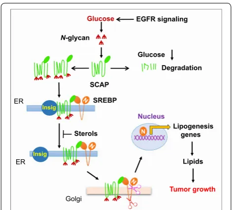
A recent series of studies in our laboratory showed that
glucose could activate SCAP/SREBP trafficking and
activation (Fig.
2
) [
55
,
103
,
104
]. We tested the effects
of glucose intermediate metabolites on different
meta-bolic pathways, including glycolysis, oxidative
phospho-rylation, and hexosamine synthesis for glycosylation. We
found that only
N
-acetylglucosamine (GlcNAc), an
inter-mediate in the hexosamine biosynthesis pathway,
acti-vates SREBPs when glucose supply is limited. We found
that inhibiting
N
-glycosylation, but not
O
-glycosylation,
abolished glucose-mediated SCAP up-regulation and
SREBP activation, indicating that glucose-mediated
N
-glycosylation of SCAP is essential for SCAP/SREBP
trafficking and activation. These findings also
demon-strated a coordinated molecular regulation mechanism
that links glucose availability and the rate of de novo lipid
synthesis (Fig.
2
) [
55
,
58
,
105
].
Glucose
Lipids
Nucleus
N Lipogenesisgenes
SREBP
Sterols
Tumor growth
Degradation EGFR signaling
ER
Golgi
Glucose
N-glycan
Insig
ER Insig
SCAP
Fig. 2 SCAP N-glycosylation is essential for SREBP trafficking and activation. SREBP activation is repressed by the ER-resident protein Insig, which binds to SCAP to prevent SREBP translocation and nuclear activation. The Nobel Prize-winning laboratories of Brown and Goldstein revealed that sterols modulate Insig interaction with SCAP to retain the SCAP/SREBP complex in the ER and inhibit SREBP [273, 274]. Our recent work has shown that glucose-mediated
SREBP activation in malignancy
The importance of SREBPs in cancer has begun to be
rec-ognized. Our group discovered that SREBP-1 is markedly
up-regulated in glioblastoma [
34
,
106
–
108
], the most
common primary brain tumor and one of the most lethal
cancers [
34
,
109
–
113
]. Glioblastomas depend strongly
on lipogenesis for rapid growth when they express the
amplified tyrosine kinase receptor called epidermal
growth factor receptor (EGFR) or its constitutively active
mutant form EGFRvIII. This mutant lacks a portion of
the extracellular ligand-binding domain [
34
,
106
,
108
,
111
,
114
,
115
]. EGFR/EGFRvIII promotes lipid synthesis
by activating SREBP-1 via PI3K/Akt signaling [
12
,
34
,
87
].
The nuclei of human glioblastoma cells display elevated
levels of SREBP-1 [
34
], suggesting that the SCAP/SREBP
complex may escape the tight repression of Insig,
lead-ing to high SREBP activation. Other groups have found
elevated SREBP-1 in various cancers, and SREBP-1 levels
in various cell lines are regulated by PI3K/Akt signaling
and mTORC1 [
116
–
122
]. How SREBP-1 is activated in
cancer cells is not entirely understood and requires
fur-ther investigation.
Inhibiting SREBPs at the genetic level or with
pharma-cological agents significantly suppresses tumor growth
and induces cancer cell death, making SREBPs promising
therapeutic targets [
28
,
34
,
123
–
137
]. However, directly
inhibiting SREBPs is challenging, as transcription
fac-tors often make poor drug targets. A more promising
approach is to inhibit SREBP translocation from the
ER to the Golgi. Along this line, fatostatin, betulin and
PF-429242 have been shown to inhibit SREBP
activa-tion and have promising anti-tumor effects in pre-clinical
studies [
126
–
131
].
SREBP-2 is up-regulated in prostate cancer [
37
,
138
].
SREBP-2 regulates 3-hydroxy-3-methylglutaryl-CoA
(HMG-CoA) reductase, the rate-limiting enzyme for
cholesterol synthesis. Inhibiting SREBP-2 has been
explored as an anti-cancer therapy [
139
–
144
]. Statins are
inhibitors of HMG-CoA reductase and are widely used
to reduce circulating cholesterol levels. The anti-cancer
effects of statins have been tested for various types of
cancers, both pre-clinically and in patients [
140
,
142
,
143
,
145
]. However, inhibition of cholesterol synthesis can
lead to feedback activation of SREBPs, making the
anti-cancer effects of statins less effective [
144
]. Thus,
combi-nation therapies that simultaneously inhibit cholesterol
synthesis and SREBP activation are being developed [
146
,
147
].
SLC25A1
A critical step for glucose-mediated de novo lipid
syn-thesis is the release of citrate from mitochondria into the
cytoplasm. Solute carrier family 25 member 1 (SLC25A1),
also referred to as citrate carrier (CIC), functions as a key
transporter to export citrate from mitochondria to the
cytoplasm, providing a key precursor for both fatty acid
and cholesterol synthesis [
148
,
149
] (Fig.
1
). SLC25A1 is
regulated by SREBP-1 [
150
] and plays an important role
in inflammation and tumor growth [
151
,
152
]. In lung
cancer cells, SLC25A1 is up-regulated by mutant p53
[
151
]. These findings, though preliminary, suggest that
specific inhibitors of SLC25A1 may have anti-tumor
effects.
ACLY
ATP citrate lyase (ACLY) converts cytoplasmic citrate
to acetyl-CoA, a precursor of lipid synthesis (Fig.
1
)
[
153
–
155
] and a substrate for protein acetylation [
153
].
ACLY is a downstream target of SREBPs [
156
–
158
], and
is up-regulated in many cancers, including glioblastoma,
colorectal cancer, breast cancer, non-small cell lung
can-cer, and hepatocellular carcinoma [
159
–
161
]. Inhibiting
ACLY at the genetic level or pharmacologically
signifi-cantly suppresses tumor growth [
162
–
164
]. The ACLY
inhibitor SB-204990 strongly inhibits tumor growth in
mice with lung, prostate or ovarian cancer xenografts
[
162
,
165
]. These results suggest that ACLY may serve as
an attractive anti-cancer target [
155
].
ACSS2
ACCs
Following the conversion of citrate and acetate to
acetyl-CoA, the ACC enzymes catalyze ATP-dependent
carboxylation of acetyl-CoA, generating malonyl-CoA
for fatty acid synthesis (Fig.
1
). Two ACC isoforms have
been identified in mammalian cells, ACC-alpha (also
termed ACC1) and ACC-beta (also known as ACC2)
[
176
,
177
]. ACC is up-regulated in several human
cancers, including glioblastoma and head and neck
squamous cell carcinoma [
34
,
178
]. Inhibiting ACCs
significantly reduces fatty acid synthesis and suppresses
tumor growth in various xenograft models [
179
–
186
].
The ACC inhibitors TOFA, soraphen A and ND646
have shown significant anti-tumor effects in xenograft
tumor models (Table
1
) [
179
–
184
].
FASN
Fatty acid synthase (FASN), a key lipogenic enzyme
cata-lyzing the last step in de novo biogenesis of fatty acids,
has been studied extensively in various cancers [
21
,
187
–
191
]. The early-generation FASN inhibitors C75,
cerulenin and orlistat (Table
1
) have been studied
pre-clinically, but their pharmacology and side effects limited
their potential for clinical use [
34
,
179
,
188
–
203
]. The
later-generation inhibitor TVB-2640 has entered clinical
trials in patients with solid tumors (Table
1
) [
21
,
191
,
204
,
205
].
SCD1
Stearoyl-CoA desaturase (SCD) is an ER-resident integral
membrane protein that catalyzes the formation of the
mono-unsaturated fatty acids oleic acid (18:1) or
palmi-toleic acid (16:1) from stearoyl-(18:0) or palmitoyl-CoA
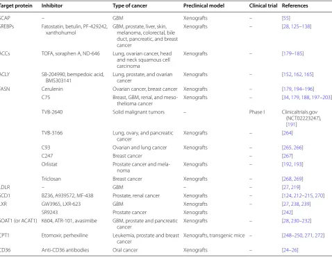
Table 1 Representative targets within the lipid metabolism pathway for anti-cancer drug development
ACCs acetyl-CoA carboxylases, ACLY ATP citrate lyase, CD36 cluster of differentiation 36, also known as fatty acid translocase (FAT), CPT1 carnitine palmitoyltransferase 1, FASN fatty acid synthase, GBM glioblastoma multiforme, LDLR low-density lipoprotein receptor, LXR liver X receptor, SCAP SREBP cleavage-activating protein, SREBPs sterol regulatory element-binding proteins
Target protein Inhibitor Type of cancer Preclinical model Clinical trial References
SCAP – GBM Xenografts – [55]
SREBPs Fatostatin, betulin, PF-429242,
xanthohumol GBM, prostate, liver, skin, melanoma, colorectal, bile duct, pancreatic, and breast cancer
Xenografts – [28, 125–138]
ACCs TOFA, soraphen A, ND-646 Lung, ovarian cancer, head and neck squamous cell carcinoma
Xenografts – [179–185]
ACLY SB-204990, bempedoic acid,
BMS303141 Lung, prostate, and ovarian cancer Xenografts – [152, 162, 165] FASN Cerulenin Ovarian cancer, breast cancer Xenografts – [179, 194–196]
C75 Breast, GBM, renal, and
meso-thelioma cancer Xenografts – [34, 179, 188, 197–203]
TVB-2640 Solid malignant tumors – Phase I Clinicaltrials.gov
(NCT02223247), [191]
TVB-3166 Lung, ovary, and pancreatic
cancer Xenografts – [264]
C93 Ovarian and lung cancer Xenografts – [265, 266]
C247 Breast cancer – [267]
Orlistat Prostate cancer and
mela-noma Xenografts – [192, 193]
Triclosan Breast cancer Xenografts – [268, 269]
LDLR – GBM – – [27, 219]
SCD1 BZ36, A939572, MF-438 Prostate, renal cancer Xenografts – [124, 212–215, 270]
LXR GW3965, LXR-623 GBM Xenografts – [27, 238, 239]
SR9243 Prostate cancer Xenografts [242]
SOAT1 (or ACAT1) K604, ATR-101, avasimibe GBM, prostate and pancreatic
cancer Xenografts – [28, 230–232]
CPT1 Etomoxir, perhexiline Leukemia, prostate and breast
cancer Xenografts, transgenic mice – [248–250, 271, 272]
(16:0) [
206
,
207
]. There are 5
SCD
genes (
SCD1
-
5
).
Humans contain the
SCD
homologs
SCD1
and
SCD5
, but
the function of SCD5 remains unknown [
208
–
210
]. The
mono-unsaturated products of SCD1 are key substrates
in the formation of membrane phospholipids,
choles-teryl esters and triglycerides, making SCD1 a promising
anti-cancer target [
75
,
211
]. The SCD1 inhibitors BZ36,
A939572 and MF-438 have shown anti-tumor effects in
pre-clinical xenograft models (Table
1
) [
212
–
215
].
Lipid uptake
CD36
In addition to de novo synthesis, lipid uptake from the
exogenous environment is another important route
through which cells acquire fatty acids. CD36 transports
fatty acids into the cell [
216
,
217
], and plays a critical role
in cancer cell growth, metastasis and the
epithelial-mes-enchymal transition [
24
–
26
]. An anti-CD36 antibody has
shown significant anti-metastatic efficacy in oral cancer
xenograft models [
25
].
LDLR
Cholesterol is an essential structural component of cell
membranes [
2
,
218
]. Cholesterol could be synthesized by
cells de novo or through internalizing low-density
lipo-protein (LDL). LDL binds to the membrane-bound LDL
receptor (LDLR) and is internalized, after which it enters
lysosomes, where free cholesterol is released [
11
,
76
].
LDLR is up-regulated in glioblastoma via EGFR/PI3K/
Akt/SREBP-1 signaling [
27
], and plays an important role
in tumor growth [
27
,
76
,
219
]. LDLR has not been
inves-tigated as an anti-cancer target.
Lipid storage/lipid droplets
SOAT1/ACAT1
When cellular lipids are in excess, they are converted
to triglycerides and cholesteryl esters in the ER,
form-ing lipid droplets [
220
–
222
]. These droplets have been
observed in various types of tumor, including
glioblas-toma, renal clear cell carcinoma, and cancers of the
prostate, colon or pancreas [
29
–
33
]. Diglyceride
acyl-transferase 1/2 (DGAT1/2) could synthesize triglyceride
from diacylglycerol and acyl-CoA (Fig.
1
) [
223
,
224
]. So
far, the role of triglycerides in cancer cells has not been
explored.
Cholesteryl esters are abundant in tumor tissue, while
they are usually undetectable in normal tissue [
225
–
229
]. Sterol
O
-acyltransferase 1 (SOAT1), also known
as acyl-CoA acyltransferase 1 (ACAT1), converts
cho-lesterol to cholesteryl esters for storage in lipid droplets
(Fig.
1
). This enzyme is highly expressed in glioblastomas
and in cancer of the prostate or pancreas; its
expres-sion level correlates inversely with patient survival [
28
,
29
,
230
–
235
]. Genetically silencing SOAT1/ACAT1 or
blocking its activity using the inhibitors K604, ATR-101
or avasimibe effectively suppresses tumor growth in
sev-eral cancer xenograft models [
28
,
230
–
232
]. These results
suggest that targeting SOAT1 and cholesteryl ester
syn-thesis may be a promising anti-cancer strategy.
Cholesterol efflux
LXR/ABCA1
Cholesterol homeostasis is critical for maintaining
cel-lular function, and is regulated by de novo synthesis,
uptake, storage, and efflux [
11
,
76
]. Increases in
choles-terol levels can trigger feedback inhibition of cholescholes-terol
biosynthesis or conversion of cholesterol into cholesteryl
esters stored in lipid droplets. Levels of 22- or
27-hydrox-ycholesterol can also increase, and these molecules
bind to and activate the liver X receptor, which turns on
expression of ATP-binding cassette proteins A1 (ABCA1)
and G1 (ABCG1) [
236
]. Both proteins are plasma
mem-brane-bound transporters that promote cholesterol
export and thereby reduce intracellular cholesterol
lev-els [
237
]. Synthetic liver X receptor agonists GW3965
and T0901317 significantly inhibit tumor growth in
animal models of glioblastoma, breast cancer or
pros-tate cancer [
7
,
27
]. Activation of the liver X receptor by
GW3965 up-regulates a ubiquitin ligase E3 that degrades
LDLR [
27
,
62
,
238
]. The highly brain-penetrant liver X
receptor agonist LXR-623 selectively kills glioblastoma
cells and prolongs survival of glioblastoma-bearing mice
[
239
]. Therefore, the combination of increasing
choles-terol efflux by activating the liver X receptor and
decreas-ing cholesterol uptake may be a promisdecreas-ing anti-cancer
strategy.
up-regulation of glycolysis and lipogenesis. It may be
more effective to simultaneously enhance cholesterol
efflux and inhibit glycolysis and lipogenesis.
Fatty acid oxidation
CPT1
Fatty acids are an important energy source for cell
growth and survival when nutrients are limiting.
Carni-tine palmitoyltransferase I (CPT1) converts fatty acids
to acylcarnitines, which are shuttled into mitochondria,
where they undergo β-oxidation and produce energy
[
21
]. Fatty acid β-oxidation plays a critical role in tumor
growth [
246
,
247
], and the CPT1 inhibitors etomoxir and
perhexiline have been tested for anti-cancer effects in
various animal models [
248
–
250
].
Lipid peroxidation and cell death
Lipids, particularly polyunsaturated fatty acids, are
sus-ceptible to oxidation by oxygen free radicals, leading to
lipid peroxidation that is harmful to cells and tissues
[
251
–
253
]. Lipid peroxides are associated with many
pathological states, including inflammation,
neuro-degenerative disease, cancer, and ocular and kidney
degeneration [
253
,
254
]. Lipid peroxidation triggers
the propagation of lipid reactive oxygen species that
can significantly alter the physical properties of cellular
membranes, or degrade into reactive compounds that
cross-link DNA or proteins, exerting further toxic effects
[
253
,
255
,
256
]. Extensive lipid peroxidation can result
in ferroptosis, a regulated form of iron-dependent,
non-apoptotic cell death [
255
,
257
]. Inducing ferroptosis may
be an anti-cancer strategy [
257
–
259
]. For example,
dis-rupting the repair of oxidative damage to bio-membranes
by inhibiting the antioxidant enzyme glutathione
peroxi-dase 4 (GPX4) could induce ferroptosis [
257
,
259
–
262
].
This has emerged as an active area of research that may
lead to new anti-cancer approaches, particularly against
metabolically active tumors.
Summary
Extensive studies have provided strong evidence for
reprogramming of lipid metabolism in cancer [
27
,
34
,
55
].
A variety of lipid synthesis inhibitors have shown
prom-ising anti-cancer effects in preclinical studies and early
phases of clinical trials [
7
,
29
,
55
,
263
]. However, major
barriers exists in developing cancer treatment by
target-ing altered lipid metabolism, mostly due to incomplete
understanding of the mechanisms that regulate lipid
syn-thesis, storage, utilization and efflux in cancer cells.
Authors’ contributions
CC performed the literature search and drafted the manuscript. FG and XC assisted in the literature search and helped draft sections of the manuscript.
DG designed and revised the manuscript. All authors read and approved the final manuscript.
Acknowledgements
The authors wish to thank Dr. Martine Torres for critical review of the manu-script and editorial assistance.
Competing interests
The authors declare that they have no competing interests.
Availability of data and materials
The data supporting the conclusions of this article are included within the article and Table 1.
Consent for publication
Not applicable.
Ethics approval and consent to participate
Not applicable.
Funding
This work was supported by NIH Grant NS079701 (DG), American Cancer Society Research Scholar Grant RSG-14-228-01-CSM (DG), OSUCCC Idea Grant (DG), an OSUCCC Translational Therapeutic Program seed grant (DG), a Peloto-nia Postdoc Fellowship (CC) and an OSU Department of Radiation-Oncology Basic Research seed Grant (CC).
Received: 12 January 2018 Accepted: 12 May 2018
References
1. Maxfield FR. Plasma membrane microdomains. Curr Opin Cell Biol. 2002;14(4):483–7.
2. Mukherjee S, Maxfield FR. Membrane domains. Annu Rev Cell Dev Biol. 2004;20:839–66. https ://doi.org/10.1146/annur ev.cellb io.20.01040 3.09545 1.
3. Pomorski T, Hrafnsdottir S, Devaux PF, van Meer G. Lipid distribu-tion and transport across cellular membranes. Semin Cell Dev Biol. 2001;12(2):139–48. https ://doi.org/10.1006/scdb.2000.0231.
4. van Meer G. Membranes in motion. EMBO Rep. 2010;11(5):331–3. https ://doi.org/10.1038/embor .2010.60.
5. van Meer G, Voelker DR, Feigenson GW. Membrane lipids: where they are and how they behave. Nat Rev Mol Cell Biol. 2008;9(2):112–24. https ://doi.org/10.1038/nrm23 30.
6. Holthuis JC, Menon AK. Lipid landscapes and pipelines in membrane homeostasis. Nature. 2014;510(7503):48–57. https ://doi.org/10.1038/ natur e1347 4.
7. Guo D, Bell EH, Chakravarti A. Lipid metabolism emerges as a promising target for malignant glioma therapy. CNS Oncol. 2013;2(3):289–99.
https ://doi.org/10.2217/cns.13.20.
8. Menendez JA, Lupu R. Fatty acid synthase and the lipogenic phenotype in cancer pathogenesis. Nat Rev Cancer. 2007;7(10):763–77. https ://doi. org/10.1038/nrc22 22.
9. Zechner R, Strauss JG, Haemmerle G, Lass A, Zimmermann R. Lipolysis: pathway under construction. Curr Opin Lipidol. 2005;16(3):333–40. 10. Efeyan A, Comb WC, Sabatini DM. Nutrient-sensing mechanisms and
pathways. Nature. 2015;517(7534):302–10. https ://doi.org/10.1038/ natur e1419 0.
11. Goldstein JL, Brown MS. A century of cholesterol and coronaries: from plaques to genes to statins. Cell. 2015;161(1):161–72. https ://doi. org/10.1016/j.cell.2015.01.036.
12. Goldstein JL, DeBose-Boyd RA, Brown MS. Protein sensors for membrane sterols. Cell. 2006;124(1):35–46. https ://doi.org/10.1016/j. cell.2005.12.022.
14. Rosen ED, Spiegelman BM. What we talk about when we talk about fat. Cell. 2014;156(1–2):20–44. https ://doi.org/10.1016/j. cell.2013.12.012.
15. Perry RJ, Samuel VT, Petersen KF, Shulman GI. The role of hepatic lipids in hepatic insulin resistance and type 2 diabetes. Nature. 2014;510(7503):84–91. https ://doi.org/10.1038/natur e1347 8. 16. Cohen JC, Horton JD, Hobbs HH. Human fatty liver disease: old
ques-tions and new insights. Science. 2011;332(6037):1519–23. https ://doi. org/10.1126/scien ce.12042 65.
17. Abramson HN. The lipogenesis pathway as a cancer target. J Med Chem. 2011;54(16):5615–38. https ://doi.org/10.1021/jm200 5805. 18. Grossi-Paoletti E, Paoletti P, Fumagalli R. Lipids in brain tumors. J
Neu-rosurg. 1971;34(3):454–5. https ://doi.org/10.3171/jns.1971.34.3.0454. 19. Podo F. Tumour phospholipid metabolism. NMR Biomed.
1999;12(7):413–39.
20. Santos CR, Schulze A. Lipid metabolism in cancer. FEBS J. 2012;279(15):2610–23. https ://doi.org/10.111 1/j.1742-4658.2012.08644 .x.
21. Rohrig F, Schulze A. The multifaceted roles of fatty acid synthesis in cancer. Nat Rev Cancer. 2016;16(11):732–49. https ://doi.org/10.1038/ nrc.2016.89.
22. Schulze A, Harris AL. How cancer metabolism is tuned for prolifera-tion and vulnerable to disrupprolifera-tion. Nature. 2012;491(7424):364–73.
https ://doi.org/10.1038/natur e1170 6.
23. Yoon S, Lee MY, Park SW, Moon JS, Koh YK, Ahn YH, et al. Up-regu-lation of acetyl-CoA carboxylase alpha and fatty acid synthase by human epidermal growth factor receptor 2 at the translational level in breast cancer cells. J Biol Chem. 2007;282(36):26122–31. https :// doi.org/10.1074/jbc.M7028 54200 .
24. Zhao J, Zhi Z, Wang C, Xing H, Song G, Yu X, et al. Exogenous lipids promote the growth of breast cancer cells via CD36. Oncol Rep. 2017;38(4):2105–15. https ://doi.org/10.3892/or.2017.5864.
25. Pascual G, Avgustinova A, Mejetta S, Martin M, Castellanos A, Attolini CS, et al. Targeting metastasis-initiating cells through the fatty acid receptor CD36. Nature. 2017;541(7635):41–5. https ://doi.org/10.1038/ natur e2079 1.
26. Nath A, Li I, Roberts LR, Chan C. Elevated free fatty acid uptake via CD36 promotes epithelial-mesenchymal transition in hepatocellular carcinoma. Sci Rep. 2015;5:14752. https ://doi.org/10.1038/srep1 4752. 27. Guo D, Reinitz F, Youssef M, Hong C, Nathanson D, Akhavan D, et al.
An LXR agonist promotes glioblastoma cell death through inhibition of an EGFR/AKT/SREBP-1/LDLR-dependent pathway. Cancer Discov. 2011;1(5):442–56. https ://doi.org/10.1158/2159-8290.CD-11-0102. 28. Geng F, Cheng X, Wu X, Yoo JY, Cheng C, Guo JY, et al. Inhibition of
SOAT1 suppresses glioblastoma growth via blocking SREBP-1-medi-ated lipogenesis. Clin Cancer Res. 2016;22(21):5337–48. https ://doi. org/10.1158/1078-0432.CCR-15-2973.
29. Geng F, Guo D. Lipid droplets, potential biomarker and metabolic target in glioblastoma. Intern Med Rev (Washington, DC). 2017. https ://doi.org/10.18103 /imr.v3i5.443.
30. Koizume S, Miyagi Y. Lipid Droplets: a key cellular organelle associ-ated with cancer cell survival under normoxia and hypoxia. Int J Mol Sci. 2016. https ://doi.org/10.3390/ijms1 70914 30.
31. Yue S, Li J, Lee SY, Lee HJ, Shao T, Song B, et al. Cholesteryl ester accu-mulation induced by PTEN loss and PI3 K/AKT activation underlies human prostate cancer aggressiveness. Cell Metab. 2014;19(3):393– 406. https ://doi.org/10.1016/j.cmet.2014.01.019.
32. Accioly MT, Pacheco P, Maya-Monteiro CM, Carrossini N, Robbs BK, Oliveira SS, et al. Lipid bodies are reservoirs of cyclooxygenase-2 and sites of prostaglandin-E2 synthesis in colon cancer cells. Cancer Res. 2008;68(6):1732–40. https ://doi.org/10.1158/0008-5472.CAN-07-1999. 33. Gaida MM, Mayer C, Dapunt U, Stegmaier S, Schirmacher P, Wabnitz
GH, et al. Expression of the bitter receptor T2R38 in pancreatic can-cer: localization in lipid droplets and activation by a bacteria-derived quorum-sensing molecule. Oncotarget. 2016;7(11):12623–32. https :// doi.org/10.18632 /oncot arget .7206.
34. Guo D, Prins RM, Dang J, Kuga D, Iwanami A, Soto H, et al. EGFR signaling through an Akt-SREBP-1-dependent, rapamycin-resistant pathway sensitizes glioblastomas to antilipogenic therapy. Sci Signal. 2009;2(101):ra82. https ://doi.org/10.1126/scisi gnal.20004 46.
35. Jeon TI, Osborne TF. SREBPs: metabolic integrators in physiology and metabolism. Trends Endocrinol Metab. 2012;23(2):65–72. https ://doi. org/10.1016/j.tem.2011.10.004.
36. Shao W, Espenshade PJ. Expanding roles for SREBP in metabolism. Cell Metab. 2012;16(4):414–9. https ://doi.org/10.1016/j.cmet.2012.09.002. 37. Ettinger SL, Sobel R, Whitmore TG, Akbari M, Bradley DR, Gleave ME,
et al. Dysregulation of sterol response element-binding proteins and downstream effectors in prostate cancer during progression to andro-gen independence. Cancer Res. 2004;64(6):2212–21.
38. Yang Y, Morin PJ, Han WF, Chen T, Bornman DM, Gabrielson EW, et al. Regulation of fatty acid synthase expression in breast cancer by sterol regulatory element binding protein-1c. Exp Cell Res. 2003;282(2):132–7. 39. Bao J, Zhu L, Zhu Q, Su J, Liu M, Huang W. SREBP-1 is an independent
prognostic marker and promotes invasion and migration in breast cancer. Oncol Lett. 2016;12(4):2409–16. https ://doi.org/10.3892/ ol.2016.4988.
40. Huang WC, Li X, Liu J, Lin J, Chung LW. Activation of androgen receptor, lipogenesis, and oxidative stress converged by SREBP-1 is responsible for regulating growth and progression of prostate cancer cells. Mol Cancer Res. 2012;10(1):133–42. https ://doi.org/10.1158/1541-7786. MCR-11-0206.
41. Yin F, Sharen G, Yuan F, Peng Y, Chen R, Zhou X, et al. TIP30 regulates lipid metabolism in hepatocellular carcinoma by regulating SREBP1 through the Akt/mTOR signaling pathway. Oncogenesis. 2017;6(6):e347.
https ://doi.org/10.1038/oncsi s.2017.49.
42. Sun Y, He W, Luo M, Zhou Y, Chang G, Ren W, et al. SREBP1 regulates tumorigenesis and prognosis of pancreatic cancer through target-ing lipid metabolism. Tumour Biol. 2015;36(6):4133–41. https ://doi. org/10.1007/s1327 7-015-3047-5.
43. Li C, Yang W, Zhang J, Zheng X, Yao Y, Tu K, et al. SREBP-1 has a prognostic role and contributes to invasion and metastasis in human hepatocellular carcinoma. Int J Mol Sci. 2014;15(5):7124–38. https ://doi. org/10.3390/ijms1 50571 24.
44. Walker AK, Jacobs RL, Watts JL, Rottiers V, Jiang K, Finnegan DM, et al. A conserved SREBP-1/phosphatidylcholine feedback circuit regu-lates lipogenesis in metazoans. Cell. 2011;147(4):840–52. https ://doi. org/10.1016/j.cell.2011.09.045.
45. Soyal SM, Nofziger C, Dossena S, Paulmichl M, Patsch W. Targeting SREBPs for treatment of the metabolic syndrome. Trends Pharmacol Sci. 2015;36(6):406–16. https ://doi.org/10.1016/j.tips.2015.04.010. 46. Muller-Wieland D, Knebel B, Haas J, Kotzka J. SREBP-1 and fatty liver.
Clinical relevance for diabetes, obesity, dyslipidemia and atherosclero-sis. Herz. 2012;37(3):273–8. https ://doi.org/10.1007/s0005 9-012-3608-y. 47. Luengo A, Gui DY, Vander Heiden MG. Targeting metabolism for cancer
therapy. Cell Chem Biol. 2017;24(9):1161–80. https ://doi.org/10.1016/j. chemb iol.2017.08.028.
48. Min HY, Lee HY. Oncogene-driven metabolic alterations in cancer. Biomol Ther (Seoul). 2017. https ://doi.org/10.4062/biomo lther .2017.211. 49. Gopal K, Grossi E, Paoletti P, Usardi M. Lipid composition of human
intracranial tumors: a biochemical study. Acta Neurochir (Wien). 1963;11:333–47.
50. Currie E, Schulze A, Zechner R, Walther TC, Farese RV Jr. Cellular fatty acid metabolism and cancer. Cell Metab. 2013;18(2):153–61. https ://doi. org/10.1016/j.cmet.2013.05.017.
51. Shimano H, Sato R. SREBP-regulated lipid metabolism: convergent physiology—divergent pathophysiology. Nat Rev Endocrinol. 2017.
https ://doi.org/10.1038/nrend o.2017.91.
52. Schlosser HA, Drebber U, Urbanski A, Haase S, Baltin C, Berlth F, et al. Glucose transporters 1, 3, 6, and 10 are expressed in gastric cancer and glucose transporter 3 is associated with UICC stage and survival. Gastric Cancer. 2017;20(1):83–91. https ://doi.org/10.1007/s1012 0-015-0577-x. 53. Sharen G, Peng Y, Cheng H, Liu Y, Shi Y, Zhao J. Prognostic value of
GLUT-1 expression in pancreatic cancer: results from 538 patients. Oncotarget. 2017;8(12):19760–7. https ://doi.org/10.18632 /oncot arget .15035 .
54. Sun HW, Yu XJ, Wu WC, Chen J, Shi M, Zheng L, et al. GLUT1 and ASCT2 as predictors for prognosis of hepatocellular carcinoma. PLoS ONE. 2016;11(12):e0168907. https ://doi.org/10.1371/journ al.pone.01689 07. 55. Cheng C, Ru P, Geng F, Liu J, Yoo JY, Wu X, et al. Glucose-mediated
growth. Cancer Cell. 2015;28(5):569–81. https ://doi.org/10.1016/j.ccell .2015.09.021.
56. Ryczko MC, Pawling J, Chen R, Abdel Rahman AM, Yau K, Copeland JK, et al. Metabolic reprogramming by hexosamine biosynthetic and golgi N-glycan branching pathways. Sci Rep. 2016;6:23043. https ://doi. org/10.1038/srep2 3043.
57. Adeva-Andany MM, Perez-Felpete N, Fernandez-Fernandez C, Dona-petry-Garcia C, Pazos-Garcia C. Liver glucose metabolism in humans. Biosci Rep. 2016. https ://doi.org/10.1042/bsr20 16038 5.
58. Guo D. SCAP links glucose to lipid metabolism in cancer cells. Mol Cell Oncol. 2016. https ://doi.org/10.1080/23723 556.2015.11321 20. 59. Dang CV, Le A, Gao P. MYC-induced cancer cell energy metabolism and
therapeutic opportunities. Clin Cancer Res. 2009;15(21):6479–83. https ://doi.org/10.1158/1078-0432.CCR-09-0889.
60. Barger JF, Plas DR. Balancing biosynthesis and bioenergetics: metabolic programs in oncogenesis. Endocr Relat Cancer. 2010;17(4):R287–304.
https ://doi.org/10.1677/ERC-10-0106.
61. Dang CV. Therapeutic targeting of Myc-reprogrammed cancer cell metabolism. Cold Spring Harb Symp Quant Biol. 2011;76:369–74. https ://doi.org/10.1101/sqb.2011.76.01129 6.
62. DeBerardinis RJ, Mancuso A, Daikhin E, Nissim I, Yudkoff M, Wehrli S, et al. Beyond aerobic glycolysis: transformed cells can engage in glutamine metabolism that exceeds the requirement for protein and nucleotide synthesis. Proc Natl Acad Sci USA. 2007;104(49):19345–50.
https ://doi.org/10.1073/pnas.07097 47104 .
63. Bergstrom J, Furst P, Noree LO, Vinnars E. Intracellular free amino acid concentration in human muscle tissue. J Appl Physiol. 1974;36(6):693–7. 64. Wishart DS, Jewison T, Guo AC, Wilson M, Knox C, Liu Y, et al. HMDB
3.0–the human metabolome database in 2013. Nucleic Acids Res. 2013;41(Database issue):D801–7. https ://doi.org/10.1093/nar/gks10 65. 65. DeBerardinis RJ, Cheng T. Q’s next: the diverse functions of glutamine
in metabolism, cell biology and cancer. Oncogene. 2010;29(3):313–24.
https ://doi.org/10.1038/onc.2009.358.
66. Bhutia YD, Babu E, Ramachandran S, Ganapathy V. Amino Acid trans-porters in cancer and their relevance to “glutamine addiction”: novel targets for the design of a new class of anticancer drugs. Cancer Res. 2015;75(9):1782–8. https ://doi.org/10.1158/0008-5472.CAN-14-3745. 67. Erickson JW, Cerione RA. Glutaminase: a hot spot for regulation of
cancer cell metabolism? Oncotarget. 2010;1(8):734–40. https ://doi. org/10.18632 /oncot arget .208.
68. Wise DR, Ward PS, Shay JE, Cross JR, Gruber JJ, Sachdeva UM, et al. Hypoxia promotes isocitrate dehydrogenase-dependent carboxylation of alpha-ketoglutarate to citrate to support cell growth and viability. Proc Natl Acad Sci USA. 2011;108(49):19611–6. https ://doi.org/10.1073/ pnas.11177 73108 .
69. Metallo CM, Gameiro PA, Bell EL, Mattaini KR, Yang J, Hiller K, et al. Reductive glutamine metabolism by IDH1 mediates lipogenesis under hypoxia. Nature. 2012;481(7381):380–4. https ://doi.org/10.1038/natur e1060 2.
70. Mullen AR, Wheaton WW, Jin ES, Chen PH, Sullivan LB, Cheng T, et al. Reductive carboxylation supports growth in tumour cells with defective mitochondria. Nature. 2012;481(7381):385–8. https ://doi. org/10.1038/natur e1064 2.
71. Le A, Lane AN, Hamaker M, Bose S, Gouw A, Barbi J, et al. Glucose-independent glutamine metabolism via TCA cycling for proliferation and survival in B cells. Cell Metab. 2012;15(1):110–21. https ://doi. org/10.1016/j.cmet.2011.12.009.
72. Comerford SA, Huang Z, Du X, Wang Y, Cai L, Witkiewicz AK, et al. Acetate dependence of tumors. Cell. 2014;159(7):1591–602. https ://doi. org/10.1016/j.cell.2014.11.020.
73. Mashimo T, Pichumani K, Vemireddy V, Hatanpaa KJ, Singh DK, Sirasanagandla S, et al. Acetate is a bioenergetic substrate for human glioblastoma and brain metastases. Cell. 2014;159(7):1603–14. https :// doi.org/10.1016/j.cell.2014.11.025.
74. Schug ZT, Peck B, Jones DT, Zhang Q, Grosskurth S, Alam IS, et al. Acetyl-CoA synthetase 2 promotes acetate utilization and maintains cancer cell growth under metabolic stress. Cancer Cell. 2015;27(1):57–71. https ://doi.org/10.1016/j.ccell .2014.12.002.
75. Peck B, Schulze A. Lipid desaturation - the next step in targeting lipogenesis in cancer? FEBS J. 2016;283(15):2767–78. https ://doi. org/10.1111/febs.13681 .
76. Goldstein JL, Brown MS. The LDL receptor. Arterioscler Thromb Vasc Biol. 2009;29(4):431–8. https ://doi.org/10.1161/ATVBA HA.108.17956 4. 77. Di Vizio D, Adam RM, Kim J, Kim R, Sotgia F, Williams T, et al.
Caveo-lin-1 interacts with a lipid raft-associated population of fatty acid synthase. Cell Cycle. 2008;7(14):2257–67. https ://doi.org/10.4161/ cc.7.14.6475.
78. Cai Y, Wang J, Zhang L, Wu D, Yu D, Tian X, et al. Expressions of fatty acid synthase and HER2 are correlated with poor prognosis of ovar-ian cancer. Med Oncol. 2015;32(1):391. https ://doi.org/10.1007/s1203 2-014-0391-z.
79. Long QQ, Yi YX, Qiu J, Xu CJ, Huang PL. Fatty acid synthase (FASN) levels in serum of colorectal cancer patients: correlation with clinical outcomes. Tumour Biol. 2014;35(4):3855–9. https ://doi.org/10.1007/ s1327 7-013-1510-8.
80. Witkiewicz AK, Nguyen KH, Dasgupta A, Kennedy EP, Yeo CJ, Lisanti MP, et al. Co-expression of fatty acid synthase and caveolin-1 in pancreatic ductal adenocarcinoma: implications for tumor progres-sion and clinical outcome. Cell Cycle. 2008;7(19):3021–5. https ://doi. org/10.4161/cc.7.19.6719.
81. Walter K, Hong SM, Nyhan S, Canto M, Fedarko N, Klein A, et al. Serum fatty acid synthase as a marker of pancreatic neoplasia. Cancer Epidemiol Biomarkers Prev. 2009;18(9):2380–5. https ://doi. org/10.1158/1055-9965.EPI-09-0144.
82. Nohturfft A, Zhang SC. Coordination of lipid metabolism in mem-brane biogenesis. Annu Rev Cell Dev Biol. 2009;25:539–66. https :// doi.org/10.1146/annur ev.cellb io.24.11070 7.17534 4.
83. Shimano H, Horton JD, Shimomura I, Hammer RE, Brown MS, Goldstein JL. Isoform 1c of sterol regulatory element binding protein is less active than isoform 1a in livers of transgenic mice and in cul-tured cells. J Clin Invest. 1997;99(5):846–54. https ://doi.org/10.1172/ JCI11 9248.
84. Shimomura I, Shimano H, Horton JD, Goldstein JL, Brown MS. Dif-ferential expression of exons 1a and 1c in mRNAs for sterol regulatory element binding protein-1 in human and mouse organs and cultured cells. J Clin Invest. 1997;99(5):838–45. https ://doi.org/10.1172/JCI11 9247.
85. Wang X, Sato R, Brown MS, Hua X, Goldstein JL. SREBP-1, a membrane-bound transcription factor released by sterol-regulated proteolysis. Cell. 1994;77(1):53–62.
86. Yokoyama C, Wang X, Briggs MR, Admon A, Wu J, Hua X, et al. SREBP-1, a basic-helix-loop-helix-leucine zipper protein that controls transcription of the low density lipoprotein receptor gene. Cell. 1993;75(1):187–97. 87. Horton JD, Goldstein JL, Brown MS. SREBPs: activators of the complete
program of cholesterol and fatty acid synthesis in the liver. J Clin Invest. 2002;109(9):1125–31. https ://doi.org/10.1172/JCI15 593.
88. Horton JD, Shah NA, Warrington JA, Anderson NN, Park SW, Brown MS, et al. Combined analysis of oligonucleotide microarray data from trans-genic and knockout mice identifies direct SREBP target genes. Proc Natl Acad Sci USA. 2003;100(21):12027–32. https ://doi.org/10.1073/ pnas.15349 23100 .
89. Horton JD, Shimomura I, Ikemoto S, Bashmakov Y, Hammer RE. Over-expression of sterol regulatory element-binding protein-1a in mouse adipose tissue produces adipocyte hypertrophy, increased fatty acid secretion, and fatty liver. J Biol Chem. 2003;278(38):36652–60. https :// doi.org/10.1074/jbc.M3065 40200 .
90. Hua X, Wu J, Goldstein JL, Brown MS, Hobbs HH. Structure of the human gene encoding sterol regulatory element binding protein-1 (SREBF1) and localization of SREBF1 and SREBF2 to chromosomes 17p11.2 and 22q13. Genomics. 1995;25(3):667–73.
91. Hua X, Yokoyama C, Wu J, Briggs MR, Brown MS, Goldstein JL, et al. SREBP-2, a second basic-helix-loop-helix-leucine zipper protein that stimulates transcription by binding to a sterol regulatory element. Proc Natl Acad Sci USA. 1993;90(24):11603–7.
92. Shimano H, Shimomura I, Hammer RE, Herz J, Goldstein JL, Brown MS, et al. Elevated levels of SREBP-2 and cholesterol synthesis in livers of mice homozygous for a targeted disruption of the SREBP-1 gene. J Clin Invest. 1997;100(8):2115–24. https ://doi.org/10.1172/JCI11 9746. 93. Lee PC, Sever N, Debose-Boyd RA. Isolation of sterol-resistant Chinese
94. Sun LP, Li L, Goldstein JL, Brown MS. Insig required for sterol-mediated inhibition of Scap/SREBP binding to COPII proteins in vitro. J Biol Chem. 2005;280(28):26483–90. https ://doi.org/10.1074/jbc.M5040 41200 . 95. Sun LP, Seemann J, Goldstein JL, Brown MS. Sterol-regulated transport
of SREBPs from endoplasmic reticulum to Golgi: Insig renders sorting signal in Scap inaccessible to COPII proteins. Proc Natl Acad Sci USA. 2007;104(16):6519–26. https ://doi.org/10.1073/pnas.07009 07104 . 96. Nohturfft A, Yabe D, Goldstein JL, Brown MS, Espenshade PJ. Regulated
step in cholesterol feedback localized to budding of SCAP from ER membranes. Cell. 2000;102(3):315–23.
97. Espenshade PJ, Li WP, Yabe D. Sterols block binding of COPII proteins to SCAP, thereby controlling SCAP sorting in ER. Proc Natl Acad Sci USA. 2002;99(18):11694–9. https ://doi.org/10.1073/pnas.18241 2799. 98. Yang T, Espenshade PJ, Wright ME, Yabe D, Gong Y, Aebersold R, et al.
Crucial step in cholesterol homeostasis: sterols promote binding of SCAP to INSIG-1, a membrane protein that facilitates retention of SREBPs in ER. Cell. 2002;110(4):489–500.
99. Adams CM, Goldstein JL, Brown MS. Cholesterol-induced conforma-tional change in SCAP enhanced by Insig proteins and mimicked by cationic amphiphiles. Proc Natl Acad Sci USA. 2003;100(19):10647–52.
https ://doi.org/10.1073/pnas.15348 33100 .
100. Adams CM, Reitz J, De Brabander JK, Feramisco JD, Li L, Brown MS, et al. Cholesterol and 25-hydroxycholesterol inhibit activation of SREBPs by different mechanisms, both involving SCAP and Insigs. J Biol Chem. 2004;279(50):52772–80. https ://doi.org/10.1074/jbc.M4103 02200 . 101. Ru P, Hu P, Geng F, Mo X, Cheng C, Yoo JY, et al. Feedback loop
regula-tion of SCAP/SREBP-1 by miR-29 modulates EGFR signaling-driven glioblastoma growth. Cell Rep. 2016;16(6):1527–35. https ://doi. org/10.1016/j.celre p.2016.07.017.
102. Ru P, Guo D. microRNA-29 mediates a novel negative feedback loop to regulate SCAP/SREBP-1 and lipid metabolism. RNA Dis. 2017. https :// doi.org/10.14800 /rd.1525.
103. Guo D. SCAP links glucose to lipid metabolism in cancer cells. Mol Cell Oncol. 2016. https ://doi.org/10.1080/23723 556.2015.11321 20. 104. Shao W, Espenshade PJ. Sugar makes fat by talking to SCAP. Cancer Cell.
2015;28(5):548–9. https ://doi.org/10.1016/j.ccell .2015.10.011. 105. Cheng C, Guo JY, Geng F, Wu X, Cheng X, Li Q, et al. Analysis of SCAP
N-glycosylation and trafficking in human cells. J Vis Exp. 2016. https :// doi.org/10.3791/54709 .
106. Guo D, Hildebrandt IJ, Prins RM, Soto H, Mazzotta MM, Dang J, et al. The AMPK agonist AICAR inhibits the growth of EGFRvIII-expressing glioblastomas by inhibiting lipogenesis. Proc Natl Acad Sci USA. 2009;106(31):12932–7. https ://doi.org/10.1073/pnas.09066 06106 . 107. Ru P, Williams TM, Chakravarti A, Guo D. Tumor metabolism of
malignant gliomas. Cancers (Basel). 2013;5(4):1469–84. https ://doi. org/10.3390/cance rs504 1469.
108. Guo D, Bell EH, Mischel P, Chakravarti A. Targeting SREBP-1-driven lipid metabolism to treat cancer. Curr Pharm Des. 2014;20(15):2619–26. 109. Wen PY, Kesari S. Malignant gliomas in adults. N Engl J Med.
2008;359(5):492–507. https ://doi.org/10.1056/NEJMr a0708 126. 110. Stupp R, Hegi ME, Mason WP, van den Bent MJ, Taphoorn MJ, Janzer
RC, et al. Effects of radiotherapy with concomitant and adjuvant temozolomide versus radiotherapy alone on survival in glioblastoma in a randomised phase III study: 5-year analysis of the EORTC-NCIC trial. Lancet Oncol. 2009;10(5):459–66. https ://doi.org/10.1016/S1470 -2045(09)70025 -7.
111. Furnari FB, Fenton T, Bachoo RM, Mukasa A, Stommel JM, Stegh A, et al. Malignant astrocytic glioma: genetics, biology, and paths to treatment. Genes Dev. 2007;21(21):2683–710.
112. Paleologos NA, Merrell RT. Anaplastic glioma. Curr Treat Options Neurol. 2012;14(4):381–90. https ://doi.org/10.1007/s1194 0-012-0177-6. 113. Ricard D, Idbaih A, Ducray F, Lahutte M, Hoang-Xuan K, Delattre JY.
Primary brain tumours in adults. Lancet. 2012;379(9830):1984–96. https ://doi.org/10.1016/S0140 -6736(11)61346 -9.
114. Huang HS, Nagane M, Klingbeil CK, Lin H, Nishikawa R, Ji XD, et al. The enhanced tumorigenic activity of a mutant epidermal growth factor receptor common in human cancers is mediated by threshold levels of constitutive tyrosine phosphorylation and unattenuated signaling. J Biol Chem. 1997;272(5):2927–35.
115. Yoshimoto K, Dang J, Zhu S, Nathanson D, Huang T, Dumont R, et al. Development of a real-time RT-PCR assay for detecting EGFRvIII in
glioblastoma samples. Clin Cancer Res. 2008;14(2):488–93. https ://doi. org/10.1158/1078-0432.CCR-07-1966.
116. Du X, Kristiana I, Wong J, Brown AJ. Involvement of Akt in ER-to-Golgi transport of SCAP/SREBP: a link between a key cell proliferative path-way and membrane synthesis. Mol Biol Cell. 2006;17(6):2735–45. https ://doi.org/10.1091/mbc.E05-11-1094.
117. Porstmann T, Griffiths B, Chung YL, Delpuech O, Griffiths JR, Downward J, et al. PKB/Akt induces transcription of enzymes involved in choles-terol and fatty acid biosynthesis via activation of SREBP. Oncogene. 2005;24(43):6465–81. https ://doi.org/10.1038/sj.onc.12088 02. 118. Porstmann T, Santos CR, Griffiths B, Cully M, Wu M, Leevers S, et al.
SREBP activity is regulated by mTORC1 and contributes to Akt-depend-ent cell growth. Cell Metab. 2008;8(3):224–36. https ://doi.org/10.1016/j. cmet.2008.07.007.
119. Duvel K, Yecies JL, Menon S, Raman P, Lipovsky AI, Souza AL, et al. Acti-vation of a metabolic gene regulatory network downstream of mTOR complex 1. Mol Cell. 2010;39(2):171–83. https ://doi.org/10.1016/j.molce l.2010.06.022.
120. Yecies JL, Zhang HH, Menon S, Liu S, Yecies D, Lipovsky AI, et al. Akt stimulates hepatic SREBP1c and lipogenesis through parallel mTORC1-dependent and inmTORC1-dependent pathways. Cell Metab. 2011;14(1):21–32.
https ://doi.org/10.1016/j.cmet.2011.06.002.
121. Peterson TR, Sengupta SS, Harris TE, Carmack AE, Kang SA, Balderas E, et al. mTOR complex 1 regulates lipin 1 localization to control the SREBP pathway. Cell. 2011;146(3):408–20. https ://doi.org/10.1016/j. cell.2011.06.034.
122. Ricoult SJ, Yecies JL, Ben-Sahra I, Manning BD. Oncogenic PI3K and K-Ras stimulate de novo lipid synthesis through mTORC1 and SREBP. Oncogene. 2015. https ://doi.org/10.1038/onc.2015.179.
123. Williams KJ, Argus JP, Zhu Y, Wilks MQ, Marbois BN, York AG, et al. An essential requirement for the SCAP/SREBP signaling axis to protect cancer cells from lipotoxicity. Cancer Res. 2013;73(9):2850–62. https :// doi.org/10.1158/0008-5472.CAN-13-0382-T.
124. von Roemeling CA, Marlow LA, Wei JJ, Cooper SJ, Caulfield TR, Wu K, et al. Stearoyl-CoA desaturase 1 is a novel molecular therapeutic target for clear cell renal cell carcinoma. Clin Cancer Res. 2013;19(9):2368–80.
https ://doi.org/10.1158/1078-0432.CCR-12-3249.
125. Li N, Zhou ZS, Shen Y, Xu J, Miao HH, Xiong Y, et al. Inhibition of the sterol regulatory element-binding protein pathway suppresses hepa-tocellular carcinoma by repressing inflammation in mice. Hepatology. 2017;65(6):1936–47. https ://doi.org/10.1002/hep.29018 .
126. Kamisuki S, Mao Q, Abu-Elheiga L, Gu Z, Kugimiya A, Kwon Y, et al. A small molecule that blocks fat synthesis by inhibiting the activation of SREBP. Chem Biol. 2009;16(8):882–92. https ://doi.org/10.1016/j.chemb iol.2009.07.007.
127. Li X, Chen YT, Hu P, Huang WC. Fatostatin displays high antitumor activ-ity in prostate cancer by blocking SREBP-regulated metabolic pathways and androgen receptor signaling. Mol Cancer Ther. 2014;13(4):855–66.
https ://doi.org/10.1158/1535-7163.MCT-13-0797.
128. Li X, Wu JB, Chung LW, Huang WC. Anti-cancer efficacy of SREBP inhibitor, alone or in combination with docetaxel, in prostate cancer harboring p53 mutations. Oncotarget. 2015;6(38):41018–32. https ://doi. org/10.18632 /oncot arget .5879.
129. Krol SK, Kielbus M, Rivero-Muller A, Stepulak A. Comprehensive review on betulin as a potent anticancer agent. Biomed Res Int. 2015;2015:584189. https ://doi.org/10.1155/2015/58418 9. 130. Gholkar AA, Cheung K, Williams KJ, Lo YC, Hamideh SA, Nnebe
C, et al. Fatostatin inhibits cancer cell proliferation by affecting mitotic microtubule spindle assembly and cell division. J Biol Chem. 2016;291(33):17001–8. https ://doi.org/10.1074/jbc.C116.73734 6. 131. Shao W, Machamer CE, Espenshade PJ. Fatostatin blocks ER exit of SCAP
but inhibits cell growth in a SCAP-independent manner. J Lipid Res. 2016;57(8):1564–73. https ://doi.org/10.1194/jlr.M0695 83. 132. Miyata S, Inoue J, Shimizu M, Sato R. Xanthohumol improves
diet-induced obesity and fatty liver by suppressing sterol regula-tory element-binding protein (SREBP) activation. J Biol Chem. 2015;290(33):20565–79. https ://doi.org/10.1074/jbc.M115.65697 5. 133. Soica C, Dehelean C, Danciu C, Wang HM, Wenz G, Ambrus R, et al.
134. Shikata Y, Yoshimaru T, Komatsu M, Katoh H, Sato R, Kanagaki S, et al. Protein kinase A inhibition facilitates the antitumor activity of xanthohumol, a valosin-containing protein inhibitor. Cancer Sci. 2017;108(4):785–94. https ://doi.org/10.1111/cas.13175 . 135. Dokduang H, Yongvanit P, Namwat N, Pairojkul C, Sangkhamanon
S, Yageta MS, et al. Xanthohumol inhibits STAT3 activation pathway leading to growth suppression and apoptosis induction in human cholangiocarcinoma cells. Oncol Rep. 2016;35(4):2065–72. https ://doi. org/10.3892/or.2016.4584.
136. Jiang W, Zhao S, Xu L, Lu Y, Lu Z, Chen C, et al. The inhibitory effects of xanthohumol, a prenylated chalcone derived from hops, on cell growth and tumorigenesis in human pancreatic cancer. Biomed Pharmacother. 2015;73:40–7. https ://doi.org/10.1016/j.bioph a.2015.05.020.
137. Monteiro R, Calhau C, Silva AO, Pinheiro-Silva S, Guerreiro S, Gartner F, et al. Xanthohumol inhibits inflammatory factor production and angio-genesis in breast cancer xenografts. J Cell Biochem. 2008;104(5):1699– 707. https ://doi.org/10.1002/jcb.21738 .
138. Li X, Wu JB, Li Q, Shigemura K, Chung LW, Huang WC. SREBP-2 promotes stem cell-like properties and metastasis by transcriptional activation of c-Myc in prostate cancer. Oncotarget. 2016;7(11):12869–84. https ://doi. org/10.18632 /oncot arget .7331.
139. Vallianou NG, Kostantinou A, Kougias M, Kazazis C. Statins and cancer. Anticancer Agents Med Chem. 2014;14(5):706–12.
140. Osmak M. Statins and cancer: current and future prospects. Cancer Lett. 2012;324(1):1–12. https ://doi.org/10.1016/j.canle t.2012.04.011. 141. Zhang J, Yang Z, Xie L, Xu L, Xu D, Liu X. Statins, autophagy and cancer
metastasis. Int J Biochem Cell Biol. 2013;45(3):745–52. https ://doi. org/10.1016/j.bioce l.2012.11.001.
142. Bathaie SZ, Ashrafi M, Azizian M, Tamanoi F. Mevalonate pathway and human cancers. Curr Mol Pharmacol. 2017;10(2):77–85. https ://doi. org/10.2174/18744 67209 66616 01121 23205 .
143. Nayan M, Punjani N, Juurlink DN, Finelli A, Austin PC, Kulkarni GS, et al. Statin use and kidney cancer survival outcomes: a systematic review and meta-analysis. Cancer Treat Rev. 2017;52:105–16. https ://doi. org/10.1016/j.ctrv.2016.11.009.
144. Pandyra AA, Mullen PJ, Goard CA, Ericson E, Sharma P, Kalkat M, et al. Genome-wide RNAi analysis reveals that simultaneous inhibition of specific mevalonate pathway genes potentiates tumor cell death. Oncotarget. 2015;6(29):26909–21. https ://doi.org/10.18632 /oncot arget .4817.
145. Nayan M, Finelli A, Jewett MA, Juurlink DN, Austin PC, Kulkarni GS, et al. Statin use and kidney cancer outcomes a propensity score analysis. Urol Oncol. 2016;34(11):487-e1–6. https ://doi.org/10.1016/j.urolo nc.2016.06.007.
146. Pandyra A, Penn LZ. Targeting tumor cell metabolism via the mevalonate pathway: two hits are better than one. Mol Cell Oncol. 2014;1(4):e969133. https ://doi.org/10.4161/23723 548.2014.96913 3. 147. Pandyra A, Mullen PJ, Kalkat M, Yu R, Pong JT, Li Z, et al.
Immedi-ate utility of two approved agents to target both the metabolic mevalonate pathway and its restorative feedback loop. Cancer Res. 2014;74(17):4772–82. https ://doi.org/10.1158/0008-5472.CAN-14-0130. 148. Catalina-Rodriguez O, Kolukula VK, Tomita Y, Preet A, Palmieri F, Wellstein
A, et al. The mitochondrial citrate transporter, CIC, is essential for mito-chondrial homeostasis. Oncotarget. 2012;3(10):1220–35. https ://doi. org/10.18632 /oncot arget .714.
149. Convertini P, Menga A, Andria G, Scala I, Santarsiero A, Castiglione Morelli MA, et al. The contribution of the citrate pathway to oxidative stress in Down syndrome. Immunology. 2016;149(4):423–31. https :// doi.org/10.1111/imm.12659 .
150. Infantino V, Iacobazzi V, De Santis F, Mastrapasqua M, Palmieri F. Transcription of the mitochondrial citrate carrier gene: role of SREBP-1, upregulation by insulin and downregulation by PUFA. Biochem Biophys Res Commun. 2007;356(1):249–54. https ://doi.org/10.1016/j. bbrc.2007.02.114.
151. Kolukula VK, Sahu G, Wellstein A, Rodriguez OC, Preet A, Iacobazzi V, et al. SLC25A1, or CIC, is a novel transcriptional target of mutant p53 and a negative tumor prognostic marker. Oncotarget. 2014;5(5):1212– 25. https ://doi.org/10.18632 /oncot arget .1831.
152. Assmann N, O’Brien KL, Donnelly RP, Dyck L, Zaiatz-Bittencourt V, Loftus RM, et al. Srebp-controlled glucose metabolism is essential for NK cell
functional responses. Nat Immunol. 2017;18(11):1197–206. https ://doi. org/10.1038/ni.3838.
153. Zhao S, Torres A, Henry RA, Trefely S, Wallace M, Lee JV, et al. ATP-citrate lyase controls a glucose-to-acetate metabolic switch. Cell Rep. 2016;17(4):1037–52. https ://doi.org/10.1016/j.celre p.2016.09.069. 154. He Y, Gao M, Cao Y, Tang H, Liu S, Tao Y. Nuclear localization of
meta-bolic enzymes in immunity and metastasis. Biochim Biophys Acta. 2017;1868(2):359–71. https ://doi.org/10.1016/j.bbcan .2017.07.002. 155. Zaidi N, Swinnen JV, Smans K. ATP-citrate lyase: a key player in
cancer metabolism. Cancer Res. 2012;72(15):3709–14. https ://doi. org/10.1158/0008-5472.CAN-11-4112.
156. Moon YA, Lee JJ, Park SW, Ahn YH, Kim KS. The roles of sterol regula-tory element-binding proteins in the transactivation of the rat ATP citrate-lyase promoter. J Biol Chem. 2000;275(39):30280–6. https ://doi. org/10.1074/jbc.M0010 66200 .
157. Sato R, Okamoto A, Inoue J, Miyamoto W, Sakai Y, Emoto N, et al. Tran-scriptional regulation of the ATP citrate-lyase gene by sterol regulatory element-binding proteins. J Biol Chem. 2000;275(17):12497–502. 158. Amemiya-Kudo M, Shimano H, Hasty AH, Yahagi N, Yoshikawa T,
Mat-suzaka T, et al. Transcriptional activities of nuclear SREBP-1a, -1c, and -2 to different target promoters of lipogenic and cholesterogenic genes. J Lipid Res. 2002;43(8):1220–35.
159. Khwairakpam AD, Shyamananda MS, Sailo BL, Rathnakaram SR, Padma-vathi G, Kotoky J, et al. ATP citrate lyase (ACLY): a promising target for cancer prevention and treatment. Curr Drug Targets. 2015;16(2):156–63. 160. Osugi J, Yamaura T, Muto S, Okabe N, Matsumura Y, Hoshino M, et al.
Prognostic impact of the combination of glucose transporter 1 and ATP citrate lyase in node-negative patients with non-small lung cancer. Lung Cancer. 2015;88(3):310–8. https ://doi.org/10.1016/j.lungc an.2015.03.004.
161. Csanadi A, Kayser C, Donauer M, Gumpp V, Aumann K, Rawluk J, et al. Prognostic value of malic enzyme and ATP-citrate lyase in non-small cell lung cancer of the young and the elderly. PLoS ONE. 2015;10(5):e0126357. https ://doi.org/10.1371/journ al.pone.01263 57. 162. Hatzivassiliou G, Zhao F, Bauer DE, Andreadis C, Shaw AN, Dhanak
D, et al. ATP citrate lyase inhibition can suppress tumor cell growth. Cancer Cell. 2005;8(4):311–21. https ://doi.org/10.1016/j.ccr.2005.09.008. 163. Lee JH, Jang H, Lee SM, Lee JE, Choi J, Kim TW, et al. ATP-citrate lyase
regulates cellular senescence via an AMPK- and p53-dependent path-way. FEBS J. 2015;282(2):361–71. https ://doi.org/10.1111/febs.13139 . 164. Hanai JI, Doro N, Seth P, Sukhatme VP. ATP citrate lyase knockdown
impacts cancer stem cells in vitro. Cell Death Dis. 2013;4:e696. https :// doi.org/10.1038/cddis .2013.215.
165. Bauer DE, Hatzivassiliou G, Zhao F, Andreadis C, Thompson CB. ATP citrate lyase is an important component of cell growth and transfor-mation. Oncogene. 2005;24(41):6314–22. https ://doi.org/10.1038/ sj.onc.12087 73.
166. Watkins PA, Maiguel D, Jia Z, Pevsner J. Evidence for 26 distinct acyl-coenzyme A synthetase genes in the human genome. J Lipid Res. 2007;48(12):2736–50. https ://doi.org/10.1194/jlr.M7003 78-JLR20 0. 167. Xu H, Luo J, Ma G, Zhang X, Yao D, Li M, et al. Acyl-CoA synthetase
short-chain family member 2 (ACSS2) is regulated by SREBP-1 and plays a role in fatty acid synthesis in caprine mammary epithelial cells. J Cell Physiol. 2018;233(2):1005–16. https ://doi.org/10.1002/jcp.25954 . 168. Sun L, Kong Y, Cao M, Zhou H, Li H, Cui Y, et al. Decreased expression of
acetyl-CoA synthase 2 promotes metastasis and predicts poor progno-sis in hepatocellular carcinoma. Cancer Sci. 2017;108(7):1338–46. https ://doi.org/10.1111/cas.13252 .
169. Gao X, Lin SH, Ren F, Li JT, Chen JJ, Yao CB, et al. Acetate functions as an epigenetic metabolite to promote lipid synthesis under hypoxia. Nat Commun. 2016;7:11960. https ://doi.org/10.1038/ncomm s1196 0. 170. Yu T, Cui L, Liu C, Wang G, Wu T, Huang Y. Expression of acetyl coenzyme
A synthetase 2 in colorectal cancer and its biological role. Zhonghua Wei Chang Wai Ke Za Zhi. 2017;20(10):1174–9.
171. Lakhter AJ, Hamilton J, Konger RL, Brustovetsky N, Broxmeyer HE, Naidu SR. Glucose-independent acetate metabolism promotes melanoma cell survival and tumor growth. J Biol Chem. 2016;291(42):21869–79. https ://doi.org/10.1074/jbc.M115.71216 6.
in hepatocellular carcinoma. J Nucl Med. 2009;50(8):1222–8. https ://doi. org/10.2967/jnume d.109.06270 3.
173. Yoshii Y, Waki A, Furukawa T, Kiyono Y, Mori T, Yoshii H, et al. Tumor uptake of radiolabeled acetate reflects the expression of cytosolic acetyl-CoA synthetase: implications for the mechanism of acetate PET. Nucl Med Biol. 2009;36(7):771–7. https ://doi.org/10.1016/j.nucme dbio.2009.05.006.
174. Li X, Yu W, Qian X, Xia Y, Zheng Y, Lee JH, et al. Nucleus-translocated ACSS2 promotes gene transcription for lysosomal biogenesis and autophagy. Mol Cell. 2017;66(5):684–97. https ://doi.org/10.1016/j.molce l.2017.04.026.
175. Li X, Qian X, Lu Z. Local histone acetylation by ACSS2 promotes gene transcription for lysosomal biogenesis and autophagy. Autophagy. 2017;13(10):1790–1. https ://doi.org/10.1080/15548 627.2017.13495 81. 176. Wang C, Rajput S, Watabe K, Liao DF, Cao D. Acetyl-CoA
carboxy-lase-a as a novel target for cancer therapy. Front Biosci (Schol Ed). 2010;2:515–26.
177. Zu X, Zhong J, Luo D, Tan J, Zhang Q, Wu Y, et al. Chemical genetics of acetyl-CoA carboxylases. Molecules. 2013;18(2):1704–19. https ://doi. org/10.3390/molec ules1 80217 04.
178. Su YW, Lin YH, Pai MH, Lo AC, Lee YC, Fang IC, et al. Association between phosphorylated AMP-activated protein kinase and acetyl-CoA carboxylase expression and outcome in patients with squamous cell carcinoma of the head and neck. PLoS ONE. 2014;9(4):e96183. https :// doi.org/10.1371/journ al.pone.00961 83.
179. Pizer ES, Thupari J, Han WF, Pinn ML, Chrest FJ, Frehywot GL, et al. Malonyl-coenzyme-A is a potential mediator of cytotoxicity induced by fatty-acid synthase inhibition in human breast cancer cells and xenografts. Cancer Res. 2000;60(2):213–8.
180. Guseva NV, Rokhlin OW, Glover RA, Cohen MB. TOFA (5-tetradecyl-oxy-2-furoic acid) reduces fatty acid synthesis, inhibits expression of AR, neuropilin-1 and Mcl-1 and kills prostate cancer cells independent of p53 status. Cancer Biol Ther. 2011;12(1):80–5. https ://doi.org/10.4161/ cbt.12.1.15721 .
181. Li S, Qiu L, Wu B, Shen H, Zhu J, Zhou L, et al. TOFA suppresses ovarian cancer cell growth in vitro and in vivo. Mol Med Rep. 2013;8(2):373–8.
https ://doi.org/10.3892/mmr.2013.1505.
182. Tan W, Zhong Z, Wang S, Suo Z, Yang X, Hu X, et al. Berberine regulated lipid metabolism in the presence of C75, compound C, and TOFA in breast cancer cell line MCF-7. Evid Based Complement Altern Med. 2015;2015:396035. https ://doi.org/10.1155/2015/39603 5.
183. Wang C, Xu C, Sun M, Luo D, Liao DF, Cao D. Acetyl-CoA carboxylase-alpha inhibitor TOFA induces human cancer cell apoptosis. Biochem Biophys Res Commun. 2009;385(3):302–6. https ://doi.org/10.1016/j. bbrc.2009.05.045.
184. Svensson RU, Parker SJ, Eichner LJ, Kolar MJ, Wallace M, Brun SN, et al. Inhibition of acetyl-CoA carboxylase suppresses fatty acid synthesis and tumor growth of non-small-cell lung cancer in preclinical models. Nat Med. 2016;22(10):1108–19. https ://doi.org/10.1038/nm.4181. 185. Luo J, Hong Y, Lu Y, Qiu S, Chaganty BK, Zhang L, et al. Acetyl-CoA
carboxylase rewires cancer metabolism to allow cancer cells to survive inhibition of the Warburg effect by cetuximab. Cancer Lett. 2017;384:39–49. https ://doi.org/10.1016/j.canle t.2016.09.020. 186. Jones JE, Esler WP, Patel R, Lanba A, Vera NB, Pfefferkorn JA, et al.
Inhibition of acetyl-CoA carboxylase 1 (ACC1) and 2 (ACC2) reduces proliferation and de novo lipogenesis of EGFRvIII human glioblastoma cells. PLoS ONE. 2017;12(1):e0169566. https ://doi.org/10.1371/journ al.pone.01695 66.
187. Menendez JA, Lupu R. Fatty acid synthase (FASN) as a therapeutic target in breast cancer. Expert Opin Ther Targets. 2017. https ://doi. org/10.1080/14728 222.2017.13810 87.
188. Menendez JA, Vellon L, Mehmi I, Oza BP, Ropero S, Colomer R, et al. Inhibition of fatty acid synthase (FAS) suppresses HER2/neu (erbB-2) oncogene overexpression in cancer cells. Proc Natl Acad Sci USA. 2004;101(29):10715–20. https ://doi.org/10.1073/pnas.04033 90101 . 189. Visca P, Sebastiani V, Botti C, Diodoro MG, Lasagni RP, Romagnoli F, et al.
Fatty acid synthase (FAS) is a marker of increased risk of recurrence in lung carcinoma. Anticancer Res. 2004;24(6):4169–73.
190. Merino Salvador M, Gomez de Cedron M, Merino Rubio J, Falagan Martinez S, Sanchez Martinez R, Casado E, et al. Lipid metabolism and
lung cancer. Crit Rev Oncol Hematol. 2017;112:31–40. https ://doi.