The Genomic Standards Consortium
Draft genome sequence of
(Y620-1) isolated from glacial ice
Yongqin Liu1*, Anyi Hu2, Liang Shen1, Tandong Yao1, Nianzhi Jiao3, Ninglian Wang4, Baiqing
Xu1
1 Key Laboratory of Tibetan Environmental Changes and Land Surface Processes, Institute of
Tibetan Plateau Research, Chinese Academy of Sciences, Beijing, China
2 Key Laboratory of Urban Environment and Health, Institute of Urban Environment,
Chi-nese Academy of Sciences, Xiamen, China
3 State Key Laboratory of Marine Environmental Science, Xiamen University, Xiamen, China 4 Cold and Arid Regions Environmental and Engineering Research Institute, Chinese
Acade-my of Sciences, Lanzhou, China
* Correspondence: Liu yongquin ([email protected])
Keywords:
ed from ice at a depth of 59 m from a high altitude glacier in China (5670 m above sea level). It is psychrotolerant with growth temperature ranges of 4 to 35°C. Here we describe the fea-tures of this organism, together with the draft genome sequence and annotation. The 5,313,963 bp long genome contains 4,828 protein-coding genes and 39 RNA genes. To the
best of our knowledge, this is the first
This study provides genetic information of this organism to identify the genes linked to its specific mechanisms for adaption to extreme glacial environment.
Introduction
Strain Y620-1 (=JCM 18589= CGMCC 1.12215T) is
the type strain of the species
has 12 species after it was first proposed by
Chelius and Triplett on 2000, and the type species
diverse environment, i.e. glacial ice, soil from the
Arctic, Colorado Plateau, farm and a ginseng field,
desert sand, freshwater and sea water, and plant
material [1-12]. So far, however, the genome
se-quences have been determined for only three
(GCA_000428845),
(GCA_000023125),
(GCA_000382205)), and only the complete
ge-nome sequence o
been published [13].
depth section of an 122 m ice core drilled from
Yuzhufeng Glacier at 5670 m above sea level,
Ti-betan Plateau, China [1]. Glacier ice is an extreme
environment with low temperature and nutrients,
but high UV radiation, and is a huge reservoir of
extremophilic microorganism that have
accumu-lated for hundreds of years [14]. Diverse isolates
were recovered from glacial ice, but the genomes
of bacteria in the extreme environment were
lim-ited [15,16]. Here, we present the genome
se-quence of psychrotolerant
Y620-1 isolated from ice core. This is the first
ge-nome sequence of a bacterial isolated from a deep
high altitude glacier ice.
Classification and features
The phylogenetic position of genu
in the
the order
Y620-1 represents a novel species of the genus
phenotypic feature [1].
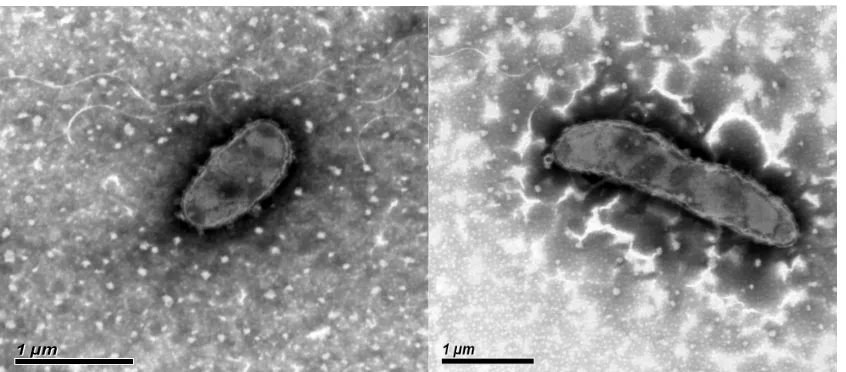
[17]. It was Gram-stain-negative, non-motile and
rod-shaped with 1 μm to 2 μm length [1] (Figure
1). Colonies are yellow, round, smooth, convex
and opaque on R2A after incubation at 30°C for 2–
3 days. Growth occurs at 4-35°C on R2A, with an
optimum at 30°C. It produces a flexirubin-like
pigment, the same as the other species in the
ge-nus. The major fatty acids are summed feature 3
(C
16: 1ω7cand/or iso-C
15: 0,2-OH), iso-C
15: 0, C
16: 1ω5cand
iso-C
17: 0 3-OH. The predominant polar lipid is
phosphatidylethanolamine [1].The important
characteristics of the strain based on literature
descriptions are summarized in Table 1. The
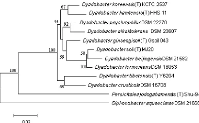
strain exhibited a 16S rRNA gene sequence
simi-larity with other members of the genus
A54 (Figure 2).
Figure 1 Transmission electron micrograph of
The utilization of carbon compounds by strain
Y620-1 was determined using Generation-III
microplates on an OmniLog phenotyping device
(BIOLOG Inc., Hayward, CA, USA). The microplates
were inoculated at 30°C with a cell suspension at a
cell density of 95-96% turbidity and dye IF-A.
Strain Y620-1 assimilates dextrin, maltose,
D-trehalose, D-cellobiose, gentiobiose, D-melibiose,
D-salicin, N-Acetyl-D-glucosamine, D-mannose,
Glycyl-L-proline, L-alanine, L-histidine, L-serine,
methyl, pyruvate, L-lactic acid, citric acid,
α-Keto-glutaric acid, L-malic acid, propionic acid and
ace-tic acid, but not stachyose,
D-raffinose,Acetyl-β-dmannosamine, Acetyl-dgalactosamine,
N-Acetyl-neuraminic acid, D-galactose, 3-methyl
885 Figure 2. Phylogenetic tree highlighting the position of
strains within the genus
GCA_000428845.1, GCF_000382205.1 and GCA_000023125.1. The type strains and their corresponding
GenBank accession numbers for 16S rRNA genes are:
DSM 16708, AJ821885
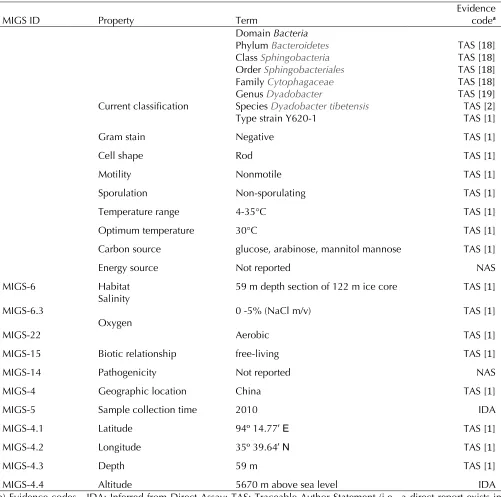
Table 1. Classification and general features of tions
MIGS ID Property Term Evidence codea
Current classification
Domain Bacteria
Phyl
Clas
Order
Famil
Genus
Species
Type strain Y620-1
TAS [18] TAS [18] TAS [18] TAS [18] TAS [19] TAS [2] TAS [1]
Gram stain Negative TAS [1]
Cell shape Rod TAS [1]
Motility Nonmotile TAS [1]
Sporulation Non-sporulating TAS [1]
Temperature range 4-35°C TAS [1]
Optimum temperature 30°C TAS [1]
Carbon source glucose, arabinose, mannitol mannose TAS [1]
Energy source Not reported NAS
MIGS-6 Habitat 59 m depth section of 122 m ice core TAS [1]
MIGS-6.3 Salinity 0 -5% (NaCl m/v) TAS [1]
MIGS-22
Oxygen
Aerobic TAS [1]
MIGS-15 Biotic relationship free-living TAS [1]
MIGS-14 Pathogenicity Not reported NAS
MIGS-4 Geographic location China TAS [1]
MIGS-5 Sample collection time 2010 IDA
MIGS-4.1 Latitude 94º 14.77′ E TAS [1]
MIGS-4.2 Longitude 35º 39.64′ N TAS [1]
MIGS-4.3 Depth 59 m TAS [1]
MIGS-4.4 Altitude 5670 m above sea level IDA
a) Evidence codes - IDA: Inferred from Direct Assay; TAS: Traceable Author Statement (i.e., a direct report exists in the literature); NAS: Non-traceable Author Statement (i.e., not directly observed for the living, isolated sample, but based on a generally accepted property for the species, or anecdotal evidence). These evidence codes are from the Gene Ontology project [20].
Genome sequencing information
Genome project history
The organism was selected for sequencing on the
basis of it from extreme deep ice core from high
altitude glacier. The shotgun genome sequencing
project was completed in December 2012 and has
been deposited at DDBJ/EMBL/GenBank under
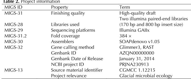
887 Table 2. Project information
MIGS ID Property Term
MIGS-31 Finishing quality High-quality draft
MIGS-28 Libraries used Two illumina paired-end libraries (170 bp and 800 bp insert size) MIGS-29 Sequencing platforms Illumina GAIIx
MIGS-31.2 Fold coverage 384 ×
MIGS-30 Assemblers SOAPdenovo v1.05
MIGS-32 Gene calling method Glimmer3, RAST
Genbank ID AZQN00000000
Genbank Date of Release January 31, 2014 NCBI project ID PRJNA230913 MIGS-13 Source material identifier CGMCC 1.12215
Project relevance Glacial microbial ecology
Growth conditions and DNA isolation
Cells of strain Y620-1 were harvested from R2A
broth following 2 days incubation at 30 °C with
shaking at 180 rpm. The genomic DNA of the
strain was extracted according to the method
pre-viously described by Marmur et al. [23].
Extrac-tion was started with 100 ml of 48 h culture,
cen-trifuged at 4 °C and 10, 000 rpm for 15 min. Then,
cells were washed three times with 5 ml sterile
water. The washed cells were resuspended in
1,128 μL Tris-HCl buffer (10 mM) containing 1
mM EDTA (pH 8.0) and 20 μg lysozyme and
incu-bated at 37 °C for 2 h. followed by adding of 6 μL
proteinase K (20 mg/mL), 4 μL DNase-free RNase
(10 mg/mL), 100 μL SDS (20% w/v) and the cell
suspension was incubated at 55 °C for 3 h. The cell
lysate was extracted twice with
phe-nol/chloroform/isoamyl alcohol (25:24:1) and
once with chloroform/isoamyl alcohol (24:1), and
the aqueous layer was separated after
centrifuga-tion at 12,000 rpm for 15 min. The DNA was
pre-cipitated with 1 volumes of frozen anhydrous
eth-anol. The purity of genomic DNA was assessed by
NanoDrop (2000c, Thermo) with OD 260:280
ra-tio of 1.8-2. The DNA was stock in TE (pH 8.0) for
genome sequencing.
Genome sequencing and assembly
The genome of strain Y620-1 was sequenced using
an Illumina GAIIx instrument with two paired-end
libraries (170 bp and 800 bp insert size). The raw
sequencing data was processed to discard reads
containing adaptor sequences, a high rate of
am-biguity, and removing the sequence reads which
were of low quality. A total of 2,041 Mb
high-quality of Illumina data were obtained, providing
approximately 384–fold coverage. The
high-quality reads were assembled in silico using
SOAPdenovo v1.05, resulting in 33 contigs (> 200
bp) with an N50 length of 797,100 bp.
Genome annotation
The coding sequences (CDS) were predicted using
Glimmer 3.02 [24], while tRNAscan-SE [25] and
RNAmmer [26] were used to identify tRNA and
rRNA, respectively. The genome sequence was
al-so uploaded into the Rapid Annotation using
Sub-system Technology (RAST) Sub-system [27] to check
the annotated sequences. The functions of
pre-dicted protein-coding genes were then annotated
through comparisons with the databases of
NCBI-NR [28], COG [29], and KEGG [30]. The program
TMHMM [31] and SignalP [32] were used to
iden-tify putative transmembrane helices and signal
peptides.
Genome properties
protein-coding genes), following by amino acids and
de-rivatives (n=272, 6.0%), cofactors, vitamins,
pros-thetic groups, pigments (n=184, 4.1%), protein
metabolism (n=143, 3.2%), membrane transport
(132, 2.9%) and respiration (131, 2.9%).
Table 3. Nucleotide content and gene count levels of the genome
Attribute Value % of totala
Genome size (bp) 5,313,963 100
DNA coding region (bp) 4,680,447 88.08
DNA G+C content (bp) 2,308,448 43.44
Total genes 4,867 100
RNA genes 39 0.80
Protein-coding genes 4,828 82.68
Genes with function prediction 2,844 58.91
Genes assigned to COGs 2,208 70.55
Genes assigned to Pfam domains 3299 68.33
Genes assigned to TIGRfam domains 2161 44.76
Genes with signal peptides 372 7.71
Genes with transmembrane helices 628 13.01
CRISPR repeats 1
a) The total is based on either the size of the genome in base pairs or the total number of protein coding genes in the annotated genome.
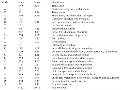
Table 4. Number of genes associated with the 25 general COG functional categories Code Value %agea Description
J 149 3.09 Translation
A 0 - RNA processing and modification
K 261 5.14 Transcription
L 130 2.69 Replication, recombination and repair B 0 - Chromatin structure and dynamics D 22 0.46 Cell cycle control, mitosis and meiosis
Y 0 - Nuclear structure
V 61 1.26 Defense mechanisms
T 217 4.49 Signal transduction mechanisms M 288 5.97 Cell wall/membrane biogenesis
N 12 0.25 Cell motility
Z 1 0.02 Cytoskeleton
W 0 - Extracellular structures
U 52 1.08 Intracellular trafficking and secretion
O 109 2.26 Posttranslational modification, protein turnover, chaperones C 201 4.16 Energy production and conversion
G 269 5.57 Carbohydrate transport and metabolism E 233 4.83 Amino acid transport and metabolism F 73 1.51 Nucleotide transport and metabolism H 182 3.77 Coenzyme transport and metabolism
I 132 2.73 Lipid transport and metabolism
P 259 5.36 Inorganic ion transport and metabolism
Q 84 1.74 Secondary metabolites biosynthesis, transport and catabolism R 410 8.49 General function prediction only
S 261 5.41 Function unknown
- 1422 29.45 Not in COGs
889
Discussion
Although there were 12 species assigned to
sequence and ar
21582. Strain Y620-1 has the smallest genome of
18053 and
6.97 Mbp and 7.37 Mbp, respectively). The GC
con-tent of strain Y620-1 is comparable to that o
than those of
and
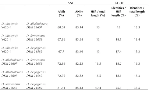
to estimate the similarity among the sequenced
ty (ANI) and Genome-to-Genome Distance
Calcula-tor (GGDC) were calculated using the software
JSpecies v1.2 [33] and GGDC v2.0 [34],
respective-ly. Table 5 shows the results of ANI and GGDC.
ANI analysis showed that strain Y620-1 shared a
low degree of similarity with other
species (< 69% ANIb and < 84% ANIm), whereas
relatively higher ANI value were obtained for
18053 and
Although the core concept of GGDC was based on
‘genome blast distance phylogeny’, which is
differ-ent from ANI [35], GGDC analysis showed similar
results. In both analyses, the highest similarity
values were observed in the comparisons o
21582. These results were in line with phylogeny
analysis based on 16S rRNA gene, which shows
tha
DSM 21582 form a cluster with
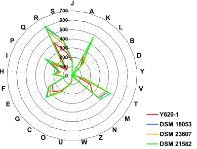
MJ20. Moreover, the comparison of distribution of
COG categories in the genome of four
strains revealed that there were significant
corre-lations between the distribution of COG categories
of strain Y620-1 and other strains (r =
0.970-0.979). However, relatively higher correlation
co-efficients were observed for
23607 and
21582 (0.991), and
Table 5. Pairwise comparisons between four
ANI GGDC
ANIb
(%) ANIm (%) HSP / total length (%)
Identities / HSP length (%)
Identities / total length
(%)
Y620-1 DSM 23607 68.04 83.14 13 18 13.3
Y620-1 DSM 18053 67.86 83.88 13 18.1 13.4
Y620-1
DSM 21582 67.7 83.46 13 17.4 13.3
DSM 23607 DSM 18053 72.89 82.23 16.5 18.2 16.3
DSM 23607 DSM 21582 72.79 82.52 16.5 18.1 16.3
Figure 3. Distribution of COG functional categories in the genome o
Y620-1,
Five cold-shock proteins were found in this
ge-nome including CspA, GyrA, RbfA, and NusA. The
proteins coded by gene RecA, RecF, RecG, RecN,
RecO, RecQ, RadA and RadC, which play a critical
role in recombinational repair of damaged DNA,
were also found [36].
Single-stranded-DNA-specific exonuclease RecJ, required for many types
of recombination events [37], and CRISPRs Cas1,
interacts with components of the DNA repair
sys-tems, also were found [38]. Phage shock protein C
existed in the genome, which may play a
signifi-cant role in the competition for survival under
nu-trient- or energy-limited conditions [39]. When
bacteria deposit on the glacier, the low
tempera-ture, high UV radiation, and desiccation could
in-duce the cold-shock and recombinational repair of
damaged DNA proteins. Additionally, oligotrophic
condition of glacial ice may induce the phage
shock protein C. The genome sequence of strain
Y620-1 provides genetic information to identify
the genes linked to its specific mechanisms for
adaption to extreme glacial environment.
Acknowledgement
This study was financially supported by the
Na-tional Natural Science Foundation of China (Grant
Nos. 41371084, 41171050, 41125003, 40930526,
41025002, 40871045), Strategic Priority Research
Program (B) of the Chinese Academy of Sciences
(XDB03030100), and the National Basic Research
Program of China (No: 2010CB951404).
References
1. Shen L, Liu Y, Yao T, Wang N, Xu B, Jiao N, Liu H, Zhou Y, Liu X, Wang Y
Int J Syst Evol Microbiol 2013; 63:3636-36
2. Chelius MK, Triplett EW. gen. nov., sp. nov., a novel gram-negative bacte-rium isolated from surface-sterilized Zea mays stems. Int J Syst Evol Microbiol 2000; 50:751-7
0 100 200 300 400 500 600
700
J
A
K
L
B
D
Y
V
T
M
N
Z
W
U
O
C
G
E
F
H
I
P
Q
R
S
891 3. Baik KS, Kim MS, Kim EM, Kim HR, Seong CN.
fresh water. Int J Syst Evol Microbiol 2007; 57:1227-1231
4. Chaturvedi P, Reddy GSN, Shivaji S.
in the Himalayas, India. Int J Syst Evol Microbiol
2005; 55:2113-21
5. Chen L, Jiang F, Xiao M, Dai J, Kan W, Fang C,
Peng F.
from high Arctic soil on the Svalbard Archipelago, Norway. Int J Syst Evol Microbiol 2013; 63:1616-1620
6. Chun J, Kang JY, Joung Y, Kim H, Joh K, Jahng KY.
seawater. Int J Syst Evol Microbiol 2013; 63:1788-1792
7. Dong Z, Guo X, Zhang X, Qiu F, Sun L, Gong H,
Zhang F.
lated from the rhizosphere of turf grasses in China.
Int J Syst Evol Microbiol 2007; 8. Lee M, Woo SG, Park J, Yoo SA.
sp. nov., a starch-degrading bacterium isolated from farm soil. Int J Syst Evol Microbiol 2010; 60:2577-2
9. Liu QM, Im WT, Lee M, Yang DC, Lee ST.
soil of a ginseng field. Int J Syst Evol Microbiol
2006; 56:1939-19
10. Reddy GSN, Garcia-Pichel F
the Colorado Plateau, USA, and an emended
de-scription of the genus
Triplett 2000. Int J Syst Evol Microbiol 2005; 55:1295-1
11. Tang Y, Dai J, Zhang L, Mo Z, Wang Y, Li Y, Ji S,
Fang C, Zheng C.
nov., isolated from desert sand. Int J Syst Evol
Microbiol 2009;
12. Zhang DC, Liu HC, Xin YH, Zhou YG, Schinner F,
Margesin R.
a psychrophilic bacterium isolated from soil. Int J
Syst Evol Microbiol 2010; 60:1640-1643
13. Lang E, Lapidus A, Chertkov O, Brettin T, Detter
JC, Han C, Copeland A, Glavina Del Rio T, Nolan M, Chen F and others. Complete genome
se-quence of
(NS114 T). Stand Genomic Sci 2009; 29; 1(2): 133–140.
14. Priscu JC, Christner BC, Foreman CM, Royston-Bishop G. Biological material in ice cores. In: Eli-as SA, editor. Encyclopedia of Quaternary Sci-ences. Volume 2: Elsevier B.V., UK.; 2007. p 1156-1166.
15. Koh HY, Lee SG, Lee JH, Doyle S, Christner BC, Kim HJ. Draft genome sequence of
philic bacterium isolated from sediment-laden stratified basal ice from Taylor Glacier, McMurdo Dry Valleys, Antarctica. J Bacteriol 2012;
194:6656-665
16. Raymond JA, Christner BC, Schuster SC. A bacte-rial ice-binding protein from the Vostok ice core.
Extremophiles 2008;
17. Reasoner DJ, Geldreich EE. A new medium for
the enumeration and subculture of bacteria from potable water. Appl Environ Microbiol 1985;
49:1-18. Garrity GM, Lilburn TG, Cole JR, Harrison SH, Euzéby J, Tindall BJ. Taxonomic Outline of the
Bacteria and
Michigan State Univer-sity Board of Trustees 2007;DOI: 10.1601/TOBA7.7
http://www.taxonomicoutline.org.
19. Skerman VBD, McGrowan V, Sneath PHA. Ap-proved list of bacterial names. Int J Syst Bacteriol: ASM Press; 1980. 225-420 p.
20. Ashburner M, Ball CA, Blake JA, Botstein D, But-ler H, Cherry JM, Davis AP, Dolinski K, Dwight SS, Eppig JT, et al. Gene ontology: tool for the unification of biology. Nat Genet 2000;
21. Bruno WJ, Socci ND, Halpern AL. Weighted neighbor joining: a likelihood-based approach to distance-based phylogeny reconstruction. Mol
Biol Evol 2000; 17:189-19
Car-denas E, Garrity GM, Tiedje JM. The ribosomal database project (RDP-II): introducing myRDP space and quality controlled public data. Nucleic
Acids Res 2007; 35:D169-D
23. Marmur J. A procedure for the isolation of deoxy-ribonucleic acid from microorganisms. J Mol Biol
1961; 3:208-218.
24. Delcher AL, Bratke KA, Powers EC, Salzberg SL.
Identifying bacterial genes and endosymbiont DNA with Glimmer. Bioinformatics 2007; 23:673-67
25. Lowe TM, Eddy SR. tRNAscan-SE: a program for improved detection of transfer RNA genes in ge-nomic sequence. Nucleic Acids Res 1997; 25:0955-0964.
26. Lagesen K, Hallin P, Rodland EA, Staerfeldt HH, Rognes T, Ussery DW. RNAmmer: consistent and rapid annotation of ribosomal RNA genes.
Nucle-ic Acids Res 2007; 35:3100-3108
27. Aziz RK, Bartels D, Best AA, DeJongh M, Disz T, Edwards RA, Formsma K, Gerdes S, Glass EM, Kubal M, et al. The RAST Server: rapid annota-tions using subsystems technology. BMC
Ge-nomics 2008; 9:
28. Benson DA, Karsch-Mizrachi I, Lipman DJ, Ostell
J, Wheeler DL. GenBank. Nucleic Acids Res 2007; 36:D25-D
29. Tatusov RL, Koonin EV, Lipman DJ. A genomic perspective on protein families. Science 1997; 278:631-6
30. Kanehisa M, Araki M, Goto S, Hattori M,
Hirakawa M, Itoh M, Katayama T, Kawashima S, Okuda S, Tokimatsu T, et al. KEGG for linking genomes to life and the environment. Nucleic
Ac-ids Res 2007; 36:D480-D48
31. Krogh A, Larsson B, von Heijne G, Sonnhammer ELL. Predicting transmembrane protein topology
with a hidden markov model: application to complete genomes. J Mol Biol 2001; 305:567-5
32. Bendtsen JD, Nielsen H, von Heijne G, Brunak S.
Improved prediction of signal peptides: SignalP
3.0. J Mol Biol 2004; 340:783-7
33. Richter M, Rosselló-Móra R. Shifting the genomic
gold standard for the prokaryotic species defini-tion. Proc Natl Acad Sci USA 2009; 106:19126-191
34. Hothorn T, Bretz F, Westfall P. Simultaneous
in-ference in general parametric models. Biom J
2008; 50:346-36
35. Auch AF, von Jan M, Klenk H-P, Göker M. Digital
DNA-DNA hybridization for microbial species delineation by means of genome-to-genome se-quence comparison. 2010.
36. Kunst F, Ogasawara N, Moszer I, Albertini AM, Alloni G, Azevedo V, Bertero MG, Bessieres P, Bolotin A, Borchert S, et al. The complete ge-nome sequence of the Gram-positive bacterium
Nature 1997; 390:249-25
37. Lovett ST, Kolodner RD. Identification and
purifi-cation of a single-stranded-DNA-specific exonuclease encoded by the recJ gene of
1989;
86:2627-2631
38. Blattner FR, Plunkett G, Bloch CA, Perna NT,
Burland V, Riley M, Collado-Vides J, Glasner JD, Rode CK, Mayhew GF, et al. The complete ge-nome sequence ofScience
1997; 277:1453-1
39. Brissette JL, Weiner L, Ripmaster TL, Model P.
Characterization and sequence of th
J Mol Biol 1991;