REVIEW
The role of extracellular vesicles
in cholangiocarcinoma
Mingzhen Bai
1, Wenkang Fu
1, Gang Su
4, Jie Cao
1, Long Gao
1, Chongfei Huang
1, Haidong Ma
1,
Jinduo Zhang
2,5,6, Ping Yue
2,5,6, Bing Bai
2,5,6, Yanyan Lin
1,2,5,6*†, Wenbo Meng
1,2,3,4,5,6*†and Xun Li
1,4,5,6,7Abstract
Cholangiocarcinoma (CCA) is a rare tumor that arises from cholangiocytes, the epithelial cells of the bile duct. The tumor is characterized by insidious onset, high degree of malignancy, poor prognosis and high recurrence rate. Due to the lack of specific biomarkers, it is difficult to diagnose CCA early and evaluate prognosis. Extracellular vesicles (EVs), which include apoptotic bodies, microvesicles and exosomes, have emerged as having important roles in cell-to-cell communication in both normal physiology and pathological conditions. Some research has found that EVs play a crucial role in the occurrence and development of CCA. EVs can carry specific molecular substances such as nucleic acids and proteins, which have potential for the diagnosis and therapy of CCA. This article reviews the current knowledge on the role of EVs in CCA. We highlight EVs and their functions in the physiology and pathophysiology of CCA, and discuss their therapeutic potential and their role as biomarkers.
Keywords: Cholangiocarcinoma, Extracellular vesicles, Exosomes, Biomarkers, Therapy
© The Author(s) 2020. This article is licensed under a Creative Commons Attribution 4.0 International License, which permits use, sharing, adaptation, distribution and reproduction in any medium or format, as long as you give appropriate credit to the original author(s) and the source, provide a link to the Creative Commons licence, and indicate if changes were made. The images or other third party material in this article are included in the article’s Creative Commons licence, unless indicated otherwise in a credit line to the material. If material is not included in the article’s Creative Commons licence and your intended use is not permitted by statutory regulation or exceeds the permitted use, you will need to obtain permission directly from the copyright holder. To view a copy of this licence, visit http://creat iveco mmons .org/licen ses/by/4.0/. The Creative Commons Public Domain Dedication waiver (http://creat iveco mmons .org/publi cdoma in/ zero/1.0/) applies to the data made available in this article, unless otherwise stated in a credit line to the data.
Introduction
Cholangiocarcinoma (CCA) is the most common malig-nancy of the biliary tree, accounting for approximately 3% of all gastrointestinal tumors and is the second most common primary liver tumor after hepatocellu-lar carcinoma (HCC) [1, 2]. The incidence of CCA var-ies geographically and demographically, and the overall incidence is still on the rise worldwide [3]. Although the 1-year survival has improved over time, the 5-year sur-vival is less than 10% [4]. Most patients with CCA do not possess exact risk factors and the clinical manifestation may be nonspecific, even in the late-stage of the disease [5]. As such, early detection could improve survival, and this highlights the requirements for novel methods to diagnose and treat CCA.
Recently, the emerging role of extracellular vesicles (EVs) in cholangiocarcinoma progression has attracted extensive attention. To date, the role of EVs has changed from being nonfunctional discards of cellular compo-nents to the current research focus [6, 7]. EVs are nano-sized, membrane-bound vesicles released from cells that can transport cargo, including DNA, RNA, and proteins, between cells as a form of intercellular communication [8, 9]. With so many contents, the nascent field of EVs has evolved to have a sharper focus, especially in oncol-ogy [10]. Therefore, EVs and their derived cargos have emerged as new biomarkers for tumor diagnosis. Tumor-derived EVs play a key role in modulating intercellular communication between tumor and stromal cells in local and distant microenvironments [11, 12]. In addition, EVs can still be used for therapeutic purposes as targets, immunomodulators and delivery vehicles [13]. In this review, we highlight and discuss about the relationship between EVs and CCA, with a special focus on their roles and potential clinical application values as biomarkers and therapeutic targets in CCA.
Open Access
*Correspondence: ldyy_linyy@lzu.edu.cn; mengwb@lzu.edu.cn †Yanyan Lin and Wenbo Meng contributed equally to this work 2 Department of Special Minimally Invasive Surgery, The First hospital of Lanzhou University, Lanzhou 730000, China
Epidemiology, risk factors, diagnosis and treatment of CCA
Cholangiocarcinoma is a devastating tumor, currently classified as intrahepatic (iCCA), perihilar (pCCA), or distal (dCCA), characterized by varying degrees of des-moplastic reaction and increasing morbidity and mor-tality worldwide [14, 15]. While it is more common in Asia, its incidence has risen significantly in Europe and North America in recent decades [16]. Approxi-mately 35,660 patients with iCCA are diagnosed each year in the United States, and the 5-year survival rate is about 10% [17, 18]. The incidence of CCA is high-est in Thailand, with 113 per 100,000 men and 50 per 100,000 women per year [19]. In most case of CCA, the etiology is unknown, but chronic inflammation and cell injury in the bile duct shown a high risk of occurrence of CCA [14]. Pathologically, the release of inflamma-tory cytokines, increased cell death and proliferation, as well as changes in the liver in fibrosis contribute to the occurrence of tumor [20]. Primary sclerosing chol-angitis is considered as the main risk factor for CCA [21]. The diagnostic basis mainly includes imaging methods (ultrasound, computed tomography, magnetic resonance imaging and fluorodeoxyglucose positron emission tomography), histological analysis of a tumor biopsy and serum nonspecific tumor biomarkers, such as carbohydrate antigen 19-9 (CA19-9) and carcinoem-bryonic antigen (CEA) [22–24]. However, the sensi-tivity of these tests in the diagnosis of CCA is limited, especially in the early stages of the disease [25]. Man-agement strategies include multispecialty treatments, with consideration of surgical resection, targeted radiation therapy, and systemic chemotherapy [24, 26, 27]. Surgical resection is the only potentially curative treatment, but the majority of patients present with advanced cancer and recurrence after resection is com-mon [16]. Lymph node metastasis is a prominent fea-ture of CCA [28]. Diagnosed CCA is usually advanced and often inoperable, leading to a poor prognosis [29].
Details about EVs
Classification of EVs
According to the current knowledge of their biogen-esis, EVs can be broadly divided into two main catego-ries: microvesicles and exosomes [30, 31]. Microvesicles (MVs, 100–1000 nm in diameter) are secreted by the shedding or outward budding of the plasma membrane [32]. The formation of microvesicles is the result of the dynamic interaction of phospholipid redistribution and cytoskeletal protein contraction [33]. Since little informa-tion about biogenesis and MV release is understood, we focused on the function of exosomes in CCA.
Characteristics of exosomes
In 1983, Johnstone et al. first isolated exosomes from the supernatant of sheep reticulocyte culture medium [34]. However, due to the role of exosomes being unclear at that time, it is thought to be a nonfunctional particle in the cell. Whereas exosomes are 40–150 nm, endosome-derived, small EVs which are secreted by most cells [35]. RNA, DNA and proteins are reported to be actively and selectively incorporated into intraluminal vesicles, which reside within multivesicular endosomes and are the pre-cursor of exosomes [36, 37]. The release of exosomes into the extracellular space is facilitated by the fusion of multivesicular bodies (MVBs) limiting the membrane with the plasma membrane [35, 38]. Then, exosomes can be carried away by extracellular fluid, such as saliva, urine, blood, semen, amniotic fluid, ascites, alveolar lav-age fluid, milk, synovial fluid and cerebrospinal fluid, and taken up by other cells [39–41]. Besides, bile included [42]. The function of exosomes depends on the type and contents of their parent cells. Exosomes from normal cells play a role in maintaining stability in vivo, while tumor cell-derived exosomes are associated with tumor progression [40, 43].
In addition, we need to identify exosomes that can dis-tinguish them from other EVs by their size and proteins markers. For instance, MVB-associated proteins (tumor susceptibility gene 101 (TSG101) and ALIX) [44, 45], fusion proteins, membrane transport proteins (flotil-lin-1) [46], tetraspanins (CD63, CD81and CD9) [36], and heat shock proteins (HSP70.1 and HSP20) [42, 47] are often used as protein markers to recognize exosomes in scientific research. Currently, with the identification of a large number of cargo molecules in exosomes, their func-tions, including regulating immune function, enhancing metastasis, and modulating intercellular communication, have also been explored in tumor cells [48–50]. Some research shows that exosomes from highly metastatic breast cancer cells (4T1 and EO771 cells) can modulate the favorable microenvironment for lung and liver metas-tasis colonization [51]. In addition, various immune cell-derived exosomes, such as natural killer cells, dendritic cells, macrophages, neutrophils, mast cells and myeloid-derived suppressors, can act on tumor cells, modulating the growth, metastasis and response to chemotherapy [48, 52]. Next, we mainly elucidate the role of exosomes in CCA.
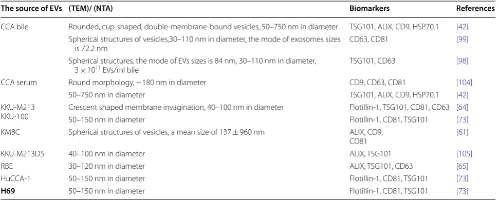
Characterization of EVs associated with CCA
used method. Accumulated evidence has reported that EVs can be extracted from the serum, bile and CCA cells of patients with bile duct carcinoma and identified by specific markers, as shown in Table 1. The morphological and molecular characteristics of these EVs indicate that they are mainly exosomes. However, the contents of EVs are numerous, and their functions are different, and these need to be further studied.
EVs regulate the progression of CCA
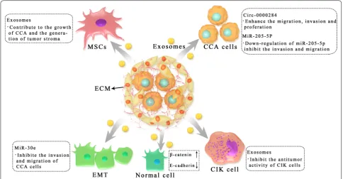
With the further development of EVs, increasing evi-dence has been presented to demonstrate the role of EVs in the progression of CCA (Fig. 1). Bile duct carcinoma usually has a dense stroma that contains immune cells (including neutrophils, tumor associated macrophages, natural killer cells, and T and B lymphocytes) and an extracellular matrix that promotes connective tissue proliferation [53, 54]. In particular, cancer-associated fibroblasts (CAFs), which communicate not only with tumor cells, but also with stromal cells, play a central role in the progression of CCA [55, 56]. CAFs influence the behavior of CCA by releasing various metabolites and soluble factors, such as vascular endothelial growth fac-tor (VEGF)-A and VEGF-C, which may lead to dilation of the lymphatic vasculature and tumor cell intravasation [57]. Therefore, it also has the property of easy transfer [18]. In this process, EVs play a crucial role in facilitating the communication of various signals (Table 2).
The function of EVs in tumor microenvironment
Cholangiocytes can be actively involved in the develop-ment of bile duct disease by stimulating the recruitdevelop-ment
and activation of inflammatory cells in the bile duct microenvironment [58]. Katsumi et al. found that acti-vated bile duct cells are involved in activating the proin-flammatory polarization of damage-associated molecular patterns (DAMPs) through the receptor for advanced gly-cation end products (RAGE) signaling pathway by releas-ing DAMPs as EV cargo [59]. Prior to this, Masyyuk et al. verified exosomes in rat bile [60]. Meanwhile, in the pro-cess of studying the cilium interaction of bile duct cells, exosomes were found to induce intracellular signals and functional responses, verifying that bile exosomes par-ticipate in intercellular signal communication [60]. This laid a foundation for the influence of bile duct carcinoma exosomes on tumor progression.
In the study of the tumor microenvironment of CCA, Haga et al. exposed marrow mesenchymal stem cells (MSCs) to CCA cell-derived EVs, which enhanced expression of alpha-smooth muscle actin mRNA and release of cytokines/chemokines such as IL-6, thus regulating the tumor microenvironment and promoting the growth of CCA. In addition, CCA cell-derived EVs can contribute to the formation of the tumor stroma through the fibroblast differentiation of MSCs [61]. This further revealed the effect of tumor cell-derived EVs on the local microenvironment. However, they did not identify the specific contents of the EVs. Moreover, proteins in exosomes are essential agents for tumor growth [62, 63]. Another study demonstrated the pro-tein spectrum of CCA-derived exosomes and their potential roles. These researchers isolated exosomes from CCA cell lines (KKU-M213 and KKU-100) and incubated them with normal bile duct cells (H69). After
Table 1 Characterization of EVs in bile, serum, cholangiocytes and CCA cells
CCA cell lines: KKU-M213, KKU-100, KKU-M213D5, KMBC, RBE and HuCCA-1. Normal human cholangiocyte cell line: H69
TSG101 tumor susceptibility gene 101, TEM transmission electron microscopy, NTA nanoparticle tracking analysis
The source of EVs (TEM)/ (NTA) Biomarkers References
CCA bile Rounded, cup-shaped, double-membrane-bound vesicles, 50–750 nm in diameter TSG101, ALIX, CD9, HSP70.1 [42] Spherical structures of vesicles,30–110 nm in diameter, the mode of exosomes sizes
is 72.2 nm CD63, CD81 [99]
Spherical structures, the mode of EVs sizes is 84 nm, 30–110 nm in diameter,
3 × 10¹¹ EVs/ml bile TSG101, CD63 [98]
CCA serum Round morphology, ~ 180 nm in diameter CD9, CD63, CD81 [104]
50–750 nm in diameter TSG101, ALIX, CD9, HSP70.1 [42]
KKU-M213
KKU-100 Crescent shaped membrane invagination, 40–100 nm in diameter50–150 nm in diameter Flotillin-1, TSG101, CD81, CD63 [Flotillin-1, CD81, TSG101 [6473]] KMBC Spherical structures of vesicles, a mean size of 137 ± 960 nm ALIX, CD9,
CD81 [61]
KKU-M213D5 40–100 nm in diameter ALIX, TSG101 [105]
RBE 30–120 nm in diameter ALIX, TSG101, CD63 [65]
HuCCA-1 50–150 nm in diameter Flotillin-1, CD81, TSG101 [73]
proteomics analysis, exosomes were found to be inter-nalized into H69 cells, resulting in migration and inva-sion of H69 cells, but failing to induce proliferation. In addition, the exosomes of KKU-M213 cells induced the expression of β-catenin and decreased the expression of E-cadherin, suggesting that exosomes might induce the migration and invasion of bile duct cells through the direct transfer of oncogene proteins between cells, thus
affecting the specific intracellular mechanism related to CCA carcinogenesis [64]. The proteomics analysis of normal bile duct cells and CCA cells confirmed the dif-ferences between these two cell types, and these differ-ences need to be further studied. Moreover, exosomes from another CCA cell line (RBE) could inhibit the antitumor activity of cytokine-induced killer (CIK) cells by downregulating the populations of CD3+, CD8+,
Fig. 1 Roles of tumor cells derived exosomes in the progression of CCA. Exosomes are critically involved in CCA progression including tumorigenesis, development, immune escape and metastasis by transferring functional biomolecules. CCA cell derived exosomes induced the expression of β-catenin and decreased the expression of E-cadherin to increase motility of normal cells. CCA cell derived exosomes interact with marrow mesenchymal stem cells (MSCs) to modulate the microenvironment and promote CCA growth. Moreover, CCA cell derived exosomes participate in immune escape by inhibiting the cytokine-induced killer (CIK) cells
Table 2 Summarize the function of EVs contents in CCA
CCA cell lines: KKU-M213, KMBC, RBE and HuCCT-1. Normal human cholangiocyte cell line: H69. A human liver stellate cell line: LX2
EVs contents Source Downstream target or recipient cells
Function References
DAMPs Actived cholangiocytes Macrophage Upregulate proinflammatory cytokines and profibrogenic factors [59] Exosomes HUCCT1 and KMBC MSCs Contribute to tumor cell growth and stromal development [61] Exosomes KKU-M213 H69 cell Induce the expression of β-catenin and reduce the expression of
E-cadherin [64]
Exosomes RBE CIK cell Inhibit the antitumor activity of CIK cells [65] MiR-205-5p KKU-M213 CCA cell Down-regulation of miR-205-5p can inhibit invasion and migration of CCA
cells [73]
NK (CD56+) and CD3+CD56+ cells and secreting TNF-α and perforin [65]. According to some research, mutations in the IDH1 gene is common in a variety of tumors, including iCCA [66, 67]. Recent studies have shown that the R132C mutation is the most common type of IDH1 mutation in iCCA. The IDH1R132C muta-tion leads to the downregulamuta-tion of P2RX7 expression, which further affects the exosome secretion of tumor cells, ultimately affecting the progression of CCA [68]. However, more functional experiments on P2RX7 are needed.
Exosomal noncoding RNAs in CCA
In addition, noncoding RNA also play a crucial role in tumor development [69]. As a functional regulatory molecule, noncoding RNAs, such as microRNAs (miR-NAs) and circular RNAs (circR(miR-NAs), could mediate cellular processes, including chromatin, transcription, posttranscriptional modification and signal transduc-tion, and, of course, predict prognosis [69–71].
Exosomal microRNAs in CCA
MiRNAs are small noncoding RNAs composed of 19–24 nucleotides [72]. Kitdumrongthum et al. found different miRNA expression profiles in the exosomes released by CCA cells and cholangiocytes, and many miRNAs with abnormal regulation had functions related to a variety of oncogenes [73]. For example, miR-205-5p, the most upregulated miRNA, down-regu-lation of miR-205-5p can inhibit invasion and migration of CCA cells [73]. Moreover, miR-205-5p has also been reported in other cancers, such as breast cancer and gastric cancer [74, 75]. However, in contrast to CCA, the expression of miR-205-5p is downregulated in breast cancer, and miR-205-5p has an antitumor effect in breast cancer [76]. Li and colleagues reported that EVs could transport miRNA species between human CCA cells and CAFs. They used LX2-derived EVs car-rying miR-195 to inhibit CCA growth and improve survival in a mouse model of CCA, demonstrating the communication between the tumor and microenviron-ment [77]. Epithelial–mesenchymal transition (EMT) is a biological process in which epithelial cells gradually change and lose epithelial characteristics and differen-tiate into mesenchymal phenotypes, and it is closely related to the invasiveness and motility of tumor cells [78–80]. CCA-derived EVs could transfer miR-30e and inhibit EMT by directly targeting the Snail in receptor cells, thus inhibiting the invasion and migration of bile duct cancer cells [81]. In addition, miR-200a/c-3p in serum exosomes was significantly positively correlated
with the CCA stage, and mainly involved in lymphatic metastasis of tumors [82].
Exosomal circRNAs in CCA
Recent research has demonstrated that circRNAs also have a biological role in CCA [83, 84]. Wang et al. found that the level of circ-0000284 was increased in CCA cell lines, tumor tissues and plasma exosomes, thus enhanc-ing the migration, invasion and proliferation of CCA cells in vivo and in vitro. The circ-0000284/miR-637/LY6E regulatory axis was involved in this process [85, 86]. In addition, exosome-mediated circ-0000284 could stimu-late the malignant behavior of surrounding normal cells and ultimately promote the progression of CCA [86].
EVs as novel biomarkers for CCA
A major reason for the poor prognosis of CCA is the lack of early detection. Late diagnosis delays optimal treat-ment and leads to lower survival. Therefore, it is neces-sary to develop new methods for the diagnosis of CCA. In recent years, studies have shown that EVs have huge potential in the diagnosis of diseases due to their unique properties and great progress has been made in study-ing of EVs as tumor diagnostic markers [41, 87, 88]. EVs in bile and blood have opened up new ideas for the early diagnosis of CCA. Compared with traditional CA-199 and CEA, EVs have higher diagnostic value in CCA, which is summarized in Fig. 2.
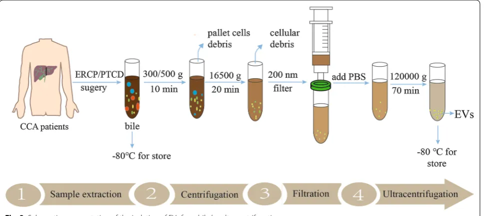
The isolation of EVs
the isolation of bile here. Bile samples of CCA are usu-ally collected by endoscopic retrograde cholangio-pancreatography (ERCP), percutaneous transhepatic biliary drainage (PTBD) and surgery, and then bile
is immediately centrifuged at 4 ℃ to remove the cell debris and filtered through a 200 nm filter. Finally, the supernatant is collected and added to an ultracentrifuge tube, diluted with PBS for further purification, mainly
Fig. 2 EVs can exist in bile and blood, which is helpful for disease detection and diagnosis
including ultracentrifugation, PEG precipitation, mem-brane filtration and affinity purification [98–100]. Some research, however, initially collected bile and then rap-idly diluted with PBS for further centrifuge [42]. The isolation of EVs were used immediately or stored at
− 80 °C until use (Fig. 3). However, research in this area is limited, and there is still much room for improve-ment in the separation and purification of exosomes.
Bile EVs
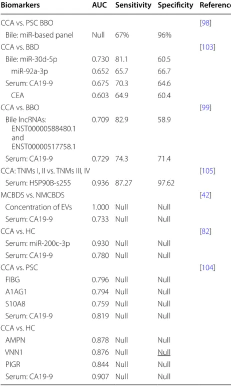
MiRNA in EVs is protected the membrane from degrad-ing enzymes, and this determines its highly stable char-acteristics in the extracellular environment, thus, miRNA is used for the diagnosis of a variety of cancers, including CCA [101, 102]. Li et al. identified and characterized EVs in human bile for the first time, and miRNA-laden EVs in human bile could be used for the diagnosis of CCA. They defined a new biliary vesicle miR-based panel (miR-191, miR-486-3p, miR-1274b, miR-16 and miR-484) for the diagnosis of CCA with a sensitivity of 67% and specificity of 96% [98]. This research initiates studying EVs in bile. Han and coworkers found that the expression levels of miR-30d-5p and miR-92a-3p in the bile of CCA patients were specifically upregulated compared with those in the bile of patients with benign biliary disease [103]. Com-pared with CEA and CA19-9, miR-30d-5p had the best effect in differentiating CCA and common bile duct dis-ease, with a sensitivity of 81.1%, specificity of 60.5% and area under the curve (AUC) value of 0.730. In addition, the identification of CCA using the combination of the two bile miRNAs and serum CA19-9 levels was weaker than that of miR-30d-5p alone [103].
Long noncoding RNAs (lncRNAs) in bile also play the same role in CCA as miRNAs. Ge et al. conducted a series of experiments on bile exosomes, and showed that the expression of the lncRNAs ENST00000588480.1 and ENST00000517758.1 in the CCA group was signifi-cantly increased compared with that of the control group. The AUC of the combined detection of the two lncR-NAs was 0.709, and the sensitivity and specificity were 82.9% and 58.9%, respectively [99]. Moreover, with the increase in the tumor TNM stage, the expression levels of ENST00000588480.1 and ENST00000517758.1 were sig-nificantly increased, and they could be potential diagnos-tic markers [99]. The content of EVs in bile can be used as a diagnostic marker, and the concentration of EVs also has potential diagnostic value. Severino et al. found that the accuracy of the bile EV count in the diagnosis of CCA was 100% with a threshold value of 9.46 × 10¹⁴
nanopar-ticles, with an AUC of 1, and this could correctly distin-guish malignant and nonmalignant common bile duct stenoses in this research [42].
Serum EVs
There are also some diagnostic markers of CCA in serum EVs. The new proteomic features found by Ander Arbelaiz in serum EVs of primary sclerosing cholangi-tis (PSC), CCA and HCC patients have potential diag-nostic value [104]. For example, fibrinogen gamma chain (FIBG), alpha-1-acid glycoprotein 1 (A1AG1) and S100A8 (S10A8) proteins have the strongest differential diagnoses of CCA and PSC, with AUC values of 0.796, 0.794 and 0.759, respectively [104]. Similarly, using pro-teomic methods, Weeraphan et al. studied the exosomal phosphoproteome of M213 and M213D5 in CCA cells, and showed that Ser255 of HSP90B was highly phos-phorylated in tissues of CCA patients with a low TNM stage (I and II) compared to those with a TNM stage of III or IV. ROC analysis showed that HSP90B-S255 was a new potential biomarker for metastatic CCA with an AUC value of 0.936 (sensitivity 87.27%, specificity 97.62%) [105]. Moreover, Shen et al. studied exosomal miRNA in peripheral blood samples from CCA patients and healthy controls. The results showed that the serum extracellular miR-200 family, especially miR-200c-3p, had the strongest diagnostic ability for CCA than that in serum CA19-9, with an AUC of 0.93, which is worth further study [82]. Meanwhile, positive of Annexin V, epithelial cell adhesion molecule (EpCAM), asialogly-coprotein receptor 1 (ASGPR1) and tumor-associated microparticles found in serum EVs were shown to have the potential to differentiate HCC and CCA from tumor-free individuals; after tumor resection, the num-ber of these microparticles decreased, which proved a correlation with the presence of the tumor [95].
Summary of the role of EVs in diagnosing CCA
not clear and how to efficiently extract and detect EVs in body fluids remains unclear.
Application of EVs in CCA therapy
To date, due to the difficulty in the early diagnosis of CCA, there are few treatment options, and radical sur-gical resection is the only effective treatment method [107, 108]. Postoperative adjuvant chemotherapy can improve the survival and cure rates, but the effect of chemotherapy is not enough [18]. With the develop-ment of research on EVs, the treatdevelop-ment of CCA has a new direction. The characteristics of tumor-secreted EVs in regulating the immune microenvironment illus-trate their clinical potential in immunotherapy, thera-peutic targeting and drug delivery [11, 109].
Therapeutic targets
Since the tumor microenvironment promotes the pro-gression and invasion of CCA, targeting the microenvi-ronment and related cells is a strategy for the treatment of CCA [110, 111]. Chen et al. found that RBE-derived exosomes could inhibit the antitumor activity of CIK cells [65]. This suggests that the effect of CIK cell-based immunotherapy is related to EVs, which may be a poten-tial therapeutic target. In addition, circ-0000284 may be a therapeutic target for CCA. Wang et al. proved that the knockdown of exosomal circ-0000284 inhib-ited CCA growth and metastasis in vivo through ani-mal experiments [86]. Besides, according to Zhang’s research, mutations in the IDH1 gene alter the function of IDH1 and affect the development of CCA by promot-ing exosome release [68]. Interestingly, IDH1 inhibitors have been reported. For example, the safety and clini-cal efficacy of mutant IDH1 inhibitor ivosidenib in the recurrence or refractory IDH1-mutated acute myeloid leukemia were demonstrated [112]. At present, inhibi-tors of IDH1 (AG120 and IDH305) are being tested in iCCA patients [113]. This provides a potential treatment of CCA. Furthermore, one study reported that inhibition of miR-205-5p in the exosomes of CCA could reduce the invasion and migration of CCA, and the miR-200 family was associated with drug resistance [73, 114].
Drug delivery
EVs are natural membrane vesicles involved in intercellu-lar communication; accumulating evidence has revealed that EVs have the characteristics of stability and low immune reactivity, and exosomes in EVs can effectively transport a variety of different types of cargo to target cells [109, 115]. As a result, EVs can also be selected as a therapeutic tool to modulate the function of CCA cells by delivering cargo media [35, 116]. Stromal derived EVs are suitable for the delivery of materials to CCA cells, and this property can be exploited for delivering antitu-mor therapy to CCA cells [117]. Li et al. proved that EVs could transport miR-195 from fibroblasts to cancer cells. In addition, fibroblasts-derived EVs loaded with miR-195, play a key role in the CCA rat model, by reducing the size of tumors and improving survival in the treated rats [77]. Moreover, after incubating the miR-30e-enriched EVs with CCA cells, Zhang found that the expression of miR-30e in receptor CCA cells increased, which ultimately regulated the invasion and migration of cells. This dem-onstrates that miR-30e-enriched EVs can be ingested by recipient cells as a means of transferring miR-30e [81]. A recent study showed that methotrexate-loaded tumor-cell-derived microvesicles were injected into the bile duct lumen of patients with extrahepatic CCA, which Table 3 EVs as diagnostic biomarkers for CCA
Biomarkers AUC Sensitivity Specificity Reference
CCA vs. PSC BBO [98]
Bile: miR-based panel Null 67% 96%
CCA vs. BBD [103]
Bile: miR-30d-5p 0.730 81.1 60.5 miR-92a-3p 0.652 65.7 66.7 Serum: CA19-9 0.675 70.3 64.6
CEA 0.603 64.9 60.4
CCA vs. BBO [99]
Bile lncRNAs: ENST00000588480.1 and
ENST00000517758.1
0.709 82.9 58.9
Serum: CA19-9 0.729 74.3 71.4
CCA: TNMs I, II vs. TNMs III, IV [105] Serum: HSP90B-s255 0.936 87.27 97.62
MCBDS vs. NMCBDS [42]
Concentration of EVs 1.000 Null Null Serum: CA19-9 0.733 Null Null
CCA vs. HC [82]
Serum: miR-200c-3p 0.930 Null Null Serum: CA19-9 0.780 Null Null
CCA vs. PSC [104]
FIBG 0.796 Null Null
A1AG1 0.794 Null Null
S10A8 0.759 Null Null
Serum: CA19-9 0.819 Null Null CCA vs. HC
AMPN 0.878 Null Null
VNN1 0.876 Null Null
PIGR 0.844 Null Null
mobilized and activated neutrophils and alleviated biliary obstruction [118]. These studies suggest that EVs carry-ing designed cargo can be used as media carriers to man-age the progression of CCA.
Conclusion and future prospects
EVs are present through the occurrence, development and metastasis of tumors, providing new clues for the diagnosis and treatment of CCA. Many substances in EVs, such as miRNAs, circRNAs, proteins and even EV concentrations, can be used as new biomarkers. It is also important to improve the early diagnosis of CCA. More-over, according to the role of EVs in the tumor microen-vironment and immunity, corresponding targeted drugs and immunotherapy can be used for the treatment of CCA. Exosomal programmed-death ligand (PD-L1) has been found to contribute to immunosuppression and has been associated with the anti-PD-1 response, although it is mainly involved in melanoma [119]. Moreover, PD-L1expression in iCCA and perihilar cholangiocarcinoma has also been reported, and is mainly expressed in tumors with a high density of tumor-infiltrating lymphocytes [120]. Immunotherapy for PD-L1 in CCA has entered clinical research, but the efficacy is not clear [121]. EVs have shown great potential in the immunotherapy of tumors, although there is not much data on the immuno-therapy of EVs in CCA and therefore, further research is needed. In summary, this review focuses on the current research status of EVs in CCA. First, the characteristics of EVs in CCA were described. Second, the mechanism of EVs in tumor growth and metastasis was discussed. Finally, we demonstrated that the contents of EVs could be used for the clinical diagnosis and treatment of CCA. Although the existing studies have partially uncovered the mechanism of EVs in CCA, there are still a few chal-lenging problems to solve. Firstly, the detailed mecha-nism to describe the role of EVs in CCA needs further clarification. In addition, standardized methods for the separation, purification and analysis of EVs in body fluids are needed. Last but not least, most of the pathophysi-ological studies are conducted through in vitro analysis, and there are few in vivo experiments based on EVs in animal models. Follow-up studies should be conducted to better apply EVs to the clinical diagnosis and treat-ment of CCA in the future. Therefore, more efforts are needed to study the role of EVs in CCA.
Abbreviations
CCA : Cholangiocarcinoma; HCC: Hepatocellular carcinoma; EVs: Extracellular vesicles; MVs: Microvesicles; MVBs: Multivesicular bodies; TSG101: Tumor susceptibility gene 101; HSP70.1: Heat shock proteins 70.1; HSP20: Heat shock proteins 20; TEM: Transmission electron microscopy; NTA: Nanoparticle track-ing analysis; CAFs: Cancer-associated fibroblasts; VEGF-A: Vascular endothe-lial growth factor-A; VEGF-C: Vascular endotheendothe-lial growth factor-C; RAGE:
Receptor for advanced glycation end products; DAMPs: Damage-associated molecular patterns; MSCs: Marrow mesenchymal stem cells; iCCA : Intrahe-patic cholangiocarcinoma; miRNAs: MicroRNAs; circRNAs: Circular RNAs; EMT: Epithelial–mesenchymal transition; CA19-9: Carbohydrate antigen 19-9; CEA: Carcinoembryonic antigen; pCCA : Perihilar cholangiocarcinoma; dCCA : Distal cholangiocarcinoma; FIBG: Fibrinogen gamma chain; A1AG1: Alpha-1-acid glycoprotein 1; S10A8: S100A8; AMPN: Aminopeptidase N; VNN1: Pantethein-ase; PIGR: Polymeric immunoglobulin receptor; ERCP: Endoscopic retrograde cholangiopancreatography; PTBD: Percutaneous transhepatic biliary drainage; AUC : Area under the curve; lncRNAs: Long noncoding RNAs; PSC: Primary sclerosing cholangitis; EpCAM: Epithelial cell adhesion molecule; ASGPR1: Asialoglycoprotein receptor 1; BBO: Benign biliary obstruction; BBD: Benign biliary disease; MCBDS: Malignant common bile duct stenoses; NMCBDS: Nonmalignant common bile duct stenoses; HC: Healthy controls; PD-L1: Programmed-death ligand.
Acknowledgements Not applicable.
Authors’ contributions
MB, WF, WM and YL designed the research. MB drafted the manuscript. XL, WM, YL, GS, WF critically revised the manuscript. MB, WF, LG, JC, CH, HM, JZ, PY and BB provided intellectual contribution. All authors read and approved the final manuscript.
Funding
This research was supported by the National Natural Science Founda-tion (Grant Number 81872036), and Lanzhou Science and Technology Bureau (Grant Number 2019-4-43), and Science and Technology Project of Chengguan District of Lanzhou City (Grant Number 2019JSCX0092 and 2019RCCX0038), and Lanzhou talent innovation project (Grant Number 2018-RC-13).
Availability of data and materials
The materials supporting the conclusions of this review are included in the article.
Ethics approval and consent to participate Not applicable.
Consent for publication Not applicable.
Competing interests
The authors declare that they have no competing interests.
Author details
1 The First Clinical Medical College, Lanzhou University, Lanzhou 730000, China. 2 Department of Special Minimally Invasive Surgery, The First hospital of Lanzhou University, Lanzhou 730000, China. 3 Division of Scientific Research and Development Planning, The First Hospital of Lanzhou University, Lanzhou 730000, China. 4 Institute of Genetics, School of Basic Medical Sci-ences, Lanzhou University, 730000 Lanzhou, China. 5 Gansu Province Institute of Hepatopancreatobiliary, 730000 Lanzhou, China. 6 Gansu Province Key Laboratory Biotherapy and Regenerative Medicine, 730000 Lanzhou, China. 7 The Second Department of General Surgery, The First Hospital of Lanzhou University, 730000 Lanzhou, China.
Received: 26 June 2020 Accepted: 29 August 2020
References
1. Rizvi S, Gores GJ. Pathogenesis, diagnosis, and management of cholan-giocarcinoma. Gastroenterology. 2013;145(6):1215–29.
3. Montomoli J, Erichsen R, Norgaard M, Hoyer M, Hansen JB, Jacobsen JB. Survival of patients with primary liver cancer in central and north-ern Denmark, 1998–2009. Clin Epidemiol. 2011;3(Suppl 1):3–10. 4. Bergquist A, von Seth E. Epidemiology of cholangiocarcinoma. Best
Pract Res Clin Gastroenterol. 2015;29(2):221–32.
5. Tyson GL, El-Serag HB. Risk factors for cholangiocarcinoma. Hepatol-ogy. 2011;54(1):173–84.
6. Maas SLN, Breakefield XO, Weaver AM. Extracellular vesicles: unique intercellular delivery vehicles. Trends Cell Biol. 2017;27(3):172–88. 7. Cocucci E, Racchetti G, Meldolesi J. Shedding microvesicles: artefacts
no more. Trends Cell Biol. 2009;19(2):43–51.
8. Zaborowski MP, Balaj L, Breakefield XO, Lai CP. Extracellular vesicles: composition, biological relevance, and methods of study. Bioscience. 2015;65(8):783–97.
9. Roma-Rodrigues C, Raposo LR, Cabral R, Paradinha F, Baptista PV, Fernandes AR. Tumor microenvironment modulation via gold nanoparticles targeting malicious exosomes: implications for cancer diagnostics and therapy. Int J Mol Sci. 2017;18(1):162.
10. Bebelman MP, Smit MJ, Pegtel DM, Baglio SR. Biogenesis and function of extracellular vesicles in cancer. Pharmacol Ther. 2018;188:1–11. 11. Becker A, Thakur BK, Weiss JM, Kim HS, Peinado H, Lyden D.
Extracel-lular vesicles in cancer: cell-to-cell mediators of metastasis. Cancer Cell. 2016;30(6):836–48.
12. Sheehan C, D’Souza-Schorey C. Tumor-derived extracellular vesicles: molecular parcels that enable regulation of the immune response in cancer. J Cell Sci. 2019. https ://doi.org/10.1242/jcs.23508 5. 13. Walker S, Busatto S, Pham A, Tian M, Suh A, Carson K, et al.
Extracel-lular vesicle-based drug delivery systems for cancer treatment. Theranostics. 2019;9(26):8001–17.
14. Sirica AE. Cholangiocarcinoma: molecular targeting strategies for chemoprevention and therapy. Hepatology. 2005;41(1):5–15. 15. Alsaleh M, Leftley Z, Barbera TA, Sithithaworn P, Khuntikeo N, Loilome
W, et al. Cholangiocarcinoma: a guide for the nonspecialist. Int J Gen Med. 2019;12:13–23.
16. Blechacz B, Cholangiocarcinoma. Current knowledge and new devel-opments. Gut Liver. 2017;11(1):13–26.
17. Zhang H, Yang T, Wu M, Shen F. Intrahepatic cholangiocarcinoma: epidemiology, risk factors, diagnosis and surgical management. Cancer Lett. 2016;379(2):198–205.
18. Sato K, Glaser S, Alvaro D, Meng F, Francis H, Alpini G. Cholangio-carcinoma: novel therapeutic targets. Expert Opin Ther Targets. 2020;24(4):345–57.
19. Rizvi S, Khan SA, Hallemeier CL, Kelley RK, Gores GJ. Cholangiocarci-noma---evolving concepts and therapeutic strategies. Nat Rev Clin Oncol. 2018;15(2):95–111.
20. Razumilava N, Gores GJ. Cholangiocarcinoma. The Lancet. 2014;383(9935):2168–79.
21. Kirstein MM, Vogel A. Epidemiology and risk factors of cholangiocar-cinoma. Visc Med. 2016;32(6):395–400.
22. Tshering G, Dorji PW, Chaijaroenkul W, Na-Bangchang K. Biomarkers for the diagnosis of cholangiocarcinoma: a systematic review. Am J Trop Med Hyg. 2018;98(6):1788–97.
23. Tsen A, Barbara M, Rosenkranz L. Dilemma of elevated CA 19-9 in biliary pathology. Pancreatology. 2018;18(8):862–7.
24. Doherty B, Nambudiri VE, Palmer WC. Update on the diagnosis and treatment of cholangiocarcinoma. Curr Gastroenterol Rep. 2017;19(1):2.
25. Tang Z, Yang Y, Zhang J, Fu W, Lin Y, Su G, et al. Quantitative prot-eomic analysis and evaluation of the potential prognostic biomarkers in cholangiocarcinoma. J Cancer. 2019;10(17):3985–99.
26. Edeline J, Benabdelghani M, Bertaut A, Watelet J, Hammel P, Joly JP, et al. Gemcitabine and Oxaliplatin Chemotherapy or Surveillance in Resected Biliary Tract Cancer (PRODIGE 12-ACCORD 18-UNICANCER GI): A Randomized Phase III Study. J Clin Oncol. 2019;37(8):658–67. 27. Dover LL, Oster RA, McDonald AM, DuBay DA, Wang TN, Jacob R.
Impact of adjuvant chemoradiation on survival in patients with resectable cholangiocarcinoma. HPB (Oxford). 2016;18(10):843–50. 28. Ji GW, Zhu FP, Zhang YD, Liu XS, Wu FY, Wang K, et al. A radiomics
approach to predict lymph node metastasis and clinical outcome of intrahepatic cholangiocarcinoma. Eur Radiol. 2019;29(7):3725–35.
29. Turgeon MK, Maithel SK. Cholangiocarcinoma: a site-specific update on the current state of surgical management and multi-modality therapy. Chin Clin Oncol. 2020;9(1):4.
30. Stahl PD, Raposo G. Exosomes and extracellular vesicles: the path forward. Essays Biochem. 2018;62(2):119–24.
31. van Niel G, D’Angelo G, Raposo G. Shedding light on the cell biology of extracellular vesicles. Nat Rev Mol Cell Biol. 2018;19(4):213–28. 32. Xie F, Zhou XX, Fang MY, Li HY, Tu YF, Su P, et al. Extracellular vesicles in
cancer immune microenvironment and cancer immunotherapy. Adv Sci. 2019;6(24):1901779.
33. Akers JC, Gonda D, Kim R, Carter BS, Chen CC. Biogenesis of extracel-lular vesicles (EV): exosomes, microvesicles, retrovirus-like vesicles, and apoptotic bodies. J Neurooncol. 2013;113(1):1–11.
34. Pan BT, Johnstone RM. Fate of the transferrin receptor during matura-tion of sheep reticulocytes in vitro: selective externalizamatura-tion of the receptor. Cell. 1983;33(3):967–78.
35. Jeppesen DK, Fenix AM, Franklin JL, Higginbotham JN, Zhang Q, Zimmerman LJ, et al. Reassessment of exosome composition. Cell. 2019;177(2):428-445.e18 (.).
36. Kowal J, Arras G, Colombo M, Jouve M, Morath JP, Primdal-Bengtson B, et al. Proteomic comparison defines novel markers to characterize heterogeneous populations of extracellular vesicle subtypes. Proc Natl Acad Sci USA. 2016;113(8):E968-77.
37. Wan Z, Gao X, Dong Y, Zhao Y, Chen X, Yang G, et al. Exosome-mediated cell-cell communication in tumor progression. Am J Cancer Res. 2018;8(9):1661–73.
38. Abels ER, Breakefield XO. Introduction to extracellular vesicles: bio-genesis, RNA cargo selection, content, release, and uptake. Cell Mol Neurobiol. 2016;36(3):301–12.
39. Street JM, Koritzinsky EH, Glispie DM, Star RA, Yuen PS. Urine exosomes: an emerging trove of biomarkers. Adv Clin Chem. 2017;78:103–22. 40. Kalluri R, LeBleu VS. The biology, function, and biomedical applications
of exosomes. Science. 2020;367(6478):eaau6977.
41. Li Y, Zhao J, Yu S, Wang Z, He X, Su Y, et al. Extracellular vesicles long RNA sequencing reveals abundant mRNA, circRNA, and lncRNA in human blood as potential biomarkers for cancer diagnosis. Clin Chem. 2019;65(6):798–808.
42. Severino V, Dumonceau JM, Delhaye M, Moll S, Annessi-Ramseyer I, Robin X, et al. Extracellular vesicles in bile as markers of malignant biliary stenoses. Gastroenterology. 2017;153(2):495–504 (e8.). 43. Makler A, Asghar W. Exosomal biomarkers for cancer diagnosis and
patient monitoring. Expert Rev Mol Diagn. 2020;20(4):387–400. 44. Jadli AS, Ballasy N, Edalat P, Patel VB. Inside(sight) of tiny
communica-tor: exosome biogenesis, secretion, and uptake. Mol Cell Biochem. 2020;467(1–2):77–94.
45. Kourembanas S. Exosomes: vehicles of intercellular signaling, biomark-ers, and vectors of cell therapy. Annu Rev Physiol. 2015;77:13–27. 46. Kowal J, Tkach M, Thery C. Biogenesis and secretion of exosomes. Curr
Opin Cell Biol. 2014;29:116–25.
47. Zhang X, Wang X, Zhu H, Kranias EG, Tang Y, Peng T, et al. Hsp20 func-tions as a novel cardiokine in promoting angiogenesis via activation of VEGFR2. PLoS ONE. 2012;7(3):e32765.
48. Greening DW, Gopal SK, Xu R, Simpson RJ, Chen W. Exosomes and their roles in immune regulation and cancer. Semin Cell Dev Biol. 2015;40:72–81.
49. Wortzel I, Dror S, Kenific CM, Lyden D. Exosome-mediated metastasis: communication from a distance. Dev Cell. 2019;49(3):347–60. 50. Seo N, Akiyoshi K, Shiku H. Exosome-mediated regulation of tumor
immunology. Cancer Sci. 2018;109(10):2998–3004.
51. Wen SW, Sceneay J, Lima LG, Wong CS, Becker M, Krumeich S, et al. The biodistribution and immune suppressive effects of breast cancer-derived exosomes. Cancer Res. 2016;76(23):6816–27.
52. Huang Y, Liu K, Li Q, Yao Y, Wang Y. Exosomes function in tumor immune microenvironment. Adv Exp Med Biol. 2018;1056:109–22.
53. Sirica AE, Gores GJ. Desmoplastic stroma and cholangiocarci-noma: clinical implications and therapeutic targeting. Hepatology. 2014;59(6):2397–402.
55. Chuaysri C, Thuwajit P, Paupairoj A, Chau-In S, Suthiphongchai T, Thuwajit C. Alpha-smooth muscle actin-positive fibroblasts promote biliary cell proliferation and correlate with poor survival in cholangio-carcinoma. Oncol Rep. 2009;21(4):957–69.
56. Vaquero J, Aoudjehane L, Fouassier L. Cancer-associated fibroblasts in cholangiocarcinoma. Curr Opin Gastroenterol. 2020;36(2):63–9. 57. Cadamuro M, Brivio S, Mertens J, Vismara M, Moncsek A, Milani C,
et al. Platelet-derived growth factor-D enables liver myofibroblasts to promote tumor lymphangiogenesis in cholangiocarcinoma. J Hepatol. 2019;70(4):700–9.
58. Pinto C, Giordano DM, Maroni L, Marzioni M. Role of inflammation and proinflammatory cytokines in cholangiocyte pathophysiology. Biochim Biophys Acta Mol Basis Dis. 2018;1864(4 Pt B):1270–8.
59. Katsumi T, Guicciardi ME, Azad A, Bronk SF, Krishnan A, Gores GJ. Activated cholangiocytes release macrophage-polarizing extracel-lular vesicles bearing the DAMP S100A11. Am J Physiol Cell Physiol. 2019;317(4):C788-C99.
60. Masyuk AI, Huang BQ, Ward CJ, Gradilone SA, Banales JM, Masyuk TV, et al. Biliary exosomes influence cholangiocyte regulatory mechanisms and proliferation through interaction with primary cilia. Am J Physiol Gastrointest Liver Physiol. 2010;299(4):G990-9.
61. Haga H, Yan IK, Takahashi K, Wood J, Zubair A, Patel T. Tumour cell-derived extracellular vesicles interact with mesenchymal stem cells to modulate the microenvironment and enhance cholangiocarcinoma growth. J Extracell Vesicles. 2015;4:24900.
62. Blomme A, Fahmy K, Peulen O, Costanza B, Fontaine M, Struman I, et al. Myoferlin is a novel exosomal protein and functional regulator of cancer-derived exosomes. Oncotarget. 2016;7(50):83669–83. 63. Kim H, Kim DW, Cho JY. Exploring the key communicator role of
exosomes in cancer microenvironment through proteomics. Proteome Sci. 2019;17:5.
64. Dutta S, Reamtong O, Panvongsa W, Kitdumrongthum S, Janpipatkul K, Sangvanich P, et al. Proteomics profiling of cholangiocarcinoma exosomes: a potential role of oncogenic protein transferring in cancer progression. Biochim Biophys Acta. 2015;1852(9):1989–99.
65. Chen JH, Xiang JY, Ding GP, Cao LP. Cholangiocarcinoma-derived exosomes inhibit the antitumor activity of cytokine-induced killer cells by down-regulating the secretion of tumor necrosis factor-alpha and perforin. J Zhejiang Univ Sci B. 2016;17(7):537–44.
66. Waitkus MS, Diplas BH, Yan H. Biological role and therapeutic potential of IDH mutations in cancer. Cancer Cell. 2018;34(2):186–95.
67. Farshidfar F, Zheng S, Gingras MC, Newton Y, Shih J, Robertson AG, et al. Integrative genomic analysis of cholangiocarcinoma identifies distinct IDH-mutant molecular profiles. Cell Rep. 2017;18(11):2780–94. 68. Zhang X, Miao R, Liu T, Xiang X, Gu J, Jia Y, et al. IDH1 as a frequently
mutated gene has potential effect on exosomes releasement by epige-netically regulating P2RX7 in intrahepatic cholangiocarcinoma. Biomed Pharmacother. 2019;113:108774.
69. Anastasiadou E, Jacob LS, Slack FJ. Non-coding RNA networks in cancer. Nat Rev Cancer. 2018;18(1):5–18.
70. Chang W, Wang Y, Li W, Geng Z. Long non-coding RNA myocardial infarction associated transcript promotes the proliferation of cholan-giocarcinoma cells by targeting miR-551b-3p/CCND1 axis. Clin Exp Pharmacol Physiol. 2020;47(6):1067–75.
71. Yu J, Zhang B, Zhang H, Qi Y, Wang Y, Wang W, et al. E2F1-induced upregulation of long non-coding RNA LMCD1-AS1 facilitates chol-angiocarcinoma cell progression by regulating miR-345-5p/COL6A3 pathway. Biochem Biophys Res Commun. 2019;512(2):150–5. 72. Esteller M. Non-coding RNAs in human disease. Nat Rev Genet.
2011;12(12):861–74.
73. Kitdumrongthum S, Metheetrairut C, Charoensawan V, Ounjai P, Janpipatkul K, Panvongsa W, et al. Dysregulated microRNA expres-sion profiles in cholangiocarcinoma cell-derived exosomes. Life Sci. 2018;210:65–75.
74. Ma C, Shi X, Guo W, Feng F, Wang G. miR-205-5p downregulation decreases gemcitabine sensitivity of breast cancer cells via ERp29 upregulation. Exp Ther Med. 2019;18(5):3525–33.
75. Yao L, Shi W, Gu J. Micro-RNA 205-5p is involved in the progression of gastric cancer and targets phosphatase and tensin homolog (PTEN) in SGC-7901 human gastric cancer cells. Med Sci Monit. 2019;25:6367–77.
76. Xiao Y, Humphries B, Yang C, Wang Z. MiR-205 dysregulations in breast cancer: the complexity and opportunities. Noncoding RNA. 2019;5(4):53.
77. Li L, Piontek K, Ishida M, Fausther M, Dranoff JA, Fu R, et al. Extracellular vesicles carry microRNA-195 to intrahepatic cholangiocarcinoma and improve survival in a rat model. Hepatology. 2017;65(2):501–14. 78. Blackwell RH, Foreman KE, Gupta GN. The role of cancer-derived
exosomes in tumorigenicity & epithelial-to-mesenchymal transition. Cancers. 2017;9(8):105.
79. Lamouille S, Xu J, Derynck R. Molecular mechanisms of epithelial–mes-enchymal transition. Nat Rev Mol Cell Biol. 2014;15(3):178–96. 80. Kalluri R, Weinberg RA. The basics of epithelial-mesenchymal transition.
J Clin Invest. 2009;119(6):1420–8.
81. Ota Y, Takahashi K, Otake S, Tamaki Y, Okada M, Aso K, et al. Extracellular vesicle-encapsulated miR-30e suppresses cholangiocarcinoma cell invasion and migration via inhibiting epithelial-mesenchymal transi-tion. Oncotarget. 2018;9(23):16400–17.
82. Shen L, Chen GP, Xia QF, Shao SJ, Fang HX. Exosomal miR-200 family as serum biomarkers for early detection and prognostic prediction of cholangiocarcinoma. Int J Clin Exp Pathol. 2019;12(10):3870. 83. Xu Y, Yao Y, Zhong X, Leng K, Qin W, Qu L, et al. Downregulated circular
RNA hsa_circ_0001649 regulates proliferation, migration and inva-sion in cholangiocarcinoma cells. Biochem Biophys Res Commun. 2018;496(2):455–61.
84. Jiang XM, Li ZL, Li JL, Xu Y, Leng KM, Cui YF, et al. A novel prognostic biomarker for cholangiocarcinoma: circRNA Cdr1as. Eur Rev Med Phar-macol Sci. 2018;22(2):365–71.
85. Louis C, Desoteux M, Coulouarn C. Exosomal circRNAs: new players in the field of cholangiocarcinoma. Clin Sci. 2019;133(21):2239–44. 86. Wang S, Hu Y, Lv X, Li B, Gu D, Li Y, et al. Circ-0000284 arouses malignant
phenotype of cholangiocarcinoma cells and regulates the biological functions of peripheral cells through cellular communication. Clin Sci. 2019;133(18):1935–53.
87. Boukouris S, Mathivanan S. Exosomes in bodily fluids are a highly stable resource of disease biomarkers. Proteomics Clin Appl. 2015;9(3–4):358–67.
88. Rahbarghazi R, Jabbari N, Sani NA, Asghari R, Salimi L, Kalashani SA, et al. Tumor-derived extracellular vesicles: reliable tools for Cancer diagnosis and clinical applications. Cell Commun Signal. 2019;17(1):73. 89. Li P, Kaslan M, Lee SH, Yao J, Gao Z. Progress in exosome isolation
tech-niques. Theranostics. 2017;7(3):789–804.
90. Yang D, Zhang W, Zhang H, Zhang F, Chen L, Ma L, et al. Progress, opportunity, and perspective on exosome isolation - efforts for efficient exosome-based theranostics. Theranostics. 2020;10(8):3684–707. 91. Kluszczynska K, Czernek L, Cypryk W, Peczek L, Duchler M. Methods
for the determination of the purity of exosomes. Curr Pharm Des. 2019;25(42):4464–85.
92. Helwa I, Cai J, Drewry MD, Zimmerman A, Dinkins MB, Khaled ML, et al. A comparative study of serum exosome isolation using differen-tial ultracentrifugation and three commercial reagents. PLoS ONE. 2017;12(1):e0170628.
93. Tang YT, Huang YY, Zheng L, Qin SH, Xu XP, An TX, et al. Comparison of isolation methods of exosomes and exosomal RNA from cell culture medium and serum. Int J Mol Med. 2017;40(3):834–44.
94. Peng Q, Zhang J, Zhou G. Comparison of plasma exosomes by differ-ential ultracentrifugation and solvent precipitation methods. Clin Lab. 2018;64(6):991–8.
95. Julich-Haertel H, Urban SK, Krawczyk M, Willms A, Jankowski K, Pat-kowski W, et al. Cancer-associated circulating large extracellular vesicles in cholangiocarcinoma and hepatocellular carcinoma. J Hepatol. 2017;67(2):282–92.
96. Bruggenwirth IMA, Porte RJ, Martins PN. Bile composition as a diagnos-tic and prognosdiagnos-tic tool in liver transplantation. Liver Transplant. 2020. https ://doi.org/10.1002/lt.25771 .
97. Reshetnyak VI. Physiological and molecular biochemical mechanisms of bile formation. World J Gastroenterol. 2013;19(42):7341–60.
•fast, convenient online submission •
thorough peer review by experienced researchers in your field • rapid publication on acceptance
• support for research data, including large and complex data types •
gold Open Access which fosters wider collaboration and increased citations maximum visibility for your research: over 100M website views per year •
At BMC, research is always in progress.
Learn more biomedcentral.com/submissions
Ready to submit your research? Choose BMC and benefit from: 99. Ge X, Wang Y, Nie J, Li Q, Tang L, Deng X, et al. The
diagnostic/prog-nostic potential and molecular functions of long non-coding RNAs in the exosomes derived from the bile of human cholangiocarcinoma. Oncotarget. 2017;8(41):69995–70005.
100. Yan IK, Berdah VX, Patel T. Isolation of extracellular RNA from bile. Meth-ods Mol Biol. 2018;1740:59–67.
101. Cortez MA, Bueso-Ramos C, Ferdin J, Lopez-Berestein G, Sood AK, Calin GA. MicroRNAs in body fluids–the mix of hormones and biomarkers. Nat Rev Clin Oncol. 2011;8(8):467–77.
102. Olaizola P, Lee-Law PY, Arbelaiz A, Lapitz A, Perugorria MJ, Bujanda L, et al. MicroRNAs and extracellular vesicles in cholangiopathies. Biochim Biophys Acta Mol Basis Dis. 2018;1864(4 Pt B):1293–307.
103. Han HS, Kim MJ, Han JH, Yun J, Kim HK, Yang Y, et al. Bile-derived circulating extracellular miR-30d-5p and miR-92a-3p as potential biomarkers for cholangiocarcinoma. Hepatobiliary Pancreat Dis Int. 2020;19(1):41–50.
104. Arbelaiz A, Azkargorta M, Krawczyk M, Santos-Laso A, Lapitz A, Perugor-ria MJ, et al. Serum extracellular vesicles contain protein biomarkers for primary sclerosing cholangitis and cholangiocarcinoma. Hepatology. 2017;66(4):1125–43.
105. Weeraphan C, Phongdara A, Chaiyawat P, Diskul-Na-Ayudthaya P, Chokchaichamnankit D, Verathamjamras C, et al. Phosphoproteome profiling of isogenic cancer cell-derived exosome reveals HSP90 as a potential marker for human cholangiocarcinoma. Proteomics. 2019;19(12):e1800159.
106. Mayr C, Beyreis M, Wagner A, Pichler M, Neureiter D, Kiesslich T. Deregu-lated microRNAs in biliary tract cancer: functional targets and potential biomarkers. Biomed Res Int. 2016;2016:4805270.
107. Cillo U, Fondevila C, Donadon M, Gringeri E, Mocchegiani F, Schlitt HJ, et al. Surgery for cholangiocarcinoma. Liver Int. 2019;39(Suppl 1):143–55.
108. Rahnemai-Azar AA, Weisbrod AB, Dillhoff M, Schmidt C, Pawlik TM. Intrahepatic cholangiocarcinoma: current management and emerging therapies. Expert Rev Gastroenterol Hepatol. 2017;11(5):439–49. 109. Vader P, Mol EA, Pasterkamp G, Schiffelers RM. Extracellular vesicles for
drug delivery. Adv Drug Deliv Rev. 2016;106:148–56.
110. Zhou G, Sprengers D, Mancham S, Erkens R, Boor PPC, van Beek AA, et al. Reduction of immunosuppressive tumor microenvironment in cholangiocarcinoma by ex vivo targeting immune checkpoint mol-ecules. J Hepatol. 2019;71(4):753–62.
111. Gentilini A, Pastore M, Marra F, Raggi C. The role of stroma in cholangiocarcinoma: the intriguing interplay between fibroblastic component, immune cell subsets and tumor epithelium. Int J Mol Sci. 2018;19(10):2885.
112. Golub D, Iyengar N, Dogra S, Wong T, Bready D, Tang K, et al. Mutant isocitrate dehydrogenase inhibitors as targeted cancer therapeutics. Front Oncol. 2019;9:417.
113. Massironi S, Pilla L, Elvevi A, Longarini R, Rossi RE, Bidoli P, et al. New and emerging systemic therapeutic options for advanced cholangiocarci-noma. Cells. 2020;9(3):688.
114. Okamoto K, Miyoshi K, Murawaki Y. miR-29b, miR-205 and miR-221 enhance chemosensitivity to gemcitabine in HuH28 human cholangio-carcinoma cells. PLoS ONE. 2013;8(10):e77623.
115. Barile L, Vassalli G. Exosomes. Therapy delivery tools and biomarkers of diseases. Pharmacol Ther. 2017;174:63–78.
116. Bunggulawa EJ, Wang W, Yin T, Wang N, Durkan C, Wang Y, et al. Recent advancements in the use of exosomes as drug delivery systems. J Nanobiotechnol. 2018;16(1):81.
117. Mendt M, Rezvani K, Shpall E. Mesenchymal stem cell-derived exosomes for clinical use. Bone Marrow Transplant. 2019;54(S2):789–92. 118. Gao Y, Zhang H, Zhou N, Xu P, Wang J, Gao Y, et al. Methotrexate-loaded
tumour-cell-derived microvesicles can relieve biliary obstruction in patients with extrahepatic cholangiocarcinoma. Nat Biomed Eng. 2020;4(7):743–53.
119. Chen G, Huang AC, Zhang W, Zhang G, Wu M, Xu W, et al. Exosomal PD-L1 contributes to immunosuppression and is associated with anti-PD-1 response. Nature. 2018;560(7718):382–6.
120. Fontugne J, Augustin J, Pujals A, Compagnon P, Rousseau B, Luciani A, et al. PD-L1 expression in perihilar and intrahepatic cholangiocarci-noma. Oncotarget. 2017;8(15):24644–51.
121. Jakubowski CD, Azad NS. Immune checkpoint inhibitor therapy in biliary tract cancer (cholangiocarcinoma). Chin Clin Oncol. 2020;9(1):2.
Publisher’s Note