ISSN: 2319-6505
SODIUM DODECYL SULFATE MODIFIED PENCIL GRAPHITE ELECTRODE SENSOR FOR
DOPAMINE OXIDATION IN PRESENCE OF ASCORBIC ACID
Umesh Chandra
1, Kumara Swamy B.E.
2*, Mahanthesha K. R.
3, and Manjunatha J. G.
4 1Department of Materials Science and Engineering, Jimma Institute of Technology, Jimma University, Ethiopia2,3Department of P.G. Studies and Research in Industrial Chemistry, Kuvempu University, Jnana Sahyadri, Shankaraghatta, 577 451 Shimoga, Karnataka, India
4Department of Chemistry, FMKMCC, Madikeri, Constituent College of Mangalore University, Karnataka, India
A R T I C L E I N F O A B S T R A C T
The graphite pencil electrode was modified with sodium dodecyl sulfate, an anionic surfactant, for simultaneous investigation of dopamine and ascorbic acid in 0.2 M phosphate buffer of pH 7.4. The modified graphite pencil electrode was stable and reproducible towards detection of dopamine. The effect of scan rate was found to be adsorption-controlled electrode process. The effect of concentration and pH effect were studied. The detection limit and quantification limit were found to be 0.1 µM and 0.333 µM. The modified electrode separated the dopamine and ascorbic acid with peak to peak separation of 110 mV. The effect of interference was done by varying the concentration of one species while other two are kept constant by using differential pulse voltammetric technique. The proposed method was applied to the detection of dopamine in injection samples.
INTRODUCTION
The pencil graphite electrode (PGE) has been successfully acting as a biosensor in modern electroanalytical field. The electrode chosen is the pencil graphite because it has a larger active electrode surface area and is therefore able to detect low concentrations of the analyte. This is significant when only small amounts of analyte are present. Moreover, disposable PGEs have been used by virtue of their high electrochemical reactivity, good mechanical rigidity, low cost, low technology, low background current, wide potential window, chemical inertness and ease of modification, renewal and miniaturization (Gao et al., 2005 and Demetriades et al., 2004).
Pencils are available everywhere with different hardness-softness and diameters and this PGE has used for several investigations (Karadeniz et al., 2003, Levent et al., 2009, Vestergaard et al., 2005, Erdem 2006, Ozsoz et al., 2003, Wang et al., 2000, Chandra et al., 2010). But very few literatures were available in the investigation of neurotransmitters by using the PGE. In order to continue our research work on the development of electrochemical sensor for the neurotransmitter investigations we selected the pencil graphite as a working electrode.
Dopamine (DA) is a well known biogenic amine acting as a neurotransmitter in the brain. It has received considerable attention because of its suspected role in a variety of neuropsychiatric disorders such as Parkinson’s disease and
Schizophrenia (Chandra et al., 2010, Gilbert et al., 2009, Chandra et al., 2009, and Chandra et al., 2010). It has been found that the dopamine possesses very strong electrochemical activity by giving dopamine-o-quinone as oxidation product.
However the determination remains a challenge because of the presence of large excess of ascorbic acid (AA). It is generally believed that direct redox reactions of this species at bare electrode are irreversible and therefore requires high over potential (Adams 1976). Moreover the direct redox reactions of this species at bare electrodes take place at very similar potential and often suffers from pronounce fouling effect, which results in rather poor selectivity and reproducibility. The ability to determine DA and AA selectively has been a major goal of electroanalytical research (Naik et al., 2009). Development of both sensitivity and selectivity are of equal importance in voltammetric procedure.
In the present work, the PGE was modified by coating its surface with sodium dodecyl sulfate (SDS), an anionic surfactant. The modified pencil electrode was shown excellent electrocatalytic activity towards the detection of DA. This work discussed about sensitivity, selectivity and reproducibility of neurotransmitter at surfactant modified pencil graphite electrode. Our surfactant modified graphite pencil electrode was successfully achieved this challenge by giving two well defined peaks for DA and AA at different potentials.
Available Online at http://journalijcar.org
International Journal
of Current Advanced
Research
International Journal of Current Advanced Research Vol 4, Issue 8, pp 237-242, August 2015
Article History:
Received 20th, July, 2015
Received in revised form 31th, July, 2015 Accepted 15th, August, 2015
Published online 28th, August, 2015
© Copy Right, Research Alert, 2015, Academic Journals. All rights reserved.
RESEARCH ARTICLE
ISSN: 2319 - 6475
Key words:
Experimental Section
Reagents
The pencil-lead rods were HB 0.5mm in diameter and 6 cm length (Camlin Ltd, Mumbai) purchased from local bookstore. DA (from Himedia) was prepared 1X10⁻⁵M stock solution by dissolving in 0.1 M perchloric acid solution. AA (from Himedia) was prepared 1X10⁻³ M stock solution by dissolving in double distilled water. Phosphate buffer solution of pH = 7.4 was used as supporting electrolyte. All Chemical mentioned above were of analytical grade.
Apparatus
The electrochemical experiments were carried out using a model-201 electroanalyser (EA-201 chemilink system, Bombay). All the experiments were carried out in a conventional three electrode system. The electrode system contained a working pencil graphite electrode, a platinum wire as counter electrode and saturated calomel electrode as reference electrode.
Preparation of SDS modified PGE (SDS/PGE)
The SDS/PGE was prepared by dipping the 3 mm length of one end of PGE in the 1X10⁻⁴M SDS solution for 60 seconds and PGE was allowed to dry at room temperature for 3 - 5 minutes. The other end made connect with the copper wire. The resulted PGE was ready to use as a working electrode.
RESULT AND DISCUSSION
The calibration of SDS/PGE
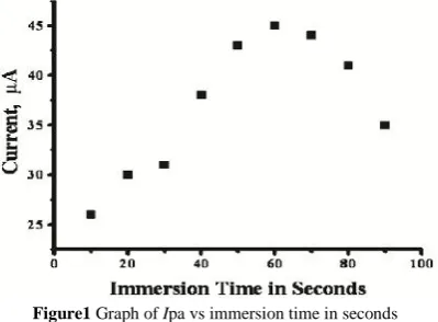
The SDS/PGE was calibrated by increasing the dipping time period from 10 to 90 seconds. The graph of anodic peak current of dopamine vs time in seconds was plotted (Figure 1). The experimental result reveals that the higher anodic peak current was obtained at 60 seconds dipping time. This shows that the accumulation of SDS on the surface of PGE was better at 60 seconds time and shows the electrochemical sensor activity.
Electrocatalytic response of DA at SDS/PGE
The cyclic voltammogram for DA was recorded at both bare PGE and SDS/PGE. DA being an easily oxidizable catecholamine, showed the voltammogram with supporting electrolyte 0.2 M phosphate buffer solution of pH = 7 at 50
mVs-1scan rate for bare PGE. Figure 2 showed a pair of one anodic peak potential at 215 mV and cathodic peak potential at 125 mV (vs SCE) for 10 µM DA at bare PGE (solid line). The separation of redox peaks (Ep) was found to be 90 mV
and the ratio of redox peak current (Ipa/Ipc) was 1.25. However, at the SDS/PGE, one anodic peak (Epa1) and two
(Epc1and Epc2) cathodic peaks were obtained for first cycle
with significant increase in both Ipa and Ipc (dashed line). To check whether the redox peak appeared for modifier or for presence of DA, we recorded the cyclic voltammogram only for phosphate buffer (dotted line).
The result shows there are no peaks obtained for the modifier. Hence the obtained redox peaks was due to the redox behavior of DA. In forward scan the DA (A) oxidized to dopamine-o-quinone (B) and the Epa1was located at 195 mV
and Epc1was found to be 160 mV. TheEp(Epa1–Epc1) was
found to be 35mV and the ratio (Ipa/Ipc) was 1.02 and
voltammogram was reversible.
The shifting of Epa towards less positive side, Epc1towards
more positive side and enhancement in redox peak currents showed the electrocatalytical activity of SDS/PGE towards oxidation of DA. The probable mechanism could be explained as; the PGE surface was uniformly covered by the anionic Figure1 Graph of Ipa vs immersion time in seconds
Scheme 1 Oxidation mechanism of DA at SDS/PGE surface Figure 2 Cyclic voltammogram of 10 µM DA in 0.2 M phosphate
buffer solution at bare PGE (solid line), at SDS/PGE (dashed line) for first cycle and at SDS/PGE (hallow circle) for second
cycle at scan rate 50 mVs-1. In the absence of DA
surfactant. These anions have well affinity towards the cationic moiety of DA and develop electrostatic attraction with the DA (Scheme 1). This made the fast electron transfer between the DA molecule and SDS/PGE, hence decrease in the over potential and increase in the current signals. The voltammogram of DA was shown another reduction peak (Epc2) was at -250 mV. This reduction peak was due to the
electron deficient oxygen atom and electron rich nitrogen atoms are present in oxidized DA molecule. When the nitrogen atom becomes deprotonated, there could be the possibilities of formation of cyclization and the obtained reduced product could be known as lucodopachrome (C). By applying the second scan a new oxidation peak (Epa2) could
be observed at around 180 mV. This peak is generated because of the oxidation of lucodopachrome to dopachrome (D) (Forzani et al., 1997, Ciszewski et al., 1999, Zhang et al., 2004, Zhang et al., 2004). The redox mechanism equations of DA were shown in Scheme 2.
To establish the existence of Epc2 and Epa2, we successive
given 10 multiple cycles were recorded. From figure 3, it was found that Epc2and Epa2still existed strongly and there is no
shift in redox peaks potentials. The current responses of all peaks were shown constant. The all redox peaks were obtained because of the DA and not for any other impurities.
The effect of scan rate on the peak potential of dopamine
Figure 4 shows the cyclic voltammogram recorded for DA at different scan rate at SDS/PGE. The scan rate has a great influence on the peak current of DA on SDS/PGE. The voltammograms showed increased in current signals with increase in scan rate from 50 to 400 mVs-1. The graph of current against scan rate was plotted. The graph shows the anodic and cathodic peak currents were directly proportional to the scan rate () in the range of 50–400 mVs−1.
The correlation coefficient obtained for anodic and cathodic peak current were 0.9984 and 0.9963, respectively (Figure 5). The difference between the anodic peak potential and the
cathodic peak potential (ΔEp) was increasing with the scan
rate. The results indicate that the electron transfer reaction was controlled by the adsorption of DA on the SDS/PGE electrode surface (Teo et al., 2014).
Effect of concentration of DA
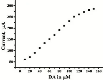
The electrocatalytic oxidation of DA was carried out by varying its concentration at SDS/PGE By increasing the concentration of DA from 10 to 150 µM, the anodic peak current was increasing. The graph of Ipa vs concentration of DA was plotted was shown in Figure 6.
The graph obtained linearly increase in peak current with increase in the DA concentration and Ipa is proportional to concentration of DA up to 110 µM and this confirms the saturation limit of DA detection at SDS/PGE. Above this concentration the anodic peak current shows almost constant in response because of kinetics limitation above the 110 µM (Teo et al., 2014). The correlation coefficient was found to be Scheme 2 Redox mechanism of DA
Figure 3 Repeated cyclic voltammograms (10 multiple scans) of 10 µM DA in PBS of pH 7.4 at SDS/PGE at scan rate 50 mVs-1
Figure 4 Cyclic voltammogram of 10 µM DA at different scan rate at SDS/PGE in 0.2 M phosphate buffer solution at pH 7.4 (a–h;
50, 100, 150, 200, 250, 300, 350 and 400 mVs-1)
Figure 5 Graph of redox peak current vs scan rate()
(r2) = 0.9999 for the DA concentration from 10 µM to 110 µM. The limit of detection (LOD) and limit of quantification (LOQ) were calculated by using the formula (1) and (2) respectively (Chandra et al., 2010). The LOD and LOQ were found to be 0.1 µM and 0.333 µM respectively. These experimentally obtained results were compared with other previously reported literatures and shown in table 1 (Sun et
al., 2007, Yixin et al., 2006, Wang et al., 2002, Fooladsaz et al., 2012, Plowman et al., 2010, Fan et al., 2011, Tsai et al.,
2011, Ma et al., 2012, Liu et al., 2012).
LOD = 3S/M (1)
LOQ = 10S/M (2)
Effect of pH on voltammetry of DA
The effect of pH plays an important role in the electrochemical response of DA at SDS/PGE. The voltammogram of DA was recorded at 0.2 M phosphate buffer solutions at different pH by using cyclic voltammetric technique. The graph current vs pH (Figure 7) showed that, the redox peak current was increased from 3.4 to 6.4 pH after that, the current was decreased for increase in pH. The pH = 7.4 was chosen for the studies because of the interest in physiological pH.
Both anodic and cathodic peak potentials were shifted to less positive side with increasing in the pH values. The Ep of
DA shifted from 300 to 120 mV by increasing the pH from 3.4 to 10.4. The potential diagram was constructed by plotting the graph of calculated E0 vs pH of the solution (Figure 8). The graph shows good linearity with a slope of 62 mV/pH.
Simultaneous determination of DA and AA
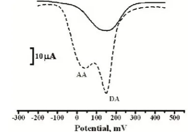
The Figure 9 showed the differential pulse voltammograms for solution containing mixture of 10 µM DA and 100 µM AA in 0.2 M phosphate buffer solution at pH = 7.4 at both bare PGE and SDS/PGE. The bare PGE (solid line) shows only one broad less sensible anodic peak for the analyte mixture. The SDS/PGE was able to separate the defined oxidation peaks of DA and AA by showing two anodic peaks (dashed line) with significantly enhanced current responses. The electrochemical response of DA and AA showed their anodic peak potentials at 150 mV and 40 mV. The peak to peak separation of DA-AA was found to be 110 mV. This potential difference was large enough to identify the DA in presence of high concentration of AA.
Interference study
The simultaneous determination of DA and AA in the mixture was carried out at SDS/PGE when concentration of one species changed, whereas the other remained constant. From the figure 10, it can be seen that the peak current of DA was proportional to its concentration, which was increased from 10 to 100 µM where AA concentration 100 µM constant.
Table 1 Comparison of different modified electrode
for DA determination
Electrode Detection limit
(µmol/L) Method Reference
IL-CPE 0.7 CV Sun et al., 2007
TA SAM/Au 0.5 DPV Yixin et al., 2006
Metallothioneins self
assembled gold electrode 6 CV Wang et al., 2002
CAT /ZnONps/CPE 3 CA, CPA Fooladsaz et al., 2012 Nanostructured gold 5 DPV Plowman et al., 2010 TiO2–graphene/GCE 2 CV Fan et al., 2011 2-Amino-thiazol (AT)
film/GCE 5 DPV Tsai et al., 2011 Graphene/GCE 0.5 CV Ma et al., 2012
CTAB/GNSP 0.6 DPV Liu et al., 2012 SDS/PGE 0.1 DPV This work
Figure 7 Graph of Ipa vs pH
Figure 8 Graph of E0(mV) vs pH
Figure 9 Differential pulse voltammogram for solution containing mixture of 10 µM DA, and 100 µM AA at bare PGE (solid line) and at
SDS/PGE (dashed line) in 0.2 M phosphate buffer solution of pH 7.4 at scan rate of 20 mVs-1
Figure 10 Differential pulse voltammogram of (a) 10 µM, (b) 20 µM, (c) 40 µM, (d) 60 µM, (e) 80 µM and (f) 100 µM DA in 0.2M phosphate buffer solution of pH 7.4 in the presence of
There were no change in the response of peak current and peak potential occurred for AA. Similarly in the figure 11 self explains the concentration effect of AA from 10 to 300 µM. These results shows that the DA and AA were exist independently in their mixtures of samples. The simultaneous study was carried out with other incorporating substances like serotonin and uric acid (Figure 12 and Figure 13). The peak to peak separation of DA with other incorporating agents and their detection limit were tabulated in table 2.
Analytical Application
The modified electrode was applied to the determination of dopamine hydrochloride injection. The DA injection sample purchased from Sterile Specialities India Private Ltd., with a specified content of DA of 40.0 mg/mL. The sample was used after suitable dilution and 0.2 M phosphate buffer was used for diluting the injection samples. The results were shown in table 3. The recovery present and R.S.D. were shown good results and the proposed methods could be efficiently used for the determination of DA in injections with recovery in the range 98.00-100.10%.
CONCLUSION
The SDS/PGE was shown strong electrocatalytic activity towards the detection of DA by enhanced both anodic and cathodic peak currents strongly and by reducing the over potential which was occurred at bare PGE. The increase in the concentration of DA results in significant enhancement of electrochemical oxidation peak current at certain concentration. The electrochemical process was found to be adsorption - controlled. The modification showed highly selective and electrocatalytic activity towards the oxidation of DA in the presence of AA and other incorporating agents. Hence, SDS/PGE acts as a good sensor for the detection of DA with detection limit and quantification limit were found to be 0.1 µM and 0.333 µM respectively. This modified electrode can be used for investigation of other neurotransmitters.
Acknowledgment
The author, Mahanthesha K. R. is grateful to the University Grants Commission, New Delhi, INDIA for awarding him a SC/ST post-doctoral fellowship. UGC-Award Letter No. F. /PDFSS-2013-14-ST-KAR-5645, Dated: 20-06-2014.
References
Adams, R. N., Probing brain chemistry with electroanalytical techniques. 1976. Anal. Chem., 48 (14): 1126A- 1138A. Chandra, U., Kumara Swamy, B. E., and Gilbert O. 2010.
Poly (amaranth) film based sensor for resolution of dopamine in the presence of uric acid: a voltammetric study. Chinese Chem. Let., 21 (12): 1490-1492.
Chandra, U., Kumara Swamy, B. E., Gilbert, O., and Sherigara, B. S. 2010. Voltammetric resolution of dopamine in the presence of ascorbic acid and uric acid at poly (calmagite) film coated carbon paste electrode. Electrochim. Acta. 55 (24): 7166-7174.
Chandra, U., Kumara Swamy, B. E., Gilbert, O., and Sherigara, B. S. 2010. Determination of dopamine in presence of ascorbic acid at eriochrome black T modified carbon paste electrode: a voltammetric study. Int. J.
Electrochem. Sci., 5 (10): 1475-1483.
Chandra, U., Kumara Swamy, B. E., Gilbert, O., Figure 11 Differential pulse voltammogram of (a) 10 µM, (b) 50 µM,
(c) 100 µM, (d) 150 µM, (e) 200 µM, (f) 250 µM and (g) 300 µ M AA/L in 0.2M phosphate buffer solution of pH 7.4 in the
presence of 10 µM DA at SDS/PGE at scan rate of 20 mVs-1
Figure 12 Differential pulse voltammogram for solution containing mixture of 10 µM DA, and 100 µM serotonine at bare PGE (solid line)
and at SDS/PGE (dashed line) in 0.2 M phosphate buffer solution of pH 7.4 at scan rate of 20 mVs-1
Figure 13 Differential pulse voltammogram for solution containing mixture of 10 µM DA, and 100 µM UA at bare PGE (solid line)
and at SDS/PGE (dashed line) in 0.2 M phosphate buffer solution of pH 7.4 at scan rate of 20 mVs-1
Table 2 Interference study of other incorporating
agent with the detection of DA
DA-Incorporating agents
Peak to peak
seperation (mV)Technique
Detection limit
mol/L DA - AA 110 DPV AA 2 DA - Uric Acid 150 DPV Uric Acid 0.9 DA - Serotonine 190 DPV Serotonine 0.5
Table 3 Detection of DA in injection samples (n = 5)
Sample Content (mg/mL) Found (mg/mL) RSD (%) Recovery (%)
1 4.0 3.92 2.8 98.00
2 4.0 3.98 2.2 99.50
Pandurangachar, M., and Sherigara, B. S. 2009. Voltammetric resolution of dopamine in presence of ascorbic acid at polyvinyl alcohol modified carbon paste electrode. Int. J. Electrochem. Sci., 4(10): 1479-1488. Ciszewski, A., and Milczarek, G. 1999. Polyeugenol-modified
platinum electrode for selective detection of dopamine in the presence of ascorbic acid. Anal. Chem., 71 (5): 1055-1061.
Demetriades, D., Economou, A., and Voulgaropoulos, A. 2004. A study of pencil-lead bismuth-film electrodes for the determination of trace metals by anodic stripping voltammetry. Anal. Chim. Acta., 519 (2): 167-172. Erdem, A., Papakonstantinou, P., and Murphy. 2006. H.
Direct DNA hybridization at disposable graphite electrodes modified with carbon nanotubes. Anal. Chem., 78 (18): 6656-6659.
Fan, Y., Lu, H.T., Liu, J.H., Yang, C.P., Jing, Q.S., Zhang, Y.X., Yang, X.K., Huang, K.J. 2011. Hydrothermal preparation and electrochemical sensing properties of TiO2-graphene nanocomposite. Colloids Surf. B, 83: 78-82.
Fooladsaz, K., Negahdary, M., Rahimi, G., Tamijani, A.H., Parsania, S., Dastjerdi, H.A., Sayad, A., Jamaleddin, A., Salahi, F., Asadi, A. 2012. Dopamine determination with a biosensor based on catalase and modified carbon paste electrode with zinc oxide nanoparticles. Int. J. Electrochem. Sci. 7: 9892-9908.
Forzani, E. S., Rivas, G. A., and Solis, V. M. 1997. Amperometric determination of dopamine on vegetal-tissue enzymatic electrodes. J. Electroanal. Chem., 435(1-2): 77-84.
Gao, W., Song, J., and Wu, N. 2005. Voltammetric behavior and square-wave voltammetric determination of trepibutone at a pencil graphite electrode. Journal of
Electroanal. Chem., 576 (1): 1-7.
Gilbert, O., Kumara Swamy, B. E., Chandra, U., and Sherigara, B. S. 2009. Simultaneous detection of dopamine and ascorbic acid using polyglycine modified carbon paste electrode: a cyclic voltammetric study. J.
Electroanal. Chem., 636 (1-2): 80-85.
Karadeniz, H., Gulmez, B., and Sahinci, F. 2003. Disposable electrochemical biosensor for the detection of the interaction between DNA and lycorine based on guanine and adenine signals. J. Pharmac. Biomed. Anal., 33( 2): 295-302.
Levent, A., Yardim, Y., and Senturk, Z., 2009. Voltammetric behavior of nicotine at pencil graphite electrode and its enhancement determination in the presence of anionic surfactant. Electrochim. Acta, 55( 1): 190-195.
Liu, S.Q., Sun, W.H., Hu, F.T. 2012. Graphene nano sheet-fabricated electrochemical sensor for the determination of dopamine in the presence of ascorbic acid using cetyltrimethylammonium bromide as the discriminating agent. Sens. Actuators B, 173: 497-504.
Ma, X.Y., Chao, M.Y., Wang, Z.X. 2012. Electrochemical detection of dopamine in the presence of epinephrine, uric acid and ascorbic acid using a graphene-modified electrode. Anal. Methods, 4: 1687-1692.
Naik, R. R., Swamy, B. E. K., Chandra, U., Niranjana, E., Sherigara, B. S., and Jayadevappa, H. 2009.Separation of ascorbic acid, dopamine and uric acid by acetone/water modified carbon paste electrode: a cyclic voltammetric study. Int. J. Electrochem. Sci., 4 (6): 855-862.
Ozsoz, M., Erdem, A., and Kerman, K. 2003. Electrochemical genosensor based on colloidal gold nanoparticles for the detection of factor V leiden mutation using disposable pencil graphite electrodes. Anal. Chem., 75(9): 2181-2187.
Plowman, B.J., Mahajan, M., O’Mullane, A.P. 2010. Bhargava, S.K. Electrochemical detection of dopamine and cytochromecat a nanostructured gold electrode. Electrochim. Acta, 55: 8953-8959.
Sun, W., Yang, M., and Jiao, K. 2007.Electrocatalytic oxidation of dopamine at an ionic liquid modified carbon paste electrode and its analytical application. Anal.
Bioanal. Chem., 389 (4): 1283-1291.
Teo, P., S., Alagarsamy, P., Huang N., M., Lim H. N., and Yusran S. 2014. Simultaneous Electrochemical Detection of Dopamine and Ascorbic Acid Using an Iron Oxide/Reduced Graphene Oxide Modified Glassy Carbon Electrode. Sensors. 14: 15227-15243.
Tsai, T.H., Huang, Y.C., Chen, S.M., Ali, M.A., Al Hemaid, F.M.A. 2011. Fabrication of multifunctional biosensor for the determination of hydrogen peroxide, dopamine and uric acid. Int. J. Electrochem. Sci., 6: 6456-6468.
Vestergaard, M., Kerman, K., and Tamiya, E. 2005. An electrochemical approach for detecting copper-chelating properties of flavonoids using disposable pencil graphite electrodes: possible implications in copper-mediated illnesses. Anal. Chim. Acta, 538 (1-2): 273-281.
Wang, J., Kawde, A. N., and Sahlin, E. 2000.Renewable pencil electrodes for highly sensitive stripping potentiometric measurements of DNA and RNA. Analyst, 125 (1): 5-7.
Wang, Q., Li, N., and Wang, W. 2002. Electrocatalytic response of dopamine at a metallothioneins self-assembled gold electrode. Anal. Sci., 18 (6): 635-639.
Yixin S., and Wang, S. F. 2006.Simultaneous determination of dopamine and ascorbic acid at a triazole self-assembled monolayer-modified gold electrode. Microchim. Acta, 154(1-2): 115-121.
Zhang, Y., Jin, G., Cheng, W., and Li, S. 2005.Poly (O-aminobenzoic acid) modified glassy carbon electrode for electrochemical detection of dopamine in the presence of ascorbic acid. Front. Biosci., 10(1): 23-29.
Zhang, Y., Jin, G., Yang, Z., and Zhao, H. 2004. Determination of dopamine in the presence of ascorbic acid using a poly(amidosulfonic acid) modified glassy carbon electrode. Microchim. Acta, 147 (4): 225-230.