Celiac Disease and Anorexia
Nervosa: A Nationwide Study
Karl Mårild, MD, PhD, a, b Ketil Størdal, MD, PhD, a, c Cynthia M. Bulik, PhD, d, e, f Marian Rewers, MD, PhD, b Anders Ekbom, MD, PhD, g Edwin Liu, MD, b Jonas F. Ludvigsson, MD, PhDd, h, i, j
abstract
BACKGROUND AND OBJECTIVE: Previous research suggests an association of celiac disease (CD) with
anorexia nervosa (AN), but data are mostly limited to case reports. We aimed to determine whether CD is associated with the diagnosis of AN.
METHODS: Register-based cohort and case-control study including women with CD (n =
17 959) and sex- and age-matched population-based controls (n = 89 379). CD (villous atrophy) was identified through the histopathology records of Sweden’s 28 pathology departments. Inpatient and hospital-based outpatient records were used to identify AN. Hazard ratios for incident AN diagnosis were estimated by using stratified Cox regression with CD diagnosis as a time-dependent exposure variable. In the secondary analyses, we used conditional logistic regression to estimate odds ratios for being diagnosed with AN before CD.
RESULTS: Median age of CD diagnosis was 28 years. During 1 174 401 person-years of
follow-up, 54 patients with CD were diagnosed with AN (27/100 000 person-years)
compared with 180 matched controls (18/100 000 person-years). The hazard ratio for later AN was 1.46 (95% confidence interval [CI], 1.08–1.98) and 1.31 beyond the first year after CD diagnosis (95% CI, 0.95–1.81). A previous AN diagnosis was also associated with CD (odds ratio, 2.18; 95% CI, 1.45–3.29). Estimates remained largely unchanged when adjusted for socioeconomic characteristics and type 1 diabetes.
CONCLUSIONS: The bidirectional association between AN diagnosis and CD warrants attention
in the initial assessment and follow-up of these conditions because underdiagnosis and misdiagnosis of these disorders likely cause protracted and unnecessary morbidity.
aDivision of Epidemiology, Norwegian Institute of Public Health, Oslo, Norway; bBarbara Davis Center, University
of Colorado, Aurora, Colorado; cDepartment of Pediatrics, Østfold Hospital Trust, Grålum, Norway; dDepartment
of Medical Epidemiology and Biostatistics and gClinical Epidemiology Unit, Department of Medicine Solna,
Karolinska Institutet, Stockholm, Sweden; Departments of ePsychiatry and fNutrition, University of North
Carolina at Chapel Hill, Chapel Hill, North Carolina; hDepartment of Pediatrics, Örebro University Hospital,
Örebro University, Örebro, Sweden; iDivision of Epidemiology and Public Health, School of Medicine, University
of Nottingham, Nottingham, United Kingdom; and jDepartment of Medicine, Columbia University College of
Physicians and Surgeons, New York, New York
Dr Mårild drafted the initial manuscript and carried out the initial analyses; Dr Ludvigsson conceptualized and designed the study, reviewed and revised the manuscript, and supervised data analyses; Drs Størdal, Rewers, Bulik, Ekbom, and Liu contributed in interpretation of the data and critically reviewed and revised the manuscript; and all authors approved the final manuscript as submitted.
DOI: 10.1542/peds.2016-4367 Accepted for publication Feb 8, 2017
Address correspondence to Karl Mårild, MD, PhD, Barbara Davis Center, University of Colorado, 1775 Aurora Ct, Aurora, CO 80045. E-mail: karlmarild@gmail.com
PEDIATRICS (ISSN Numbers: Print, 0031-4005; Online, 1098-4275).
To cite: Mårild K, Størdal K, Bulik CM, et al. Celiac Disease and Anorexia Nervosa: A Nationwide Study. Pediatrics. 2017;139(5):e20164367
WhaT’s KnOWn On ThIs subjecT: Case reports suggest an association of celiac disease with anorexia nervosa, but there are few large-scale studies. This is additionally complicated by the clinical similarities between the illnesses that may lead to misclassification, delay in diagnosis, and improper treatment.
Celiac disease (CD) is an inflammatory disorder in which gluten ingestion causes small-intestinal villous atrophy; the treatment consists of a lifelong strict adherence to a gluten-free diet.1
The disease occurs in 1% to 2% of the Western population2 and is
more prevalent in women. Common presenting symptoms include gastrointestinal complaints, loss of appetite, fatigue, and, in children, stunted growth and pubertal delay.3 There are also numerous
extraintestinal manifestations of CD, including psychiatric comorbidity.4
Anorexia nervosa (AN) is an eating disorder associated with underweight, fear of weight gain, and disturbance in the way in which one’s body weight or shape is experienced, undue influence of body weight on self-evaluation, or persistent lack of recognition of the seriousness of the low weight.5 The disorder typically
affects girls during adolescence and young adulthood and is associated with elevated psychiatric and somatic morbidity and mortality.6 Diseases
with dietary constraints, such as food allergy and type 1 diabetes, 7, 8
have been associated with AN. It is conceivable that a restrictive diet, in susceptible individuals, may trigger obsessive eating patterns; for others, the diet may cause a prolonged negative energy balance that could be the tipping point for developing AN.9 Previous research
has indicated that AN may be linked to CD, but data have mostly been limited to case reports.10–16 The only
earlier population-based study in this field found CD to be associated with a significant two-to-threefold increased diagnosis of AN and vice versa.17 However, that study was
restricted to patients requiring hospital admission for CD and may therefore have included patients with a high degree of comorbidity, which may have caused exaggerated risk estimates.
The primary aim of this nationwide study was to test the hypothesis that individuals with biopsy-verified CD were at increased risk of AN before or after CD diagnosis. Our secondary aim was to examine if any observed association with AN diagnosis was specific to CD or also present in patients with milder small-intestinal histopathology. We therefore examined the association of diagnosed AN also among biopsied individuals without CD.
MeThODs
Using the personal identity number18
assigned to all Swedish residents, we linked histopathology data on individuals undergoing small-intestinal biopsy to the National Patient Register19 to determine their
risk for AN diagnosis compared with population-based matched controls.
study sample
Individuals with CD, defined as the presence of small-intestinal villous atrophy (Marsh grade III), 20 were
identified by using the computerized histopathology records of Sweden’s 28 pathology departments; an earlier evaluation has shown that 95% of Swedish individuals with villous atrophy have a clinical CD diagnosis.21 The time of diagnosis
was defined as the time of small-intestinal biopsy, ranging from 1969 to 2008.18
We also identified individuals undergoing biopsy showing small-intestinal inflammation (Marsh grade I–II) or normal mucosa (Marsh grade 0) but with a positive CD serology test (antigliadin, antiendomysial, or transglutaminase antibodies).22 Data
on CD serology originated from 8 university hospitals providing care for approximately half of the Swedish population.22
By using the same dataset as previously reported, 23 this study
restricted participation to women
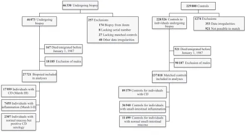
living in Sweden in 1987 and later: 17 959 with CD, 7455 with small-intestinal inflammation, and 2307 women with normal small-intestinal mucosa, but positive CD serology. Through the government agency Statistics Sweden, each individual undergoing biopsy was matched by sex, age, calendar period of birth, and county of residence with up to 5 controls from the general population (n = 137 818) (Fig 1). Our main analyses were restricted to women to eliminate wrongful AN diagnoses in men.
anorexia nervosa
Diagnosis of AN was defined24, 25 by
any of the following International Classification of Diseases, Eighth Revision (ICD-8), International Classification of Diseases, Ninth Revision (ICD-9), and International Classification of Diseases, Tenth Revision (ICD-10) codes recorded in the National Patient Register: 8, 306.50; 9, 307B; ICD-10, F50.0 and F50.1. To eliminate diagnostic misclassification from eating-related disorders of young children, we restricted AN to diagnoses recorded at age 6 years or later.26 The National Patient Register
started in 1964, became nationwide in 1987, and includes hospital-based outpatient care since 2001.19 The
date of diagnosis was determined by the first appearance of an AN diagnosis.
covariates
We obtained data from Statistics Sweden on the highest attained education level and socioeconomic status until the end of 2009; for children, we used the highest category attained by their parents. Socioeconomic status was defined by occupational status according to the European Socioeconomic Classification system.27 We also
30 years (ICD-8/ICD-9, 250; ICD-10, E10). We used an age cutoff because ICD-8 and ICD-9 do not distinguish between type 1 and type 2 diabetes. Covariates are categorized as shown in Table 1.
statistical analyses
The associations of the diagnosis of AN were estimated by using Cox regression and logistic regression models for matched data.
Cox Regression: Subsequent Diagnosis of AN After CD
We used stratified Cox proportional-hazards regression, with age in days as the time metric, to estimate hazard ratios (HRs) for incident diagnosis of AN with associated 95% confidence intervals (CIs). The HRs were modeled separately within each stratum consisting of 1 woman undergoing biopsy and up to 5 matched controls; this stratumwise analysis eliminates the effect of the matching variables (ie, age, sex, calendar period of birth, and county of residence). Individuals were followed from January 1, 1987, or birth for children born thereafter, until AN diagnosis, emigration, death, or December 31, 2009, whichever occurred first. The follow-up started in 1987, when the National Patient Register became nationwide, for all participants at risk for AN. We excluded 73 individuals diagnosed with AN before 1987 (start of follow-up) and 319 with a record FIGuRe 1
Flowchart illustrating the formation of the study sample. Individuals undergoing small-intestinal biopsy were identified by using the computerized histopathology records of Sweden’s pathology departments. The study sample was restricted to women living in Sweden in 1987 (when the National Patient Register became nationwide) and later.
TabLe 1 Characteristics of Women Undergoing Small-Intestinal Biopsy
CD Inflammation Normal Mucosa With
Positive CD Serology
Total 17 959 7455 2307
Year of birth, median (interquartile range)
1970 (1945–1989) 1951 (1933–1969) 1963 (1947–1978)
Age (y) at time of biopsy
Median (interquartile range) 28 (6–52) 46 (31–63) 38 (23–53)
Age 0–19, n (%) 7428 (41) 717 (10) 459 (20)
Age 20–39, n (%) 3649 (20) 2196 (29) 755 (33)
Age 40–59, n (%) 3862 (22) 2304 (31) 740 (32)
Age 60+, n (%) 3020 (17) 2238 (30) 353 (15)
Calendar year at time of biopsy
Median (interquartile range) 1999 (1993–2003) 2000 (1994–2004) 2001 (1998–2004)
≤1986, n (%) 1210 (7) 464 (6) 0 (0)
1987–1999, n (%) 8601 (48) 3238 (43) 819 (36)
≥2000, n (%) 8148 (45) 3753 (50) 1488 (64)
Type 1 diabetesa, n (%) 526 (3) 59 (1) 35 (2)
Histopathology findings indicating CD (villous atrophy, Marsh III), inflammation (Marsh I–II), and normal mucosa (Marsh 0), but with positive CD serology 180 days before and until 30 days after biopsy (antigliadin, antiendomysial, or antitransglutaminase antibodies). Controls are not included in the table because their age, sex, and calendar year distributions were identical to those of the individuals undergoing biopsy (due to matching).
of AN before undergoing biopsy (or corresponding date among controls). Hence, the Cox regression analyses included 17 924 individuals with CD, 7425 with inflammation, 2294 with normal mucosa but positive CD serology, and 137 504 matched controls.
The proportional-hazards assumption was found to be valid based on graphical presentation of the data. The time of biopsy (and corresponding date for matched controls) was included as a time-dependent variable; individuals with CD, inflammation, or normal mucosa but positive CD serology were defined as exposed from time of biopsy and thereafter considered exposed throughout follow-up. Individuals biopsied before January 1, 1987 were considered to be exposed from the start of the follow-up period. Hazard ratios were adjusted for education level, socioeconomic status, and type 1 diabetes.
For individuals with CD and their controls, we conducted separate analyses for children (age ≤19 years) and adults and by calendar period of biopsy (≤1994 and ≥1995). The HR for AN beyond the first year of CD diagnosis was modeled by introducing a 1-year lag time during which an individual was considered unexposed until 1 year after diagnosis and exposed thereafter. To test the ascertainment of AN, we performed a separate analysis restricted to AN listed as the main diagnosis in the National Patient Register.
Logistic Regression: Diagnosis of AN Before CD
We used conditional logistic regression to estimate odds ratios (ORs) for previous diagnosis of AN among individuals undergoing biopsy compared with their matched controls.28 Analyses
were performed stratumwise and therefore conditioned on age, sex,
calendar period of birth, and county of residence. In parallel with the above, we performed adjusted analyses, accounting for education level, socioeconomic status, and type 1 diabetes. In individuals with CD and their controls, we also performed analyses stratified by age and calendar year of birth. In a post hoc analysis, we also examined AN and CD in men (CD,
n = 11 025; controls, n = 54 774). Statistical significance was defined as 95% CIs for HRs and ORs not including 1.00. Data were analyzed by using Stata version 14 (Stata Corp, College Station, TX).
ethics
This study was approved by the Regional Ethical Vetting Board in Stockholm, Sweden, which deemed that individual informed consent was not required.29 Data used in this
study were deidentified.
ResuLTs
Among the 17 959 women with CD in this study, the median age at CD diagnosis was 28 years (interquartile range, 6–52 years) (see Table 1 for population characteristics). There were 353 individuals diagnosed with AN at a median age of 17 years (interquartile range, 15–22 years).
cD and Future an Diagnosis
From 1987 through 2009, during 1 174 401 person-years of follow-up, 54 patients with CD were diagnosed with AN compared with 180 matched controls. The incidence rate for AN after a CD diagnosis was 27/100 000 person-years (95% CI, 21–36) compared with 18/100 000 person-years (95% CI, 16–21) among matched controls; this corresponded to an HR for AN diagnosis of 1.46 (95% CI, 1.08–1.98). Adjustments for education level, socioeconomic status, and type 1 diabetes yielded a largely unchanged HR (Fig 2). Analyses stratified by age at CD
diagnosis and by calendar period of biopsy are presented in Fig 2. Among women with a main diagnosis of AN in the National Patient Register, the HR was 1.34 (95% CI, 0.97–1.86).
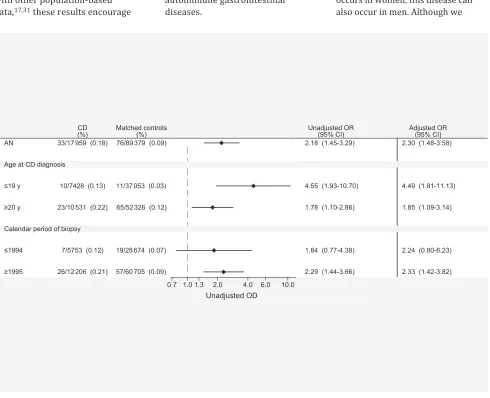
Diagnosis of an before cD
A total of 33 individuals with CD (0.18%) and 76 matched controls (0.09%) had a record of AN before CD diagnosis (and corresponding date among controls); the OR for a previous diagnosis of AN was 2.18 (95% CI, 1.45–3.29) among individuals diagnosed with CD compared with controls. There were significantly increased ORs for a previous history of AN when stratifying by age at CD diagnosis and calendar period of diagnosis (Fig 3). When restricting our analysis to those with AN listed as the main diagnosis in the National Patient Register, the OR was 2.13 (95% CI, 1.37–3.32).
an in biopsied Patients Without cD
In secondary analyses, we tested the association of AN with (1) small-intestinal inflammation without villous atrophy and (2) normal small-intestinal mucosa, but with positive CD serology. Both groups had a significantly increased risk for diagnosis of AN before as well as after the time of biopsy compared with matched controls (Tables 2 and 3).
Post hoc analysis: an and cD in Men
There was no significantly increased risk for subsequent AN among males with CD (HR, 0.95; 95% CI, 0.21–4.32). This HR was based on 12 men being diagnosed with AN. Nor did we find a significantly increased OR for previous AN among men with CD (OR, 2.50; 95% CI, 0.63–10.00).
DIscussIOn
CD diagnosis. This bidirectional association should encourage physicians to closely monitor these patients and calls for heightened understanding of factors that contribute to their cooccurrence.
Potential Mechanisms of action
We propose that a number of factors may contribute to this bidirectional association.
First, on initial examination, individuals with underlying CD or AN may be misdiagnosed with the other condition. In patients who receive an initial erroneous CD diagnosis, it should soon become evident that the gluten-free diet is not effective and additional
evaluation would reveal the eating disorder. In patients who receive an initial erroneous diagnosis of AN and who present with ongoing symptoms of fatigue and abdominal complaints, additional workups should be initiated to rule out CD or other gastrointestinal illnesses. Such persistent symptoms should be followed up because untreated CD leads to complications and a poorer quality of life.4
Second, we cannot rule out that our results have been influenced by surveillance bias. Before and after the time of diagnosis, individuals with either CD or AN are typically more extensively investigated for other diseases compared with individuals in the general
population. For example, diarrhea due to laxative misuse in AN as well as liver enzyme abnormalities can both lead to a work-up for CD. The positive associations found between AN and small-intestinal inflammation but no villous atrophy and with having a biopsy with normal mucosa but positive CD serology strongly indicate that surveillance bias at least partly plays a role here because, in Sweden, a gluten-free diet is not typically recommended for these conditions.
Third, shared risk factors, including genetic susceptibility, may play a role.30 Recent genomewide
association studies of AN have indicated genetic regions shared FIGuRe 2
with type 1 diabetes and other autoimmune illnesses. Together with other population-based data, 17, 31 these results encourage
a closer examination of the genetic relationship between AN and autoimmune gastrointestinal diseases.
clinical Relevance
The positive association of CD with AN, both before and after CD diagnosis, should spur a careful initial assessment and follow-up of these illnesses. It should also be appreciated that the 2 conditions can complicate each other. Having AN makes it harder to follow a gluten-free diet, and it cannot be excluded that some AN patients knowingly consume gluten-containing products to lose weight.11 The treatment of AN and
CD require different competences and a multidisciplinary approach to management is important.
Although the vast majority of AN occurs in women, this disease can also occur in men. Although we
FIGuRe 3
OR for AN before CD diagnosis. Participants were matched for age, sex, calendar period of birth, and county of residence. Adjusted analyses accounted for education level, socioeconomic status, and type 1 diabetes.
TabLe 2 Association of Future AN Diagnosis Among Biopsied Women Without CD HR for Future ANa
No. of Incident AN
Incidence Rate of AN per 10 000
PYR
Unadjusted HR for Future AN (95% CI)
Adjustedb HR for Future AN (95% CI)
Matched controls 20 6 (4–9) Ref Ref
Inflammationc 9 13 (7–25) 2.12 (0.97–4.67) 1.41 (0.55–3.58)
Matched controls 19 20 (13–31) Ref Ref
Normal mucosa but CD+ serologyd
9 47 (24–90) 2.45 (1.10–5.45) 2.59 (1.10–6.10)
PYR, person-years; Ref, reference.
a HRs were estimated by using stratified Cox proportional-hazard regression and ORs were estimated with conditional
logistic regression. Controls were matched for age, sex, calendar period of birth, and county of residence.
b Adjusted analyses accounting for education level, socioeconomic status, and type 1 diabetes. c Small-intestinal inflammation without villous atrophy (Marsh I–II).
d Normal mucosa (Marsh 0) but with positive CD serology 180 days before and until 30 days after biopsy (antigliadin,
found no significant association between AN and CD in men, this does not mean that the risk of CD is different in women and men with AN. Our male-restricted analysis had low statistical power, and we urge caution when interpreting those findings.
comparison With earlier Literature
Research on CD and AN almost exclusively consists of case-series.10–15
The largest case series so far was published by Leffler et al11 in 2007
and included 10 female patients with CD and eating disorders. All patients in this study were first diagnosed with AN or bulimia nervosa and only later with CD. Only 1 previous study by Basso et al16 specifically screened for
CD in patients with AN; 1 out of 177 (0.6%) screened patients with AN had biopsy-verified CD, but this study was unable to calculate any relative risk estimates because it had no control group, nor did the investigators examine the temporality between CD and AN.
Recently, however, an English register-based study, including hospitalized AN patients, found a significant threefold increased risk of later CD diagnosis and a twofold risk increase for AN in CD patients.17
Although that study identified patients requiring hospital admission for CD, our study includes both in- and outpatients with CD. This distinction may be important because inpatient registers are often limited to patients
with more severe CD and more susceptible to surveillance bias, which could overinflate risk estimates.
strengths and Limitations
The main strength of our paper is the population-based approach with almost 18 000 CD patients. The large numbers allowed us to examine subsets of patents with CD stratified by age and calendar period of diagnosis.
We used small-intestinal biopsy reports with villous atrophy to identify individuals with CD. Throughout the study period, biopsy was the gold standard for CD diagnosis, and >96% of Swedish pediatric and adult gastroenterologists performed a biopsy before assigning a CD diagnosis.21 Non-celiac causes for
villous atrophy are uncommon in Sweden, and when we examined 114 randomly selected charts of patients with villous atrophy, 108 (95%) had CD.21 In a subset of
patients with available data at the time of CD diagnosis, 88% were positive for either transglutaminase, endomysium, or antigliadin
antibodies. Of note, a large
proportion of patients in our sample were diagnosed when gliadin was the only available antibody and before transglutaminase and endomysium antibodies were readily available in the clinic.
Although we are unaware of any validation of AN as registered in the
National Patient Register (and we did not have access to medical records to verify the AN diagnoses), the overall validity of this register is high with 85% to 95% of diagnoses being correct.19 To increase the specificity
of the AN diagnosis, we carried out a sensitivity analysis restricting our outcome to individuals with AN listed as the main diagnosis. This analysis also showed a positive association, although the HR was somewhat lower (1.34 vs 1.46 for our main analysis) and was not statistically significant.
This study has some limitations. We did not screen for undiagnosed CD or subthreshold AN. It is therefore likely that some individuals with long-standing eating disturbances were first screened for CD, and then subsequently diagnosed with AN only when the gluten-free diet did not help. A related limitation is our lack of clinical data on symptoms or signs that would enable us to differentiate between subtypes of AN or differences in CD presentation. Moreover, in patients with an initial diagnosis of AN followed by a subsequent diagnosis of CD, it is also possible that the AN diagnosis may represent a misdiagnosis of CD.
In our earlier validation study of CD, 21 1 in 3 patients with CD
suffered from weight loss or growth failure, which are symptoms resembling AN. It is not likely that the association is due to gastrointestinal changes in AN patients being misinterpreted as CD on biopsy. Not even AN with severe underweight has demonstrated mucosal atrophy.10, 32
In addition, data on ethnicity and diet were lacking. The latter prevented us from studying whether strict adherence to a gluten-free diet mediated the association with AN. As part of an earlier validation study of our cohort, 71 out of 86 (83%) randomly selected patients with CD revealed dietary adherence according
TabLe 3 Association of AN Diagnosis Among Biopsied Women Without CD, Odds Ratio for Prior AN OR for Previous ANa
Undergoing Biopsy (%)
Matched Controls (%)
Unadjusted OR for Previous AN (95% CI)
Adjustedb OR for Previous AN (95% CI) Inflammationc 30/7455 (0.40) 45/36 940 (0.12) 3.35 (2.11–5.33) 3.39 (2.09–5.49) Normal mucosa
but CD+ serologyd
13/2307 (0.56) 23/11 499 (0.20) 2.86 (1.44–5.68) 3.22 (1.49–6.96)
a HRs were estimated by using stratified Cox proportional-hazard regression and ORs were estimated with conditional
logistic regression. Controls were matched for age, sex, calendar period of birth, and county of residence.
b Adjusted analyses accounting for education level, socioeconomic status, and type 1 diabetes. c Small-intestinal inflammation without villous atrophy (Marsh I–II).
d Normal mucosa (Marsh 0) but with positive CD serology 180 days before and until 30 days after biopsy (antigliadin,
to patient chart data.21 Hence, we
cannot isolate a restricted diet as the mechanism underlying the association between CD and later AN; however, given the high adherence to gluten-free diets, we speculate that dietary restrictions may trigger AN in susceptible individuals.
Finally, we cannot rule out that residual confounding factors may have influenced our results, although we accounted for important potential confounders, such as age, birth year, country of residence, education,
socioeconomic status, and type 1 diabetes.
cOncLusIOns
The bidirectional association between diagnosis of AN and CD warrants attention in the initial assessment and the follow-up of women with these illnesses. This is important because the presentation of these conditions may mimic each other and the misdiagnosis of either disorder likely causes protracted and unnecessary morbidity.
ReFeRences
1. Lebwohl B, Ludvigsson JF, Green PH. Celiac disease and non-celiac gluten sensitivity. BMJ. 2015;351:h4347 2. Walker MM, Murray JA, Ronkainen
J, et al. Detection of celiac disease and lymphocytic enteropathy by parallel serology and histopathology in a population-based study.
Gastroenterology. 2010;139(1):
112–119
3. Leffler DA, Green PH, Fasano A. Extraintestinal manifestations of coeliac disease. Nat Rev Gastroenterol
Hepatol. 2015;12(10):561–571
4. Zingone F, Swift GL, Card TR, Sanders DS, Ludvigsson JF, Bai JC. Psychological morbidity of celiac disease: a review of the literature. United European
Gastroenterol J. 2015;3(2):136–145
5. American Psychiatric Association; DSM-5 Task Force. Diagnostic and Statistical Manual of Mental Disorders:
DSM-5. 5th ed. Washington, DC:
American Psychiatric Association; 2013 6. Franko DL, Keshaviah A, Eddy KT,
et al. A longitudinal investigation of mortality in anorexia nervosa and
bulimia nervosa. Am J Psychiatry. 2013;170(8):917–925
7. Teufel M, Biedermann T, Rapps N, et al. Psychological burden of food allergy. World J Gastroenterol. 2007;13(25):3456–3465
8. Jones JM, Lawson ML, Daneman D, Olmsted MP, Rodin G. Eating disorders in adolescent females with and without type 1 diabetes: cross sectional study.
BMJ. 2000;320(7249):1563–1566 9. Bulik CM. Towards a science of
eating disorders: replacing myths with realities: the fourth Birgit Olsson lecture. Nord J Psychiatry. 2016;70(3):224–230
10. Martínez-Olmos MA, Peinó R, Prieto-Tenreiro A, et al. Intestinal absorption and pancreatic function are preserved in anorexia nervosa patients in both a severely malnourished state and after recovery. Eur Eat Disord Rev. 2013;21(3):247–251
11. Leffler DA, Dennis M, Edwards George JB, Kelly CP. The interaction between eating disorders and celiac disease: an exploration of 10
cases. Eur J Gastroenterol Hepatol. 2007;19(3):251–255
12. Yucel B, Ozbey N, Demir K, Polat A, Yager J. Eating disorders and celiac disease: a case report. Int J Eat Disord. 2006;39(6):530–532
13. Ricca V, Mannucci E, Calabrò A, Bernardo MD, Cabras PL, Rotella CM. Anorexia nervosa and celiac disease: two case reports. Int J Eat Disord. 2000;27(1):119–122
14. Karwautz A, Wagner G, Berger G, Sinnreich U, Grylli V, Huber WD. Eating pathology in adolescents with celiac disease. Psychosomatics. 2008;49(5):399–406
15. Wright K, Smith MS, Mitchell J. Organic diseases mimicking atypical eating disorders. Clin Pediatr (Phila). 1990;29(6):325–328
16. Basso MS, Zanna V, Panetta F, et al. Is the screening for celiac disease useful in anorexia nervosa? Eur J Pediatr. 2013;172(2):261–263
17. Wotton CJ, James A, Goldacre MJ. Coexistence of eating disorders and autoimmune diseases: record linkage
abbRevIaTIOns
AN: anorexia nervosa CD: celiac disease CI: confidence interval HR: hazard ratio
ICD-8: International Classification of Diseases, Eighth
Revision
ICD-9: International Classification of Diseases, Ninth Revision
ICD-10: International
Classification of Diseases, Tenth Revision
OR: odds ratio
Copyright © 2017 by the American Academy of Pediatrics
FInancIaL DIscLOsuRe: The authors have indicated they have no financial relationships relevant to this article to disclose.
FunDInG: Dr Størdal was funded through an unrestricted grant from the OAK foundation, Geneva, Switzerland. Dr Bulik acknowledges funding from the Swedish Research Council (reference number: 538-2013-8864). The funding sources did not influence any aspect of the study or approval of the manuscript and decision to submit the manuscript for publication.
POTenTIaL cOnFLIcT OF InTeResT: Dr Bulik is a grant recipient from and a consultant for Shire; the other authors have indicated they have no potential conflicts of interest to disclose.
cohort study, UK. Int J Eat Disord. 2016;49(7):663–672
18. Ludvigsson JF, Otterblad-Olausson P, Pettersson BU, Ekbom A. The Swedish personal identity number: possibilities and pitfalls in healthcare and medical research. Eur J Epidemiol. 2009;24(11):659–667
19. Ludvigsson JF, Andersson E, Ekbom A, et al. External review and validation of the Swedish national inpatient register.
BMC Public Health. 2011;11:450
20. Marsh MN. Gluten, major histocompatibility complex, and the small intestine. A molecular and immunobiologic approach to the spectrum of gluten sensitivity (‘celiac sprue’). Gastroenterology. 1992;102(1):330–354
21. Ludvigsson JF, Brandt L, Montgomery SM, Granath F, Ekbom A. Validation study of villous atrophy and small intestinal inflammation in Swedish biopsy registers. BMC Gastroenterol. 2009;9:19
22. Ludvigsson JF, Brandt L, Montgomery SM. Symptoms and signs in individuals
with serology positive for celiac disease but normal mucosa. BMC
Gastroenterol. 2009;9:57
23. Ludvigsson JF, Montgomery SM, Ekbom A, Brandt L, Granath F. Small-intestinal histopathology and mortality risk in celiac disease. JAMA. 2009;302(11):1171–1178
24. Karamanis G, Skalkidou A, Tsakonas G, et al. Cancer incidence and mortality patterns in women with anorexia nervosa. Int J Cancer. 2014;134(7):1751–1757 25. Michels KB, Ekbom A. Caloric
restriction and incidence of breast cancer. JAMA. 2004;291(10):1226–1230 26. Yao S, Kuja-Halkola R, Thornton LM,
et al. Familial liability for eating disorders and suicide attempts: evidence from a population registry in Sweden. JAMA. Psychiatry. 2016;73(3):284–291
27. Rose D, Harrison E. Social Class in Europe: An Introduction to the European Socio-Economic Classification. London, NY: Routledge; 2010
28. Rothman KJ, Greenland S, Lash TL. Modern Epidemiology. 3rd ed. Philadelphia, PA: Wolters Kluwer Health/Lippincott Williams & Wilkins; 2008
29. Ludvigsson JF, Håberg SE, Knudsen GP, et al. Ethical aspects of registry-based research in the Nordic countries. Clin
Epidemiol. 2015;7:491–508
30. Mostowy J, Montén C, Gudjonsdottir AH, et al. Shared genetic factors involved in celiac disease, type 2 diabetes and anorexia nervosa suggest common molecular pathways for chronic diseases. PLoS One. 2016;11(8):e0159593
31. Raevuori A, Haukka J, Vaarala O, et al. The increased risk for autoimmune diseases in patients with eating disorders. PLoS One. 2014;9(8):e104845 32. Crenn P, Vahedi K, Lavergne-Slove
DOI: 10.1542/peds.2016-4367 originally published online April 3, 2017;
2017;139;
Pediatrics
Liu and Jonas F. Ludvigsson
Karl Mårild, Ketil Størdal, Cynthia M. Bulik, Marian Rewers, Anders Ekbom, Edwin
Celiac Disease and Anorexia Nervosa: A Nationwide Study
Services
Updated Information &
http://pediatrics.aappublications.org/content/139/5/e20164367
including high resolution figures, can be found at:
References
http://pediatrics.aappublications.org/content/139/5/e20164367#BIBL
This article cites 29 articles, 2 of which you can access for free at:
Subspecialty Collections
http://www.aappublications.org/cgi/collection/eating_disorders_sub
Eating Disorders
http://www.aappublications.org/cgi/collection/nutrition_sub
Nutrition
http://www.aappublications.org/cgi/collection/gastroenterology_sub
Gastroenterology following collection(s):
This article, along with others on similar topics, appears in the
Permissions & Licensing
http://www.aappublications.org/site/misc/Permissions.xhtml
in its entirety can be found online at:
Information about reproducing this article in parts (figures, tables) or
Reprints
http://www.aappublications.org/site/misc/reprints.xhtml
DOI: 10.1542/peds.2016-4367 originally published online April 3, 2017;
2017;139;
Pediatrics
Liu and Jonas F. Ludvigsson
Karl Mårild, Ketil Størdal, Cynthia M. Bulik, Marian Rewers, Anders Ekbom, Edwin
Celiac Disease and Anorexia Nervosa: A Nationwide Study
http://pediatrics.aappublications.org/content/139/5/e20164367
located on the World Wide Web at:
The online version of this article, along with updated information and services, is
by the American Academy of Pediatrics. All rights reserved. Print ISSN: 1073-0397.