Themed Section: Science and Technology
Study of Effect of Etching Time and Concentration on
Microstructure of SG 400/12 Grade Ductile Cast Iron
Nilesh T. Khot*, Anil S. Morkane*, Umesh V. Borkar*, Sanket S. Maske*, Prof. B. S. Kamble#
*UG students at Department of Production Engineering, KIT’s College of Engg., Kolhapur, Maharashtra, India
# Assistant Professor atDept.of Production Engg., KIT’s College of Engg., Kolhapur, Maharashtra, India
ABSTRACT
The study of material microstructure is known as Metallography. This is very critical in estimating the material properties. Metallography can be a very useful quality control tool too. By examining the micrographs one can determine the quality of the product. Micro structural examination is generally done on a sample whose surface has been grinded or polished to a mirror-like finish. SG cast iron is characterized by nodules-like graphite distribution when seen under an optical microscope. The as-polished samples are also etched with various etchants to look at the underlying phases (e.g. ferrite, pearlite, cementite), which also have an effect on the properties of the SG cast-iron. If a casting has predominant ferrite phase, then it would be soft. Ample presence of carbide will lead to a hard material. The study reported here deals with the etching of SG cast iron (400/12 Grade) samples for short etching time (4 – 10 sec) and also for long etching time (30 sec and 60 sec). For the purpose of observing the microstructure we have used Scanning Electron Microscope (SEM). The optimal solution is taken by the use of regression analysis technique. Etching is the selective corrosion of the surface under consideration to reveal the underlying features. The purpose of etching is to reveal certain features (e.g. grain boundary and phases) which are not visible in the as-polished condition.
Keywords : Sampeling, Polishing, Etching, Microscopic examination, Nital, Picral
I. INTRODUCTION
Metallography has been described as both a science and an art. Traditionally, metallography has been the study of the microscopic structure of metals and alloys using optical metallographs, electron microscopes or other surface analysis equipment. It can be more precisely defined as the scientific discipline of observing and determining the chemical and atomic structure and spatial distribution of the constituents, inclusions or phases in metallic alloys. The surface of a metallographic specimen is prepared by various methods of grinding, polishing, and etching. After preparation, it is often analysed using optical or
electron microscopy. Using only metallographic techniques, a skilled technician can identify alloys, predict material properties and its performance and reliability. Thus metallography is used in materials development, incoming inspection, production and manufacturing control, and for failure analysis; in other words, product reliability.
II. LITERATURE REVIEW
International Journal of Scientific Research in Science and Technology (www.ijsrst.com)
248
an optimum etch at 70 sec of etching which can go up to 80 sec of etch and the concentration of Picral may be between 3% - 4%. David A. Scott (3) had observed that the grey cast iron contains 4% carbon and a little phosphorus in early 20th century. Before etching, the outlines of some of the thick graphite. T.M. Hegazy (4)
has undergone study of effect of NaOH concentration and etching duration on some properties of γ-irradiated PADC and it is revelled that bulk etching rate shows weak dependence on the etching time. David P. B. Samuelsso (6) had used digital image
correlation technique (DIC). This method is mostly used for determination of strains on a macroscopic level e.g. testing of a tensile test piece or any other test piece or component that is exposed to a load. And it is found that it is possible to investigate strain-fields on a microscopic scale, different phases of cast irons and their strain response during loading and to investigate the effect of eutectic cells and primary austenite on the strain distribution. Donald Zipperian (7) has
enlisted different types of etchants used for etching are mentioned. George Vander Voort (21) has studied
the microstructure of CI using different etchants namely Nital and Picral at various concentration and time rates. Also discussed about the how to prepare the sample specimen for metallographic study without damage.
III. EXPERIMENTAL PROCEDURE
Figure 1. Test Speciman of SG Iron grade 400/12
The SG Iron material of grade 400/12 is taken as sample under study. The initial material which in the form of rod cut into the length 10 * Ø15 mm as shown in Figure with the help Semi-automatic Hack Saw Machine.
Figure 2. Speciman samples cut of SG Iron grade 400/12 to carry out buffing operation
The surface on which the polishing operations have to be done, initially they are made free from burs and sharp edges with the help of file. Manual polishing with successively 100, 200, 300 and 400 grade sand papers are used. After the semi finishing of the specimens now they are prepared for actual metallography. To achieve the mirror like surface finish they are placed on the Buffing Machine.
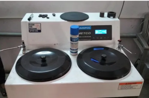
Figure 3. Buffing Machine
For this experimental study, the most commonly used Nitric acid and Picric acid were used as etchants. Nital and Picral with 2% and 4% concentration were used with acid dipping dwell period of 4,10,30,60 seconds period. The 2% Nital was prepared by adding 2 ml of concentrated Nitric acid (HNO3) in 100 ml of 99%
ethanol respectively. Every time freshly prepared etchant were used for each study.
Table .1 Experimental parameters and combination Exp.No. Etchant Etchant
Concentration
Etching Time (Sec) 1
Nital 2%
4
2 10
3 30
4 60
5
Nital 4%
4
6 10
7 30
8 60
9
Picral 2%
4
10 10
11 30
12 60
13
Picral 4%
4
14 10
15 30
16 60
After the preparation of specimen, these are dipped in the etchants of various concentrations. Then the microstructural images are observed under SEM (Scanning Electron Microscope).
(a) (b) (c) (d)
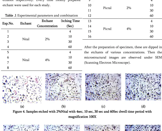
Figure 4. Samples etched with 2%Nital with 4sec, 10 sec, 30 sec and 60Sec dwell time period with magnification 100X
(a) (b) (c) (d)
Figure 5. Samples etched with 4%Nital and with 4sec, 10 sec, 30 sec and 60Sec dwell time period with magnification 100X
(a) (b) (c) (d)
International Journal of Scientific Research in Science and Technology (www.ijsrst.com)
250
(a) (b) (c) (d)
Figure 7. Samples etched with 4%Picral and with 4sec, 10 sec, 30 sec and 60Sec dwell time period with magnification 100X
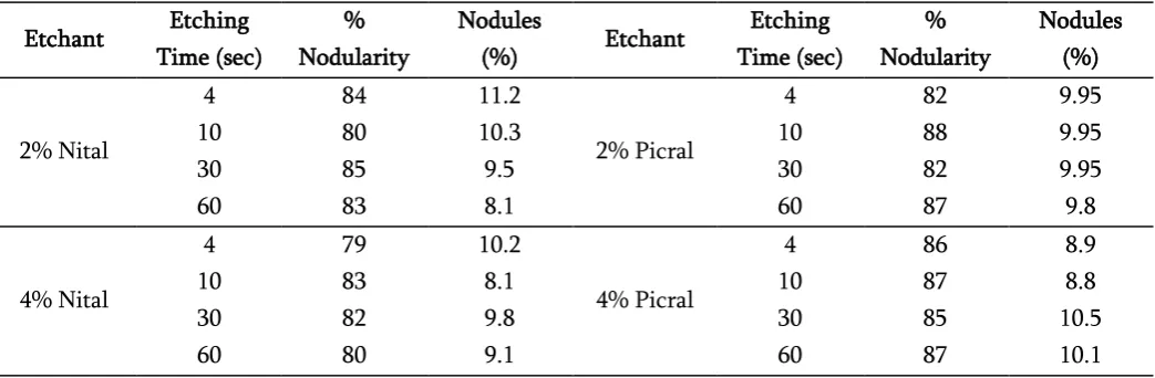
Table .2 Percentage of Nodularity and nodules observed under SEM Etchant Etching
Time (sec)
% Nodularity
Nodules
(%) Etchant
Etching Time (sec)
% Nodularity
Nodules (%)
2% Nital
4 84 11.2
2% Picral
4 82 9.95
10 80 10.3 10 88 9.95
30 85 9.5 30 82 9.95
60 83 8.1 60 87 9.8
4% Nital
4 79 10.2
4% Picral
4 86 8.9
10 83 8.1 10 87 8.8
30 82 9.8 30 85 10.5
60 80 9.1 60 87 10.1
IV. MICROSTRUCTURAL OBSERVATIONS
The magnified images from the etched samples with 2% Nitric acid are as shown in figure 4. It is clearly observed changes in the pearlite and Ferritic grain boundry visibility with an increase in the etching time from 4 sec to 60 sec. Figure 4a clearly shows signs of Insufficient etching concentration with no any clear visibility of phase as well as Ferritic grain boundry. With increase in etching holding time the microstructural contrast also increase. With 2% Nital concentration and etching holding time of 4 to 60 sec., an average of 9.7% Nodules are observed .Similarly magnified images from the etched samples with 4% Nitric acid are as shown in figure 5. Here also it is observed that there is increased contrast with increase in acid concentration and etching holding time. An average of 9.3% Nodules are observed with 4% Nital concentration and time of 4 to 60 sec. With 4% Nital
concentration and time of 30 and 60 sec there is clear visibility of Ferritic grain boundry. There is clear observation of Pearlitic matrix with 9.40% pearlite & 82.50% Ferrite.
The magnified images from the etched samples with 2% Picric acid are as shown in figure 6. There is no sign of visibility of Ternary phosphorous eutectic (carbides) in structure. With increase in etching holding time there is visible Contrast among the different phase with increased darkness. With 2 and 4% Nitric and Picric acid Ternary phosphorous eutectic (carbides) are not observed in structure.
V. CONCLUSION
nodules. With extended etching duration period of 30 sec with 4% Nitric acid gives optimum and clear microstructure visibility. Picric acid provides better results with 4% and 4 sec etching duration time.
VI. REFERENCES
[1] S. Chakraborty and S. Nadimuthu, November 2013, Study Of Etchants in Grey Cast Iron – Effect Of Etching Time, Indian Foundry Journal, volume-59, No. 11, Page No. 19-25.
[2] Cast Iron Handbook, Edited by Joseph R. Davis, ASM International.
[3] E.E.T. ELSawy,, M.R. EL-Hebeary, I.S.E. El Mahallawi, July 2017, Effect of manganese, silicon and chromium additions on microstructure and wear characteristics of grey cast iron for sugar industries applications, Volume-320.
https://doi.org/10.1016/j.wear.2017.07.007 [4] David A. Scott, Metallography and
Microstructure of Ancient and Historic Metals. [5] Chemical Etching, Donald Zipperian, Quality
Matters Newsletter,Pace Technologies, Vol. 2, Issue 5, 2003.
[6] T.M. Hegazy, M.Y. Shoeib, G.M. Hassan, September 2013, Study on the effect of NaOH concentration and etching duration on some properties of g-irradiated PADC, BENI - SUEF university journal of basic and applied sciences 2, page No. 36-40.
[7] Deborah A. Redford, Brian H. Clarkson, Mark Jensen, March 1986, The effect of different etching times on the sealant bond strength, etch depth, and pattern in primary teeth, The American Academy of Pediatric Dentistry, Vol. 8 No. 1.
[8] David P.B. Samuelsson, Analysis of microstructural strain-fields in grey cast iron, Diploma work No. 47/2011.
[9] Diploma work No. 47/2011, September 2003, Chemical Etching, PACE Technologies, Volume II, Issue 5.
[10] B.S. Motagi, Ramesh Bhosle, July 2012, Effect of Heat Treatment on Microstructure and Mechanical Properties of Medium Carbon Steel, International Journal of Engineering Research and Development, Volume 2, Issue 1, Page No. 7-13.
[11] F. Hairer, A. Karelová, C. Krempaszky, E. Werner, T. Hebesberger, A. Pichler, Etching Techniques For The Microstructural Characterization Of Complex Phase Steels By Light Microscopy, Page No. 50-54.
[12] Francesco Iacoviello, Daniela Iacoviello, Vittori Di Cocco, Alberto De Santis, Laura D Agostino, lassification of ductile cast iron specimens based on image analysis and support vector machine XXIV Italian Group of Fracture Conference, 1-3 March 2017, Urbino, Italy
[13] Jacques Lacaze, Jon Sertucha, Lena Magnusson Aberg, Microstructure of As-cast Ferritic-Pearlitic Nodular Cast Irons ISIJ 1606, ISIJ International, Vol. 56 (2016), No. 9, pp. 1606– 1615
[14] Abhijit Malage, Priti P. Rege, Manoj J. Rathod, Automatic quantitative analysis of microstructure of ductile cast iron using digital image processing Association of Metallurgical Engineers of Serbia (AMES) Scientific paper UDC: 620.18:669.13
[15] Brij Kumar Dhindaw,Effect of Processing Parameters on the Mechanical Properties of Heavy Section Ductile Iron Hindawi Publishing Corporation, Journal of Metallurgy, Volume 2015, Article ID 931535, 11 pages
International Journal of Scientific Research in Science and Technology (www.ijsrst.com)
252
Niobium Additions Materials Research. 2014;17(5): 1167-117
[17] George Vander Voort, Metallographic
preparation of tool steels-
https://vacaero.com/information- resources/metallography-with-george-vander- voort/1168-metallographic-preparation-of-tool-steels.html
[18] George Vander Voort, Microstructure of ferrous alloys- https://vacaero.com/information- resources/metallography-with-george-vander-voort/894-microstructure-of-ferrous-alloys.html [19] Cees van de velde, Etchants used to revealing the Microstructure of Ductile Cast Iron- http://www.ceesvandevelde.eu/etchants.html [20] George Vander Voort. Metallographic imaging
modes. VAC AERO International Inc.
https://vacaero.com/information- resources/metallography-with-george-vander-voort/1432-metallographic-imaging-modes.html [21] George Vander Voort. Safety in the metallography Laboratory. VAC AERO International
Inc.https://vacaero.com/information- resources/metallography-with-george-vander- voort/1426-safety-in-the-metallography-laboratory.html
[22] Miss. Shilpa Godbole, Dr. (Mrs).V. Jayshree “Microstructure analysis of SGI using hybrid image processing approach” ISSN: 2278 – 1323 [23] Mrs. Vasu Shinde,Mrs.B.Ravi “Effect of
orientation , thickness and composition on properties of Ductile iron castings’’International Journal of Cast Metals Research 2012 VOL 000 [24] Alex Escobar, Diego Celentano, Marcela
Cruchaga and Bernd Schulz “On the Effect of Pouring Temperature on Spheroidal Graphite Cast Iron Solidification”Received: 31 December 2014 / Accepted: 27 March 2015 / Published: 20 April 2015
[25] Lisa Shifani Madtha, Prof.B.R Narendra Babu “Experimental Behavioural Study Of Ductile