BMC Dermatology 2002,
2 x
Research article
Functional expression of NF1 tumor suppressor protein: association
with keratin intermediate filaments during the early development
of human epidermis
Maria Malminen
1,2
, Sirkku Peltonen*
1,2
, Jussi Koivunen
3
and
Juha Peltonen
1,3,4
Address: 1Department of Medical Biochemistry, University of Turku, Turku, Finland, 2Department of Dermatology, University of Turku, Turku,
Finland, 3Department of Anatomy and Cell Biology, University of Oulu, Oulu, Finland and 4Department of Dermatology, University of Oulu,
Oulu, Finland
E-mail: Maria Malminen - [email protected]; Sirkku Peltonen* - [email protected]; Jussi Koivunen - [email protected]; Juha Peltonen - [email protected]
*Corresponding author
Abstract
Background: NF1 refers to type 1 neurofibromatosis syndrome, which has been linked with mutations of the large NF1 gene. NF1 tumor suppressor protein, neurofibromin, has been shown to regulate ras: the NF1 protein contains a GTPase activating protein (GAP) related domain which functions as p21rasGAP. Our studies have previously demonstrated that the NF1 protein forms a high affinity association with cytokeratin 14 during the formation of desmosomes and hemidesmosomes in cultured keratinocytes.
Methods: The expression of NF1 protein was studied in developing human epidermis using western transfer analysis, indirect immunofluorescence, confocal laser scanning microscopy, immunoelectron microscopy, and in situ hybridization.
Results: The expression of NF1 protein was noted to be highly elevated in the periderm at 8 weeks estimated gestational age (EGA) and in the basal cells at 8–14 weeks EGA. During this period, NF1 protein was associated with cytokeratin filaments terminating to desmosomes and hemidesmosomes. NF1 protein did not display colocalization with α-tubulin or actin of the cytoskeleton, or with adherens junction proteins.
Conclusions: These results depict an early fetal period when the NF1 tumor suppressor is abundantly expressed in epidermis and associated with cytokeratin filaments. This period is characterized by the initiation of differentiation of the basal cells, maturation of the basement membrane zone as well as accentuated formation of selected cellular junctions. NF1 tumor suppressor may function in the regulation of epidermal histogenesis via controlling the organization of the keratin cytoskeleton during the assembly of desmosomes and hemidesmosomes.
Published: 29 August 2002
BMC Dermatology 2002, 2:10
Received: 15 May 2002 Accepted: 29 August 2002
This article is available from: http://www.biomedcentral.com/1471-5945/2/10
Background
The epidermis is derived from the embryonic ectoderm and is first detectable at 4 weeks estimated gestational age (EGA). At this time, the epidermis is composed of two dis-tinct layers of cells. The surface layer, or the periderm, forms the outer limits of the embryo facing the amniotic fluid. Underneath the periderm lies the basal cell layer of the primitive epidermis. The maturation of the epidermis includes development of the intermediate cell layers si-multaneously with the maturation of the basement mem-brane zone between 9 and 16 weeks EGA, shedding of the periderm layer and final maturation into stratified squa-mous epithelium by the beginning of the third trimester [1–3]. The development of cellular junctions takes place concomitantly with the morphological maturation [2,4– 6]. These events include the formation of desmosomes, adherens junctions, tight junctions and hemidesmo-somes. The formation of cellular junctions is important for the proper development of epidermis, since e.g. condi-tional ablation of α-catenin, a cytoplasmic plaque protein of adherens junctions, in skin results in dramatic altera-tions in the morphogenesis and differentiation of epider-mis [7]. The formation of hemidesmosomes and subsequent attachment of the keratinocytes to the under-lying basement membrane is essential for the polarization of the basal cells of epidermis. The protein plaques of con-tact sites are connected to a specific set of cytoskeletal fil-aments. Specifically, the junctional proteins of desmosomes and hemidesmosomes are connected to in-termediate filaments while adherens and tight junctions are linked with actin microfilaments.
NF1 refers to type 1 neurofibromatosis syndrome, which has been linked with mutations of the large NF1 gene [8– 10]. The hallmarks of NF1 include café au lait pigment spots of the epidermis, skin freckling, cutaneous neurofi-bromas, and Lisch nodules of the iris [9,11]. Other find-ings often associated with NF1 include learning disabilities, varying osseous dysplasias, optic pathway gli-omas and predisposition to malignancies. Molecular cloning of the entire coding sequence of the NF1 gene and subsequent analysis of the corresponding peptide se-quence has lead to recognition of NF1 protein (neurofi-bromin) as a regulator of ras, or p21rasGAP [8,10,12,13]. NF1 protein regulates the levels of biologically active ras-GTP. Ras activity is associated with the regulation of cell growth and differentiation, including control of cytoskel-etal organization and formation of cell-cell junctions [7,14]. The NF1 protein has been referred to as a tumor suppressor as cells of malignant peripheral nerve sheath tumors of neurofibromatosis patients may display loss of heterozygosity of the NF1 gene [15]. In addition, somatic mutations of NF1 gene have also been found in colon ad-enocarcinoma, myelodysplastic syndrome, and anaplastic astrocytoma tissues of otherwise healthy persons [16,17].
Furthermore, the levels of NF1 protein and/or mRNA have been reported to be altered in certain proliferative diseases, such as transitional cell carcinoma, basal cell car-cinoma, astrocytoma, pheochromocytoma, meningioma, and psoriasis [18–22].
Several studies implicate that NF1 protein has a crucial role during the development. Mice carrying a ho-mozygous null mutation at the NF1 locus die in utero ap-parently due to the severe malformations in heart [23]. Unlike in adult tissues, NF1 protein is ubiquitously ex-pressed during the development. The expression level and pattern of NF1 protein have been noted to change mark-edly and rapidly during the development in mice and men [24,25]. These rapid changes have been considered to be related to some of the major morphological changes oc-curring in tissues e.g. heart [26,27].
In order to reveal the functions of NF1 protein at the mo-lecular level, interactions of the NF1 protein with cy-toskeletal and plasma membrane components have been studied. Recent keratinocyte culture studies have demon-strated that the NF1 tumor suppressor factor forms a high affinity association with cytokeratin 14 during the short period when the formation of desmosomes and hemidesmosomes takes place [28]. NF1 protein becomes rapidly associated with intermediate filament cytoskele-ton (cytokeratin 14), desmoplakin, and β4 integrin, when cultured human keratinocytes are induced to differentiate and form cell-cell junctions by increasing Ca2+ concentra-tion of the cell culture medium. On the other hand, Li et al. [29] have demonstrated differential localization of the
NF1 gene product with the F-actin or microtubule cy-toskeleton during the differentiation of telencephalic neu-rons. Furthermore, Hsueh et al. [30] have demonstrated that NF1 tumor suppressor can form a bipartite interac-tion with transmembrane proteoglycans syndecans 1–4.
The purpose of the present study was to investigate the in-teractions of NF1 protein with cytoskeleton during the morphogenesis of human epidermis. Developing epider-mis provided a unique opportunity to visualize cytoskele-tal and junctional proteins together with NF1 protein in vivo. The results indicated interaction of NF1 protein with cytokeratin 14 during the polarization of basal cells and the maturation of intercellular junctions.
Methods
Tissue samples and keratinocyte cell cultures
skin samples were obtained from plastic surgical opera-tions carried out for cosmetic reasons from healthy per-sons at the University Hospital of Turku, Finland. All the tissue samples were obtained with the appropriate ap-proval of the Joint Ethical Committee of the University Hospital of Turku and the University of Turku, Finland. The tissue samples were used for the following experi-ments. Primary cultures of normal human keratinocytes were established from adult skin samples and cultured as described earlier by Pummi et al. [6].
Primary antibodies
A commercial polyclonal affinity-purified rabbit antibody NF1GRP(D) raised against synthetic peptide correspond-ing to amino acids 2798 – 2818 of the predicted NF1 gene product was used for Western blotting, indirect immun-ofluorescence, immunoelectron microscopy, avidin-bi-otin immunolabeling, and coimmunoprecipitation (sc-67, Santa Cruz Biotechnology, Santa Cruz, CA) [8]. The dilution used for indirect immunofluorescence varied from 1:10 in neonatal and adult skin to 1:500 in fetal samples. Other antibodies specific for NF1 protein were the affinity-purified polyclonal NF1ab67 recognizing a peptide sequence encoded by exon 23a and the polyclo-nal antiserum NF1as159 recognizing a peptide sequence at the carboxyterminal end of NF1 protein. These antibod-ies have been characterized earlier by Hermonen et al. [20] and Koivunen et al. [28]. Other primary antibodies used in indirect immunofluorescence were mouse monoclonal antibodies to human E-cadherin (13-1700; Zymed Labo-ratories Inc., San Francisco, CA); actin (1378 996) and desmoplakin I and II (881 147) both from Boehringer Mannheim Biochemica, Mannheim, Germany; α-actinin (A-5044), cytokeratin 14 (C-8791), and α-tubulin (T-9026) from Sigma Chemical Company, St. Louis, MO; β4 integrin (12088-019, Gibco BRL Life Technologies Inc.); and syndecan-1 (CD138, MCA681H, Serotec, Kidlington, UK).
Western transfer analysis
Cultured keratinocytes, adult epidermal and fetal skin samples were homogenized in RIPA buffer [1% Igepal CA-630, 0.5% sodium deoxycholate, and 0.1% sodium do-decyl suphate (SDS) in phosphate buffered saline (PBS)] supplemented with protease inhibitors (Complete, Mini, EDTA-free Protease Inhibitors Cocktail Tablets, Boehring-er Mannheim GmbH). Protein concentrations of soluble fraction were detected with DC Protein Assay (Bio-Rad Laboratories, Hercules, CA). 40 µg of cultured keratinoc-yte and fetal preparations and 80 µg of adult epidermal preparation were loaded on 6% SDS polyacrylamide gel. After electrophoresis, proteins were transferred to Immo-bilon-P filter (Millipore Corporation, Bedford, MA) and immunolabeled with NF1GRP(D) antibody at the dilu-tion of 1:1000. Peroxidase-linked donkey-anti-rabbit (NA
934) (Amersham International plc, Little Chalfont, Buck-inghampshire, England) was used as secondary antibody at the dilution of 1:10 000. Proteins were detected with ECL (Amersham Life Sciences, Little Chalfont, England) and the filter was exposed to autoradiographic film (East-man Kodak, New York, NY).
Co-immunoprecipitation
Cultured keratinocytes were extracted with CB-buffer (Coimmunoprecipitation buffer: 20 mM Tris, pH 7.4, 150 mM NaCl, 0.5% Igepal CA-630, and Complete, Mini, EDTA-free Protease Inhibitors Cocktail Tablets, Boehring-er Mannheim) and the protein concentrations of soluble fraction were detected with DC Protein Assay (Bio-Rad Laboratories). 650 µg of the cell lysate was used for the coimmunoprecipitation. The lysate was first precleared with 50 µl of G-sepharose (Boehringer Mannheim) for 3 h and centrifuged. 10 µl of antibody for syndecan-1 (0.1 mg/ml) was added to the supernatant and incubated for 1 h. 50 µl of G-sepharose was added and further incubated for 12 h. The lysate was centrifuged and the pellet was washed with CB-buffer for four times. Precipitated pro-teins were separated from the sepharose by adding Lae-mmli buffer and boiling the sample for 4 min. After centrifugation, the supernatant was loaded on SDS poly-acrylamide gel, electrophoresed, transferred to Immo-bilon-P filter, immunolabeled with NF1GRP(D), and detected with ECL as described above with Western blot-ting.
Indirect immunofluorescence and confocal laser scanning microscopy
(115-025-146, Jackson ImmunoResearch Laboratories Inc., West Grove, PA). Confocal laser scanning microscopy was car-ried out using Leica TCS SP Spectral confocal laser scan-ning microscope equipped with an air-cooled Argon-Krypton ion-laser system (Leica Microsystems Heidelberg GmbH, Heidelberg, Germany) and Leica TCS NT software (Version 1.6.551 Heidelberg, Germany).
Immunoelectron microscopy
The immunoelectron microscopy was performed using a post-embedding method as described earlier by Pummi et al. [6]. In short, 1 × 1 mm pieces of fetal skin were fixed with 4% paraformaldehyde and embedded in L.R. White resin (London Resin Company Limited, Berkshire, Eng-land). NF1GRP(D) (dilution 1:100) was used as primary antibody and goat-anti-rabbit IgG coupled to 12 nm gold particles (Jackson ImmunoResearch Laboratories Inc.) was used as secondary antibody at the dilution of 1:30. The sections were examined and photographed using JEOL 1200SX electron microscope.
Avidin-Biotin immunolabeling
Formalin fixed and paraffin-embedded specimens were immunolabeled with avidin-biotin method using Histo-stain-Plus Kit (Zymed Laboratories Inc.) according to the protocol supplied with the kit by the manufacturer. NF1GRP(D) was used as the primary antibody at the dilu-tion of 1:200. 3.3'-diaminobenzidine tetrahydrochloride (DAB-Plus Kit, Zymed Laboratories Inc.) was used to vis-ualize the antibody localization. Slides were counter-stained with Mayer's haematoxylin (Oy Reagena LTD, Kuopio, Finland).
In situ hybridization
In situ hybridization was performed as described previous-ly in detail by Ylä-Outinen et al. [31]. In short, a 650 bp mouse NF1 cDNA fragment corresponding to bases 230– 880 of human NF1 cDNA (acc:M89914) was used as a template for the synthesis of digoxigenin labeled sense and antisense cRNA probes. Paraffin-embedded fetal skin sections were deparaffinized, pretreated with 0.2 M HCl, washed in 2 mg/ml glysin-PBS, and acetylated in 0.1 M tri-ethanolamine (pH 8.0) 0.25% acetic anhydride solution. Slides were heated to 90°C for 5 min, cooled over ice, and prehybridized in solution containing 50% formamide, 0.6 M NaCl, 2 mM Tris-HCl, pH 7.4, 1 mM EDTA, 1.0 mg/ ml BSA, 0.02% PVP, 0.02% Ficoll, 10 mM DTT, 0.2 mg/ ml ssDNA, and 10% dextran sulphate. Hybridization was performed at 45°C overnight in a mixture containing pre-hybridization solution and 300 ng of cRNA probe for each sample. All samples were prepared as duplicate and hy-bridized separately with sense and antisense probes.
After hybridization, the samples were washed in 4 × SSC and unbound RNA probe was removed with RNase
treat-ment. The samples were washed to the final stringency of 0.1 × SSC, 1 mM EDTA, and 1 mM DTT at 45°C. DIG-la-beled cRNA probe was detected with sheep anti-digoxi-genin antibody (dilution 1:100) coupled to alkaline phosphatase (Roche Diagnostics), which was used with color forming substrate solution according to the protocol provided by manufacturer.
Results
The expression of NF1 protein in epidermis and in cul-tured keratinocytes was first assessed by western transfer analysis. The results revealed two bands of 240 kDa and 250 kDa representative of NF1 protein in all samples, al-though the band of 240 kDa was less prominent in cul-tured keratinocytes (Fig 1, lane 1) and in adult epidermis (Fig 1, lane 7) compared to the fetal skin samples (Fig 1, lanes 2–6). 240 and 250 kDa bands positive for NF1 pro-tein in western transfer analysis have earlier been de-scribed in other tissues, also [8,27,32]. The expression level of the NF1 protein was higher in cultured keratinoc-ytes and fetal skin samples compared to the adult epider-mis.
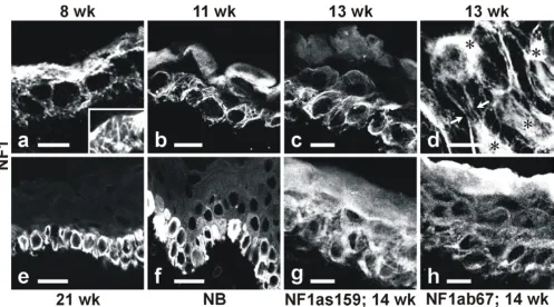
Indirect immunofluorescence labeling and confocal laser scanning microscopy (CLSM) was effective in visualizing NF1 protein in different layers of the epidermis, and in demonstrating variable subcellular distribution patterns of the protein within individual cells. At 8 weeks EGA, ep-idermis consisted of two cell layers, the basal and the peri-derm layers. Both cell layers were intensely labeled with the C terminus-specific antibody NF1GRP(D) which re-vealed a fibrillar labeling pattern (Fig 2a). The periderm cells displayed a more intense immunosignal compared to the basal cells at this two-layer stage of epidermal devel-opment. A third, the intermediate, cell layer was detecta-ble between the basal and the periderm cell layers at 11 weeks EGA (Fig 2b). All three cell layers were positive for NF1 protein. The labeling pattern remained fibrillar in the basal cells, while labeling of periderm cells had changed into a diffuse distribution within the cytoplasm. Cells of Figure 1
the intermediate layer showed less intense immunoreac-tion for NF1 protein.
At 13 weeks EGA, the basal cells retained the fibrillar labe-ling pattern with NF1GRP(D) antibody (Fig 2c). Analyses of the transverse sections of the basal cells were particular-ly informative. Specificalparticular-ly, the results demonstrated that NF1 antibody labeled a web of fibrils extending from a perinuclear location towards the cell periphery and desmosomes (Fig 2d; see also Fig 5B:c). At 13 weeks EGA, the periderm cells revealed a faint and a more diffuse la-beling pattern for the NF1 compared to the basal cells (Fig 2c). Samples of 14 weeks EGA were also labeled with two other NF1 specific antibodies (NF1as159 and NF1ab67) (Fig 2g,2h). Both antibodies labeled all three types of ep-idermal cell layers. The NF1as159 antibody showed a
fi-brillar pattern in the basal cell cytoplasm (Fig 2g). NF1 isoform type II specific antibody ab67 labeled all cell lay-ers in a more punctuate manner (Fig 2h). Between 13 and 15 weeks EGA, the expression of NF1 protein faded out in periderm cells (data not shown).
Four distinct layers of epidermis, the basal, the spinous, the granular, and the cornified cell layers were detectable at 21 weeks EGA. The periderm cells had disappeared by that time. By 21 weeks EGA and in the neonatal human epidermis, the fibrillar labeling for the NF1 protein in ba-sal cells was no longer detectable. The labeling pattern had changed from fibrillar into more diffuse cytoplasmic staining (Fig 2e,2f). Even the highest magnifications failed to reveal the fibrillar labeling (not shown).
Figure 2
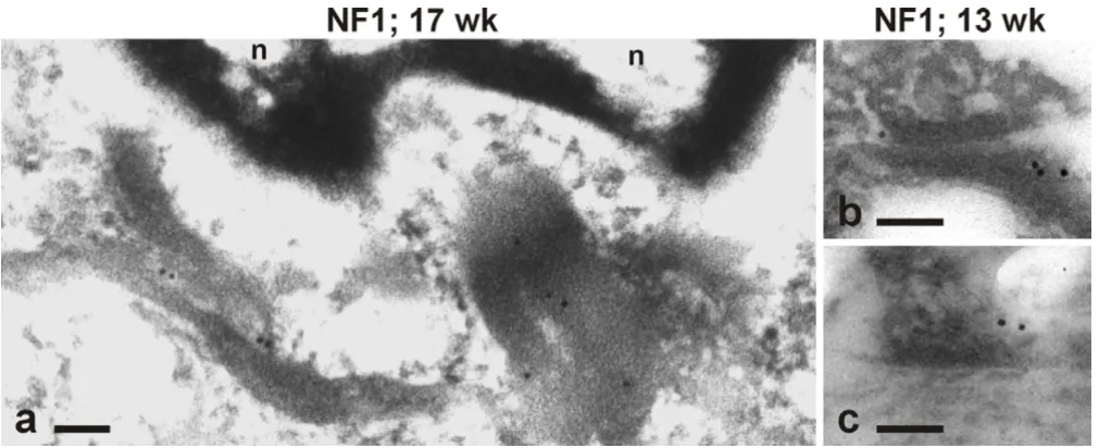
In order to localize NF1 protein at the ultrastructural level, post-embedding immunoelectron microscopy of devel-oping skin was performed using NF1GRP(D) antibody. The results revealed that in basal keratinocytes, the NF1 protein was located on the bundles of cytokeratin fila-ments, which formed a tight web around the nucleus (Fig 3a). Gold particles representative of NF1 protein were de-tected also on cytokeratin filaments terminating to desmosomes (Fig 3b) and hemidesmosomes (Fig 3c).
In further analyses, the expression of NF1 gene was stud-ied by in situ hybridization and avidin-biotin immunola-beling of paraffin embedded samples. Avidin-biotin immunolabeling revealed a pattern corresponding to that detected by indirect immunofluorescence. At 13 weeks EGA, the basal and the periderm cell layers were intensely labeled with the NF1GRP(D) antibody (Fig 4a). Also the intermediate cells showed positive reaction located in the cell periphery. At 18 weeks EGA, the basal cell layer re-vealed the most intense labeling while the other layers were less positive (Fig 4d). In situ hybridization revealed NF1 mRNA in developing epidermis. At 13 weeks EGA, NF1 mRNA was noted in the basal and intermediate cell layers (Fig 4c). Subcellularly, NF1 mRNA was located at the basal aspect of basal cells and at the cell periphery of intermediate cells. At 18 weeks EGA, NF1 mRNA was de-tectable near the cell membranes in the suprabasal cell layers, while the basal cells expressed NF1 mRNA at a very low level (Fig 4f).
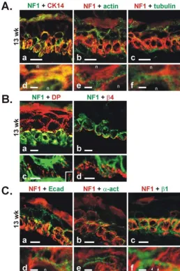
To investigate the fibrillar labeling pattern of NF1 protein in early developing epidermis in more detail, double im-munolabelings with NF1 protein and cytoskeletal marker proteins were carried out. At 13 weeks EGA, cytokeratin 14 antibody labeled cytoskeletal intermediate filaments of the basal cells (Fig 5A:a,d). Some labeling was also noted in the periderm. Immunosignals for the NF1 protein and cytokeratin 14 showed marked colocalization on many locations in basal cells (Fig 5A:a,d). Actin antibody la-beled the peripheral cytoplasm of all epidermal and peri-derm cells (Fig 5A:b,e). As the NF1 protein positive fibrils were located closer to the nuclei and actin labeling was de-tected more peripherally, these two components showed separate labeling (Fig 5A:b,e). α-tubulin antibody labeled also all epidermal cell layers, but did not colocalize with NF1 protein (Fig 5A:c,f).
In order to evaluate the spatial relationship of NF1 posi-tive cytokeratin fibrils with desmosomes and hemidesmo-somes, double immunolabelings of NF1 protein with desmosomal and hemidesmosomal marker proteins were carried out. At 13 weeks EGA, desmoplakin, a desmosom-al cytoplasmic plaque protein, was present in a typicdesmosom-al punctuate pattern at the cell-cell junction sites in all cell layers (Fig 5B:a). However, the intercellular junction sites of basal cells showed weaker labeling for desmoplakin. Higher magnification revealed that NF1 positive cytoker-atin fibrils terminated to dots of desmoplakin (Fig 5B:c). Antibody for β4 integrin, a transmembrane component of hemidesmosomes, labeled the basal aspects of the basal cells as distinct dots (Fig 5B:b,d). Higher magnification re-Figure 3
vealed that NF1 positive cytokeratin fibrils were directed towards the β4 integrin dots (Fig 5B:d). By 16 weeks EGA, labeling of β4 integrin was intensified and changed into a more linear labeling pattern (not shown).
The localization of NF1 protein and selected adherens junction proteins were investigated using double immu-nolabelings in developing epidermis at 13 weeks EGA. E-cadherin antibody revealed the cell-cell junction sites of the basal and intermediate cells (Fig 5C:a), but high mag-nification showed that the immunosignals for E-cadherin and NF1 protein were not colocalized (Fig 5C:d). Also, α -actinin and NF1 protein were mutually exclusive (Fig 5C:b,e). β1 integrin labeled the cell membranes of the
ba-sal cells, including the baba-sal aspect of baba-sal cells (Fig 5C:c). Higher magnification showed that NF1 protein positive fibrils were terminating between the β1 integrin labeling at cell-cell junction sites (Fig 5C:f). Thus, immu-nosignals for NF1 protein and adherens junction compo-nents were separate.
Recent data by Hsueh et al. [30] demonstrated interaction of NF1 protein and syndecans 1–4. To evaluate whether these proteins were in close contact in keratinocytes, dou-ble immunolabeling was performed. In developing epi-dermis at 14 weeks EGA, syndecan-1 was localized to the plasma membrane of keratinocytes in all epidermal layers (Fig 6a). Potential colocalization or close spatial relation-Figure 4
Figure 5
A. Double immunolabeling and CLSM analysis of NF1 protein with NF1GRP(D) antibody and cytoskeletal marker proteins at 13 weeks EGA in developing human epidermis. (a) NF1 protein (green) and cytokeratin 14 (red) show colocalization (yellow) on many locations of the basal cells. (d) High magnification reveals a marked degree of colocalization of NF1 protein and cytok-eratin 14 labeling patterns. (b) NF1 protein (red) and actin (green) antibodies show separate labeling patterns. (e) Higher mag-nification reveals that the NF1 positive fibrils (red) are located closer to the nuclei (n), while actin (green) is located more peripherally. (c) NF1 protein (red) and α-tubulin (green) antibodies show separate labeling. (f) High magnification of labeling for
ship of syndecan-1 with NF1 protein was noted on some basal cells (Fig 6a,6c). However, in adult epidermis, im-munosignals for the NF1 protein and syndecan-1 were en-tirely separate (Fig 6b,6d). Immunoprecipitation of keratinocyte culture lysates with syndecan-1 specific anti-body and subsequent western analysis with NF1GRP(D) antibody demonstrated that syndecan-1 was connected to a small fraction of NF1 protein (Fig 7).
Discussion
The results of the present study demonstrate an early fetal period when the NF1 tumor suppressor is abundantly ex-pressed and associated with bundles of intermediate fila-ments of the basal keratinocytes. This emphasizes that the
NF1 gene is developmentally regulated also in human skin. These findings also strengthen the view that the large NF1 protein exerts its functions through multiple molec-ular interactions. Specifically, NF1 tumor suppressor fac-tor associates and functions with proteins located with the plasma membrane (p21ras, syndecans), or with cytoskel-etal components associated with selected cellular junc-tions (desmosomes, hemidesmosomes) [12,28,30]. The action of NF1 protein may be analogous to e.g. fatty acid synthase, which conducts its tasks using different sites of the same molecule. It should also be noted that the activ-ity of ras is associated with the regulation of cell growth
and differentiation, including control of cytoskeletal or-ganization and formation of cell-cell junctions [7,14].
Western transfer analysis, immunolocalization and in situ
hybridization showed that the NF1 gene was abundantly expressed in developing human epidermis. In contrast to adult epidermis, fetal basal cells displayed a fibrillar labe-ling pattern for NF1 protein between 8 and 14 weeks EGA. Later during the development, the fibrillar labeling of ba-sal cells changed into a more diffuse cytoplasmic pattern. The fibrillar labeling pattern suggested that the NF1 pro-tein was associated with cytoskeleton. Double immunola-belings demonstrated a relatively high degree of colocalization of NF1 protein and cytokeratin 14 at light microscopic level, and immunoelectron microscopy lo-cated NF1 protein on perinuclear keratin bundles. In de-veloping human epidermis, NF1 protein did not show detectable association with actin microfilaments or tubu-lin, although in other studies and conditions, NF1 protein has been shown to interact with these cytoskeletal compo-nents [29].
Concomitantly with the association of NF1 protein to the cytokeratin, the epidermis undergoes profound changes: the stratification of epidermis, maturation of basement membrane zone, and the formation of desmosomes and hemidesmosomes is activated. At 9–16 weeks EGA, kera-tin filaments become more prominent, and the number of desmosomes increase [4]. First hemidesmosomes have Figure 6
Double immunolabeling and CLSM analysis of developing and adult epidermis for NF1 protein and syndecan-1. (a) At 14 weeks EGA, the NF1 protein (red) shows fibrillar pattern in basal cells while syndecan-1 (green) is localized to the plasma membrane of keratinocytes in all epidermal layers. Close spa-tial distribution or colocalization (yellow) is detected on some basal cells. (b) In adult epidermis the immunosignals for the NF1 protein (red) and syndecan-1 (green) are completely separate. Scale bars 10 µm in a,b, 2 µm in c,d.
Figure 7
been noted at ~9 weeks EGA. Between 9 and 15 weeks EGA the number of hemidesmosomes increases by about fourfold, and the morphology of hemidesmosomes ma-tures simultaneously with increased contact with interme-diate keratin filaments [2]. NF1 protein was detected also near the desmosomes and hemidesmosomes as indicated by both immunoelectron microscopy and double immu-nolabeling of NF1 protein with desmoplakin and β4 in-tegrin. These in vivo observations are in good agreement with the earlier in vitro study by Koivunen et al. [28]. They showed the association of NF1 protein with CK14-con-taining keratin intermediate filaments during the forma-tion of desmosomes in cultured human keratinocytes. The present study demonstrates a similar association of NF1 protein and intermediate filaments during the formation of intercellular junctions in vivo. Developing epidermis thus provides a good model to study differentiation of ke-ratinocytes in vivo, since the interactions of various cy-toskeletal and junction proteins can be readily visualized.
The finding that NF1 specific antibodies raised against dif-ferent peptide segments of the protein results in difdif-ferent labeling is in agreement with earlier studies [20,21,28,33]. Specifically, NF1GRP(D) antibody labeled most intensely the basal cells, while other antibodies used, NF1ab67 and NF1as159, labeled all the developing cell layers indicating that NF1 protein is present in the intermediate cell layers, too. One plausible explanation is that NF1GRP(D) anti-body may recognize only a specific type of NF1 protein conformation.
Expression of NF1 mRNA was studied at 13 and 18 weeks EGA using in situ hybridization. At 13 weeks, the localiza-tion of NF1 mRNA corresponded with the immunosignal for the NF1 protein. However, at 18 weeks, NF1 mRNA and protein displayed differential spatial distribution within the epidermis. Specifically, NF1 mRNA was detect-ed in the suprabasal cells but not in the basal layer. Al-though NF1 protein remained detectable in the basal cells at 18 weeks EGA, the labeling pattern for NF1 protein in the basal cells had changed to a more diffuse cytoplasmic staining. It is thus possible that the more diffuse localiza-tion of NF1 protein in the basal cells is associated with downregulation of the NF1 mRNA level. These results are in concordance with previous studies detecting NF1 mRNA in suprabasal cell layers in late developing rat epi-dermis while basal cells contained no detectable NF1 mRNA at this stage of rat epidermal development [33].
NF1 mRNA was intracellularly distributed to the basal as-pects of the basal cells and to the cell peripheries of inter-mediate cells at 13 weeks EGA. These are the sites where desmosome and hemidesmosome formation takes place during this stage of development. Previously, Ylä-Outinen
et al. [31] have shown that NF1 mRNA is targeted to
cell-cell contact zone in cultured human keratinocytes when they are induced to differentiate. This might be true also
in vivo, since NF1 mRNA was accounted at cell-cell and cell-basement membrane contact zones.
Periderm cells showed profound changes in the expres-sion pattern of NF1 protein. Our study revealed that, at 8 weeks EGA, when the desmosome formation is active in the periderm, the periderm cells showed fibrillar labeling for NF1 protein. By the 11th week of EGA, labeling for NF1 protein revealed a more diffuse pattern in the peri-derm cells, diminished thereafter, and disappeared by the 16th week EGA. This correlates with the cessation of peri-derm cell proliferation at about 12 weeks EGA [3] and dis-ruption of intermediate keratin filaments at 15–17 weeks EGA [4]. After the periderm cells cease dividing, they syn-thesize little if any protein and are gradually shed into the amniotic fluid by the end of the third trimester [3]. The ex-pression of NF1 protein in the periderm thus correlates well with the periderm cell proliferation and cell junction forming activity. This is also in concordance with previous study on developing rat skin demonstrating NF1 protein first in the basal and periderm cells, but later mostly in the spinous cell layer [33].
Even though a bipartite interaction between transmem-brane syndecans and NF1 protein has been characterized in neurons by two hybrid method [30], the results of the present study demonstrated only little of potential colo-calization of NF1 protein and syndecan-1 in epidermal keratinocytes during the development and in adult. Thus, the conditions when NF1 protein and syndecans may in-teract, and the cell biological significance of this interac-tion remain to be elucidated.
List of abbreviations
BSA, Bovine serum albumin; CLSM, confocal laser scan-ning microscopy; EGA, estimated gestational age; GAP, G-protein activating G-protein; NF1, type 1 neurofibromatosis; PBS, phosphate buffered saline; SDS, sodium dodecyl sul-phate.
Competing interests None declared.
Authors' Contributions
Acknowledgements
This work was supported by Turku University Foundation, Turku Univer-sity Central Hospital (grant #13338), Oulu UniverUniver-sity Hospital, Finnish Cul-tural Foundation, Finnish Cancer Societies, and Finnish Society of Dermatopathology.
References
1. Holbrook KA, Odland GF: The fine structure of developing hu-man epidermis: light, scanning, and transmission electron microscopy of the periderm.J Invest Dermatol 1975, 65:16-38 2. McMillan JR, Eady RA: Hemidesmosome ontogeny in digit skin
of the human fetus.Arch Dermatol Res 1996, 288:91-97
3. Holbrook KA: Development of human skin. Retinoids 1997,
13:47-53
4. Dale BA, Holbrook KA, Kimball JR, M Hoff, Sun TT: Expression of epidermal keratins and filaggrin during human fetal skin de-velopment.J Cell Biol 1985, 101:1257-1269
5. Hentula M, Peltonen J, Peltonen S: Expression profiles of cell-cell and cell-matrix junction proteins in developing human epi-dermis.Arch Dermatol Res 2001, 293:259-267
6. Pummi K, Malminen M, Aho H, Karvonen SL, Peltonen J, Peltonen S:
Epidermal tight junctions: ZO-1 and occludin are expressed in mature, developing, and affected skin and in vitro differen-tiating keratinocytes.J Invest Dermatol 2001, 117:1050-1058 7. Vasioukhin V, Bauer C, Degenstein L, Wise B, Fuchs E:
Hyperprolif-eration and defects in epithelial polarity upon conditional ab-lation of α-catenin in skin.Cell 2001, 104:605-617
8. Gutmann DH, Wood DL, Collins FS: Identification of the neurofi-bromatosis type 1 gene product.Proc Natl Acad Sci U S A 1991,
88:9658-9662
9. Gutmann DH, Aylsworth A, Carey JC, Korf B, Marks J, Pyeritz RE, Ru-benstein A, Viskochil D: The diagnostic evaluation and multidis-ciplinary management of neurofibromatosis 1 and neurofibromatosis 2.JAMA 1997, 278:51-57
10. Marchuk DA, Saulino AM, Tavakkol R, Swaroop M, Wallace MR, An-dersen LB, Mitchell AL, Gutmann DH, Boguski M, Collins FS: cDNA cloning of the type 1 neurofibromatosis gene: complete se-quence of the NF1 gene product.Genomics 1991, 11:931-940 11. Riccardi VM: Von Recklinghausen neurofibromatosis.N Engl J
Med 1981, 305:1617-1627
12. Xu GF, Lin B, Tanaka K, Dunn D, Wood D, Gesteland R, White R, Weiss R, Tamanoi F: The catalytic domain of the neurofi-bromatosis type 1 gene product stimulates ras GTPase and complements ira mutants of S. cerevisiae.Cell 1990, 63:835-841 13. Bollag G, McCormick F: Ras regulation. NF is enough of GAP.
Nature 1992, 356:663-664
14. Mercer JA: Intercellular junctions: downstream and upstream of Ras?Semin Cell Dev Biol 2000, 11:309-314
15. Legius E, Marchuk DA, Collins FS, Glover TW: Somatic deletion of the neurofibromatosis type 1 gene in a neurofibrosarcoma supports a tumour suppressor gene hypothesis. Nat Genet
1993, 3:122-126
16. Li Y, Bollag G, Clark R, Stevens J, Conroy L, Fults D, Ward K, Fried-man E, Samowitz W, Robertson M: Somatic mutations in the neurofibromatosis 1 gene in human tumors.Cell 1992, 69: 275-281
17. Johnson MR, Look AT, DeClue JE, Valentine MB, Lowy DR: Inactiva-tion of the NF1 gene in human melanoma and neuroblasto-ma cell lines without impaired regulation of GTP-Ras.Proc Natl Acad Sci U S A 1993, 90:5539-5543
18. Gutmann DH, Geist RT, Rose K, Wallin G, Moley JF: Loss of neu-rofibromatosis type I (NF1) gene expression in pheochromo-cytomas from patients without NF1.Genes Chromosomes Cancer
1995, 13:104-109
19. Gutmann DH, Giordano MJ, Mahadeo DK, Lau N, Silbergeld D, Guha A: Increased neurofibromatosis 1 gene expression in astro-cytic tumors: positive regulation by p21-ras.Oncogene 1996,
12:2121-2127
20. Hermonen J, Hirvonen O, Ylä-Outinen H, Lakkakorpi J, Björkstrand AS, Laurikainen L, Kallioinen M, Oikarinen A, Peltonen S, Peltonen J:
Neurofibromin: expression by normal human keratinocytes
in vivo and in vitro and in epidermal malignancies.Lab Invest
1995, 73:221-228
21. Peltonen J, Karvonen SL, Ylä-Outinen H, Hirvonen O, Karvonen J:
Lesional psoriatic epidermis displays reduced neurofibromin immunoreactivity.J Invest Dermatol 1995, 105:664-667
22. Aaltonen V, Boström PJ, Söderström KO, Hirvonen O, Tuukkanen J, Nurmi M, Laato M, Peltonen J: Urinary bladder transitional cell carcinogenesis is associated with down-regulation of NF1 tu-mor suppressor gene in vivo and in vitro. Am J Pathol 1999,
154:755-765
23. Brannan CI, Perkins AS, Vogel KS, Ratner N, Nordlund ML, Reid SW, Buchberg AM, Jenkins NA, Parada LF, Copeland NG: Targeted dis-ruption of the neurofibromatosis type-1 gene leads to devel-opmental abnormalities in heart and various neural crest-derived tissues.Genes Dev 1994, 8:1019-1029
24. Daston MM, Ratner N: Neurofibromin, a predominantly neuro-nal GTPase activating protein in the adult, is ubiquitously ex-pressed during development.Dev Dyn 1992, 195:216-226 25. Hirvonen O, Lakkakorpi J, Aaltonen V, Hirvonen H, Rossi M,
Karvo-nen SL, Ylä-OutiKarvo-nen H, Kalimo H, PeltoKarvo-nen J: Developmental reg-ulation of NF1 tumor suppressor gene in human peripheral nerve.J Neurocytol 1998, 27:939-952
26. Baizer L, Ciment G, Hendrickson SK, Schafer GL: Regulated ex-pression of the neurofibromin type I transcript in the devel-oping chicken brain.J Neurochem 1993, 61:2054-2060
27. Huynh DP, Nechiporuk T, Pulst SM: Differential expression and tissue distribution of type I and type II neurofibromins during mouse fetal development.Dev Biol 1994, 161:538-551
28. Koivunen J, Ylä-Outinen H, Korkiamäki T, Karvonen SL, Pöyhönen M, Laato M, Karvonen J, Peltonen S, Peltonen J: New function for NF1 tumor suppressor.J Invest Dermatol 2000, 114:473-479
29. Li C, Cheng Y, Gutmann DA, Mangoura D: Differential localiza-tion of the neurofibromatosis 1 (NF1) gene product, neurofi-bromin, with the F-actin or microtubule cytoskeleton during differentiation of telencephalic neurons.Brain Res Dev Brain Res
2001, 130:231-248
30. Hsueh YP, Roberts AM, Volta M, Sheng M, Roberts RG: Bipartite in-teraction between neurofibromatosis type I protein (neu-rofibromin) and syndecan transmembrane heparan sulfate proteoglycans.J Neurosci 2001, 21:3764-3770
31. Ylä-Outinen H, Koivunen J, Nissinen M, Björkstrand AS, Paloniemi M, Korkiamäki T, Peltonen S, Karvonen SL, Peltonen J: NF1 tumor sup-pressor mRNA is targeted to cell-cell contact zone in Ca2+ -induced keratinocyte differentiation.Lab Invest 2002, 82: 353-361
32. Hirvonen O, Lakkakorpi J, Aaltonen V, Hirvonen H, Rossi M, Karvo-nen SL, Ylä-OutiKarvo-nen H, Kalimo H, PeltoKarvo-nen J: Developmental reg-ulation of NF1 tumor suppressor gene in human peripheral nerve.J Neurocytol 1998, 27:939-952
33. Malhotra R, Ratner N: Localization of neurofibromin to kerati-nocytes and melakerati-nocytes in developing rat and human skin.
J Invest Dermatol 1994, 102:812-818
Pre-publication history
The pre-publication history for this paper can be accessed here:
http://www.biomedcentral.com/1471-5945/2/10/prepub
Publish with BioMed Central and every scientist can read your work free of charge
"BioMedcentral will be the most significant development for disseminating the results of biomedical research in our lifetime."
Paul Nurse, Director-General, Imperial Cancer Research Fund
Publish with BMCand your research papers will be:
available free of charge to the entire biomedical community
peer reviewed and published immediately upon acceptance
cited in PubMed and archived on PubMed Central
yours - you keep the copyright
[email protected] Submit your manuscript here:
http://www.biomedcentral.com/manuscript/