E D I T O R I A L
Open Access
High time to omit oxygen therapy in ST
elevation myocardial infarction
Ardavan Khoshnood
1,2Abstract
Supplemental oxygen (O
2) therapy in patients with chest pain has been a cornerstone in the treatment of suspected
myocardial infarction (MI). Recent randomized controlled trials have, however, shown that supplemental O
2therapy
has no positive nor negative effects on cardiovascular functions, mortality, morbidity or pain in normoxic patients with
suspected MI and foremost patients with ST Elevation Myocardial Infarction (STEMI). O
2therapy in normoxic STEMI
patients should therefore be omitted. More studies are needed in discussing hemodynamically unstable STEMI
patients, as well as patients with non-STEMI, unstable angina and other emergency conditions.
Keywords:
Oxygen, Oxygen therapy, ST elevation myocardial infarction, STEMI, Physiology, Pathology, Emergency medicine
Main text
ST Elevation Myocardial Infarction (STEMI) is the most
serious manifestation of Acute Coronary Syndrome
(ACS) and is by the Fourth Universal Definition of
Myo-cardial Infarction deemed to be a Type I myoMyo-cardial
in-farction, caused by an acute atherothrombotic coronary
artery disease (CAD) [
1
]. The consequence of CAD is a
partial or complete occlusion of a coronary artery thus
contributing to the termination of oxygen (O
2) supply to
the myocardium, giving rise to ischemia [
1
,
2
].
Because of its seriousness, prompt and rapid diagnosis
and treatment is of high importance to reduce both
mortality and morbidity [
3
]. The most important
treat-ment in STEMI is Percutaneous Coronary Intervention
(PCI), which should be performed as soon as possible
after that the condition is diagnosed [
4
–
6
].
International guidelines also emphasize on treatment
with dual antiplatelets in the emergency phase before
the start of the PCI [
4
–
6
]. Previous guidelines also
rec-ommended the immediate administration of O
2to
pa-tients with diagnosed or suspected ACS, without any
respect to the blood O
2saturation [
7
–
10
]. In discussing
current guidelines, only the 2017 ESC Guidelines for the
management of patients with STEMI [
5
] states that O
2should not routinely be administrated to patients with
STEMI, and that only those with a blood O
2saturation
< 90% or PaO
2< 60 mmHg should receive O
2therapy.
With an overwhelming scientific evidence that O
2therapy has no positive (nor negative) effects in patients
with STEMI, all international, regional and local
guide-lines should be updated and omit O
2therapy in
nor-moxic (
≥
90%) STEMI patients.
Oxygen therapy
The history of O
2as a medicine dates back to 1775 when
the British chemist Joseph Priestly discovered O
2and
stated that it could be used as a medicine [
11
]. It was,
however, in 1900 that the first publication on the role of
O
2therapy in patients with chest pain was published. It
was a short letter by Dr. Charles Steele, in which he
deemed that O
2therapy had relieved chest pains in one
single patient he believed to have angina [
12
]. Ever since
this letter by Dr. Steele, several studies have tried to
an-swer how supplemental O
2therapy in both healthy and ill
patients affect their cardiovascular system.
The rationale behind O
2therapy has been that by adding
O
2to the patient
’
s blood, the myocardium can be
oxygen-ated, which in turn will contribute to a diminished ischemic
area and infarct size, thereby minimizing the risk for lethal
arrhythmias [
13
,
14
]. Studies on canines [
14
–
16
] have given
some support to this theory, showing that O
2therapy
de-creases infarct size and ischemia in these animals. A recent
study on swine, however, showed that hyperoxemia can
ag-gravate and worsen myocardial ischemia [
17
].
Correspondence:[email protected]
1Department of Clinical Sciences, Faculty of Medicine, Lund University, Lund,
Sweden
2Department of Emergency and Internal Medicine, Skåne University Hospital
Lund, Lund, Sweden
In healthy individuals, experimental studies have
shown that hyperoxemia because of supplemental O
2therapy, may contribute to negative cardiovascular
ef-fects like a decrease in coronary blood flow, arterial
vasoconstriction, diminished cardiac output, an increase
in the systemic vascular resistance as well as impaired
blood flow to organs and tissues [
18
–
26
].
In patients with suspected as well as confirmed
myocar-dial infarction, the role of O
2were for a long time highly
inconclusive. Our knowledge gap in this matter was not
because of lack of studies, but rather because of the poor
methodologies used in these studies. Ever since 1900,
sev-eral studies have been published on the role of O
2in
pa-tients with chest pain, coronary artery disease, cardiac
failure as well as suspected and confirmed myocardial
in-farction. All of them have unfortunately had serious
limi-tations and have therefore not been able to correctly
answer the question of how O
2therapy affects the
cardio-vascular system in both healthy patients and patients with
myocardial infarction and cardiac failure. The studies have
either been case studies or small reports including only a
few patients, thus not being generalizable, or small studies
[
18
–
20
,
27
–
43
]. Furthermore, the vast majority of the
studies was conducted in the pre-PCI era and even before
Troponin was used as an important part in the diagnosis
of myocardial infarction.
Because of the above limitations, a Cochrane report
from 2013 [
44
] called for randomized controlled trials to
once for all answer the question about which role
sup-plemental O
2therapy should have in patients with chest
pain and suspected myocardial infarction. The authors
of the report stated that
“
A definitive randomised
con-trolled trial is urgently required [
…
].
”
Randomized controlled trials
Before the Cochrane reports call for a definitive
random-ized controlled trial (RCT) in 2013, there were already
four RCTs on the role of supplemental O
2in patients
with myocardial infarction; Rawles et al. from 1976 [
45
],
Wilson et al. from 1997 [
42
], Ukholkina et al. from 2005
[
46
] and Ranchord et al. from 2012 [
47
].
The two first studies were conducted in the pre-PCI
era. While Wilson et al. [
42
] found no significant
differ-ences between the patients with myocardial infarction
randomized to supplemental O
2or air, Rawles et al. [
45
]
showed that patients with myocardial infarction
receiv-ing supplemental O
2, had a larger infarct size as
mea-sured with serum aspartate aminotransferase.
The study by Ukholkina et al. [
46
] is the only randomized
study showing a positive effect of supplemental O
2therapy
in patients with myocardial infarction. The study is thus
highly biased because of a limited methodology [
44
].
Rancord et al. [
47
] have a sound methodology, and
should be considered the first modern RCT on the role
of supplemental O
2in patients with STEMI. The authors
found no significant differences between the two arms
(supplemental O
2vs titrated O
2) with regard to infarct
size as measured by cardiac Troponin T, as well as
car-diac MRI (CMRI) close to one month after inclusion.
After the Cochrane report from, three more RCTs
have been published discussing the role of supplemental
O
2therapy in myocardial infarction; the AVOID study
[
48
,
49
], the SOCCER study [
50
–
53
] and the DETO2X
study [
54
–
57
].
The main publication of the AVOID study was
con-ducted by Stub et al. [
48
] in which 441 STEMI patients
were randomized to supplemental O
2therapy or air.
Even though the study found no significant difference in
infarct size as measured by cardiac Troponin, a subset of
the patients undergoing CMRI after six months, showed
that those randomized to supplemental O
2therapy, had
a larger infarct size as measured in absolute mass but
not in percent of the left ventricle. A sub study [
49
] of
the AVOID trial showed later that patients randomized
to the O
2arm, had significantly higher cardiac Troponin
rates than those randomized to the air arm.
The SOCCER study was conducted in Sweden by
Khoshnood et al. and aimed to evaluate the effects of
supplemental O
2in normoxic first-time STEMI patients
accepted for PCI. Patients were randomized to either
supplemental O
2therapy or air. All patients underwent
CMRI, while only a subset of patients underwent
echo-cardiography. Their chest pain was scored and assessed
prehospital and in-hospital with the Visual Analog Scale
(VAS) [
50
]. Ninety-four patients underwent CMRI which
showed no significant difference between the two arms
in discussing infarct size, myocardium at risk and
myo-cardial salvage index [
51
]. Of the 87 patients undergoing
echocardiography, no significant differences could be
measured between the two arms in discussing left
ven-tricular ejection fraction and wall motion score index
[
52
]. In a recently published sub study, 111 patients were
assessed in regard to chest pain to evaluate the analgesic
effect of O
2therapy. Those randomized to the
supple-mental O
2group had significantly higher median VAS
and also received significantly higher amounts of
mor-phine. The study could not show that supplemental O
2diminished chest pain [
53
].
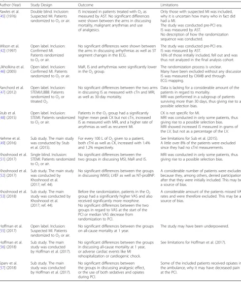
Table 1
A summary of the randomized controlled trials studying the effects of O
2therapy in patients with suspected or confirmed
myocardial infarctions
Author (Year) Study Design Outcome Limitations
Rawles et al. [45] (1976)
Double blind. Inclusion: Suspected MI. Patients randomized to O2or air.
IS increased in patients treated with O2as measured by AST. No significant differences were shown between the arms in discussing mortality, malignant arrythmias and use of analgesics.
Only those with suspected MI was included, why it is uncertain how many who in fact did had a MI.
The study was conducted pre-PCI era. IS was measured by AST.
No description of how the randomization sequence was conducted.
Wilson et al. [42] (1997)
Open label. Inclusion: Confirmed MI. Patients randomized to O2or air.
No significant differences were shown between the arms in discussing arrhythmias as well as ST segment changes in the ECG.
The study was conducted pre-PCI era. IS was measured by AST.
16% of those initially included, fell out and was thus not analyzed in the final analysis cohort.
Ukholkina et al. [46] (2005)
Open label. Inclusion: Confirmed MI. Patients randomized to O2or air.
MaR, IS and arrhythmias were significantly lower in the O2group.
The randomization process is unclear.
Many have been excluded without any discussion. IS was measured by CKMB and through
ECG mapping.
Ranchord et al. [47] (2012)
Open label. Inclusion: STEMI/LBBB. Patients randomized to O2or titrated O2.
No significant differences between the two arms in discussing IS as measured with cTn and MRI, as well as 30-day mortality.
Data is lacking for a considerable amount of the patients in regard to mortality.
MRI was performed in a subgroup of patients surviving more than 30 days, thus giving rise to a possible selection bias.
Stub et al. [48] (2015)
Open label. Inclusion: STEMI. Patients randomized to O2or air.
Patients in the O2group had a significantly higher mean peak CK but not cTn, increased IS as measured with MRI, and a higher rate of arrythmias as well as recurrent MI.
CK is not specific for MI.
MRI was conducted in only some patients, thus giving rise to a possible selection bias. MRI showed increased IS measured in grams of the LV, but not as a percentage of the LV.
Nehme et al. [49] (2016)
Sub study. The main study was conducted by Stub et al. (2015).
For every 100 L of O2given to a patient, both cTnI as well as CK, increased with 1.4% and 1.2% respectively.
See limitations for Sub et al. (2015).
A little over 8% of the patients were excluded since they had no cTnI measurements.
Khoshnood et al. [51] (2017)
Single blind. Inclusion: STEMI. Patients randomized to O2or air.
No significant differences between the two groups in discussing MSI, MaR and IS.
MRI was conducted in only some patients, thus giving rise to a possible selection bias.
Khoshnood et al. [52] (2017)
Sub study. The main study was conducted by Khoshnood et al. (2017; ref. 44).
No significant differences between the groups in discussing WMSI, LVEF as well as NT-proBNP.
A considerable number of patients were excluded because they, among others, denied participation after that they were initially included. This may be a source of bias.
Khoshnood et al. [53] (2018)
Sub study. The main study was conducted by Khoshnood et al. (2017; ref. 44).
Before the randomization, patients in the O2 group had a significantly higher VAS and also received significantly more morphine. No significant differences between the two groups in regard to VAS at the start of the PCI or median VAS decrease from randomization to PCI.
A considerable amount of the patients missed VAS rates and were therefore excluded. This may be a source of bias.
Hoffman et al. [55] (2017)
Open label. Inclusion: Suspected MI. Patients randomized to O2or air.
No significant differences between the groups on all-cause mortality at 1 year.
The study may have been underpowered.
Hoffman et al. [56] (2018)
Sub study. The main study was conducted by Hoffman et al. (2017).
No significant differences between the groups in discussing all-cause mortality at 1 year, or adverse cardiac events like MI rehospitalization or cardiogenic chock.
See limitations for Hoffman et al. (2017).
Sparv et al. [57] (2018)
Sub study. The main study was conducted by Hoffman et al. (2017).
No significant differences between the groups in discussing analgesic effect, or the use of both sedatives and opiates during PCI.
Some of the included patients received opiates in the ambulance, why it may have decreased pain at the PCI.
myocardial infarction and cardiogenic shock [
56
]. In a
re-cent published DETO2X sub study by Sparv et al. [
57
] on
the analgesic effect of supplemental O
2therapy in patients
with suspected myocardial infarction, there were no
sig-nificant differences between the two arms in regard to
pain nor the amount of morphine and sedatives received
during PCI.
Table
1
summarizes all the RCTs.
Omit supplemental O
2therapy in STEMI
The above RCTs clearly show that O
2therapy has so
positive nor negative cardiovascular effects, when used
in normoxic patients with STEMI both prehospital and
in-hospital. Two recent reviews and meta-analysis on
the role of supplemental O
2therapy in acute myocardial
infarction, showed also no benefit of using O
2therapy in
these patients [
58
,
59
].
In discussing supplemental O
2therapy in normoxic
STEMI patients, the evidences are clear and consistent,
why all guidelines must be reformed to state that
supple-mental O
2therapy in these patients should be omitted.
It is, however, of high importance to point that patients
diagnosed with STEMI, and who have a low blood
oxy-gen saturation, should receive supplemental O
2. It is the
routine use of O
2therapy, with no respect to blood
oxy-gen saturation, that should be omitted (Fig.
1
). With this
said, it is important to point out that the RCTs
pre-sented above does also have some limitations as the
ma-jority of them have had a small cohort, and the focus
have been stable and normoxic STEMI patients. These
limitations might reduce the generalizability of the
stud-ies. More studies are therefore needed in discussing
sup-plemental O
2therapy in hemodynamic unstable STEMI
patients, patients with non-STEMI as well as unstable
angina. This is especiCally of importance since some
studies argue that supplemental O
2therapy
adminis-trated to acutely ill patients can be toxic and increase
mortality and morbidity [
60
,
61
].
Abbreviations
ACS:Acute Coronary Syndrome; CAD: Coronary Artery Disease; CMRI: Cardiac Magnetic Resonance Imaging; MI: Myocardial Infarction; O2: Oxygen;
PCI: Percutaneous Coronary Intervention; RCT: Randomized Controlled Trials; STEMI: ST Elevation Myocardial Infarction; VAS: Visual Analog Scale
Acknowledgements
Not applicable.
Funding
Not applicable.
Availability of data and materials
Not applicable.
Author’s contribution
The author read and approved the final manuscript.
Authors’information
Dr. Ardavan Khoshnood is a senior resident in Emergency Medicine, and holds a PhD in Clinical Medicine, Emergency Medicine. He is a lecturer at the Medical Faculty at Lund University, and also serves as an Associate Editor for the BMC Emergency Medicine.
Ethics approval and consent to participate
Not applicable.
Consent for publication
Not applicable.
Competing interests
Dr. Khoshnood have authored several articles on the role of supplemental O2
therapy in patients with myocardial infarction and STEMI. Dr. Khoshnood is also an Associate Editor at the BMC Emergency Medicine.
Publisher
’
s Note
Springer Nature remains neutral with regard to jurisdictional claims in published maps and institutional affiliations.
Received: 30 August 2018 Accepted: 11 October 2018
References
1. Thygesen K, Alpert JS, Jaffe AS, Chaitman BR, Bax JJ, Morrow DA, White HD, Group ESCSD. Fourth universal definition of myocardial infarction (2018). Eur Heart J. 2018:ehy462.
2. Heusch G, Gersh BJ. The pathophysiology of acute myocardial infarction and strategies of protection beyond reperfusion: a continual challenge. Eur Heart J. 2017;38(11):774–84.
3. Scholz KH, Maier SKG, Maier LS, Lengenfelder B, Jacobshagen C, Jung J, Fleischmann C, Werner GS, Olbrich HG, Ott R, et al. Impact of treatment delay on mortality in ST-segment elevation myocardial infarction (STEMI) patients presenting with and without haemodynamic instability: results from the German prospective, multicentre FITT-STEMI trial. Eur Heart J. 2018;39(13):1065–74. 4. O'gara PT, Kushner FG, Ascheim DD, Casey DE, Chung MK, De Lemos JA,
Ettinger SM, Fang JC, Fesmire FM, Franklin BA: 2013 ACCF/AHA guideline for the management of ST-elevation myocardial infarction: executive summary: a report of the American College of Cardiology Foundation/American Heart Association task force on practice guidelines. J Am Coll Cardiol 2013, 61(4): 485–510.
5. Ibanez B, James S, Agewall S, Antunes MJ, Bucciarelli-Ducci C, Bueno H, Caforio ALP, Crea F, Goudevenos JA, Halvorsen S, et al. 2017 ESC guidelines for the management of acute myocardial infarction in patients presenting with ST-segment elevationThe task force for the management of acute myocardial infarction in patients presenting with ST-segment elevation of the European Society of Cardiology (ESC). Eur Heart J. 2018;39(2):119–77. 6. Excellence NIfHaC. Myocardial infarction with ST-segment elevation: acute
management. In: NICE guideline (CG167); 2013.
7. Arntz H-R, Bossaert L, Filippatos GS. European resuscitation council guidelines for resuscitation 2005. Resuscitation. 2005;67:S87–96. 8. Pollack CV Jr, Diercks DB, Roe MT, Peterson ED. 2004 American College of
Cardiology/American Heart Association guidelines for the Management of Patients with ST-elevation myocardial infarction: implications for emergency department practice. Ann Emerg Med. 2005;45(4):363–76.
9. Nolan JP, Deakin CD, Soar J, Böttiger BW, Smith G. European resuscitation council guidelines for resuscitation 2005. Resuscitation. 2005;67:S39–86. 10. Anderson JL, Adams CD, Antman EM, Bridges CR, Califf RM, Casey DE,
Chavey WE, Fesmire FM, Hochman JS, Levin TN, et al. ACC/AHA 2007 guidelines for the Management of Patients with Unstable Angina/non– ST-elevation myocardial infarction: a report of the American College of Cardiology/American Heart Association task force on practice guidelines (writing committee to revise the 2002 guidelines for the Management of Patients with Unstable Angina/non–ST-elevation myocardial infarction) developed in collaboration with the American College of Emergency Physicians, the Society for Cardiovascular Angiography and Interventions, and the Society of Thoracic Surgeons endorsed by the American Association of Cardiovascular and Pulmonary Rehabilitation and the Society for Academic Emergency Medicine. J Am Coll Cardiol. 2007;50(7):e1–e157. 11. Priestly J. Experiments and observations on different kinds of air, vol. 2.
London: St. Paul’s Church-Yard; 1775.
12. Steele C. Severe angina pectoris relieved by oxygen inhalations. The British Med J. 1900:1568–8.
13. Madias J, Hood W Jr. Reduction of precordial ST-segment elevation in patients with anterior myocardial infarction by oxygen breathing. Circulation. 1976;53(3 Suppl):I198.
14. Maroko PR, Radvany P, Braunwald E, Hale SL. Reduction of infarct size by oxygen inhalation following acute coronary occlusion. Circulation. 1975;52(3):360–8. 15. Sayen J, Sheldon W, Horwitz O, Kuo PT, Peirce G, Zinsser HF, Mead J. Studies of
coronary disease in the experimental animal. II. Polarographic determinations of local oxygen availability in the dog's left ventricle during coronary occlusion and pure oxygen breathing. J Clin Invest. 1951;30(9):932–40.
16. Kelly RF, Hursey TL, Parrillo JE, Schaer GL. Effect of 100% oxygen
administration on infarct size and left ventricular function in a canine model of myocardial infarction and reperfusion. Am Heart J. 1995;130(5):957–65. 17. Guensch DP, Fischer K, Shie N, Lebel J, Friedrich MG. Hyperoxia exacerbates
myocardial ischemia in the presence of acute coronary artery stenosis in swine. Circ Cardiovasc Interv. 2015;8(10):e002928.
18. Milone SD, Newton GE, Parker JD. Hemodynamic and biochemical effects of 100% oxygen breathing in humans. Can J Physiol Pharmacol. 1999;77(2):124–30.
19. Waring WS, Thomson AJ, Adwani SH, Rosseel AJ, Potter JF, Webb DJ, Maxwell SR. Cardiovascular effects of acute oxygen administration in healthy adults. J Cardiovasc Pharmacol. 2003;42(2):245–50.
20. Rousseau A, Bak Z, Janerot-Sjöberg B, Sjöberg F. Acute hyperoxaemia-induced effects on regional blood flow, oxygen consumption and central circulation in man. Acta Physiol Scand. 2005;183(3):231–40.
21. Bergofsky EH, Bertun P. Response of regional circulations to hyperoxia. J Appl Physiol. 1966;21(2):567–72.
22. Kenmure AC, Murdoch WR, Hutton I, Cameron AJ. Hemodynamic effects of oxygen at 1 and 2 Ata pressure in healthy subjects. J Appl Physiol. 1972; 32(2):223–6.
23. Thomson AJ, Drummond GB, Waring WS, Webb DJ, Maxwell SR. Effects of short-term isocapnic hyperoxia and hypoxia on cardiovascular function. J Appl Physiol. 2006;101(3):809–16.
24. Whitehorn W, Edelmann A, Hitchcock FA. The cardiovascular responses to the breathing of 100 per cent oxygen at normal barometric pressure. Am J Physiol-Legacy Content. 1946;146(1):61–5.
25. Bodetoft S, Carlsson M, Arheden H, Ekelund U. Effects of oxygen inhalation on cardiac output, coronary blood flow and oxygen delivery in healthy individuals, assessed with MRI. Eur J Emerg Med. 2011;18(1):25–30. 26. Eggers G Jr, Paley H, Leonard J, Warren J. Hemodynamic responses to
oxygen breathing in man. J Appl Physiol. 1962;17(1):75–9.
27. Levy RL, Barach AL. The therapeutic use of oxygen in coronary thrombosis. J Am Med Assoc. 1930;94(18):1363–5.
28. EW B. Oxygen in high concentrations for relief of pain: in coronary thrombosis and severe angina pectoris. J Am Med Assoc. 1940;114(16):1512–4. 29. HI R, FD R, CF N. One hundred per cent oxygen in the treatment of acute
myocardial infarction and severe angina pectoris. J Am Med Assoc. 1950; 144(5):373–5.
30. Neill WA. Effects of arterial hypoxemia and hyperoxia on oxygen availability for myocardial metabolism: patients with and without coronary heart disease. Am J Cardiol. 1969;24(2):166–71.
31. Haque WA, Boehmer J, Clemson BS, Leuenberger UA, Silber DH, Sinoway LI. Hemodynamic effects of supplemental oxygen administration in congestive heart failure. J Am Coll Cardiol. 1996;27(2):353–7.
32. McNulty PH, King N, Scott S, Hartman G, McCann J, Kozak M, Chambers CE, Demers LM, Sinoway LI. Effects of supplemental oxygen administration on coronary blood flow in patients undergoing cardiac catheterization. Am J Phys Heart Circ Phys. 2005;288(3):H1057–62.
33. Saadjian A, Paganelli F, Levy S. Hemodynamic response to oxygen Administration in Chronic Heart Failure: role of Chemoreflexes. J Cardiovasc Pharmacol. 1999;33(1):144–50.
34. Daly WJ, Behnke RH. Hemodynamic consequences of oxygen breathing in left ventricular failure. Circulation. 1963;27(2):252–6.
35. Mak S, Azevedo ER, Liu PP, Newton GE. Effect of hyperoxia on left ventricular function and filling pressures in patients with and without congestive heart failure. Chest. 2001;120(2):467–73.
36. Bourassa MG, Campeau L, Bois MA, Rico O. The effects of inhalation of 100 per cent oxygen on myocardial lactate metabolism in coronary heart disease. Am J Cardiol. 1969;24(2):172–7.
37. Foster GL, Casten GG, Reeves T, Hurst wSADC. The effects of oxygen breathing in patients with acute myocardial infarction. Cardiovasc Res. 1969;3(2):179–89. 38. Ganz W, Donoso R, Marcus H, Swan H. Coronary hemodynamics and
myocardial oxygen metabolism during oxygen breathing in patients with and without coronary artery disease. Circulation. 1972;45(4):763–8. 39. Kenmure A, Murdoch W, Beattie A, Marshall J, Cameron A. Circulatory and
metabolic effects of oxygen in myocardial infarction. Br Med J. 1968;4(5627):360–4. 40. Stone GW, Martin JL, de Boer M-J, Margheri M, Bramucci E, Blankenship JC,
Metzger DC, Gibbons RJ, Lindsay BS, Weiner BH. Effect of supersaturated oxygen delivery on infarct size after percutaneous coronary intervention in acute myocardial infarction. Circ Cardiovasc Interv. 2009;2(5):366–75. 41. Lancaster R, McNicol M. Oxygen therapy in myocardial infarction. Postgrad
Med J. 1967;43(505):706.
42. Wilson A, Channer K. Hypoxaemia and supplemental oxygen therapy in the first 24 hours after myocardial infarction: the role of pulse oximetry. J R Coll Physicians Lond. 1997;31(6):657–61.
43. Madias JE, Madias NE, Hood WB. Precordial ST-segment mapping. 2. Effects of oxygen inhalation on ischemic injury in patients with acute myocardial infarction. Circulation. 1976;53(3):411–7.
44. Cabello JB, Burls A, Emparanza JI, Bayliss S, Quinn T. Oxygen therapy for acute myocardial infarction. The Cochrane database of systematic reviews. 2013;2013(8):CD007160.
45. Rawles J, Kenmure A. Controlled trial of oxygen in uncomplicated myocardial infarction. Br Med J. 1976;1(6018):1121–3.
46. Ukholkina GBKI, Kuchkina NV, Grendo EP, Gofman I. Oxygen therapy in combination with endovascular reperfusion during the first hours of acute myocardial infarction: clinical and laboratory findings. Int J Interv Cardioangiology. 2005;9:45–51.
47. Ranchord AM, Argyle R, Beynon R, Perrin K, Sharma V, Weatherall M, Simmonds M, Heatlie G, Brooks N, Beasley R. High-concentration versus titrated oxygen therapy in ST-elevation myocardial infarction: a pilot randomized controlled trial. Am Heart J. 2012;163(2):168–75.
48. Stub D, Smith K, Bernard S, Nehme Z, Stephenson M, Bray JE, Cameron P, Barger B, Ellims AH, Taylor AJ: Air versus oxygen in ST-segment elevation myocardial infarction. Circulation 2015:CIRCULATIONAHA. 114.014494. 49. Nehme Z, Stub D, Bernard S, Stephenson M, Bray JE, Cameron P, Meredith
IT, Barger B, Ellims AH, Taylor AJ. Effect of supplemental oxygen exposure on myocardial injury in ST-elevation myocardial infarction. Heart. 2016; heartjnl-2015-308636.
50. Khoshnood A, Carlsson M, Akbarzadeh M, Bhiladvala P, Roijer A, Bodetoft S, Höglund P, Zughaft D, Todorova L, Erlinge D. The effects of oxygen therapy on myocardial salvage in ST elevation myocardial infarction treated with acute percutaneous coronary intervention: the supplemental oxygen in catheterized coronary emergency reperfusion (SOCCER) study. Cardiology. 2015;132(1):16–21.
51. Khoshnood A, Carlsson M, Akbarzadeh M, Bhiladvala P, Roijer A, Nordlund D, Höglund P, Zughaft D, Todorova L, Mokhtari A. Effect of oxygen therapy on myocardial salvage in ST elevation myocardial infarction: the randomized SOCCER trial. Eur J Emerg Med. 2018;25(2):78–84.
52. Khoshnood A, Akbarzadeh M, Roijer A, Meurling C, Carlsson M, Bhiladvala P, Höglund P, Sparv D, Todorova L, Mokhtari A. Effects of oxygen therapy on wall-motion score index in patients with ST elevation myocardial infarction—the randomized SOCCER trial. Echocardiography. 2017;34(8):1130–7.
with ST elevation myocardial infarction: results from the randomized SOCCER trial. Scand Cardiovasc J. 2018;52(2):69–73.
54. Hofmann R, James SK, Svensson L, Witt N, Frick M, Lindahl B, Östlund O, Ekelund U, Erlinge D, Herlitz J. DETermination of the role of OXygen in suspected acute myocardial infarction trial. Am Heart J. 2014;167(3):322–8. 55. Hofmann R, James SK, Jernberg T, Lindahl B, Erlinge D, Witt N, Arefalk G,
Frick M, Alfredsson J, Nilsson L, et al. Oxygen therapy in suspected acute myocardial infarction. N Engl J Med. 2017;377(13):1240–9.
56. Hofmann R, Witt N, Lagerqvist B, Jernberg T, Lindahl B, Erlinge D, Herlitz J, Alfredsson J, Linder R, Omerovic E, et al. Oxygen therapy in ST-elevation myocardial infarction. Eur Heart J. 2018;39(29):2730–9.
57. Sparv D, Hofmann R, Gunnarsson A, James S, Hedberg C, Lauermann J, Torild P, Omerovic E, Bergström K, Haugen E et al: The analgesic effect of oxygen in suspected acute myocardial infarction.ASubstudy of the DETO2X-AMI Trial 2018, 11(16):1590–1597.
58. Abuzaid A, Fabrizio C, Felpel K, Al Ashry HS, Ranjan P, Elbadawi A, Mohamed AH, Barssoum K, Elgendy IY. Oxygen therapy in patients with acute myocardial infarction: a systemic review and meta-analysis. Am J Med. 2018;131(6):693–701. 59. Sepehrvand N, James SK, Stub D, Khoshnood A, Ezekowitz JA, Hofmann R.
Effects of supplemental oxygen therapy in patients with suspected acute myocardial infarction: a meta-analysis of randomised clinical trials. Heart. 2018. 60. Girardis M, Busani S, Damiani E, et al. Effect of conservative vs conventional oxygen therapy on mortality among patients in an intensive care unit: the oxygen-icu randomized clinical trial. JAMA. 2016;316(15):1583–9. 61. Chu DK, Kim LHY, Young PJ, Zamiri N, Almenawer SA, Jaeschke R, Szczeklik W,