RESEARCH
MicroRNA-720 promotes in vitro cell
migration by targeting Rab35 expression
in cervical cancer cells
Yunlan Tang
1, Yi Lin
1, Chuang Li
1, Xunwu Hu
1, Yi Liu
1, Mingyang He
1, Jun Luo
2, Guihong Sun
3, Tao Wang
4,
Wenxin Li
1and Mingxiong Guo
1*Abstract
Background: MicroRNA-720 (miR-720), a nonclassical miRNA, is involved in the initiation and progression of several tumors. In our previous studies, miR-720 was shown to be significantly upregulated in cervical cancer tissues com-pared with normal cervical tissues. However, the precise biological functions of miR-720, and its molecular mecha-nisms of action, are still unknown.
Results: Microarray expression profiles, luciferase reporter assays, and western blot assays were used to validate Rab35 as a target gene of miR-720 in HEK293T and HeLa cells. The regulation of Rab35 expression by miR-720 was assessed using qRT-PCR and western blot assays, and the effects of exogenous miR-720 and Rab35 on cell migration were evaluated in vitro using Transwell® assay, wound healing assay, and real-time analyses in HeLa cells. The influ-ences of exogenous miR-720 on cell proliferation were evaluated in vitro by the MTT assay in HeLa cells. In addition, expression of E-cadherin and vimentin associated with epithelial-mesenchymal transition were also assessed using western blot analyses after transfection of miR-720 mimics and Rab35 expression vectors. The results showed that the small GTPase, Rab35, is a direct functional target of miR-720 in cervical cancer HeLa cells. By targeting Rab35, overexpression of miR-720 resulted in a decrease in E-cadherin expression and an increase in vimentin expression and finally led to promotion of HeLa cell migration. Furthermore, reintroduction of Rab35 3′-UTR(−) markedly reversed the induction of cell migration in miR-720-expressing HeLa cells.
Conclusions: The miR-720 promotes cell migration of HeLa cells by downregulating Rab35. The results show that miR-720 is a novel cell migration-associated gene in cervical cancer cells.
Keywords: miR-720, Cervical cancer cells, Rab35, Cell migration
© 2015 Tang et al. This article is distributed under the terms of the Creative Commons Attribution 4.0 International License (http://creativecommons.org/licenses/by/4.0/), which permits unrestricted use, distribution, and reproduction in any medium, provided you give appropriate credit to the original author(s) and the source, provide a link to the Creative Commons license, and indicate if changes were made. The Creative Commons Public Domain Dedication waiver (http://creativecommons.org/ publicdomain/zero/1.0/) applies to the data made available in this article, unless otherwise stated.
Background
Discovered in 1993, microRNAs (miRNAs) are a class of short, non-coding RNAs that are highly efficient gene expression regulators in various cellular processes [1–5]. They modulate gene expression predominantly through an interaction with the 3′-UTR of their target mRNAs [6, 7]. Increasing evidence suggests that dysfunctions of miRNAs are involved in the initiation and progres-sion of cancer [8, 9], as well as in animal developmental
processes [10]. It has been reported that miR-720 is not a classic miRNA, but is probably a fragment of tRNA [11]. Hara et al. identified miR-720 as a novel miRNA regula-tor in the differentiation of dental pulp cells [12]. Other studies suggested that circulating miR-720 was a novel serum biomarker in some tumors, such as colorectal cancers [13] and myelomas [14]. Furthermore, miR-720 could act as a colorectal cancer-promoting factor, and could be a marker for the prognosis of colorectal cancer [15]. Previous results have reported that miR-720 is also frequently decreased in breast cancer and functions as an anti-metastatic gene by downregulating TWIST1 [16]. Our previous studies suggested that miR-720 expression
Open Access
*Correspondence: guomx@whu.edu.cn
1 College of Life Sciences and State Key Laboratory of Virology, Wuhan University, 430072 Wuhan, People’s Republic of China
is significantly upregulated in cervix uteri squamous cell carcinoma tissues, when compared with normal cervix uteri tissues [17]. Cervical cancer is the third most com-monly diagnosed cancer, and the fourth most common type of cancer among women, worldwide [18]. However, the roles of miR-720 in the initiation and progression of cervical cancer are still largely unknown.
The small GTPase, Rab35, is a member of the RAS onco-gene family. It regulates many essential cellular processes, such as recycling from endosomes for neurite outgrowth [19–25], exosome release [26], cytokinesis [27], and actin cytoskeleton organization [28–31] by cycling between a GTP-bound active form and a GDP-bound inactive form [32]. In addition, Rab35 is involved in the early stage of FcγR-mediated phagocytosis in macrophages [33] and also
acts as a regulator of vesicle transport required specifically for phagocytosis [34]. Although Rab35 has been shown to regulate various cellular processes, its precise roles in these processes are not fully understood. Moreover, several recent studies have reported that dysfunctions of Rab35 also exert vital functions, such as regulation of myoblast fusion and cadherin-dependent adherens junction for-mation [35]. Some studies showed that Wnt5a promotes breast cancer cell migration via the Dvl2/Rab35/Rac1 sign-aling pathway [36], and Rab35 maintains cadherins at the cell surface to promote cell–cell adhesion [37].
In the present study, we therefore sought to deter-mine the role of miR-720 in cervical cancer cells. We first defined miR-720 as a new, in vitro cell migration-associated miRNA in HeLa cells. And overexpression or inhibition of miR-720 did not significantly affect HeLa cell proliferation. Moreover, we identified Rab35, a key regulator of endosomal membrane trafficking, as a direct and functional target of miR-720. The newly identified miR-720/Rab35 axis provides a molecular mechanism for abnormal cell migration in cervical cancer cells via its effects on the expression of an epithelial marker, E-cad-herin, and a mesenchymal marker, vimentin.
Results
miR‑720 promotes cell migration but does not affect cell proliferation in HeLa cells
In our previous study, we found the expression level of miR-720 was significantly upregulated in cervical can-cer tissues compared to normal adjacent tissues [17]. To further explore its functions in the development and progression of cervical cancer, we assessed the influence of miR-720 on cell migration and cell proliferation in HeLa cells, by gain- and loss-of-function analyses. First, we showed that miR-720 mimics could lead to a signifi-cant increase in miR-720 expression (Fig. 1a). Then, cell migration assays were performed after HeLa cells were
Fig. 1 MiR-720 promotes cell migration but does not affect cell
transfected for 24 h with the miR-720 mimic (M-720) or the miR-mimic negative control (M-NC). Overexpres-sion of miR-720 in HeLa cells significantly promoted migration ability, as determined by wound healing assays, showing that the abilities of migrated cells filling a scratch was significantly enhanced in miR-720-overex-pressed cells than the M-NC cells (p < 0.01) (Fig. 1b). The induction effect of miR-720 on cell migration was further confirmed by Transwell® assays. The representative pho-tographs of Transwell® migration, plus the histogram of the results, both showed that the M-720 had a significant improvement on cell migration (Fig. 1c). A real-time cell analysis is a new method to accurately detect cell migra-tion in real time. Using this method, we further demon-strated that upregulation of miR-720 could induce cell migration in HeLa cells (Fig. 1d).
In addition, inhibition by miR-720 inhibitors resulted in a dramatic decrease in miR-720 expression (Fig. 1e). We therefore used these transfected cells to conduct 3-4,5-dimethyl-2-thiazolyl)-2,5-diphenyl-2-H-tetrazo-lium bromide (MTT) assays to assess the effects of miR-720 on cell proliferation. The results showed that neither the miR-720 mimic nor the miR-720 inhibitor had signifi-cant effects on cell proliferation (Fig. 1f, g). Thus, we next studied the role of miR-720 in cell migration of HeLa cells.
Identification of the miR‑720 targets by integrative bioinformatics analysis
In order to characterize the molecular mechanism of miR-720 on cell migration, the potential mRNA targets of
miR-720 were identified. To investigate the potential target genes and their binding sites on the seed region of miR-720, we used the TargetScan program ( http://www.tar-getscan.org/) [7, 38, 39] and the miRanda program (http:// www.microrna.org/) [40]. The TargetScan and miRanda program predicted respectively 827 and 1328 candidate genes, which were possible targets of miR-720 (Fig. 2b).
Recent studies have shown that miRNAs can reduce the levels of many of their target transcripts, and not just protein expression deriving from these transcripts [41]. Based on these observations, we used a high throughout genome mRNA microarray to identify potential target genes of miR-720. We performed global microarray gene expression profiling using the Human Genome U133 Plus 2.0 Array (Affymetrix, Santa Clara, CA, USA) in HEK293T cells transfected with pre-miR-720 or negative control mimics. Twenty-four hours after transfection, the expression level of miR-720 (relative to endogenous U6 RNA) in HEK293T cells was determined by qRT-PCR. The expression level of miR-720 was increased about 550-fold as compared to the negative control. The micro-array results showed that when compared with the con-trols, 216 probes, representing 195 genes (three of these genes are still unnamed and not included) were downreg-ulated by ≥2-fold (p < 0.05) in pre-miR-720 transfected HEK293T cells (Fig. 2a; Additional file 1: Table S1). Using a Gene List Venn Diagram, we identified 10 potential
tar-get genes as CHP, INSIG1, INTS7, KCTD15, METTL2B,
NRXN3, PER2, RAB35, SGK3, and TET2 among the
microarray results and the putative miR-720 target gene list (as predicted by TargetScan and miRanda) (Fig. 2c).
Fig. 2 Identification of miR-720 targets. a Microarray assays were performed on HEK293T cells transfected with pre-miR-720 and the
Identification of miR‑720 targets by the luciferase reporter assay
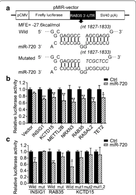
Using luciferase reporter assays, we next sought to verify direct regulation of these candidate targets by miR-720. Among these candidate target genes, except for KCTD15 with two predicted miR-720 binding sites in 3′-UTR, the rest of the target genes had only a predicted target site in 3′-UTR. We subcloned the partial 3′-UTRs contain-ing the miR-720-bindcontain-ing sites of these candidate target genes, such as Rab35, into the luciferase-based reporter vector pMIR-REPORT (Ambion, Austin, Texas, US), and cotransfected the reporter constructs in HEK293T cells with the pre-miR-720 precursor or negative control (Fig. 3a). Among these reporter constructs, miR-720 sig-nificantly suppressed the luciferase activity of the reporter vector containing binding sites in INSIG1, KCTD15, METTL2B, NRXN3, Rab35, RASAL2, or TET2, but not in CHP, PER2, or SGK3 for miR-720 targeting (Fig. 3b). To further confirm the predicted miRNA binding sites, seven base pair mutations of the miR-720 binding sites on the INSIG1, KCTD15, METTL2B, NRXN3, Rab35, RASAL2, and TET2 genes were generated by site-directed mutagen-esis. Because KCTD15 has two predicted binding sites in its 3′-UTR, we performed molecular cloning for single site mutations to obtain vectors named MUT1-KCTD15, MUT2-KCTD15, and double site mutation vectors named MUT1,2-KCTD15. As expected, once the miR-720 bind-ing sites in the 3′-UTR of these candidate target mRNAs were mutated, the luciferase activities could not be sup-pressed by miR-720 (Fig. 3c). Taken together, the results of these reporter assays showed that miR-720 could directly bind to the 3′-UTR of INSIG1, KCTD15, and RAB35.
Rab35 is a target of miR‑720 in HeLa cells
The studies suggested that INSIG1 is an insulin-induced gene and encodes an endoplasmic reticulum (ER) mem-brane protein that plays a critical role in regulating cho-lesterol concentrations in cells [42–46]; and Kctd15 has a role in regulating the neural crest formation in the embryo [47, 48]. And Rab35 was involved in breast can-cer cell migration processes [36] and promotes cell–cell adhesion [37]. Because miR-720 could induce cell migra-tion in HeLa cells, we focus our initial research empha-sis on Rab35. Firstly we further confirmed whether the mRNA and protein expression levels of Rab35 could have been regulated by miR-720 in cervical cancer HeLa cells. The results showed that mRNA expression of Rab35 was not significantly affected by both the overexpression and suppressed expression of miR-720 (Fig. 4a, b). How-ever, the protein level of Rab35 was negatively regulated by miR-720 in HeLa cells as expected (Fig. 4c). Thus, we propose that miR-720 plays a role in the regulation of Rab35 at the translational level in HeLa cells.
Rab35 can suppress cell migration of HeLa cells
Then we wondered whether Rab35 had negative effects on cell migration in HeLa cells compared with miR-720. So we constructed a miR-720 insensitive Rab35 expres-sion vector (pcDNA3.0-Rab35) that only included the coding sequence of Rab35 on the base of the
pcDNA3.0-Vector, to escape miR-720 targeting. The overexpression level of this vector was assessed by west-ern blotting, and the results showed that the vector suc-cessfully induced Rab35 overexpression (Fig. 5a). Then, we assayed the impact of forced expression of Rab35 on cell migration of HeLa cells. Using the wound healing assay, the overexpression of Rab35 inhibited the healing of the scratch when compared to the control group trans-fected with the empty pcDNA3.0-Vector (Fig. 5b). In the other two types of cell migration assays (Transwell® migration assay and real-time cell analysis), we con-sistently found that Rab35 had the ability to inhibit cell migration of HeLa cells (Fig. 5c, d). These results showed that Rab35 has an opposite effect on HeLa cell migration, in comparison with miR-720, indicating that it may be a functional target of miR-720 in HeLa cells.
miR‑720 mediates cell migration by targeting Rab35 in HeLa cells
In the previous experiments, overexpression of miR-720 could promote cell migration of HeLa, and forced expres-sion of Rab35 could suppress cell migration. To further demonstrate whether Rab35 is the actual functional tar-get of miR-720 acting on cell migration of HeLa cells, we cotransfected the miR-720 mimic and the miR-720 insensitive Rab35 vector into HeLa cells, then conducted a Transwell® migration assay. Even though there is over-expression of miR-720, the miR-720 insensitive Rab35
vector can still restore Rab35 expression and antagonize the positive effect of miR-720 on cell migration of HeLa cells, finally resulting in inhibition of cell migration, com-pared with the negative control group transfected with the miR-720 mimic and the empty pcDNA3.0-Vector (Fig. 6a, b). Thus, we postulate that miR-720 exerts its positive function on cell migration of HeLa cells by nega-tively regulating Rab35, which can suppress cell migra-tion of HeLa cells.
Molecular mechanism of the miR‑720 effect on cell migration of HeLa cells
vimentin levels; E-cadherin was increased and vimentin was decreased (Fig. 7b, c). Collectively, these results sug-gest that the intrinsic mechanism of miR-720 promotion on cell migration by downregulation of Rab35 is essentially through the activation of the EMT in HeLa cells.
Discussion
Tumor metastasis remains a major cause of cancer-related death. Metastasis is comprised of various
as coordinated regulators of ECM remodeling during cancer cell migration and invasion [53]. We previously reported that miR-720 expression is significantly upreg-ulated in cervix uteri squamous cell carcinoma tissue when compared to normal cervix uteri tissue, indicating
that miR-720 is closely related to the pathological pro-cesses of cervical cancer [17]. Some studies have reported that miR-720 impedes cell invasion and migration, and then inhibits metastasis in breast cancer by directly tar-geting TWIST1 [16]. In the present study, we showed that overexpression of miR-720 leads to a reduction of E-cadherin and an increase of vimentin protein levels in cervical cancer cell lines, which indicates that miR-720 can induce EMT in cervical cancer cells and promote cell migration. In combination with our previous studies and reports from other investigators, we propose that miR-720 may be an important regulator of cell migration and invasion in various cancers, in spite of its functional dif-ferences that are dependent on distinct cellular microen-vironments. It was reported that individual miRNA has the potential to regulate hundreds of targets [54]. In addi-tion, a miRNA can not only directly affect the levels of target mRNA but also indirectly affect the levels of tar-get mRNA, in other words, a miRNA can have secondary effects or even tertiary effects on its target mRNA [55]. Besides, the effects of a miRNA on regulating its targets crucially depends on its expression levels and the regula-tion happened to have a cell-type dependent effect [56–
58]. Therefore, we propose that miR-720 may regulate the cell migration processes in various cancer cells by distinct molecular mechanism.
Since miR-720 was discovered, it has been widely studied as a miRNA. A study showed that miR-720 could negatively regulate p63 involved in an autoregula-tory feedback loop, which then contributed to epithelial
Fig. 6 Restoration of Rab35 expression reverses the promoting effect of miR-720 on cell migration. Representative photographs (a) and statistical
results (b) of Transwell® assays show that while transfecting miR-720 mimics could lead to a significant increase in cell migration ability compared with cells transfected with the microRNA mimic negative control, cotransfecting pcDNA3.0-Rab35 and miR-720 mimics resulted in a dramatic decrease in cell migration ability compared with the negative group cotransfected with miR-720 mimics and the pcDNA3.0-Vector. *p< 0.05
Fig. 7 Detection of E-cadherin and vimentin expression by western
development processes [59]. The miR-720 could sup-press cell migration and invasion in breast cancer by directly targeting TWIST1 [16]. Furthermore, miR-720 participates in the control of the stem cell phenotype of human dental pulp cells by directly repressing NANOG levels [12]. miR-720 is closely related to the patho-logical processes of various tumors [17]. A more recent study showed that overexpression of miR-720 in pri-mary human CD8+ T cells inhibits the T cell receptor (TCR) stimulation-induced proliferation in patients with chronic hepatitis B virus (HBV) infection, which sug-gests that therapies targeting miR-720 may help restore impaired immunity in chronic HBV-infected patients [60]. However, another study reported that miR-720 is probably a fragment from a tRNA, rather than an miRNA [11]. And other report showed that, as a tRNA-derived miRNA, miR-720 is one of the four most abun-dant MCF7 extracellular vesicle miRNAs, suggesting that miR-720 may be a new biomarker in breast cancer cells [61]. Although there is controversy over whether miR-720 is a miRNA or a tRNA-derived RNA fragment (tRF), functional studies of miR-720 will provide valuable infor-mation concerning the possible clinical significance of this small non-RNA molecule.
As a key regulator of endosomal membrane trafficking and the actin cytoskeleton, Rab35 in this study was found to be directly regulated by miR-720 at the posttranscrip-tional level. The small GTPase, Rab35, a member of the RAS oncogene family, regulates many essential cellular processes, such as recycling from endosomes, exosome release, cytokinesis, and actin cytoskeleton organiza-tion by cycling between a GTP-bound active form and a GDP-bound inactive form. Active Rab35 controls recy-cling pathways to increase cell adhesion and decrease cell migration [32]. It is noteworthy that many studies have demonstrated a close relationship between Rab35 and Arf6, which is also a small GTPase. Both these two small GTPases are involved in functionally related cell pro-cesses, such as cell migration and cytokinesis, endocyto-sis, actin remodeling, sorting, and recycling of adhesion molecules. However, the activities of Rab35 and Arf6 are inversely coordinated [24, 27, 62, 63]. To the best of our knowledge, this is the first study to provide experimental evidences that Rab35 is posttranscriptionally regulated by miR-720, which then contributes to the regulation of cell migration. Based upon these observations, it is important to further validate the functions of the miR-720/Rab35 axis in tumor metastasis and invasion in animal models and clinical tissues in future studies.
Conclusions
Our results show that miR-720 promotes cell migra-tion in cervical cancer cells by directly targeting
Rab35. Targeting the miR-720/Rab35 axis is therefore a promising therapeutic strategy for cervical cancer treatment.
Methods Cell culture
HeLa cells and HEK293T cells were obtained from the Type Culture Collection of the Chinese Academy of Sci-ences (Shanghai, China). HeLa cells were cultured in Minimum Essential Medium (Gibco/Life Technologies, US) and HEK293T cells were incubated in Dulbecco’s Modified Eagle’s Medium (Gibco/Life Technologies, US). The base medium was supplemented with 10 % fetal bovine serum (Gibco/Life Technologies, US) to make the complete growth medium, and the cells were incubated at 37 °C with 5 % CO2.
Oligonucleotide transfection
The pre-miR-720, miR-720 mimics, inhibitor, and cor-responding controls were designed and synthesized by RiboBio (Guangzhou, China). HEK293T cells were trans-fected with pre-miR-720 or pre-miR-control at a final concentration of 100 nM. HeLa cells were transfected with mimics at a final concentration of 100 nM, or trans-fected with inhibitor at a final concentration of 200 nM using Lipofectamine® RNAiMAX reagent (Invitrogen, US), following the manufacturer’s instructions. After 48 h, the cells were harvested for further analysis.
RNA extraction and quantitative real‑time PCR
Total RNA was extracted using TRIzol® Reagent (Life Technologies, US) according to the manufacturer’s pro-tocol. One µg of total RNA was reverse transcribed into cDNA with a FastQuant RT Kit (Tiangen Biotech, Bei-jing, China). Quantitative analyses of miR-720 were performed using the Bulge-LoopTM miRNA quantita-tive RT-PCR primer (RiboBio, Guangzhou, China) and/ or a S-Poly(T) real-time PCR assay of microRNAs assays [64], and SYBR Select Master Mix (Life Technologies) on an ABI Prism 7500 real-time PCR system (Applied Bio-systems, Foster City, CA, USA). Quantitative analysis of Rab35 was performed using designed qPCR primers
(for-ward, 5′-ACGACCACCTCTTCAAGCTG-3′; reverse,
5′-CCGGATCTTGAAATCCACTC-3′) and the same
Microarray analysis
After the experimental HEK293T group was transfected with pre-miR-720 and the control HEK293T group was transfected with the pre-miR-control for 24 h, the HEK293T cells were lysed with TRIzol® reagent (Life Technologies, US) and total RNA was extracted according to the manufacturer’s protocol. The RNA samples were then sent to the Shanghai Biotechnology Corporation to perform microarray analysis (Affymetrix GeneChip® Human Genome U133 Plus 2.0 Array). Each group sam-ple set was comprised of three replicates. The Affymetrix GeneChip® system was used for hybridization, staining, scanning, and imaging of the arrays. Raw data were ana-lyzed with the Affymetrix GeneChip® Operating Soft-ware (GCOS1.4) using the manufacturer’s default analysis settings and global scaling as the normalization method. One-way analysis of variance (ANOVA) was performed with GeneSpring 7.31 software (Agilent, Santa Clara, CA, USA) to identify genes whose expression changed signifi-cantly between the pre-miR-720 group and the control.
Luciferase reporter assay
The putative binding sites of the 3′-UTR of the human genes for miR-720 targeting were predicted by TargetS-can Human (http://www.targetscan.org/) computational methods.
The partial 3′-UTR fragment (about 250 bp) of these potential targets, including predicted binding sites, as our previous report [65], were separately cloned into
the pMIR-REPORT™ miRNA Expression Reporter
Vec-tor (Applied Biosystems) at the Mlu I/Hind III site. The primers were designed as Table 1.
The mutant 3′UTR inserts, with an opposite mutation in the miRNA seed sequence binding sites, were gener-ated by overlapping PCR methods [66] using the mutated primers, listed as Table 1.
Wild-type and mutation inserts were confirmed by DNA sequencing. HEK293T cells were seeded in triplicate at a density of 2 × 10⁵ cells per 24-well plate. The
follow-ing day, cells were transfected with 100 ng wild-type or mutated firefly reporter vector, 10 ng of the control vector containing Renilla luciferase, and pRL-TK (Promega, US), at a final concentration of 50 nM miRNA, or negative con-trol (NC) precursors, using 1.5 μL Lipofectamine® 2000
(Invitrogen) per well. Cells were assayed 48 h after trans-fection with the Dual Luciferase Assay (Promega) accord-ing to the manufacturer’s instructions. Each experiment was repeated at least in triplicate. The efficiency of trans-fection was normalized using the Renilla luciferase activity.
The pcDNA expression plasmids and plasmid transfection
The coding sequence of Rab35 was ampli-fied from HeLa cell cDNA by PCR using primers:
forward, 5′-CGCGGATCCATGGCCCGGGACTACGAC
CA-3′; reverse, 5′-CCGCTCGAGTTAGCAGCAGCGT
TTCTTTC-3′. The PCR fragment was inserted using
BamH I/Xho I into the pcDNA3.0-Vector to generate pcDNA3.0-Rab35. The empty pcDNA3.0-Vector was used as the negative control. HeLa cells were transfected with plasmid using the Lipofectamine® 2000 Transfec-tion Reagent (Invitrogen) according to the manufac-turer’s protocol. After 48 h, the cells were harvested for further analysis.
Western blot analysis
Total cell extracts (40 µg) were resolved on a 10 % SDS-PAGE and transferred to nitrocellulose membranes (Mil-lipore, Germany). Membranes were blocked with 5 % skim milk (BioSharp, US) and further incubated with the following primary antibodies: Rab35 and the mesenchy-mal cell marker vimentin (1:2000; San Ying Biotechnol-ogy, China), the epithelial cell marker E-cadherin (1:200; BD Biosciences, US), and GAPDH (1:10,000; Ray Anti-body Beijing, China). After washing, membranes were incubated with a 1:10,000 dilutions of HRP-conjugated secondary antibodies (Santa Cruz Biotechnology, Santa Cruz, CA, USA). After washing six times with TTBS, with each wash lasting 5 min, the membranes were incubated with Pierce ECL western blotting substrate (Thermo Fisher Scientific, US), and the protein bands were visualized by autoradiography and quantified by Image J software (NIH, Bethesda, MD, USA).
Cell proliferation assay
For the HeLa cell proliferation assay, cells were tran-siently transfected for 24 h, and then seeded into 96-well plates (6 × 10³ cells/well), with each group comprised of
six replicates. The cell growth was regarded as the initial value when the cells had been seeded for 2 h. At indi-cated times, cells were incubated with 10 μL of 5 mg/
mL 3-4,5-dimethyl-2-thiazolyl)-2,5-diphenyl-2-H-tetra-zolium bromide (MTT) for 4 h at 37 °C, and then the generated crystals were dissolved by dimethyl sulfoxide (DMSO). Absorbance was measured at 450 nm using a microplate reader (Biotech) every 24 h for four consecu-tive days.
Wound healing assay
After transfection for 24 h in 6-well plates, cells were replated into 12-well plates (2.5 × 10⁵ cells per well), in
subjected to photography using a light microscope. After incubation for 24 h, additional photographs were taken. Wound healing ability was determined by measuring the scraped area alteration because of cell migration using Image J software.
Transwell® assay
After transfection for 24 h, HeLa cells (2.5 × 10⁴
cells) were suspended in 300 µL of Minimal Essential Medium (MEM) without serum and then reseeded into
the upper chamber of the Transwell® inserts (8 µm pore size; Corning, USA) of a companion 24-well plate in pre-warmed containing 10 % FBS. After incubating at 37 °C for 24 h, cells on the top surface of the insert were removed by gentle wiping with a cotton swab, and the cells which migrated to the bottom surface of the insert were fixed in 4 % paraformaldehyde for 15 min, stained with 0.1 % crystal violet (Sigma-Aldrich, St. Louis, MO, USA) for 15 min, rinsed in PBS, and then photographed. Cell migration ability was determined
Table 1 Primers used in this study
Underlined characters indicate regions of the miR-720 seed sequences for mutation in luciferase reporter assay
Name Sequences (5′–3′)
3′UTR-CHP-S CAAAACGCGTCTGTCAACACAAACCTGC
3′UTR-CHP-A CGTAAAGCTTGCCAGACCCCTGAACTAT
3′UTR-INSIG1-S GCTTACGCGTGAGGGAAATGTCTTGGAG
3′UTR-INSIG1-A CGCCAAGCTTGATGTTCAAATCTGGTAG
3′UTR-INTS7-S ATCGACGCGTACAGTTTGGTTTTTCATA
3′UTR-INTS7-A ACTCAAGCTTAAACAAGAAAGAACAATC
3′UTR-KCTD15-S CAACACGCGTATCGTCACAGGAGAAGGT
3′UTR-KCTD15-A AGACAAGCTTTCCAACCCCGCCCCAAC
3′UTR-METTL2B-S ATATACGCGTCCCGTTGTGTTTCCGAGC
3′UTR-METTL2B-A CGGCAAGCTTAGCCCTTCACTAACCCCT
3′UTR-NRXN3-S ACTTACGCGTCTGAGGGGAAAAATGGCT
3′UTR-NRXN3-A TAACAAGCTTTGGCTGGGTGAATGGAC
3′UTR-RAB35-S TTAAACGCGTAAGGAGGGTGCTGTGGAG
3′UTR-RAB35-A ACACAAGCTTCCCATAACCCCCATTCAT
3′UTR-SGK3-S ATATACGCGTGAGACCATCCTGGGCAAC
3′UTR-SGK3-A CCGCAAGCTTCCAGTTATTCTCCTTTAC
3′UTR-RASAL2-S ACTAACGCGTCGACTTCCAAGGTCAATG
3′UTR-RASAL2-A CAGAAAGCTTGTCCAACCAGAACCAGTC
3′UTR-TET2-S CGAGACGCGTAGACTGGTAAAGTGTGGT
3′UTR-TET2-A CACCGAAGCTTATTCACTATTTCTGCCA
INSIG1-MUT-F GAAGTTATTAGATGAAAGGTCGCTCTGAATCTTTAAAACAGAC
INSIG1-MUT-R GTCTGTTTTAAAGATTCAGAGCGACCTTTCATCTAATAACTTC
KCTD15-site1-MUT-F TGTGGACTCCTCCCAGTTGTCGCTCTATGTGCTTTGCCGGGAG
KCTD15-site1-MUT-R CTCCCGGCAAAGCACATAGAGCGACAACTGGGAGGAGTCCACA
KCTD15-site2-MUT-F GAGGGTCCAAAGCTGGCCGTCGCTCCACCAGGGTCCCAGGTGT
KCTD15-site2-MUT-R ACACCTGGGACCCTGGTGGAGCGACGGCCAGCTTTGGACCCTC
METTL2B-MUT-F GTCCAGCCTGGGCAAAATTCGCTCTGACCCTGAATCTGAAAGT
METTL2B-MUT-R ACTTTCAGATTCAGGGTCAGAGCGAATTTTGCCCAGGCTGGAC
NRXN3-MUT-F AGGAAAAAAACTCAAAACAAATCGCTCTGAGACTATTGCCATA
NRXN3-MUT-R TATGGCAATAGTCTCAGAGCGATTTGTTTTGAGTTTTTTTCCT
RAB35-MUT-F CCTCTGAGCGATCAGGCCTCCGGTCGCTCCGTGTGCTTGCAAATTC
RAB35-MUT-R GAATTTGCAAGCACACGGAGCGACCGGAGGCCTGATCGCTCAGAGGC
RASAL2-MUT-F AGTAGGAACTGTTGTGTGTCGCTCTCATGAGCCTGTAGGTTCA
RASAL2-MUT-R TGAACCTACAGGCTCATGAGAGCGACACACAACAGTTCCTACT
TET2-MUT-F ATGTAGAAGACTCTTATGTCGCTCTTAATGCAGAGAAGGCCTT
by counting the stained cells in the bottom surface of the insert under a light microscope (100×) in eight ran-domly selected fields. The average number of cells in the eight fields was taken as the final result. Each group had three replicates and each experiment was repeated three times.
Real‑time cell analysis
For real-time cell analysis (RTCA), HeLa cells were transiently transfected for 24 h, and 6 × 10⁴ cells
were suspended in 100 µL of MEM without serum in the upper chamber (ACEA Biosciences; Hangzhou, China) according to the basic cell migration assay pro-tocol of the RTCA xCELLigence system (ACEA Bio-sciences). Each group had four replicates, and the cell migration index was calculated by real-time detection for 48 h.
Statistical analysis
Data were expressed as mean ± SEM. Student’s t test (unpaired, two tailed) was used in the analysis of dif-ferences between the experimental groups and control groups, with p < 0.05 considered statistically significant. Statistical analyses were performed using GraphPad Prism, version 5.0 software.
Authors’ contributions
MG and YT designed experiments and analyzed data; YT, YLin, CL, XH, YLiu, MH and JL performed experiments; MG, YT and GS wrote the manuscript; MG, WL, TW and GS supervised the project. All authors read and approved the final manuscript.
Author details
1 College of Life Sciences and State Key Laboratory of Virology, Wuhan Univer-sity, 430072 Wuhan, People’s Republic of China. 2 Department of Pathology, Zhongnan Hospital, Wuhan University, 430071 Wuhan, People’s Republic of China. 3 School of Basic Medical Sciences, Wuhan University, 430071 Wuhan, People’s Republic of China. 4 Department of Respiratory and Critical Care Medicine, Tongji Hospital, Tongji Medical College, Huazhong University of Sci-ence and Technology, 430030 Wuhan, China.
Acknowledgements
This work was supported by National Natural Science Foundation of China Grants 30871245 and 31271511 (to M. Guo), 81170049 and 81470252 (to T. Wang), 31370187 (to G. Sun) and Fundamental Research Funds for the Central Universities Grant 2042014KF0243 (to M. Guo).
Compliance with ethical guidelines
Competing interests
The authors declare that they have no competing interests.
Received: 2 August 2015 Accepted: 22 September 2015
Additional file
Additional file 1.Table S1. Results of microarray analysis.
References
1. Bushati N, Cohen SM. microRNA functions. Annu Rev Cell Dev Biol. 2007;23:175–205.
2. Bartel DP. MicroRNAs: genomics, biogenesis, mechanism, and function. Cell. 2004;116(2):281–97.
3. Ambros V. The functions of animal microRNAs. Nature. 2004;431(7006):350–5.
4. Wightman B, Ha I, Ruvkun G. Posttranscriptional regulation of the hetero-chronic gene lin-14 by lin-4 mediates temporal pattern formation in C. elegans. Cell. 1993;75(5):855–62.
5. Lee RC, Feinbaum RL, Ambros V. The C. elegans heterochronic gene lin-4 encodes small RNAs with antisense complementarity to lin-14. Cell. 1993;75(5):843–54.
6. Bartel DP. MicroRNAs: target recognition and regulatory functions. Cell. 2009;136(2):215–33.
7. Lewis BP, Burge CB, Bartel DP. Conserved seed pairing, often flanked by adenosines, indicates that thousands of human genes are microRNA targets. Cell. 2005;120(1):15–20.
8. Croce CM. Causes and consequences of microRNA dysregulation in cancer. Nat Rev Genet. 2009;10(10):704–14.
9. Garzon R, Calin GA, Croce CM. MicroRNAs in cancer. Annu Rev Med. 2009;60:167–79.
10. Alvarez-Garcia I, Miska EA. MicroRNA functions in animal development and human disease. Development. 2005;132(21):4653–62.
11. Schopman NC, Heynen S, Haasnoot J, Berkhout B. A miRNA-tRNA mix-up: tRNA origin of proposed miRNA. RNA Biol. 2010;7(5):573–6.
12. Hara ES, Ono M, Eguchi T, Kubota S, Pham HT, Sonoyama W, Tajima S, Takigawa M, Calderwood SK, Kuboki T. miRNA-720 controls stem cell phenotype, proliferation and differentiation of human dental pulp cells. PLoS One. 2013;8(12):e83545.
13. Nonaka R, Miyake Y, Hata T, Kagawa Y, Kato T, Osawa H, Nishimura J, Ikenaga M, Murata K, Uemura M et al. Circulating miR-103 and miR-720 as novel serum biomarkers for patients with colorectal cancer. Int J Oncol. 2015;47(3):1097–102.
14. Jones CI, Zabolotskaya MV, King AJ, Stewart HJ, Horne GA, Che-vassut TJ, Newbury SF. Identification of circulating microRNAs as diagnostic biomarkers for use in multiple myeloma. Br J Cancer. 2012;107(12):1987–96.
15. Wang X, Kuang Y, Shen X, Zhou H, Chen Y, Han Y, Yuan B, Zhou J, Zhao H, Zhi Q, et al. Evaluation of miR-720 prognostic significance in patients with colorectal cancer. Tumour Biol J Int Soc Oncodev Biol Med. 2015;36(2):719–27.
16. Li LZ, Zhang CZ, Liu LL, Yi C, Lu SX, Zhou X, Zhang ZJ, Peng YH, Yang YZ, Yun JP. miR-720 inhibits tumor invasion and migration in breast cancer by targeting TWIST1. Carcinogenesis. 2014;35(2):469–78.
17. Lin Y, Zeng Y, Zhang F, Xue L, Huang Z, Li W, Guo M. Characterization of microRNA expression profiles and the discovery of novel microRNAs involved in cancer during human embryonic development. PLoS One. 2013;8(8):e69230.
18. Jemal A, Bray F, Center MM, Ferlay J, Ward E, Forman D. Global cancer statistics. CA Cancer J Clin. 2011;61(2):69–90.
19. Etoh K, Fukuda M. Structure-function analyses of the small GTPase Rab35 and its effector protein centaurin-beta2/ACAP2 during neurite outgrowth of PC12 cells. J Biol Chem. 2015;290(14):9064–74.
20. Kobayashi H, Fukuda M. Rab35 establishes the EHD1-association site by coordinating two distinct effectors during neurite outgrowth. J Cell Sci. 2013;126(Pt 11):2424–35.
21. Kanno E, Ishibashi K, Kobayashi H, Matsui T, Ohbayashi N, Fukuda M. Comprehensive screening for novel Rab-binding proteins by GST pull-down assay using 60 different mammalian Rabs. Traffic (Copenhagen, Denmark). 2010;11(4):491–507.
22. Kobayashi H, Etoh K, Ohbayashi N, Fukuda M. Rab35 promotes the recruitment of Rab8, Rab13 and Rab36 to recycling endosomes through MICAL-L1 during neurite outgrowth. Biol Open. 2014;3(9):803–14. 23. Kobayashi H, Etoh K, Fukuda M. Rab35 is translocated from Arf6-positive
perinuclear recycling endosomes to neurite tips during neurite out-growth. Small GTPases. 2014;5:e29290.
25. Chevallier J, Koop C, Srivastava A, Petrie RJ, Lamarche-Vane N, Pres-ley JF. Rab35 regulates neurite outgrowth and cell shape. FEBS Lett. 2009;583(7):1096–101.
26. Hsu C, Morohashi Y, Yoshimura S, Manrique-Hoyos N, Jung S, Lauterbach MA, Bakhti M, Gronborg M, Mobius W, Rhee J, et al. Regulation of exo-some secretion by Rab35 and its GTPase-activating proteins TBC1D10A-C. J Cell Biol. 2010;189(2):223–32.
27. Chesneau L, Dambournet D, Machicoane M, Kouranti I, Fukuda M, Goud B, Echard A. An ARF6/Rab35 GTPase cascade for endocytic recycling and successful cytokinesis. Curr Biol CB. 2012;22(2):147–53.
28. Zhang J, Fonovic M, Suyama K, Bogyo M, Scott MP. Rab35 controls actin bundling by recruiting fascin as an effector protein. Science. 2009;325(5945):1250–4.
29. Dambournet D, Machicoane M, Chesneau L, Sachse M, Rocancourt M, El Marjou A, Formstecher E, Salomon R, Goud B, Echard A. Rab35 GTPase and OCRL phosphatase remodel lipids and F-actin for successful cytoki-nesis. Nat Cell Biol. 2011;13(8):981–8.
30. Chua CE, Lim YS, Tang BL. Rab35–a vesicular traffic-regulating small GTPase with actin modulating roles. FEBS Lett. 2010;584(1):1–6. 31. Marat AL, Ioannou MS, McPherson PS. Connecdenn 3/DENND1C binds
actin linking Rab35 activation to the actin cytoskeleton. Mol Biol Cell. 2012;23(1):163–75.
32. Chaineau M, Ioannou MS, McPherson PS. Rab35: GEFs, GAPs and effec-tors. Traffic (Copenhagen, Denmark). 2013;14(11):1109–17.
33. Egami Y, Fukuda M, Araki N. Rab35 regulates phagosome formation through recruitment of ACAP2 in macrophages during FcgammaR-mediated phagocytosis. J Cell Sci. 2011;124(Pt 21):3557–67.
34. Shim J, Lee SM, Lee MS, Yoon J, Kweon HS, Kim YJ. Rab35 mediates trans-port of Cdc42 and Rac1 to the plasma membrane during phagocytosis. Mol Cell Biol. 2010;30(6):1421–33.
35. Charrasse S, Comunale F, De Rossi S, Echard A, Gauthier-Rouviere C. Rab35 regulates cadherin-mediated adherens junction formation and myoblast fusion. Mol Biol Cell. 2013;24(3):234–45.
36. Zhu Y, Shen T, Liu J, Zheng J, Zhang Y, Xu R, Sun C, Du J, Chen Y, Gu L. Rab35 is required for Wnt5a/Dvl2-induced Rac1 activation and cell migra-tion in MCF-7 breast cancer cells. Cell Signal. 2013;25(5):1075–85. 37. Allaire PD, Seyed Sadr M, Chaineau M, Seyed Sadr E, Konefal S, Fotouhi M,
Maret D, Ritter B, Del Maestro RF, McPherson PS. Interplay between Rab35 and Arf6 controls cargo recycling to coordinate cell adhesion and migra-tion. J Cell Sci. 2013;126(Pt 3):722–31.
38. Friedman RC, Farh KK, Burge CB, Bartel DP. Most mammalian mRNAs are conserved targets of microRNAs. Genome Res. 2009;19(1):92–105. 39. Grimson A, Farh KK, Johnston WK, Garrett-Engele P, Lim LP, Bartel DP.
MicroRNA targeting specificity in mammals: determinants beyond seed pairing. Mol Cell. 2007;27(1):91–105.
40. John B, Enright AJ, Aravin A, Tuschl T, Sander C, Marks DS. Human Micro-RNA targets. PLoS Biol. 2004;2(11):e363.
41. Lim LP, Lau NC, Garrett-Engele P, Grimson A, Schelter JM, Castle J, Bartel DP, Linsley PS, Johnson JM. Microarray analysis shows that some microRNAs downregulate large numbers of target mRNAs. Nature. 2005;433(7027):769–73.
42. Ren R, Zhou X, He Y, Ke M, Wu J, Liu X, Yan C, Wu Y, Gong X, Lei X, et al. Crystal structure of a mycobacterial Insig homolog provides insight into how these sensors monitor sterol levels. Science. 2015;349(6244):187–91. 43. Burg JS, Powell DW, Chai R, Hughes AL, Link AJ, Espenshade PJ. Insig
regulates HMG-CoA reductase by controlling enzyme phosphorylation in fission yeast. Cell Metab. 2008;8(6):522–31.
44. Sun LP, Seemann J, Goldstein JL, Brown MS. Sterol-regulated transport of SREBPs from endoplasmic reticulum to Golgi: Insig renders sorting signal in Scap inaccessible to COPII proteins. Proc Natl Acad Sci USA. 2007;104(16):6519–26.
45. Radhakrishnan A, Ikeda Y, Kwon HJ, Brown MS, Goldstein JL. Sterol-regulated transport of SREBPs from endoplasmic reticulum to Golgi: oxysterols block transport by binding to Insig. Proc Natl Acad Sci USA. 2007;104(16):6511–8.
46. Goldstein JL, DeBose-Boyd RA, Brown MS. Protein sensors for membrane sterols. Cell. 2006;124(1):35–46.
47. Zarelli VE, Dawid IB. The BTB-containing protein Kctd15 is SUMOylated in vivo. PLoS One. 2013;8(9):e75016.
48. Zarelli VE, Dawid IB. Inhibition of neural crest formation by Kctd15 involves regulation of transcription factor AP-2. Proc Natl Acad Sci USA. 2013;110(8):2870–5.
49. Polyak K, Weinberg RA. Transitions between epithelial and mesenchymal states: acquisition of malignant and stem cell traits. Nat Rev Cancer. 2009;9(4):265–73.
50. Miettinen PJ, Ebner R, Lopez AR, Derynck R. TGF-beta induced transdiffer-entiation of mammary epithelial cells to mesenchymal cells: involvement of type I receptors. J Cell Biol. 1994;127(6 Pt 2):2021–36.
51. Quail DF, Joyce JA. Microenvironmental regulation of tumor progression and metastasis. Nat Med. 2013;19(11):1423–37.
52. Chaffer CL, Weinberg RA. A perspective on cancer cell metastasis. Sci-ence. 2011;331(6024):1559–64.
53. Zhou L, Liu F, Wang X, Ouyang G. The roles of microRNAs in the regula-tion of tumor metastasis. Cell Biosci. 2015;5:32.
54. Vidigal JA, Ventura A. The biological functions of miRNAs: lessons from in vivo studies. Trends Cell Biol. 2015;25(3):137–47.
55. Cloonan N, Brown MK, Steptoe AL, Wani S, Chan WL, Forrest AR, Kolle G, Gabrielli B, Grimmond SM. The miR-17-5p microRNA is a key regulator of the G1/S phase cell cycle transition. Genome Biol. 2008;9(8):R127. 56. Lu LF, Gasteiger G, Yu IS, Chaudhry A, Hsin JP, Lu Y, Bos PD, Lin LL, Zawislak
CL, Cho S, et al. A Single miRNA-mRNA Interaction Affects the Immune Response in a Context- and Cell-Type-Specific Manner. Immunity. 2015;43(1):52–64.
57. Tay Y, Tan SM, Karreth FA, Lieberman J, Pandolfi PP. Characterization of dual PTEN and p53-targeting microRNAs identifies microRNA-638/Dnm2 as a two-hit oncogenic locus. Cell Rep. 2014;8(3):714–22.
58. Denzler R, Agarwal V, Stefano J, Bartel DP, Stoffel M. Assessing the ceRNA hypothesis with quantitative measurements of miRNA and target abun-dance. Mol Cell. 2014;54(5):766–76.
59. Chikh A, Matin RN, Senatore V, Hufbauer M, Lavery D, Raimondi C, Ostano P, Mello-Grand M, Ghimenti C, Bahta A, et al. iASPP/p63 autoregulatory feedback loop is required for the homeostasis of stratified epithelia. EMBO J. 2011;30(20):4261–73.
60. Wang Y, Zhang Z, Ji D, Chen GF, Feng X, Gong LL, Guo J, Li ZW, Chen CF, Zhao BB, et al. Regulation of T cell function by microRNA-720. Sci Rep. 2015;5:12159.
61. Guzman N, Agarwal K, Asthagiri D, Yu L, Saji M, Ringel MD, Paulaitis ME. Breast Cancer-Specific miR Signature Unique to Extracellular Vesi-cles Includes “microRNA-like” tRNA Fragments. Mol Cancer Res MCR. 2015;13(5):891–901.
62. Uytterhoeven V, Kuenen S, Kasprowicz J, Miskiewicz K, Verstreken P. Loss of skywalker reveals synaptic endosomes as sorting stations for synaptic vesicle proteins. Cell. 2011;145(1):117–32.
63. Hanono A, Garbett D, Reczek D, Chambers DN, Bretscher A. EPI64 regu-lates microvillar subdomains and structure. J Cell Biol. 2006;175(5):803–13. 64. Kang K, Zhang X, Liu H, Wang Z, Zhong J, Huang Z, Peng X, Zeng Y, Wang Y, Yang Y, et al. A novel real-time PCR assay of microRNAs using S-Poly(T), a specific oligo(dT) reverse transcription primer with excellent sensitivity and specificity. PLoS One. 2012;7(11):e48536.
65. Lin Y, Li D, Liang Q, Liu S, Zuo X, Li L, Sun X, Li W, Guo M, Huang Z. miR-638 regulates differentiation and proliferation in leukemic cells by targeting cyclin-dependent kinase 2. J Biol Chem. 2015;290(3):1818–28. 66. Ho SN, Hunt HD, Horton RM, Pullen JK, Pease LR. Site-directed