R E S E A R C H
Open Access
Horizontal transfer of
vanA
between
probiotic
Enterococcus faecium
and
Enterococcus faecalis
in fermented soybean
meal and in digestive tract of growing pigs
Ning Li
1,2, Haitao Yu
1,2, Hongbin Liu
1,2, Yuming Wang
1,2, Junyan Zhou
1,2, Xi Ma
1,2, Zheng Wang
3,
Chengtao Sun
3and Shiyan Qiao
1,2*Abstract
Background:The aim of this study was to investigate the intergeneric transfer of vancomycin resistance genevanA between probiotic enterococci in the fermentation progress of soybean meal and in the digestive tract of growing pigs. OnevanAgenotype vancomycin resistantE. faeciumstrain, Efm4, and one chloramphenicol-resistantE. faecalis strain, Efs2, were isolated from twenty-nine probiotic basis feed material / additive samples. Forin vitroconjugation, Efm4 and Efs2 were used as starter to ferment soybean meal. Forin vivoconjugation, thirty growing pigs were randomly assigned to five groups (n= 6), treated with a basic diet, or supplemented with 10% fermented soybean meal, 1% Efm4, 5% Efs2 or a combination of 1% Efm4 + 5% Efs2 for 7 d, respectively. Fecal samples of pigs in each group were collected daily for the isolation and dynamic analysis of Efm4, Efs2 and transconjugants. The sequence types (STs) of Efm4, Efs2 and transconjugants were analyzed by multilocus sequence typing (MLST). The vanA harboring plasmid in Efm4 and transconjugants was analyzed by S1-pulsed field gel electrophoresis (PFGE) and further verified by multiple alignments.
Results: The results showed that, in FSBM, transconjugants were detected 1 h after the fermentation, with a conjugation frequency of ~ 10−3 transconjugants / recipient. Transconjugants proliferated with Efm4 and Efs2 in the first 8 h and maintained steadily for 10 d till the end of the experiment. Additionally,in vivoexperiment showed that transcojugants were recovered in one of six pigs in both FSBM and Efm4 + Efs2 groups, with conjugation frequency of ~ 10−5 and ~ 10−4, respectively. MLST revealed the ST of Efm4, Efs2 and transconjugants was ST1014, ST69 and ST69, respectively. S1-PFGE confirmed the existence of thevanA-harboring, 142,988-bp plasmid, which was also a multi-drug resistant plasmid containingTn1546-like transposon.
Conclusions:The findings revealed the potential safety hazard existing in the commercial probiotic enterococci in China, because the horizontal transfer from farm to fork could potentially pose a safety risk to the public.
Keywords:Chloramphenicol, Digestive tract, Enterococci, Fermented soybean meal, Growing pigs, Probiotics, Transconjugants,vanA, Vancomycin
© The Author(s). 2019Open AccessThis article is distributed under the terms of the Creative Commons Attribution 4.0 International License (https://creativecommons.org/licenses/by/4.0/), which permits unrestricted use, distribution, and reproduction in any medium, provided you give appropriate credit to the original author(s) and the source, provide a link to the Creative Commons license, and indicate if changes were made. The Creative Commons Public Domain Dedication waiver (https://creativecommons.org/publicdomain/zero/1.0/) applies to the data made available in this article, unless otherwise stated.
* Correspondence:[email protected]
1
State Key Laboratory of Animal Nutrition, College of Animal Science and Technology, China Agricultural University, Beijing 10093, China
2Beijing Key Laboratory of Bio-feed Additives, China Agricultural University,
Beijing 10093, China
Background
Enterococci species are widely distributed in the environ-ment and enable to colonize in the gastrointestinal tract in humans and most animals [1]. For decades, some entero-cocci strains like Enterococcus faecium and Enterococcus faecalisare used as probiotics in human and farm animals as a kind of lactic acid bacteria [2]. Previous studies have proven that enterococci can maintain a balance among the intestinal flora [3], promote the absorption of nutrients [4– 6] and improve the host immunity [7–9]. In spite of those probiotic properties, however, enterococci have also been known as one of the multiresistant pathogen, ranking among the leading causes of hospital-acquired infections worldwide [10–12].
Vancomycin therapy is one of the most efficient treat-ments for enterococci infections. While the emergence and rapid spread of vancomycin-resistant enterococci (VRE) represents a particular challenge [13,14], as there are few remaining therapeutic options for infections caused by VRE [15,16]. Glycopeptide resistance genesvanis proven to be the major vancomycin resistant mechanism known in the enterococci. Within all the ninevangene types [17],
vanAis the most frequently indentified one among entero-cocci isolates from clinical settings [18,19]. The ability of enterococci to acquire and exchange plasmids and mobile genetic elements that carry antimicrobial resistance gens (like vanA) has contributed to their role as multiresistant pathogens within both human and animals.
The spread of VRE as well as vanA is not restricted only clinically but they appear also in the community and natural environment as well as animal sectors. For instance, the emergence of VRE in food animal in Eur-ope is proven to have a high association with the use of avoparcin, a glycopeptide antibiotic as growth promoting agent in animal feed [20]. Avoparcin was never approved as a feed additive in China, and worldwide reports on VRE in the livestock industry is limited.
Recently, growing attention has been focused on the worrisome situation of VRE in hospitals or community care centers, which might be traced back to the consumed food, such as yogurt and pork, and eventually to the diet that farm animals intake [21]. Therefore, the aim of this study was to determine the VRE strains in commercial available probiotic enterococci in Beijing, China. More-over, to assess the potential security issue of these bacteria as an accumulator of antibiotic resistance genes (ARGs), horizontal dissemination ofvanAbetweenE. faeciumand
E. faecalisin fermented soybean meal (FSBM) and the di-gestive tract of growing pigs were also analyzed.
Materials and methods Bacterial isolates
Enterococci based FSBM and silage corn samples, as well as microbial feed additive samples were purchased
in swine farms and local husbandry and veterinary sta-tions in Beijing, China, during June to December, 2016. For FSBM and silage corn samples, enterococci enrich-ment was performed by adding 25 g sample into 225 mL of Buffered Peptone Water (Beijing Land Bridge, China), vortexed, and then incubated at 37 °C for 24 h. Then 1 mL of the enrichment was added into 9 mL Bile Esculin Azide (BEA; Beijing Land Bridge, China) broth and then incubated 37 °C for 24 h. The changing color of BEA broth from transparent dark brown to opaque black was a sign of the existence of enterococci. A loop of enrich-ment was streaked on BEA agar plate, incubated 37 °C for 24 h. For feed additive samples, 1 g or 1 mL of the sample was dissolved in 9 mL normal saline, then a loop of the solution was streaked on BEA agar plate, incu-bated 37 °C for 24 h.
The identification of suspected strains were performed by VITEK 2 Compact Automatic Bacterial Identification and Drug Sensitivity Analysis System (bioMerieux, France) using specific Gram-positive bacteria identifica-tion cards (bioMerieux, France), followed by further confirmation of 16S rDNA-based polymerase chain reac-tion (PCR) screen using universal primers 27-F and 1,492-R, and specific primers for E. faecium and E. fae-calis(Additional file1: Table S1).
Antimicrobial susceptibility tests
Isolates confirmed asE. faeciumor E. faecaliswere sub-jected to antimicrobial susceptibility testing to determine their resistance to twenty common antibiotics of nine categories (Additional file 1: Table S2) in accordance with the Clinical and Laboratory Standards Institute (CLSI) [22]. Each isolate was inoculated into 1 mL of sterilized normal saline by picking 3 to 5 colonies using cotton swab from an overnight culture on BHI agar to visually match a McFarland turbidity standard of 0.5. The bacteria liquid was evenly coated onto the surface of un-supplemented Mueller-Hinton (Oxoid, UK) agar plates using cotton swab. Each plate was pasted with five different antimicrobial disks (Beijing Tiantan, China), and then incubated at 37 °C for 24 h. The diameter of in-hibition zones was measured to the nearest millimeter and interpreted according to the CLSI standards and previous studies [22,23]. TheE. faecalisreference strain ATCC 29212 was used as the quality control.
Choice of donor and recipient strains
and Efs2 were extracted using Wizard® Genomic DNA Purification Kit (Promega, USA) according to manu-facturer’s protocol and further evaluated by PCR for the detection of van and chl genes. (Additional file 1: Table S1).
Amplified PCR products were purified using Wizard® SV Gel and PCR Clean-Up System (Promega, USA). Cleaned fragments were submitted to Sangon Biotech (Beijing, China) and sequenced from both the forward and reverse stands. The sequence was then analyzed using BLAST.
Freeze-dried powder of Efm4 and Efs2 were produced by National Feed Engineering Technology Research Center (Beijing, China), with initial bacteria counts ~ 1010CFU/g, respectively, then stored at −20 °C. Before use, the bacterial activity would be detected by measur-ing bacterial colony-formmeasur-ing ability after incubation for 24 h at 37 °C on BHI agar plates.
In vitroconjugation: soybean meal fermentation and sample collection
Fresh bacteria liquid cultures of Efm4 and Efs2 were pro-duced by overnight incubation in BHI broth at 37 °C, re-spectively, and sub-cultured before use. The initial concentration was adjusted to ~ 108CFU/mL for both Efm4 and Efs2. FSBM was conducted in aseptic fermen-tation bags (5 L, provided by Ministry of Agricultural Feed Industry Centre, Beijing, China) using the following ingredients: sterilized soybean meal (SBM), 500 g; glu-cose, 20 g; sterilized ddH2O, 435 mL; yeast, 5 g; bacteria
liquid, 50 mL, with the volume ratio was 1:5 (v/v, Efm4 / Efs2) [24, 25]. Two control groups with the same ingre-dients supplemented with 50 mL Efm4 liquid or 50 mL Efs2 liquid, respectively, were set.
All fermentation ingredients except Efm4 were pre-confirmed to be negative for vanA. BHI agar plates containing either 128 mg/L vancomycin or 64 mg/L chloramphenicol were used for detection of Efm4 and Efs2, respectively. BHI agar plates supple-mented with vancomycin and chloramphenicol were used for the detection of transconjugants. Five repli-cates were maintained for each treatment. Efm4, Efs2 and transconjugants were counted through serial plat-ing on selective BHI agar plates at 0, 1, 2, 4, 6, 8, 12, and 24 h, and then daily from day 2 to day 10.
On each selective plate, five clones were randomly se-lected, cultured, identified by species assessment as de-scribed above. Putative transconjugants obtained on double resistant selective plates were further confirmed by PCR amplification on the detection of van and chl
genes [26,27].
The conjugant frequency was calculated using the equation described by Liu et al. [28]:
Conjugation
ð Þfrequency¼Clones of Transconjugants CFUð =plateÞ Clones of Recipients CFUð =plateÞ
In vivoconjugation: animals, feeding and sample collection
All procedures used in these experiments were con-ducted in accordance with the Chinese Guidelines for Animal Welfare and were approved by the China Agri-cultural University Institutional Animal Care and Use Committee (Beijing, China) [29]. The pig cages, pigpens and feeding appliances were pre-disinfected, then con-firmed the absence of enterococci as well asvan andchl
genes using methods described previously [30].
A total of thirty barrows (Duroc × Landrace × York-shire) at the age of 60 d, with initial body weight of 15.38 ± 2.8 kg were individually housed and provided with a non-medicated corn-soybean meal basal diet and sterilized waterad libitum for 10 d. Fresh fecal samples were collected daily to confirm the absence VRE and
vanA. After acclimatization, pigs were randomly assigned to one control group (n= 6), fed with the same basal diet previously described; and four treatment groups (n= 6), fed with basal diet supplemented with 10% FSBM, 1% Efm4, 5% Efs2 or a combination of 1% Efm4 + 5% Efs2 for 7 d, respectively.
The estimation of FSBM consumption was about 5 kg/ d, so 5 bags of FSBM were performed daily, from −1 d to 6 d of the experiment as described above. The fer-mentation process lasted for 2 d. At this time point, the bacteria concentrate of Efm4 and Efs2 was both ~ 109 CFU/g FSBM. Then 5 kg fresh FSBM were uniformly dispersed into 45 kg basal diet to feed FSBM group bar-rows. Meanwhile, the initial bacteria concentrate of Efm4 in Efm4 group and Efm4 + Efs2 group was ~ 1.0 × 10⁸CFU/g feed, and the initial bacteria concentrate of Efs2 in Efs2 group and Efm4 + Efs2 group was ~ 5.0 × 10⁸CFU/g feed, respectively.
Fresh rectal feces sample of each pig was collected once every 24 h for serial plating and counting of Efm4, Efs2, and transconjugants using selective BHI agar plates. Donor, recipient and transconjugant strains on selective plates and then resistant genes were identified as described above.
Multilocus sequence typing
S1-PFGE and southern hybridization
S1-PFGE was performed as described previously [2]. Overnight incubated cells of Efm4, Efs2 and transconju-gants were embedded in InCert Agarose (Lonza, USA), respectively, followed by digestion with S1 nuclease (TaKaRa, Japan). The DNA restriction fragment were separated in 1% SeaKem Gold® Agarose (Lonza, USA) using a pulse gel electrophoresis apparatus (Bio-Rad, USA). Genomic DNA ofSalmonella serovarBraenderup strain H9812, digested with XbaI (TaKaRa, Japan), was used as a molecular standard.
Southern hybridization was performed on the S1-PFGE gel using DIG High Prime DNA Labeling and Detection Starter Kit II (Roche, Germany) with digoxin-labeled DNA probes specific for vanA, followed by man-ufacturer’s protocol.
Plasmid sequencing
The whole genome of Efm4 including vanA-harboring plasmid was sequenced using the Pacific Biosciences RS II (Pacific Biosciences, Menlo Park, CA, USA) sequen-cing platform.De novo assemblies of Pacbio Reads were performed by SMRT Analysis pipeline v2.3.0 in conjunc-tion with RS_HGAP_Assembly. Three protocols and additional assemblies were performed by minimus 2 from the AMOS package [32]. The open reading frames (ORFs) were identified and annotated with the prodigal.
The vanA-carrying plasmids from transconjugants ob-tained from FSBM and Efm4 + Efs2 group were partially sequenced (~ 13,000 bp) to verify their transfer into Efs2. By targeting the regions adjacent vanA, primers (Add-itional file2: Supplemental data 1) were designed based on the sequence ofvanA-carrying plasmid in Efm4. Mul-tiple alignment of acquire sequence was conducted using SnapGene (Version 4.2.6) against the sequence of
vanA-carrying plasmid Efm4.
Results
Antimicrobial susceptibility
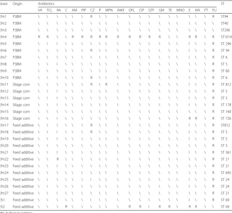
Among twenty-nine enterococci isolates, two strains (6.90%, 2/29) were susceptible to all the twenty antibi-otics tested; the remaining strains (93.10%, 27/29) show-ing resistance to at least one antibiotic (Table 1). An E. faecium strain, Efm4, exhibited a high MIC to vanco-mycin (> 1,024μg/mL) harboring vanA, while an E. fae-calis strain, Efs2, resistant to chloramphenicol (128μg/ mL) carrying catA1 on genome were selected as donor and recipient strains (Table2).
In vitroconjugation
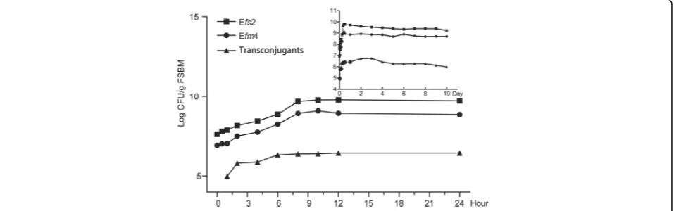
During the fermentation process, the donor strain Efm4 and the recipient strain Efs2 both grew well and had similar growth kinetics. Concentrations of Efm4 reached a maximum of 1.25 × 10⁹CFU/g FSBM at hour 10, with
the concentrations of Efs2 reached a maximum of 6.0 × 10⁸CFU/g FSBM as well (Fig. 1). Transconjugants were detected at the first detection point of fermentation (1 h), with the cell count at 0.94 × 10⁵CFU/g FSBM, and the conjugation frequency being 1.2 × 10⁻³ (0.94 × 10⁵ CFU/g FSBM / 7.8 × 10⁷CFU/g FSBM, transconjugants/ Efs2). The number of transconjugants increased continu-ously and then peaked at day 3, at 5.8 × 10⁶CFU/g FSBM. Moreover, the transconjugants remained consist-ently at ~ 106CFU/g FSBM during day 1 (from hour 6) to day 10 (Fig.1).
In vivoconjugation
No pigs were colonized by vancomycin or chloramphenicol-resistant enterococci prior to the study (day−10 to −1, data not shown). Besides, dur-ing the test period, no VRE strain was detected in the fecal samples collected from the control group fed with basil diet.
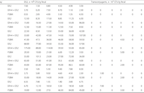
The donor, recipient, and transconjugants exhibited similar growth kinetics, indicating that these two strains could stably reside the digestive tract of growing pigs, and therefore there was no growth defect that could have affected the results (Fig. 2a and b). In the fecal samples obtained from Efm4 + Efs2 group, the concen-tration of Efm4 and Efs2 reached a maximum of 6.44 × 10⁷CFU/g fecal and 9.28 × 10⁷CFU/g fecal at day 4, re-spectively (Fig.2a). Similarly, the concentrations of Efm4 and Efs2 peaked at 4.88 × 10⁷CFU/g and 7.28 × 10⁷ CFU/g on day 3 and day 5 in the FSBM group, respect-ively (Fig. 2b). Notably, transconjugants were observed in both two groups, although only positively detected in one pig of six for each group. In Efm4 + Efs2 group, the conjugation frequency reached to 1.97 × 10⁻⁴and 6.25 × 10⁻⁴ at day 6 and day 7 (Fig. 2c, Table 3), respectively. In contrast, the transconjugant was more frequently oc-curred, with a conjugation frequency of 9.4 × 10⁻⁵, 4.60 × 10⁻⁴, 2.75 × 10⁻⁵, 5.0 × 10⁻⁵and 9.4 × 10⁻⁵, from day 3 to day 7, respectively (Fig.2b, Table3).
The presence vanA and catA1in transconjugants was confirmed by further PCR bothly in vitro and in vivo
experiments.
Molecular typing of probiotic enterococci isolates
The MLST analysis identified that the ST of Efm4, Efs2 and transconjugants was ST1014, ST69 and ST69, re-spectively (Table1).
S1-PFGE and southern hybridization
Southern hybridization confirmed that vanA hybrid-ized on the visible plasmid of Efm4 with a size of ~ 140 kb, which was subsequently named pQZ67 in this study.
Analysis of the plasmid pQZ67
The complete DNA sequence of plasmid pQZ67 was ob-tained by whole-genome sequencing (Fig.4). 142,988-bp pQZ67 consisted of 38,000 amino acid residues and con-taining 185 potential edcoding sequences (CDSs), with a
G + C content of 34.03%. The vanRSHAX gene cluster was located between two insertion sequences which were IS21and IS256. It was a Tn1546-like compound transposon, which played a crucial role in horizontal transmission and might assist in the horizontal transfer ofvanAin Enterococcaceae.
Multiple alignment showing the vanA-carrying frag-ments (~ 13,780 bp) from the FSBM as well as Efm4 + Efs2 group were identical to the corresponding region of
Table 1Antimicrobial susceptibilities and sequence types of enterococci isolated from feed/feed additives
Strain Origin Antibiotics ST
VA TCL RA C AM PIP CZ P MPN AMX OFL CIP GTF GM TE MNO E KIA FT FU
Efm1 FSBM \ \ \ \ \ \ R \ \ \ \ \ \ \ \ \ \ \ \ R ST94
Efm2 FSBM \ \ \ \ \ \ \ \ \ \ \ \ \ \ \ \ \ \ \ \ ST40
Efm3 FSBM \ \ \ \ \ \ \ \ \ \ \ \ \ \ \ \ \ \ \ \ ST296
Efm4 FSBM R R \ \ R R R R R R R R R R \ \ R R \ R ST1014
Efm5 FSBM \ \ I \ \ \ \ \ \ \ \ \ \ \ \ \ I \ \ R ST 296
Efm6 FSBM \ \ \ \ \ \ R \ \ \ \ \ \ \ \ \ I \ \ R ST 94
Efm7 FSBM \ \ \ \ \ \ \ \ \ \ \ \ \ \ \ \ I \ \ R ST 6
Efm8 FSBM \ \ I \ \ \ \ \ \ \ \ \ \ \ \ \ \ \ \ R ST 5
Efm9 FSBM \ \ \ \ \ \ \ \ \ \ \ \ \ \ \ \ \ \ \ R ST 60
Efm10 FSBM \ \ \ \ \ \ R \ \ \ \ \ \ \ \ \ \ \ \ R ST 6
Efm11 Silage corn \ \ \ \ \ \ R \ R \ \ \ \ \ \ \ I \ \ R ST 812
Efm12 Silage corn \ \ I \ \ \ \ \ \ \ \ \ \ \ \ \ \ \ \ R ST 5
Efm13 Silage corn \ \ I \ \ \ \ \ \ \ \ \ \ \ \ \ I \ \ R ST 5
Efm14 Silage corn \ \ I \ \ \ \ \ \ \ \ \ \ \ \ \ I \ \ R ST 178
Efm15 Silage corn \ \ \ \ \ \ \ \ \ \ \ \ \ \ \ \ \ \ \ R ST 160
Efm16 Silage corn \ \ I \ \ \ \ \ \ \ \ \ \ \ \ \ R R \ R ST 726
Efm17 Feed additive \ \ \ \ \ \ R \ \ \ \ \ \ \ \ \ I \ \ R ST812
Efm18 Feed additive \ \ I \ \ \ R \ \ \ \ \ \ \ \ \ I \ \ R ST 5
Efm19 Feed additive \ \ I \ \ \ \ \ \ \ \ \ \ \ \ \ I \ \ R ST 5
Efm20 Feed additive \ \ \ \ \ \ \ \ \ \ \ \ \ \ \ \ \ \ \ R ST 5
Efm21 Feed additive \ \ I \ \ \ \ \ \ \ \ \ \ \ \ \ I \ \ R ST 361
Efm22 Feed additive \ \ R \ \ \ \ \ \ \ \ \ \ \ \ \ \ \ \ R ST 21
Efm23 Feed additive \ \ I \ \ \ \ \ \ \ \ \ \ \ \ \ \ \ \ R ST 21
Efm24 Feed additive \ \ \ \ \ \ \ \ I \ \ \ \ \ \ \ \ \ \ R ST 695
Efm25 Feed additive \ \ \ \ \ \ I \ \ \ \ \ \ \ \ \ I \ \ R ST 24
Efm26 Feed additive \ \ \ \ \ \ \ \ \ \ \ \ \ \ \ \ I \ \ R ST 24
Efm27 Feed additive \ \ I \ \ \ \ \ \ \ \ \ \ \ \ \ \ \ \ R ST 21
Efs1 Feed additive \ \ \ \ \ \ I \ \ \ \ \ \ \ \ \ I \ \ R ST 69
Efs2 Feed additive \ \ \ R \ \ \ \ \ \ R R I R R \ R R \ \ ST 69
Efm,E. faeciumisolates; Efs,E. faecalisisolates; \, susceptible; R, resistant; I, intermediate; ST, sequence type;
VA, Vacomycin; TCL, Teicoplanin; RA, Rifampicin; C, Chloramphenicol; AM, Ampicillin; PIP, Piperacillin; CZ, Cefamedin; P, Penicillin; MPN, Meropenem; AMX, Amoxicillin; OFL, Ofloxacin; CIP, Ciprofloxacin; GTF, Gatifloxacin; GM, Gentamicin; TE, Tetracycline; MNO,
vanA-carrying plasmid in Efm4, expect single base muta-tion withinvanAgene in transconjugants (Additional file
2: Supplemental data 1).
Discussion
To the best of our knowledge, this is the first time of the observation on the transfer of vanA of probiotic origin occurred in the process of FSBM and in the digestive tract of growing pigs. Over the past few decades, with the extensive emergence of antibiotic-resistant strains, probiotics are increasingly used in human and food ani-mals as suitable replacements of antibiotics [33, 34]. As traditional lactic acid bacteria,E. faecium andE. faecalis
have been wildly used in foods, drugs, dietary supple-ments, microbial feed and feed additives [35]. However, some prospective studies showed that these bacteria can serve as an accumulator of antibiotic resistance genes (ARGs) potentially providing them to pathogens, raising the problem of pathogen resistance [36,37]. For the past two decades, the production and consumption of micro-bial products associated with the increase in intensive livestock farming, and the safety of these products is im-portant for food safety. Previous studies have shown
concern on probiotics applied in feed containing trans-ferable ARGs [38, 39]. Using filter mating experiments, Nawaz et al. [40] found thatermB and tetMfrom lacto-bacillus strains could be successfully transferred to E. faecalis, with conjugation frequency ranged from 10−5 to 10−6. Moreover, Ma et al. [41] usedLactobacillus del-brueckii strain harboring mcrC as donor strain, and E. faecalis ATCC 29212 as recipient strain, resulting in a conjugation frequency as high as 2.2 × 10⁻². Similarly, our study found out that, during the production of FSBM, the vanAcould be transferred easily at the early stage (detected only 1 h after the fermentation began), and the transconjungations increased steadily during the whole fermentation process of FSBM. Neverthe-less, the conjugation frequency ranged from ~ 10−3to ~ 10−4, which was about 10 to 100 fold higher than primary studies [42, 43]. It might be because that FSBM provided a suitable environment for conjuga-tions to happen, where three necessary condiconjuga-tions were need: full contact, absolute stationary and suffi-cient nutrition [44, 45].
Most of the previous experiments of resistance genes transfer in vivo were performed on specific pathogen
Table 2Antimicrobial susceptibilities and sequence type of donor strain Efm4 and recipient strain Efs2
Strain Origin Antibiotics ST
VA TCL RA C AM PIP CZ P MPN AMX OFL CIP GTF GM TE MNO E KIA FT FU
Efm4 FSBM R R \ \ R R R R R R R R R R \ \ R R \ R ST1014
Efs2 Feed additive \ \ \ R \ \ \ \ \ \ R R I R R \ R R \ \ ST69
Efm,E. faeciumisolates; Efs,E. faecalisisolates; \, susceptible; R, resistant; I, intermediate; ST, sequence type
VA, Vacomycin; TCL, Teicoplanin; RA, Rifampicin; C, Chloramphenicol; AM, Ampicillin; PIP, Piperacillin; CZ, Cefamedin; P, Penicillin; MPN, Meropenem; AMX, Amoxicillin; OFL, Ofloxacin; CIP, Ciprofloxacin; GTF, Gatifloxacin; GM, Gentamicin; TE, Tetracycline; MNO,
Minocycline; E, Erythromycin; GI, Kitasamycin; FT, Nitrofurantoin; FU, Furazolidone
Fig. 1Growth kinetics of Efm4, Efs2 and transconjugants during the fermentation process of soybean meal. The solid circular represents the growth kinetics of the donor strain Efm4. The solid square represents the growth kinetics of the donor strain Efm4; the solid minute triangle represents the growth kinetics of the transconjugants. Efm4,vanApositive strain Enterococcus faeciumEfm4; Efs2, chloramphenicol-resistant strain
free (SPF) or gnotobiotic animals. Bourgeois- Nicolaos et al. [46] revealed that vanA could transfer from pig de-rived E. faecium to human derived E. faecalis in germ-free mice intestines, but only 2 colonies of trans-conjugants were detected after administration donor and receiver strains intragastrically for 14 d. Moreover, the transfer of vanA-harboring plasmid from poultry and pig derived strain to human fecal E. faecium strains in digestive tract of germ-free mice was observed by Dahl et al. [47], indicating that even transient colonization
strains might provide a significant reservoir to transfer resistance genes to permanent commensal bacteria. A study by Lester et al. [48] demonstrated that conjugation could occur in the human gastrointestinal tract. In this study, three of six volunteers who ingested ~ 109CFU
vanA-harboring E. faecium bacteria in 250 mL milk had transconjugant bacteria in their feces at a mean con-jugation frequency of ~ 10−7 transconjugants / recipi-ent. This was in line with the observations in our results, in particular for the Efm4 + Efs2 group, as the Fig. 2Viable count of Efm4, Efs2 and transconjugants in feceal samples of growing pigs. (a): The growth kinetics of donor strain Efm4 in Efm4 group, Efm4+ Efs2 group and FSBM group; (b): The growth kinetics of receiver strain Efs2 in Efs2 group, Efm4+ Efs2 group and FSBM group; (c): The growth kinetics of transconjugants in Efm4+ Efs2 group and FSBM group. In both Efm4+ Efs2 group and FSBM group, the transconjugants were only detected in one pig out of six. Results from fecal samples obtained at 24 h before the experiment are plotted as day zero. FSBM, fermented soybean meal; Efm4,vanApositive strainEnterococcus faeciumEfm4; Efs2, chloramphenicol-resistant strainEnterococcus faecalisEfs2
Table 3Colony count of Efs2 and transconjugants from fecal samples ofin vivoconjugation
Day Efs2, × 106CFU/g fecal Transconjugants, × 103CFU/g fecal
1 Efs2 7.60 1.50 0.80 4.50 4.90 3.30
Efm4 + Efs2 3.70 0.20 7.50 8.70 1.10 2.30 0 0 0 0 0 0
FSBM 0.55 2.85 4.90 5.50 1.35 6.50 0 0 0 0 0 0
2 Efs2 12.50 8.20 17.50 8.85 11.25 6.30
Efm4 + Efs2 15.00 16.50 27.00 14.50 55.00 86.00 0 0 0 0 0 0
FSBM 12.30 15.00 11.30 12.50 7.50 8.50 0 0 0 0 0 0
3 Efs2 22.50 8.50 13.50 55.00 36.00 42.00
Efm4 + Efs2 33.00 42.00 47.50 14.50 73.00 107.00 0 0 0 0 0 0
FSBM 41.00 47.5 36.00 46.00 58.00 59.50 0 0 0 4.50 0 0
4 Efs2 68.00 77.00 34.50 55.50 36.00 57.00
Efm4 + Efs2 175.00 88.00 114.00 39.50 55.00 85.00 0 0 0 0 0 0
FSBM 20.50 19.00 21.00 6.00 12.20 3.50 0 0 0 5.00 0 0
5 Efs2 55.00 101.5 23.00 27.00 72.00 36.00
Efm4 + Efs2 65.00 51.00 41.00 35.5 65.00 4.00 0 0 0 0 0 0
FSBM 43.00 65.00 87.00 95.00 86.5 60.00 0 0 0 2.00 0 0
6 Efs2 3.50 1.85 5.30 9.40 7.80 8.30
Efm4 + Efs2 3.75 5.80 9.50 4.60 4.50 2.30 0 1.00 0 0 0 0
FSBM 55.00 18.00 14.00 34.00 27.00 92.00 0 0 0 2.00 0 0
7 Efs2 6.55 7.80 1.55 1.80 3.80 6.75
Efm4 + Efs2 6.70 12.10 18.50 5.50 18.50 6.00 0 7.00 0 0 0 0
FSBM 10.00 12.00 27.0 46.50 84.00 45.00 0 0 0 3.50 0 0
transconjugants were obtained 5 d after ingesting Efm4 together with Efs2, and the conjugation fre-quency was ~ 10−4 to ~ 10−5.
It was noteworthy that transconjugants in the FSBM passed through the digestive tract of growing pigs suc-cessfully and could be detected after 3 d of feeding. Moreover, this cycle was 2 d sooner than feeding Efm4 and Efs2 directly. In addition, in commercial production facility, the SBM fermentation process usually lasted only for 2 to 5 d before further processing, and the in-oculation amount of bacteria was much more than we used in the current study [49]. This emphasized the pos-sibility that the resistance genes harbored by commercial probiotics used in feed and feed additives can be dissem-inated in digestive tracts of animal, which may cause fur-ther infections under extreme conditions.
Many researchers explored the transferability of resist-ance genes from farm to fork [50, 51]. Nawaz et al. [40] analyzed the antibiotic resistance in lactic acid bacteria from retail fermented foods in Xi’an, China. Not only did they identifytetSgene fromLactobacillus brevisand
Lactobacillus kefiri for the first time, but they also
observe the ermB gene from L. fermentum and L. sali-varius. Furthermore, the tetM gene from L. plantarum
and L. breviswas also observed to be successfully trans-ferred to E. faecalis by filter mating, with a conjugation frequency as ~ 10−6to ~ 10−5. In addition, He et al. [52] analyzed the oxazolidinone / phenicol resistance gene
optrA from 17 non-relatedE. faecalis isolates from hu-man and animal origin, and the IS1216E elements on the plasmid were also found to contribute the most in the dissemination of the optrAgene. The IS 1216E ele-ments were also detected on pQZ67 plasmid in our study, demonstrating the likelihood of in vitro and in vivo transfer of vanA between enterococci and other Gram-positive bacteria from food or feed to animal or human via food chainin vitroandin vivo.
In this study, the diversity of STs showed that the commercial probiotic enterococci strains were from multiple sources. Additionally, it should be noted that the sequence type of VRE strain Efm4 was ST1014 (Fig.
3). ST1014 was evolved out of ST78, which was the dominant clone complex (CC) in most cities in China, causing the prevalence and spread of VRE [53]. Fig. 3S1-PFGE analysis of Efm4, Efs2 and transconjutions and southern hybridization. Line M, molecular weight markerSalmonella braenderup
H9812; Line 1 and 6, donor strainE. faeciumEfm4; Line 2 and 7, recipient strainE. faecalisEfs2; Line 3 and 8, transconjugants obtained from FSBM; Line 4 and 9, transconjugants obtained from Efm4+ Efs2 group; Line 5 and 10, transconjugants obtained from FSBM group. FSBM, fermented soybean meal; PFGE, pulsed-field gel electrophoresis; Efm4, vanA positive strainEnterococcus faeciumEfm4; Efs2, chloramphenicol-resistant strain
Furthermore, another reported vancomycin resistant E. faecium strain with the same ST1014 was isolated in a hospital in Shandong Province, China, in 2013 [53]. Al-though there was no evidence showing the direct link between that isolate and Efm4, the possible affiliation be-tween ST1014 and ST78 (CC17) still rang the alarms of the safety of probiotic enterococci applied in feed and food.
Ideally, probiotics used in food and feed production should harbor none of the transferable resistance genes and should be sensitive to pathogen related antibiotics [54]. European Food Safety Authority (EFSA) recom-mended that bacterial strains harboring virulence factors or transferable ARGs should not be used in animal feeds, probiotic and fermented foods for human [55,56]. The transmission of ARGs in digestive tracts is a major
health concern related to the probiotic application. Unfor-tunately, until now, ARGs are not included in the standard screening assays before production and application in foods and feeds. Most of the fermentation starter probiotic strains that used for food and animal feed processing were lacking of thorough and rigorous assessments. The donor strain Efm4 used in this study was isolated from a retail FSBM product, revealing the risk of dissemination mul-tiple resistant plasmids was not only through the ingestion of food animals, but also via further feed processing like SBM fermentation. In addition to vanA, the macrolide-resistant geneerm and two aminoglycoside re-sistant genes aadK and aphA were also identified in pQZ67 (Fig.4), explained the resistant of Efm4 to erythro-mycin, kitasamycin and gentamicin. This also indicated that pQZ67 was a multidrug resistance plasmid.
In the past decade, the consumption of FSBM in Asian countries has increased sharply accompanied with the rapid development of animal husbandry [57]. To date, although there has been no report of VRE or other antibiotic-resistance probiotics in FSBM and in the in-testines of growing pigs, the potential risk of horizontal transfer of resistance genes is still hanging in cliff, be-cause the consumption is huge while the surveillance is lacked.
Conclusion
In conclusion, our study illustrated thatvanAcould suc-cessfully transfer among enterococci during the fermen-tation process of soybean meal and also in the digestive tract of growing pigs. Thus, it is suggested that when considering an Enterococcus strain as a starter for pro-biotics, each specific strain should be carefully evaluated to determine the presence of all known virulence factors.
Additional files
Additional file 1:Table S1.Primers used for PCR and DNA sequencing in this study and size of the PCR-targeted products.Table S2.Name, abbreviation and drug concentration of 20 antibiotics. (DOCX 19 kb)
Additional file 2:Primers used for partially sequencing of transconjugations and size of the PCR-targeted products. (DOCX 96 kb)
Abbreviations
ARGs:Antibiotic resistance genes; BEA: Bile Esculin Azide; BHI: Brain heart infusion; CC: Clonal Complex; Efm:Enterococcus faecium; Efs:Enterococcus
faecalis; FSBM: Fermented soybean meal; MIC: Minimum inhibitory concentration; PCR: Polymerase chain reaction; PFGE: Pulsed-field gel electrophoresis; SBM: Soybean meal; VRE: Vancomycin-resistant enterococci
Acknowledgements
We are grateful to Dr. Yongzhi Yang and Dr. Yingbo Shen for their excellent technical assistance.
Funding
This study was funded by National Key Research and Development Program of China (Grant number 2016YFD0501308) and Agro-scientific Research in the Public Interest (Grant number 201403047).
Availability of data and materials
All data generated or analyzed during this study are included in this published article and its supplementary information files.
Accession number(s)
The sequence of the 142,988-bp sequence of the van-carrying plasmid pQZ67 has been deposited in the GenBank database under accession No.CP025427.
Authors’contributions
SQ: Designed the experiments; NL and HY: Performed the experiments; NL, HY and HL: Analyzed the data; NL: wrote the paper, which was edited by XM, CW and SQ; ZW, CHS: Contributed reagents and analysis tools. SQ: Resourced the project. All authors read and approved the final manuscript.
Ethics approval and consent to participate
All procedures used in experiments of our lab were performed in accordance with the China Agricultural University Animal Care and Use Committee guidelines (ID: SKLAB-B-2010-003).
Consent for publication
Not applicable.
Competing interests
The authors declare that they have no competing interests.
Author details
1
State Key Laboratory of Animal Nutrition, College of Animal Science and Technology, China Agricultural University, Beijing 10093, China.2Beijing Key
Laboratory of Bio-feed Additives, China Agricultural University, Beijing 10093, China.3National Center for Veterinary Drug Safety Evaluation, College of
Veterinary Medicine, China Agricultural University, Beijing 100193, China.
Received: 6 December 2018 Accepted: 7 March 2019
References
1. Arias CA, Murray BE. The rise of theEnterococcus: beyond vancomycin resistance. Nat Rev Microbiol. 2012;10:266–78.https://doi.org/10.1038/nrmicro2761. 2. Liu Y. The dissemination mechanism of the multi-resistant genecfrin
Enterococcus spp. from animal origin. [dissertation/docter’s thesis]. Beijing: Chinese Agricultural University; 2013.
3. Thacker PA. Alternatives to antibiotics as growth promoters for use in swine production: a review. J Anim Sci Biotechnol. 2014;4:35–46.https://doi.org/ 10.1186/2049-1891-4-35.
4. Mallo JJ, Rioperez J, Honrubia P. The addition ofEnterococcus faeciumto diet improves piglet's intestinal microbiota and performance. Livest Sci. 2010;133:176–8.https://doi.org/10.1016/j.livsci.2010.06.057.
5. Capcarova M, Hascik P, Kolesarova A, Kacaniova M, Mihok M, Pal G. The effect of selected microbial strains on internal milieu of broiler chickens after peroral administration. Res Vet Sci. 2010;91:132–7.https://doi.org/10. 1016/j.rvsc.2010.07.022.
6. Wang Y, Lu WQ, Li DF, Liu XT, Wang HL, Niu S, et al. Energy and ileal digestible amino Acid concentrations for growing pigs and performance of weanling pigs fed fermented or conventional soybean meal. Asian Austral J Anim.2014;27:706–16.https://doi.org/10.5713/ajas.2013.13612.
7. Sun P, Wang JQ, Jiang YM. Effects ofEnterococcus faecium(SF68) on immune function in mice. Food Chem. 2010;123:63–8.https://doi.org/10. 1016/j.foodchem.2010.03.128.
8. Wang Y, Yang GR, Zhang GG. Effects ofEnterococcus faeciumon growth performance, intestinal flora and immune function of weaner piglets. Chinese J Ani Nutr. 2013;25:1069–76.https://doi.org/10.3969/j.issn.1006-267x.2013.05.023. 9. Kim YR, Kim EY, Choi SY, Hossain MT, Oh R, Heo WS, et al. Effect of a
probiotic strain,Enterococcus faecium, on the immune responses of olive flounder (Paralichthys olivaceus). J Microbiol Biotechnol. 2010;22:526–9. 10. Sydnor ER, Perl T. M. Hospital epidemiology and infection control in acute care
settings. Clin Microbiol Rev. 2011;24:141–73.https://doi.org/10.1128/CMR.00027-10. 11. Miller WR, Munita JM, Arias CA. Mechanisms of antibiotic resistance in
enterococci. Expert Rev Anti Infect Ther. 2014;12:1221–36.https://doi.org/10. 1586/14787210.2014.956092.
12. Jahan M, Zhanel GG, Sparling R, Holley AR. Horizontal transfer of antibiotic resistance fromEnterococcus faeciumof fermented meat origin to clinical isolates ofE. faeciumandEnterococcus faecalis. Int J Food Microbiol. 2015; 199:78–85.https://doi.org/10.1016/j.ijfoodmicro.2015.01.013.
13. Leclercq R, Derlot E, Duval J, Courvalin P. Plasmid mediated resistance to vancomycin and teicoplanin inEnterococcus faecium. N Engl J Med. 1988; 319:157–61.https://doi.org/10.1056/NEJM198807213190307.
14. Cetinkaya Y, Falk P, Mayhall CG. Vancomycin-resistant enterococci. Clin Microbiol Rev. 2000;13:686–707.https://doi.org/10.1128/CMR.13.4.686-707.2000. 15. Weigel LM, Clewell DB, Gill SR, Clark NC, McDougal LK, Flannagan SE, et al.
Genetic analysis of a high-level vancomycin-resistant isolate of
Staphylococcus aureus. Science. 2003;302:1569–71.https://doi.org/10.1126/ science.1090956.
16. Fremin BJ, Bhatt AS. Allied commensal forces against vancomycin-resistant enterococci. Cell Host Microbe. 2017;21:559–60.https://doi.org/10.1016/j. chom.2017.04.015.
17. Chen CH, Sun JY, Guo Y, Lin DF, Guo QL, Hu FP, et al. High prevalence of
18. Hammerum AM, Baig S, Kamel Y, Roer L, Pinholt M, Gumpert H, et al. Emergence ofvanA Enterococcus faeciumin Denmark, 2005–15.
J Antimicrob Chemother. 2017;72:2184–90.https://doi.org/10.1093/jac/dkx138. 19. European Centre for Disease Prevention and Control (ECDC). Data from:
Summary of the Latest Data on Antibiotic Resistance in the European Union. (2016). https://ecdc.europa.eu/en/publications-data/summary-latest-data-antibiotic-resistance-eu-2016
20. Klare I, Heier H, Claus H, Reissbrodt R, Witte W.VanA-mediated high-level glycopeptide resistance inEnterococcus faeciumfrom animal husbandry. Fems Microbiol Lett. 1995;125:165–171. doi: 0378–1097(94)00493–5. 21. Ubeda C, Taur Y, Jenq RR, Equinda MJ, Son T, Samstein M, et al. Vancomycin
resistantEnterococcusdomination of intestinal microbiota is enabled by antibiotic treatment in mice and precedes bloodstream invasion in humans. J Clin Invest.2010;120:4332–41.https://doi.org/10.1172/JCI43918. 22. Clinical and Laboratory Standards Institute. Methods for dilution antimicrobial
susceptibility tests for bacteria that grow aerobically. 27th ed; 2015. 23. Raghavendra GA, Felicia G, Shi XR, Jose S, Sanjeev KN, Mike DT, et al.
Antimicrobial resistance of Enterococcus faecium strains isolated from commercialprobiotic products used in cattle and swine. J Anim Sci. 2018;96: 912–20.https://doi.org/10.1093/jas/sky056.
24. Wang Y, Liu XT, Wang HL, Li DF, Piao XS, Lu WQ. Optimization of processing conditions for solid-state fermented soybean meal and its effects on growth performance and nutrient digestibility of weanling pigs. Livest Sci. 2014;5:697–702.https://doi.org/10.1016/j.livsci.2014.07.020. 25. Yang GH, Guan JJ, Wang JS, Jia F. Optimization of multi-strain solid state
fermentation to improve the content of soybean meal protein by response surface analysis. Adv Mater Res. 2013;3:1224–1228.https://doi.org/10.4028/ www.scientific.net/AMR.690-693.1234.
26. Roberts MC, No DB, Marzluff JM, Delap JH, Turner R. Vancomycin resistant
Enterococcus spp.from crows and their environment in metropolitan Washington State, USA: Is there a correlation between VRE positive crows and the
environment? Vet Mic. 2016;194:48–54.https://doi.org/10.1016/j.vetmic.2016.01.022. 27. Kim M, Kwon TH, Jung SM, Cho SH, Jin SY, Park NH, et al. Antibiotic resistance of bacteria isolated from the iInternal organs of edible snow crabs. PLoS ONE. 2013;8:e70887.https://doi.org/10.1371/journal.pone.0070887.
28. Liu YY, Wang Y, Walsh TR, Yi LX, Zhang R, Spencer J, et al. Emergence of plasmid-mediated colistin resistance mechanism MCR-1 in animals and human beings in China: a microbiological and molecular biological study. Lancet Infect Dis. 2016;16:161–8. https://doi.org/10.1016/S1473-3099(15)00424-7.
29. General Administration of Quality Supervision, Inspection and Quarantine of the People’s Republic of China and China National Standardization Management Committee. Laboratory animal—guideline for ethical review of animal welfare. GB/T 35892–2018.
30. Peng ZX, Wang W, Hu YJ, Li FQ. Development and optimization of rapid detection ofEnterococcus spp. in retailed raw pork and environment samples. J Food Saf Food Qual. 2016;7:2240–6.
31. Florence D, Bruno P, Patrice C. Detection of thevanalphabet and identification of enterococci and staphylococci at the species level by multiplex PCR. J Clin Microbiol. 2004;42:5857–60.https://doi.org/10.1128/ JCM.42.12.5857-5860.2004.
32. Treangen TJ, Sommer DD, Angly FE, Koren S, Mihai P. Next generation sequence assembly with AMOS. Curr Protoc Bioinformatics. 2011;33:Unit11.8.
https://doi.org/10.1002/0471250953.bi1108s33.
33. Quigley EM, Quera R. Small intestinal bacterial overgrowth: roles of antibiotics, prebiotics, and probiotics. Gastroenterology. 2006;130:78–90.
https://doi.org/10.1053/j.gastro.2005.11.046.
34. Angelakis E, Merhej V, Raoult D. Related actions of probiotics and antibiotics on gut microbiota and weight modification. Lancet Infect Dis. 2013;13:889– 99.https://doi.org/10.1016/S1473-3099(13)70179-8.
35. Silbergeld EK, Graham J, Lance B. Industrial food animal production, antimicrobial resistance, and human health. Annu Rev Publ. Health. 2008;29: 151–69.https://doi.org/10.1146/annurev.publhealth.29.020907.090904. 36. Yoo SJ, Sung H, Cho YU, Kim MN, Pai CH, Kim YS, et al. Role of horizontal
transfer of the transposonTn1546 in the nosocomial spread ofvanA
vancomycin-resistant enterococci at a tertiary care hospital in Korea. Infect. Control Hosp. Epidemiol. 2006;27:1081–7.https://doi.org/10.1086/507279. 37. Zhang QQ, Ying GG, Pan CG, Liu YS, Zhao JL. Comprehensive evaluation of
antibiotics emission and fate in the river basins of China: source analysis, multimedia modeling, and linkage to bacterial resistance. Environ Sci. Technol. 2015;49:6772–82.https://doi.org/10.1021/acs.est.5b00729.
38. Robredo B, Singh KV, Baquero F, Murray BE, Torres C. Vancomycin- resistant enterococci isolated from animals and food. Int J Food Microbiol. 2000;54: 197–204.https://doi.org/10.1016/S0168-1605(99)00195-6.
39. Gousia P, Economou V, Bozidis P, Papadopoulou C. Vancomycin-resistance phenotypes, vancomycin-resistance genes, and resistance to antibiotics of enterococci isolated from food of animal origin. Food Borne Pathog Dis. 2015;12:214–33.https://doi.org/10.1089/fpd.2014.1832.
40. Nawaz M, Wang J, Zhou A, Ma C, Wu X, Moore JE, et al. Characterization and transfer of antibiotic resistance in lactic acid bacteria from fermented food products. Curr Microbiol. 2011;62:1081–9.https://doi.org/10.1007/ s00284-010-9856-2.
41. Ma X, Sun P, He PL, Han PF, Wang JJ, Qiao SY, et al. Development of monoclonal antibodies and a competitive ELISA detection method for glycinin, an allergen in soybean. Food Chem. 2010;121:546–51.https://doi. org/10.1016/j.foodchem.2009.12.045.
42. Niamh T, Declan B, Séamus F. Characterisation and transferability of antibiotic resistance genes from lactic acid bacteria isolated from Irish pork and beef abattoirs. Research in Microbiology. 2010;161:127–35.https://doi. org/10.1016/j.resmic.2009.12.010.
43. Conwell M, Daniels V, Naughton PJ, Dooley JSG. Interspecies transfer of vancomycin, erythromycin and tetracycline resistance amongEnterococcus speciesrecovered from agrarian sources. BMC Microbio. 2017;17:19–26.
https://doi.org/10.1186/s12866-017-0928-3.
44. Werner G, Klare I, Witte W. Large conjugativevanAplasmids in vancomycin-resistantEnterococcus faecium. J Clin Microbiol. 1999;37:2383–6.
45. Wiedenbeck J, Cohan FM. Origins of bacterial diversity through horizontal genetic transfer and adaptation to new ecological niches. FEMS MicrobiolRev. 2011;35:957–76.https://doi.org/10.1111/j.1574-6976.2011.00292.x.
46. Bourgeois-Nicolaos N, Moubareck C, Mangeney N, Butel MJ, Doucet-Populaire F. Comparative study ofvanAgene transfer fromEnterococcus faeciumtoEnterococcus faecalisand toEnterococcus faeciumin the intestine of mice. FEMS Microbiol Lett. 2006;254:27–36. https://doi.org/10.1111/j.1574-6968.2005.00004.x.
47. Dahl KH, Mater DD, Flores MJ, Johnsen PJ, Midtvedt T, Corthier G, et al. Transfer of plasmid and chromosomal glycopeptide resistance determinants occurs more readily in the digestive tract of mice thanin vitroand exconjugants can persist stably invivoin the absence of glycopeptide selection. J Antimicrob Chemother. 2007;59:478–86.https://doi.org/10.1093/jac/dkl530.
48. Lester CH, Frimodt-Møller N, Sørensen TL, Monnet DL, Hammerum AM.In vivotransfer of thevanAresistance gene from anEnterococcus faecium
isolate of animal origin to anE. faeciumisolate of human origin in the intestines of human volunteers. Antimicriob. Agents Ch. 2006;50:596–600.
https://doi.org/10.1128/AAC.50.2.596-599.2006.
49. Pangeni D, Jendza JA, Menon DR, Anil L, Yang X, Baidoo SK. Effect of replacing conventional soybean meal with low oligosaccharide soybean meal fed to weanling piglets. J Anim Sci. 2017;95:320–6.https://doi.org/10.2527/jas.2016.0780. 50. Verraes C, Boxstael SV, Meervenne EV, Coillie EV, Butaye P, Catry B, et al.
Antimicrobial resistance in the food chain: A review. Int J Environ Res Public Health. 2013;10:2643–69.
51. Meervenne EV. Antibiotic resistance transfer during food production and preservation. [dissertation/docter’s thesis]. Ghent: Ghent University. 52. He T, Shen Y, Schwarz S, Cai J, Lv Y, Li J, et al. Genetic environment of the
transferable oxazolidinone/phenicol resistance geneoptrAinEnterococcus faecalis
isolates of human and animal origin. J Antimicrob Chemother. 2016;71:1466–73. 53. Yan JG, Dong CZ, Zhao GM, Zhou WL, Sun X, Wang YL, et al. Drug
resistance genes in vancomycin resistant Enterococcus faecium and MLST genotyping. Chin J Nosocomio. 2016;26:4092–7.https://doi.org/10.11816/cn. ni.2016-152595.
54. Founou LL, Founou RC, Essack SY. Antibiotic resistance in the food chain: A developing country perspective. Front Microbiol. 2016;7:1881–99. 55. European Food Safety Authority. Opinion of the scientific committee on a
request from EFSA on the introduction of a qualified presumption of safety (QPS) approach for assessment of selected microorganisms referred to EFSA. EFSA J. 2007;187:1–16.
56. European Food Safety Authority. Scientific opinion on the maintenance of the list of QPS biological agents intentionally added to food and feed. EFSA J. 2012;10:1–84.

![Fig. 4 Genetic map of the vanA harboring plasmid pQZ67. The circles display (from the outside to inside): (i) size in bp; (ii) positions of thepredicted coding sequences transcribed in clockwise orientation; (iii) position of predicted coding sequences transcribed in counterclockwiseorientation; (iv) GC content plotted against 50%, with brown indicating> 50% and purple indicating< 50%; and (v) GC skew [(G + C)/(G-C)] in awindow of 300 bp](https://thumb-us.123doks.com/thumbv2/123dok_us/398553.1532655/9.595.57.540.87.477/thepredicted-transcribed-orientation-sequences-transcribed-counterclockwiseorientation-indicating-indicating.webp)