Open Access
Research
Small but versatile: the extraordinary functional and structural
diversity of the
β
-grasp fold
A Maxwell Burroughs
1,2, S Balaji
1, Lakshminarayan M Iyer
1and L Aravind*
1Address: 1National Center for Biotechnology Information, National Library of Medicine, National Institutes of Health, Bethesda, MD 20894, USA
and 2Bioinformatics Program, Boston University, Boston, MA 02215, USA
Email: A Maxwell Burroughs - [email protected]; S Balaji - [email protected]; Lakshminarayan M Iyer - [email protected]; L Aravind* - [email protected] * Corresponding author
Abstract
Background: The β-grasp fold (β-GF), prototyped by ubiquitin (UB), has been recruited for a strikingly diverse
range of biochemical functions. These functions include providing a scaffold for different enzymatic active sites (e.g. NUDIX phosphohydrolases) and iron-sulfur clusters, RNA-soluble-ligand and co-factor-binding, sulfur transfer, adaptor functions in signaling, assembly of macromolecular complexes and post-translational protein modification. To understand the basis for the functional versatility of this small fold we undertook a comprehensive sequence-structure analysis of the fold and developed a natural classification for its members.
Results: As a result we were able to define the core distinguishing features of the fold and numerous
elaborations, including several previously unrecognized variants. Systematic analysis of all known interactions of the fold showed that its manifold functional abilities arise primarily from the prominent β-sheet, which provides an exposed surface for diverse interactions or additionally, by forming open barrel-like structures. We show that in the β-GF both enzymatic activities and the binding of diverse co-factors (e.g. molybdopterin) have independently evolved on at least three occasions each, and iron-sulfur-cluster-binding on at least two independent occasions. Our analysis identified multiple previously unknown large monophyletic assemblages within the β-GF, including one which unifies versions found in the fasciclin-1 superfamily, the ribosomal protein L25, the phosphoribosyl AMP cyclohydrolase (HisI) and glutamine synthetase. We also uncovered several new groups of β-GF domains including a domain found in bacterial flagellar and fimbrial assembly components, and 5 new UB-like domains in the eukaryotes.
Conclusion: Evolutionary reconstruction indicates that the β-GF had differentiated into at least 7 distinct
lineages by the time of the last universal common ancestor of all extant organisms, encompassing much of the structural diversity observed in extant versions of the fold. The earliest β-GF members were probably involved in RNA metabolism and subsequently radiated into various functional niches. Most of the structural diversification occurred in the prokaryotes, whereas the eukaryotic phase was mainly marked by a specific expansion of the ubiquitin-like β-GF members. The eukaryotic UB superfamily diversified into at least 67 distinct families, of which at least 19–20 families were already present in the eukaryotic common ancestor, including several protein and one lipid conjugated forms. Another key aspect of the eukaryotic phase of evolution of the β-GF was the dramatic increase in domain architectural complexity of proteins related to the expansion of UB-like domains in numerous adaptor roles.
Reviewers: This article was reviewed by Igor Zhulin, Arcady Mushegian and Frank Eisenhaber.
Published: 2 July 2007
Biology Direct 2007, 2:18 doi:10.1186/1745-6150-2-18
Received: 2 June 2007 Accepted: 2 July 2007
This article is available from: http://www.biology-direct.com/content/2/1/18
© 2007 Burroughs et al; licensee BioMed Central Ltd.
Background
The discovery of covalent modification of eukaryotic
pro-teins by the conjugation of ubiquitin to the ε-amino
groups of target lysines has spawned some of the most exciting directions of research in current molecular biol-ogy [1-3]. Ubiquitin (Ub) itself is a small polypeptide of 76 residues, and its crystal structure revealed a distinctive
fold dominated by a β-sheet with 5 anti-parallel β-strands
and a single helical segment [4,5] (Figure 1A). Pioneering investigations of Kraulis, Overington and Murzin showed that this fold was not unique to Ub, but was also present in several other proteins with biologically distinct func-tions. These included the staphylococcal enterotoxin B, the streptococcal immunoglobulin (Ig)-binding protein G and 2Fe-2S ferredoxins [6-8]. The common fold shared by
these proteins was termed the β-grasp, because the β-sheet
appears to grasp the helical segment in this domain [7]. These early studies provided the first indications that,
despite its small size, the β-grasp fold (β-GF) might serve
as a multi-functional scaffold in diverse biological con-texts.
The centrality of Ub conjugation in eukaryotic molecular biology has led to numerous investigations on Ub and Ub-related domains [9,10]. These studies have resulted in a large body of data on the properties of the Ub-like
ver-sions of the β-GF. The key emerging findings were that
several other Ub-like proteins (Ubl), such as Urm1 [11], Apg12 [12], Nedd8 [13], and SUMO [14,15] are also cov-alently linked to target polypeptides, just as Ub itself. In contrast, some Ub-related domains, like the Ubx domain
or Ub-like domains of IκB kinases, play adaptor roles in
Ub-signaling [16-19]. These studies also showed that eukaryotes possess a distinctive enzymatic apparatus for Ub-modification, comprised of a cascade of three enzymes: E1, E2 and E3. These enzymes successively acti-vated Ub/Ubls for transfer using the free energy derived from ATP hydrolysis, relayed it via thiocarboxylate link-ages involving the C-terminal residue of Ub/Ubls, and finally transferred it to lysines on target polypeptides [1,10,20-22]. Eukaryotes were also shown to contain an elaborate apparatus for removal of covalently linked Ub/ Ubls and proteasomal degradation of Ub-modified pro-teins [23-27].
Concomitantly, structural studies also uncovered several
new versions of the β-GF in a variety of domains, greatly
widening its horizon of biological functions. Examples of
such β-GF domains are: 1) the TGS domain, an
RNA-bind-ing domain found in aminoacyl tRNA synthetases and other translation regulators (PDB: 1QF6 [28,29]). 2) The doublecortin (DCX) (PDB: 1MJD [30]), RA (PDB: 1C1Y [31]), PB1 (PDB: 1IPG [32]), and FERM N-terminal domains (PDB: 1EF1 [33]), which function as adaptors in animal signaling proteins and apoptosis regulators by
mediating protein-protein interactions. 3) The soluble
lig-and-binding β-GF (SLBB) domain involved in binding
vitamin B12 and other solutes in animals and bacteria
(PDB: 2BBC, 2FUGS [34-36]). 4) Various toxins related to the staphylococcal enterotoxin B including superantigens involved in the toxic shock syndrome (PDB: 1ESF [37]). 5) Functionally obscure subunits of various enzymatic complexes, like TmoB of the aromatic monooxygenase oxygenase complex (PDB: 1T0S [38]) and RnfH of the Rnf dehydrogenases [39]. 6) Conserved domains, perhaps involved in RNA binding, in the archaeo-eukaryotic RNA polymerase RPB2 subunit [40] and bacterial translation initiation factor IF3 (PDB: 1TIF [41-43]). 7) Staphyloki-nases and streptokiStaphyloki-nases which are fibrinolytic enzymes of low GC Gram-positive bacteria (PDB: 2SAK [44]). 8) MutT/nudix enzymes – a group of phosphohydrolases acting on diverse substrates [45]. These observations
sug-gested that the β-GF is indeed a widely utilized structural
scaffold, with an underappreciated versatility and an evo-lutionary history rich in adaptive radiations.
One notable evolutionary question in this regard was the origin of eukaryotic Ub and its relationships to other
domains with the β-GF. The first major advances in this
direction came with the identification of the sulfur trans-fer proteins, ThiS and MoaD, respectively involved in thi-amine and Molybdenum cofactor (MoCo) biosynthesis,
which contained β-GFs closely related to Ub [46,47].
Fur-thermore, it was demonstrated that their C-terminal resi-dues formed thiocarboxylates, just like Ub, and this was catalyzed by enzymes (ThiF and MoeB), which are very similar to the E1 enzymes involved in Ub-conjugation [46-50]. More recently, research from our group showed that the Ub-conjugation systems might not be an
exclu-sive feature of eukaryotes. Proteins with Ub-like β-GF
domains, and functionally linked enzymes related to E1, E2 and deubiquitinating peptidases of the JAB domain superfamily were found in several, phylogenetically diverse bacteria. We presented evidence that though some of these systems are likely to be involved in sulfur transfer reactions in metabolite biosynthesis, akin to ThiS and
MoaD, others might potentially function as bona fide
con-jugation systems that transfer β-GF proteins to target
polypeptides [39]. Hence, the eukaryotic Ub-conjugation system might have evolved from more ancient precursors that were present in bacteria prior to the origin of eukary-otes.
With some clarity emerging on issue of the origin of Ub/ Ubls and the associated biochemical networks, we sought to investigate the broader issue of the adaptive radiations
of the entire β-GF. In particular we were interested in a
number of problems from structural and evolutionary stand points: 1) Establishing the entire gamut of structural
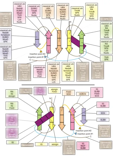
Topology diagrams of selected β-GF members Figure 1
Topology diagrams of selected β-GF members. A generalized representative is shown in (A) with the key structural features found in certain lineages of the fold labeled, while (B) depicts idealized versions of specific lineages, the names of which are given above the diagrams. Strands are shown as arrows with the arrowhead at the C-terminal end. Strands belonging to the 4-stranded β-GF core are colored green, the additional strand found in the 5-stranded assemblage is colored yellow, strands forming a conserved insert within the β-GF scaffold are colored magenta, and other strands specific to a certain lineage are colored grey and outlined with a broken line. Helices are depicted as rectangles, with the core absolutely conserved helix colored orange and other helices specific to a certain lineage colored grey and outlined with a broken line. The diagrams are grouped and labeled in a manner consistent with the structural classes described in the text, with members of the eukaryotic UB-like superfamily nested within other members of the 5-stranded assemblage. The 2Fe-2S cluster of the ferredoxins is shown as four small ovals bound to cysteine residues represented by the letter "C".
Ubiquitin MoaD ThiS
CAD PB1
TGS
2Fe-2S ferredoxin
Streptokinase/Staphylokinase
Doublecortin
Superantigen toxins Glutamine synthase
N terminal domain
TmoB
Nudix (MutT) hydrolases
SLBB
L25 ribosomal protein
L-proline dehydrogenase-type
oxidoreductase (L-proDH alpha) AOR-N Molydopterin-dependent
oxidoreductase
FimD
S4
5-stranded Assemblage
Four-stranded barrelizing versions
Eukaryote-ubiquitin superfamily
N C
C C
C
C
C
N
N N
N C
N C C
N
C
N C
N C
N
C
N
C N
Fasciclin
N C
HisI
C
N
C
N
C C C
N N
C
C
C
N
N C
N C
N C
N C Immunoglobulin binding
domain/BofC Translation initiation factor, If3
N-teminal domain Yml108w POZ domain
4-stranded basal assemblage
N C
N C
N
C C
N
C
N
lateral shelf
connector arm
5-stranded assemblage additional strand
A.
B.
N C Archeo-eukaryotic RNA
polymerase β-subunit
N
C UFD of E1
S1 S2 S4 S3 S5
2) Identifying any unifying structural themes that might exist across most or all functionally diverse versions of the fold. 3) Determination of the lineage-specific sequence-structure correlates for the varied functional adaptations
of the β-GF. 4) Developing a higher order evolutionary
classification for the β-GF and using it as a scaffold to
identify the major temporal phases of adaptive radiation. 5) Identifying instances of drastic shifts in biological or biochemical functions in specific monophyletic lineages
of the β-GF. One example of such a functional shift is seen
in the evolution of the classical Ub-like proteins, where a unique post-translational modification system emerged from a core metabolic sulfur transfer system. 6) Identify-ing previously unrecognized members of the fold, if any, and thereby expanding the functional spectrum or provid-ing a rationale for function prediction of uncharacterized
members of the fold. 7) We also hoped that the β-GF
might provide a model for understanding the more gen-eral problem of how certain small protein folds tend to be extensively deployed in a whole diversity of functional contexts.
In this article we present the results of our systematic
anal-ysis of the β-GF with the objective of addressing the above
points.
Results and Discussion
Identification of β-GF domainsAs the β-GF is small in size and its representatives very
divergent, it is not possible to exhaustively identify all members through sequence or structure similarity searches initiated from a single starting point. Accord-ingly, we used a multi-pronged strategy of sequence, struc-ture, and topological similarity searches. As a starting
point, we used all the currently available structures of β
-GF proteins from the Protein Data Bank (PDB) [51]. This set was compiled by collecting all structures already
clas-sified under the β-GF in the SCOP database [6], their
rel-atives from the PDB database that are not present in SCOP, and new versions which were detected in our recent studies [34,39]. These representatives were used as seeds for initiating sequence profile searches of the NCBI NR (non-redundant) database with the PSI-BLAST pro-gram [52] (see materials and methods for details). Statis-tically significant hits (e < 0.01) recovered in these searches were used to generate alignments for further HMM searches of individual genome databases and repre-sentatives used for transitive PSI-BLAST searches of the NR database. All newly-identified clusters of domains distinct from previously identified sequence families containing
the β-GF were aligned and used to predict secondary
struc-ture with the JPRED program [53]. The predicted second-ary structure and the conservation pattern were superimposed onto the secondary structure and
conserva-tion patterns of the known β-GF sequence families to
ascertain the validity of the newly-detected versions (see Additional file 1 for alignments and complete list of recovered sequences).
All available structures of bona fide β-GF domains were
compared in order to establish a unique core template
topology that discriminated the β-GF from all other folds
(Figure 1A; Table 1; see below for further details). Then
the representative structures of β-GF domains were used
as queries to search a local current version of the PDB database for structurally similar domains using the DALILITE program [54,55]. All hits were evaluated through reciprocal DALILITE searches of the PDB data-base to determine if their best matches included any
known β-GF proteins. The hits were also further evaluated
for congruence to the unique topological template. In addition to the match to the core structural template, we also systematically documented all unique features of each newly-detected structure. Through these searches we were able to identify around ten previously unknown
families/superfamilies of domains containing the β-GF,
including certain structurally distinctive variants. Com-parisons of the distributions of previously characterized globular domains in proteins from sequenced genomes suggests that our procedures have identified a major
frac-tion of conserved lineages of the β-GF.
Core conserved topology, structural variation, and derivatives of the β-GF
A comparison of the available β-GF structures revealed a
common core of 4 strands forming an anti-parallel sheet, and a single helical region (see Table 1, Fig. 1A). The char-acteristic topological feature is that the first and last strands are adjacent and parallel to each other, and the remaining two strands of the conserved core are anti-par-allel and flank the former two strands on either side. The first and last strands are invariably located in the center of the sheet with a cross-over occurring via the single helical element. This helical region is packed against one face of the sheet, typically leaving the other face exposed. The chief interacting positions between sheet and the helical segment and the pattern of key stabilizing hydrophobic interactions are conserved throughout the fold,
support-ing its monophyletic origin. The β-GF domains found in
IF3 and the second largest subunit (β-subunit orthologs)
of the archaeo-eukaryotic RNA polymerase more or less
correspond to this conserved core (Figure 1B). Several β
-GF domains display simple structural elaborations of this basic 4-stranded core. The simplest of these is the seen in a small family of yeast proteins typified by Yml108w from
S. cerevisiae (PDB: 1N6Z [56]). This version has a large insert between the first two strands and an additional hel-ical extension at the C-terminus (Figure 1B). Another
notable variant of the basic 4-stranded form of the β-GF
Biology Direct
2
007,
2
:18
http
://www.biol
ogy-di
rect.com/content/2/1/1
Pa
ge 5 of
(page number not for citation purposes)
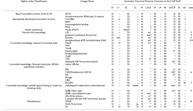
S1 L1 S2 L2 H L3/LS S3 L4 S4 L5/CA S5 tail notes
Basal 4-stranded versions of the β-GF IF3-N S1 -- S2 -- H -- S3 -- O O S5
--Archeo-eukaryotic RNA poly. β-subunit S1 -- S2 -- H -- S3 -- O O S5
--Sporadically-distributed 4-stranded versions Yml108w S1 cc S2 -- H -- S3 -- O O S5 h
BofC S1 -- S2 -- H -- S3 -- O O S5
--Immunoglobulin-binding S1 -- S2 -- H -- S3 -- O O S5
--POZ S1 -- S2 -- H h S3 -- O O S5
--Nudix superfamily Nudix (MutT) S1 -- S(ee)2 -- H * S3 -- O O S5 e
Fasciclin-like assemblage L25 S1 -- S2 -- H ee* S3 -- O O S5 -- 3
glutamine synthetase N-terminal S1 -- S2 -- H eee* S3 -- O O S5 -- 3
fasciclin S1 hhh S2 -- H ee* S3 -- O O S5 -- 3
phosphoribosyl AMP cyclohydrolase (HisI) S1 -- S2 -- H ee* S3 -- O O S5 -- 3,4
5-stranded assemblage: classical 5-stranded clade MoaD S1 H S2 -- H h** S3 -- S4 * S5
--ThiS S1 -- S2 -- H * S3 -- S4 * S5
--TmoB S1 -- S2 -- H * S3 -- S4 * S5
--Superantigen S1 -- S2 -- H * S3 -- S4 h* S5
--Strepto/Staphylokinase S1 -- S2 -- H * S3 -- S4 * S5
--YukD S1 -- S2 -- H * S3 -- S4 * S5
--TGS S1 -- S2 -- H h* S3 -- S4 * S5
--Aldehyde OR2 N-terminal domain S1 -- S2 -- H * S3 -- S4 eh* S5
--5-stranded assemblage: Selected eukaryote UB-like superfamily members
classic UB-like S1 -- S2 -- H * S3 -- S4 * S5
--PB1 S1 -- S2 -- H * S3 -- S4 h* S5
--CAD/Doublecortin (DCX) S1 -- S2 -- H * S3 -- S4 [h]* S5 -- 6
RA S1 -- S2 -- H * S3 -- S4 h* S5
--Elongin S1 -- S2 -- H * S3 -- S4 * S5
--UBX S1 -- S2 -- H * S3 -- S4 * S5
--E1/UFD O -- S2 -- H * S3 -- S4 * S5 S6 7
5-stranded assemblage: soluble ligand binding or metal ion chelating clade
molydopterin-dependent oxidoreductase S1 -- S2 hehee H * S3 -- S4 eee* S5
--SLBB: Nqo1-type S1 -- S2 -- H * S3 -- S4 hh* S5 -- 5
SLBB: transcobalamin-type S1 -- S2 -- H eee* S3 -- S4 * S5
--2Fe-2S ferredoxin S1 -- S2 -- H cc* S3 -- S4 * S5
--L-proline DH-like OR2 N-terminal domain S1 -- S2 -- H ee* S3 -- S4 * S5
--Miscellaneous WWE S1 -- S2 -- H e* S3 -- O O S5 e 8
FimD N-terminal S1 -- S2 ee H * S3 -- O O S5
--S4 O O O O H h* S3 -- S4 * S5
--1. S: Strand, L: Loop, H: Helix, LS: Lateral Shelf, CA: Connector Arm, O: absence of given feature, --: presence of a loop feature, *: presence of LS or CA, h: insert in helical conformation, e: insert in extended conformation (strand-like), cc: long coil insert.
2. OR: oxidoreductase.
3. Versions form barrel through insertion of strands at the lateral shelf.
4. Barrel is less pronounced in this version, strands are inserted more upstream relative to the other 3 versions. 5. Two small helices are present in ascending arm.
6. Single helix found at ascending arm in several members.
(MutT) hydrolases. Here, the middle of the second strand of the conserved core is interrupted by a peculiar insert that projects out to form a distinctive "outflow". This out-flow often assumes a hairpin-like configuration stabilized by hydrogen bonding between segments in an extended conformation (Figure 1B).
All other versions of the β-GF are characterized by major
modifications to the 4-stranded core in the form of dis-tinct inserts that add new secondary structure elements. The first of these is a previously uncharacterized variation containing an insertion of one or more strands between the helical segment and strand 3. The conserved inserted strand seen in all domains with this version forms a hair-pin with the connector segment between the helical seg-ment and strand 3 which also assumes an extended conformation. This hairpin, together with any additional strands in the insert results in these versions of the fold assuming barrel-like structures with differing degrees of openness (Figure 1, Table 1). Examples of this version of
the β-GF domain are observed in the ribosomal protein
L25 (PDB: 1B75 [57]), fasciclin (PDB: 1O70 [58]), and glutamine synthetase (PDB: 1LGR, 2GLS [59,60]). We
uncovered yet another novel variant of the β-GF in the
N-terminal domain of the periplasmic pilus assembly pro-tein FimD (PDB: 1ZE3, chain D [61]). This version is typ-ified by a unique insert N-terminal to the helical segment which results in the formation of a barrel-like configura-tion comparable to the above structural variants.
The most common version of the β-GF is typified by the
presence of an additional strand that packs against the
conserved third strand at the margin of the core β-sheet.
The acquisition of this additional strand has resulted in the emergence of a connector arm that joins it to the ter-minal conserved strand of the core sheet (Figure 1, Table
1). All ubiquitin-like β-GF domains, including sulfur
car-rier proteins like MoaD and ThiS, contain this 5-stranded version of the fold. The connector arm is variable in struc-ture and length and assumes a wide range of conforma-tions ranging from coils to structured elements in different versions of the fold (Figure 1B, Table 1). A deriv-ative of this Ub-like 5-stranded version is found as a C-ter-minal domain (UFD) in most eukaryotic E1 Ub-conjugating enzymes [62,63] – here a circular permuta-tion appears to have displaced the N-terminus to the C-terminus. Given that the N- and C-terminal strands of the
β-GF are adjacent to each other, the C-terminal strand in
the permuted version occupies the same position as the N-terminal strand of the classical versions, but is oriented in the opposite direction (Figure 1, Table 1).
The 5-stranded versions may show further variations due to inserts at different points in the conserved core. One prominent example is the 2Fe-2S ferredoxin, which
tains an insert before the third conserved strand with con-served cysteines for chelating the Fe ion. Similarly, a long insert adopting an extended conformation is observed at a comparable position in several versions of the SLBB domain [34] and the molybdopterin-dependent oxidore-ductases (Figure 1B, Table 1). In the SLBB domain, the
curved β-strands from the insert along with the strands of
the β-GF domain core contribute to the formation of a
barrel-like structure (PDB: 2BBC [34]). In the middle domain of molybdopterin-dependent oxidoreductases (PDB: 1SOX [64], chain A) there is an additional insert of
2 β-strands associated with the connector arm, which
results in an even more complex 3-layered structure, with the two inserts forming a barrel-like element within it. Another previously unknown variant is seen in the N-ter-minal domain of the aldehyde oxidoreductases (AOR-N) (PDB: 1AOR [65]), wherein the connector arm assumes an extended conformation and packs as an additional
strand at the fringe of the β-sheet adjacent to the strand-4
which is a specific feature of the 5-stranded versions
(Fig-ure 1B). In the AOR-Ns, two of these variant β-GF
domains stack via the exposed surface of the β-sheet and
form a 4-layered sandwich module.
Our structure similarity searches identified a few struc-tures which, despite lacking the core conserved topology
of the classical β-GF, aligned well with a part thereof.
Reciprocal searches indicated that β-GF domains were the
best hits for these structures. Additionally, these structures were not representatives of any other previously identified folds. These structures include the S4 RNA-binding domain (PDB: 1c05 [66]), the WWE domain (PDB: 2A90 [67]), and the POZ domain (PDB: 1BUO [68]). Previous structural studies had noted a region of local structural
similarity, termed the α-L motif, between the S4 and the
TGS domain [69]. Given the functional similarity (RNA-binding) and close structural congruence between the shared elements of these two domains, it is quite likely S4 domain is a degenerate variant of the 5-stranded TGS-like
β-GF domain, which has emerged through partial loss of
the N-terminal part of the domain including the first two strands. The WWE domain and the POZ domain are found only in eukaryotes [70], suggesting that they could have potentially emerged from pre-existing folds through rapid divergence. Given its general structural similarity
with the β-GF domains, it is likely to have been derived
from the 5-stranded version of this fold. The WWE domain appears to have acquired an additional strand after the terminal strand which is inserted in the middle of the core sheet. The pre-strand 3 region in this domain also adopts a peculiar structure which makes it appear very
dif-ferent from the classical β-GF domains. In contrast, the
POZ domain appears to have been derived from a
degra-dation of the penultimate strand on the fringe of the sheet.
Natural classification of β-GF domains
In order to address the prime evolutionary questions
about the β-GF, we attempted to construct a classification
that most closely approximates the higher-order evolu-tionary relationships of the members of this fold. The small size of the majority of the versions of this domain often precludes sufficient resolution of relationships using conventional phylogenetic tree methods, some-times even within superfamilies that display significant sequence similarity. This difficulty is further compounded by the extreme sequence divergence even between ver-sions having highly similar tertiary structures (e.g. ubiqui-tin and ThiS). Hence, we had to rely to a greater extent on structure similarity-based clustering, shared derived struc-tural characters, and phyletic patterns of sequence super-families to reconstruct the evolutionary history. Thus, we produced the classification using the following general steps: 1) sequence similarity-based clustering with the BLASTCLUST program [71] helped in identifying the
cores of all major sequence families of β-GF domains. 2)
Subsequent comparison of the individual sequence con-servation profiles led to the establishment of the most inclusive higher-order assemblages of these families (termed superfamilies) based on shared derived features. 3) The next level of relationships beyond what could be resolved through sequence comparisons was established using structural similarity. This was done both by con-structing distance trees based on pairwise Z-scores for structure similarity and deriving the most parsimonious tree based on shared structural features (see Table 1 for major structural features). This procedure, while allowing reasonable resolution of the higher-order relationships, might on occasions produce relatively flat hierarchies for lower-level clusters where none of the methods offer reli-able resolution of relationships. A summary of this classi-fication is presented in Figures 2, 3 and Additional file 2.
Basal versions and other sporadically distributed 4-stranded versions of the β-GF
The above analysis of the structural diversity of the fold suggests that the 4-stranded version is the simplest form from which all other versions could have been derived through accretion of inserts and additional secondary structure elements. Two structurally close superfamilies of
the 4-stranded β-GF domain, namely the IF3-N and the
archaeo-eukaryotic RNA polymerase domain, are respec-tively universally conserved in the bacterial and archaeal-eukaryotic branches of life. This, taken together with their shared general functional connection to RNA metabo-lism, suggests that they arose from a similarly structured precursor that can be traced back to the last universal com-mon ancestor (LUCA). This structurally simple
represent-ative of the β-GF is likely to represent one of the most
basal lineages of the fold. The remaining sequence clusters BofC, yeast Yml108w, and immunoglobulin-binding pro-teins of low GC Gram-positive bacteria with structurally
comparable, simple 4-stranded β-GF domains show
extremely limited phyletic patterns (Additional file 2), suggesting a probable recent derivation from the more ancient versions. The versions in the Ig-binding proteins and BofC are restricted to Gram-positive bacteria, and the former might have been derived in pathogenic forms from BofC, which is a secreted developmental signaling mole-cule widely distributed in free-living Gram-positive bacte-ria [72,73]. The eukaryote-specific POZ domain might represent another derivative of a more widely-distributed 4-stranded version, which has accreted an additional C-terminal helical bundle to form a distinctive globular structure (Figures 1, 2 and Table 1).
The Nudix (MutT) superfamily
The remaining versions of the β-GF fold appear to form a
monophyletic clade unified by the presence of an ances-tral "lateral shelf" or "flange" that forms an extended con-nector between the helical segment and the remaining portion of the sheet after the topological cross-over (Fig-ure 3 and Table 1). Of these versions, the Nudix super-family appears to be one of the early branches given that
its β-sheet retains the ancestral 4-stranded core. All
mem-bers of this superfamily share the above-described insert or "outflow" in the middle of strand 2 which forms a dis-tinctive shelf for accommodating substrates. This super-family is also unified by the presence of a conserved PXG motif in strand-2, immediately after the "outflow", and a unique constellation of conserved residues in the helical segment which form the phosphohydrolase active site [74,75]. The Nudix superfamily represents a rare instance
of adaptation of the β-GF as a scaffold for catalytic activity.
Its phyletic patterns suggest an ancestral presence in all three superkingdoms implying that it might have been present in the LUCA (Figure 3, Additional file 2).
Fasciclin-like assemblage
A structurally distinct subgroup of β-GF domains which
was uncovered as a result of our analysis unifies previ-ously unrecognized versions of the fold, namely the fasci-clin domain (PDB: 1O70 [58]), the ribosomal protein L25 (PDB: 1B75 [57]), and the phosphoribosyl AMP cyclohy-drolase (HisI) (PDB: 1ZPS [76]) with the glutamine syn-thetase N-terminal domain. The unique insert and associated structural peculiarities such as the barrel-like configuration shared by these domains strongly suggests that they form a higher-order monophyletic cluster within
the β-GF termed the fasciclin-like assemblage. The similar
super-families within this assemblage appear to bind small mol-ecules or soluble ligands. The fasciclin domain binds sugar moieties of cell-surface glycoproteins [58], the HisI domain binds phosphoribosyl AMP [77], and the glutamine synthetase N-terminal domain contributes to the substrate binding pocket of the enzyme [59]. The L25 domain binds 5S RNA (PDB: 1DFU [78]), although there is no evidence that it does so in a comparable manner as the other members of this assemblage. Given the above observations, it is possible that the ancestral version of this assemblage had small-molecule binding capabilities. Despite the distinctive structural innovations, the
con-served core of the β-GF domain in this assemblage is a
4-stranded version with a "lateral shelf" suggesting that it represents an early branch of the clade unified by the latter derived feature (Figure 3). Of the sequence superfamilies
of this assemblage, the glutamine synthetase N-terminal domain is traceable to LUCA. Hence, the fasciclin-like
ver-sion of the β-GF domain might have diverged from other
major lineages of the fold prior to LUCA.
The FimD superfamily, while containing a unique struc-tural variant of the fold, shows greatest strucstruc-tural similar-ity to the above assemblage. Its phyletic pattern is limited, being found only in proteobacteria and deinococci (Addi-tional file 2). Thus, it could have been derived from the above assemblage in a lineage-specific manner.
The 5-stranded assemblage
The 5-stranded assemblage is unified by the addition of the fifth strand to the core sheet and the consequent emer-gence of the "connector arm" linking the additional Cartoon representations of distinct β-GF domains
Figure 2
Cartoon representations of distinct β-GF domains. Critical residues in MutT and HisI that are involved in enzyme catalysis are also shown.
4-stranded version, IF3-N (PDB: 1TIF) 5-stranded version, MoaD (PDB: 1FM0D)
Molydopterin-dependent oxidoreductase version (PDB: 1SOXA)
Cicularly-permutated version, UFD of E1 (PDB: 1Y8Q)
Dimeric HisI version (PDB: 1ZPS) (side view) N
C
N C
N C
Nudix/MutT version (PDB: 1IRYA)
N C
C86 C102
C109 E43
R51, E52 E55, E56
N
strand to the terminal strand (Figure 1A). The strong con-servation of this unique structural feature, in conjunction with the exclusive grouping of these versions in structure similarity-based clustering, suggests that they form a monophyletic assemblage. This version of the fold is most prevalent, both in terms of number of distinct super-families contained within it and universal representation found across all life forms. At least 4 monophyletic line-ages of this assembly, namely the TGS domain, the ThiS
and MoaD proteins, and the 2Fe-2S ferredoxins can be traced to LUCA. Beyond these, there are several lineages that are conserved in a single superkingdom or distributed more sporadically within a superkingdom. On the whole, two major clades can be recognized within the 5-stranded
assemblage. The first of these, termed the classical
5-stranded clade, unites the three ancient lineages TGS, ThiS, and MoaD and several other closely-related versions. This clade is also supported by the presence of a highly con-Reconstructed evolutionary history of β-grasp fold
Figure 3
Reconstructed evolutionary history of β-grasp fold. Individual lineages are listed to the left of the figure grouped according to classifications given in the text, with their inferred evolutionary depth traced by solid horizontal lines across the relative tem-poral epochs representing major evolutionary transitional periods shown as vertical lines. The horizontal lines are color-coded according to their observed phyletic distributions, the key for this coloring scheme is given at the bottom of the figure. Dashed lines indicate uncertainty in terms of the origins of a lineage, while grey ellipses group lineages of relatively restricted phyletic distribution with more broadly distributed lineages, indicating that the former likely underwent rapid divergence from the lat-ter. Major predicted structural/functional transitions of the fold are marked by green ellipses with a brief description given. Colored, labeled squares immediately to the left of the lineage names represent broad functional categories: E, enzymatic activ-ity; LMB, ligand or metal-binding; CO, conjugated versions; AD, mediator of protein-protein interactions; RNA, RNA metabo-lism-related.
CO Ancestral
β-grasp domain
LUCA Bacteria/Archaea
Diversification
Origin of
Eukaryotes OrganismsExtant
Nqo1 (SLBB) transcobalamin (SLBB) 2Fe-2S ferredoxin SOX
ThiS MoaD
TGS AOR-N
Nudix (MutT) GS-N
Archaea+Bacteria Bacteria+Eukarya
Universal
Bacteria Eukarya
L25
HisI
IF3-N
IGB BofC Yml108w
A-E RNA polymerase β-subunit FimD-N
Fasciclin_I WWE
POZ S4
5-stranded assemblage
Fasciclin-like assemblage
basal versions
L-proDH alpha
Archaea+Eukarya
Eukaryote UB-like (see fig. 3)
Mut7-C fusion UB-like family Prokaryotic UB-like families
Nudix superfamily
classical 5-stranded
clade soluble ligand or metal ion binding
clade
metal-chelating sulfur-carrying precursor
metal-chelating, ligand-binding precursor
sulfur-transfer precursor
addition of fifth strand
barrel-forming insertions acquired
likely RNA-associated ancestral β-GF version
AD
CO
AD
LMB LMB
E
AD AD
AD
AD
AD
AD
AD
AD
LMB
E
E E
RNA RNA RNA
CO
RnfH
AD
RNA
RNA RNA RNA RNA
YukD
TmoB
SupAnt SPK
AD
AD
AD
FliD-FlgL/K Urm-1
CO
sporadic 4-stranded
served alcoholic residue at the transition between the N-terminal hairpins and the helical segment of the fold [39].
The UB-like β-GF domains are derived from the ThiS and
MoaD-like versions and comprise the most diverse super-family within the classical 5-stranded clade.
Eukaryotic representatives of the UB-like superfamily β-GF domains
In eukaryotes, this superfamily has undergone explosive diversification with at least 19–20 distinct families which can be traced back to the last eukaryotic common ancestor (LECA). These families include six conjugated versions (ubiquitin, Urm1, Apg8/Aut7, Apg12, Ufm1 and SUMO/ SMT3) [79,80] and several known or predicted to func-tion as adapters in multi-domain proteins, like the tubu-lin cofactor B (TBCB) [81], Ub/Ubl conjugating E1 enzymes [62,63] and phosphatidyl-inositol 3 kinase (PI3K) [82]. Overall, in the course of eukaryotic evolu-tion, at least 67 distinct sequence families appear to have emerged within this superfamily with some restricted to particular eukaryotic kingdoms like animals or plants. We identified several previously uncharacterized eukaryotic families such as NPL4p, the UB-like domains of the BMI1/ Posterior Sex Combs family of chromatin associated E3 ligases, a family with the UB-like domain fused to a cyto-chrome b5 domain, and the auxin response factor (BIPOSTO) in plants (see Additional file 1 for align-ments). On the whole, comparisons of sequence
conser-vation profiles showed that β-GF domains related to the
classical ubiquitin domain form a large monophyletic assemblage within the superfamily, including several dis-tinct families such as Nedd8, SUMO, ubiquitin, NPL4, BAG, the Ubx domain, the tubulin co-factors or chaper-ones (TBCB and TBCE), Bat3/Dsk and Apg12/Gate16 (Fig. 3). The circularly permuted C-terminal UFD of eukaryotic E1s, which distinguishes them from the prokaryotic E1-related enzymes, also appears to have been derived from this lineage. Sequence comparisons also showed that the RA, FERM N-terminal module, and PI3K adapter domain families form another distinct higher-order monophyletic lineage. The remaining lineages typified by ECR1/UBA1 and BM-002, while structurally close to the rest, formed distinct sequence families that could not be placed into the any of the above larger assemblages of families (see Additional file 2 for details).
Bacterial representatives of the UB-like superfamily and the classical 5-stranded assemblage
In bacteria, Ub-like superfamily includes several sporadi-cally distributed UB-like families which have been previ-ously described in considerable detail [39]. Several other sporadic bacterial lineages also belong to the classical 5-stranded clade, such as the fibrinolytic adapters of several Gram-positive bacteria (e.g. streptokinase), the superanti-gen/toxin domains, the RnfH proteins and subunits of aromatic compound monooxygenases like TmoB. Our
searches also identified a previously unknown version of the classical 5-stranded clade in a group of bacterial flag-ellar assembly proteins typified by FliD, FlgL and FlgK, and related bacteriophage-tail proteins found in a range of Mu-like caudoviruses (see Additional file 1). Sequence searches indicate that RnfH is closest to the TGS domains and is likely to be an offshoot of that superfamily (Fig. 3). The superantigen/toxin versions and the streptokinase/ staphylokinases appear to form a monophyletic cluster, as they are both secreted versions and interact with sub-strates similarly (See below). However, barring RnfH, the exact relationships of these more sporadic bacterial line-ages to the more ancient lineline-ages of the classical 5-stranded clade remain unclear.
The soluble ligand or metal-binding clade of the 5-stranded assemblage
The second major clade of the 5-stranded assemblage
uni-fies a group of β-GF domains whose interrelationships
were previously unknown. This clade is unified by the presence of a set of inserts that are associated with binding soluble ligands or chelating metal ions. While the inserts themselves are poorly conserved in sequence, their posi-tion, especially in relation to the bound ion or ligand, is well conserved. The main sequence superfamilies in this clade are the 2Fe-2S ferredoxins, the SLBB domains, and the molybdopterin-dependent oxidoreductase domains. As recently shown, the SLBB superfamily is of bacterial provenance [34]. The molybdopterin-dependent oxidore-ductases, typified by the sulfite oxidase (SOX), are widely distributed in all the three superkingdoms but show no evidence in phylogenetic analysis for being present in LUCA. Given that the eukaryotic versions localize to the mitochondrion [83], they appear to have probably been derived from the bacterial progenitor of the mitochon-dria. The N-terminal domain of the L-proline dehydroge-nase-type oxidoreductase (PDB: 1Y56 [84]) is another family of proteins belonging to this clade of the 5-stranded assemblage. Sequence profile analysis showed a statistically significant relationship between these domains and the 2Fe-2S ferredoxins, suggesting that they belong to the same superfamily. They appear to have been derived from the more universally distributed 2Fe-2S ferredoxins through loss of the metal-chelating conserved cysteines relatively early in bacterial evolution.
The N-terminal module of the aldehyde oxidoreductases
A distinctive superfamily of the 5-stranded assemblage that we discovered in our analysis was the N-terminal module of the aldehyde oxidoreductase (AOR-N) (PDB: 1AOR [65]) that contains two tandem, distantly related
copies of the β-fold. These are unified by the modified
the 5-stranded assemblage. It should be noted that they lack any unique structure or sequence feature unifying
them to the sulfite oxidase-like molybdopterin-binding β
-GF domains. Hence, it is possible that they arose from a MoaD-like precursor that evolved an ability to bind met-allopterins specifically (See below). Phyletic patterns indi-cate a potential bacterial origin for this superfamily. The above-mentioned structural similarity of the universally distributed S4 RNA-binding domain with the TGS domain suggests that the former might be another highly divergent lineage that was derived from a TGS-like
classi-cal 5-stranded β-GF domain prior to LUCA.
The relative timeline of major adaptive radiations and functional transitions of the β-GF domains
The pre-LUCA phase and inference of the ancestral function of the
β-GF
The inference of at least 7 β-GF or β-GF-derived (the S4
domain) lineages in LUCA suggests that there was a major diversification of the fold even before LUCA (Figure 3). In structural terms, the inferred representatives in LUCA span all major variants of the fold, from the simplest 4-stranded versions to the barrel-like forms (GS-N domain) to simple and elaborated versions the 5-stranded form. This suggests that the major structural variations were already in place as a result of the early diversification events of the pre-LUCA phase. In functional terms, ver-sions close to the primitive state of both the 4- and 5-stranded forms, the RNA polymerase/IF3-N domain and
the TGS domain, respectively, as well as the possible β-GF
derivative, the S4 domain, have functions related to RNA metabolism or RNA-binding [29,43,85]. Even members of the Nudix clade are known to interact with nucleic acids or chemically-related molecules such as nucleoside diphosphate derivatives [74]. RNA metabolism-associ-ated functions are also sporadically observed in later-derived lineages such as the L25 ribosomal proteins in the fasciclin-like assemblage, the family of prokaryotic UB-related domains fused to the Mut-7C-like RNAses [39], and several eukaryotic UB-like domains like those found in eIF3 p135/Clu-1 (see Additional file 1 for an align-ment), RBBP6 (DWNN domain) [86], and prp21/Splicing factor 3 [87]. Given that the at least 4 of the seven main lineages traceable to LUCA, including some of the inferred basal lineages, have a RNA/ribonucleoprotein associated
role, it appears likely that the ancestral version of the β-GF
was probably involved in RNA-binding. The distribution of RNA-related roles (Fig. 3, Fig, 4) implies that this func-tion seems to have been retained or re-acquired in some sense in several later derived versions of the fold.
A corollary to the inference of the ancestral function of the fold is that there were major functional innovations even in the pre-LUCA period. These are most prominently seen in the 5-stranded assemblage, and appear to be associated
with the emergence of distinctive roles in sulfur delivery and scaffolding of Fe-S clusters. Previous observations have shown biochemical links between the formation of metal-sulfur clusters and sulfur transfer, including path-ways in which ThiS and MoaD-like proteins participate [88]. This observation raises the intriguing possibility that the earliest functional shift involved recruitment of a
5-stranded β-GF domain for a shared general role in both
sulfur transfer and generation of Fe-S clusters. It is quite possible that the subsequent specialization of such a generic precursor spawned the two paralogous families of sulfur transfer proteins (MoaD and ThiS) on one hand and the 2Fe-2S ferredoxins on the other. The rise of the 2Fe-2S ferredoxins probably coincided with the emer-gence of the precursors of the electron transfer chains of respiratory metabolism. The early divergence of MoaD and ThiS suggests that some basic aspects of the biosyn-thetic pathways for complex sulfur-containing metabo-lites like molybdenum/tungsten cofactor and thiamine evolved prior to LUCA.
The post-LUCA phase: the prokaryotic superkingdoms
probably occurred from an ancestral soluble ligand-bind-ing state. However, emergence of catalysis in the Nudix superfamily appears to be a likely extension of the original nucleic acid-binding properties of the fold.
This phase also saw the recruitment of several forms of the
β-GF domain for mediating specific protein-protein
inter-actions in the assembly or stabilization of multi-protein
complexes. Different distantly related β-GF domains were
recruited in the biogenetic systems of flagella and analo-gous structures, the pili. The FimD protein has an
N-termi-nal β-GF domain fused to a C-terminal outer
membrane-spanning domain [90]. This β-GF domain serves as an
adapter to recruit the fimbrial subunit chaperone FimC while the C-terminal domain serves as a platform on which the fimbrial subunits assemble to form the pilus
[91,92]. Likewise, novel versions of the classical
5-stranded β-GF domain, which we discovered in FliD and
FlgL/FlgK, are likely to play roles in the assembly of flagel-lum (FliD) and its hook (FlgL/K), while their relatives in Mu-like bacteriophages might similarly help in assembly of the viral tail (see Additional file 1). Pathogenic bacteria appear to have sporadically adapted both 4- and 5-stranded versions in roles related to interaction with host proteins as a part of their virulence. The strepto/staphy-lokinases which interact with plasmin, and the superanti-gens which interact with vertebrate T-cell receptors [93] from the 5-stranded assemblage and the immunoglobu-lin-binding domains [94] of the 4-stranded assemblage appear to represent multiple convergent recruitments for virulence-related interactions. The classical 5-stranded clade in particular appears to have given rise to several lin-Reconstructed evolutionary history of eukaryotic ubiquitin superfamily
Figure 4
Reconstructed evolutionary history of eukaryotic ubiquitin superfamily. Similar to Figure 3, however, major evolutionary tran-sitions are now shown as horizontal lines and the maximum depth to which these individual lineages can be traced is now shown with solid vertical lines. Functional categories are the same as described in Figure 3.
animals
crown group
kineto-plastids/ Naegleria
LECA
BM-002/Ufm1 RA FERM PI3KN UBX Classical
UBs
SUMO/SMT3
NIP45/RENi Rad23-N Nedd8 Elongin
B
Ubl5 HubA UBP7/UBP14 Ublcp1 Bat3/Dsk TbcB Tbce
Oasl2-C
BAG-N Sf3a/prp21 Sacsin AT
23465p-C
F
A
T10/Diubiquitin
AN1
HOPSP
AD AD AD AD
CO AD AD CO AD CO CO AD AD AD AD AD AD
NIRF-N
AD
Dwnn
HOIL-1
AD
Parkin
AD
S30-N
AD
BMSC
Midnolin DC-C Herp-1 NUB1L
AD
CO
AD
classical UB-like RA/FERM/
PI3K/Dwnn
TRS4-N
AD
U1
1/U12-like
CO
all eukaryotes
kinetoplastids+apicomplexa+crown group
apicomplexa+crown group
crown group
animals plants
CO
USP48/USP26-C
AD
GDX-N
CAD Doublecortin CAD/ DCX
AD AD
Usp40-N
UBP1
1
/GGNB1
HIP7P-N
VCPIP1
UFD
of
E1
AD
PB1
AD
BIPOST
O/ARF
ISG-15
CO
At5g35690-N
Ddi1
AD
Sin3a/SAP18
AD
Rb1cc1
IKK
MUBs Ubiquilin Gpsn2-N
AD
Bmi1/Psc
Wdr48-C
Apg12 APG5
Urm-1
CO
NPL4-N
AD
CLU1/eIF-3
TUG-UBL1-N
AD
classical UB-like clade
CO
eages that seem to function as protein interaction adapt-ers, assembly or stability factors in very different biochemical contexts. For example, the TmoB family might function in stabilizing the proteobacterial aromatic monooxygenase complex [38], different members of the RnfH family might play roles in protein stability or assem-bly of the Rnf oxidoreductase complex, and YukD in the assembly of the ESAT-type export systems of Firmicutes [39].
However, the most important innovation in the bacteria was the emergence of potential conjugation systems that
covalently linked ubiquitin-like β-GF domains to other
proteins (predecessors of the eukaryotic conjugation sys-tems). In functional terms, this process represents a collu-sion of the sulfur-transfer aspect with the protein interaction function which was also widely emerging in members of the fold. The preliminary analysis of these bacterial UB-like systems suggests that they might have already acquired roles related to protein stability and sig-naling. The details of the bacterial antecedents of the eukaryotic UB-conjugation system have already been dis-cussed in a recent work [39] and are not dwelt upon here.
The eukaryotic phase: expansion of the ubiquitin-like domains
Genomic and cell biological evidence suggests that the eukaryotes emerged as a result of a basic endosymbiotic event between a proteobacterium and an archaeon (most likely a euryarchaeon) [95-97]. Consequently, eukaryotes
inherited several versions of the β-GF domain found in
both their archaeal and bacterial (mitochondrial) precur-sors (see Figure 2 and Additional file 2). The currently available data implies that in eukaryotes there was no
diversification of the β-GF domain comparable to what
happened in bacterial evolution that resulted in emer-gence of fundamentally new biochemical activities. Eukaryotes, however, showed an explosive development of the ubiquitin-like lineage resulting in forms that occu-pied biological functional niches across the entire cell. Most of these functions depend on the ancient property of the classical ubiquitin-like 5-stranded version to mediate protein-protein interactions, particularly in relation to the assembly or stabilization of complexes. These functions were performed either via conjugation of UB/UBLs to tar-get proteins and phosphatidylethanolamine, or as domains within multi-domain proteins. The biochemical diversification of the UB-like clade to perform multiple biological roles appears to have been notable even in LECA (Figure 4). These adaptations include: 1) conjuga-tion to proteins destined for degradaconjuga-tion (classical UB). 2) Tagging of proteins for altering interactions and localiza-tion (e.g. SUMO/SMT3) [14,15] 3) conjugalocaliza-tion to both a protein target (Apg5p) and the amino group of the lipid phosphatidylethanolamine (Agp8p/Aut7p) in regulation of the distinctly eukaryotic process of autophagy. 4)
Pos-sible recognition of proteins with conjugated UB moieties (e.g. NPL4) [98]. 5) Binding of E2s to present them to the active site of E1s for conjugation of UB/UBls (the UFD of E1s [62,63]). 6) Assembly of tubulin polymers (TBCB) [81] and microtubule-binding (DCX domains [30]). 7) Protein-protein interactions in modification (e.g. Ub-like domains in Ub-deconjugating enzymes Ub-like Ubp7/ Ubp14 and the Bmi1/Posterior Sex Combs-like E3s) and other signaling pathways (e.g. PI3 Kinase N-terminal domain) [82]. The ancestral eukaryotic member of the UB-like clade is likely to have been a conjugated version because: 1) conjugated forms are seen across the entire diversity of the eukaryotic UB-like clade, which includes at least 5 versions traceable to LECA and 2) they preserve the basic thiocarboxylate-forming chemistry seen in their even more ancient precursors like ThiS or MoaD. Given the inferred presence of multiple non-conjugated forms in LECA, multiple early functional shifts resulting in non-conjugated appear to have occurred prior to the diver-gence of extant eukaryotes from LECA, but after the emer-gence of the first eukaryotic cell.
namely the POZ and WWE domain through major
struc-tural modification of the core β-GF domains.
Evolutionary trends in the domain architectures of β-GF domains
Previous studies on domains occurring in diverse architec-tural contexts in multi-domain proteins have hinted at a strong relationship between domain architectures and functional constraints [104]. We systematically analyzed
the domain architectures of the β-GF domains and their
conservation across evolution to identify these constraints and any role they might have in predicting functions of uncharacterized versions of the domain. Both the sulfur-carrier function and conjugation to other proteins require
the free carboxy-terminus of the standalone β-GF domain.
As a result, the standalone copies of the 5-stranded UB-like version have been preserved across all three superkingdoms since LUCA. But an alternative strategy to this, observed primarily in eukaryotes, is the generation of free C-termini through post-translational proteolytic cleavage as seen in the polyubiquitins and APG8p (Aut7p). This raises that possibility that there might be other as yet undiscovered versions which are released for conjugation by proteolytic processing, as has been previ-ously proposed for the DWNN domain [86]. In this con-text, it remains to be seen if the Ub-like domain in the eukaryotic DDI1p-like proteins [39], which is connected via a glycine-rich linker to the rest of the protein (Fig. 5) might be processed by the C-terminal aspartyl peptidase domain release a free UB-like polypeptide.
In contrast, versions involved in protein and nucleic acid interactions are under no major constraints to remain as standalone forms of the domain. Hence, we find
numer-ous instances of β-GF domains involved in this function
occurring in multi-domain architectures. The ribosomal proteins tend to be small and usually one or two-domain proteins. Accordingly, there is not much architectural diversity seen in case of forms like L25. The forms found in the DNA-dependent RNA polymerase represent some
of the most complex architectures wherein the β-grasp
domain is inserted within an RRM-fold domain which in turn is inserted within a larger, multi-domain scaffold [43]. In most cases, the multi-domain architectures of RNA metabolism-related proteins are well-conserved across entire superkingdoms or even the three superking-doms of Life because of the universality of these functions in their respective phyletic ranges. Multi-domain architec-tures associated with signaling or small-molecule interac-tions are often more restricted in their phyletic range and show lineage-specific diversity [105,106]. Consistent with this, considerable lineage-specific diversity is observed in
prokaryotic β-GF domains involved in small
molecule-binding like the cobalamin-molecule-binding SLBB domains and fasciclin domains and certain enzymes such as the molyb-dopterin-dependent oxidoreductases (Figure 5). All these
domains are typically encountered in secreted proteins and form highly variable multi-domain architectures in various bacteria. In some instances two distinct versions
of the β-GF domain might occur in the same polypeptide:
for example, the fasciclin domain and the molybdopterin-dependent oxidoreductase domains occur in certain secreted enzymes (Figure 5). Conversely, the small
mole-cule-binding β-GF in certain highly conserved
intracellu-lar enzymes like glutamine synthetase and aldehyde oxidoreductases do not show much diversity in domain architectures.
To objectively assess the trends in domain architectural complexity, we made use of the previously devised com-plexity quotient (CQ) [19]. The CQ provides a measure of the complexity of domain architectures in which a given domain occurs (Figure 5). Specifically, it is defined as the product of the number of different types of domains that
co-occur with β-grasp domain containing proteins and the
average number of domains detected in these proteins. The complexity quotient was plotted against the total
number of proteins containing β-GF domains in a given
organism. This was done for 19 completely sequenced species of prokaryotes and 19 eukaryotic proteomes span-ning the entire currently available phyletic spectrum of organisms with sequenced genomes. In the case of prokaryotes the plot reveals a more or less flat line with an approximately constant domain architectural complexity
across all prokaryotes, irrespective of the number of β-GF
proteins they possessed (Figure 5). The plot only showed a few anomalous points: there was a greater than expected
paucity of β-GF proteins in the highly reduced genome of
Mycoplasma and an inexplicably high architectural
com-plexity in Thermotoga maritima. Thus, barring very few
exceptions, the main tendency in prokaryotes is a wide
variability in the number of proteins with β-GF domains
rather than any concerted increase in architectural com-plexity.
Eukaryotes not only have greater numbers of β-GF
domain proteins, but also appear to display greater diver-sity of domain architectures relative to the prokaryotes.
The complexity of the β-GF proteins as well as their
num-bers appear to increase throughout eukaryotic evolution with the highest figures observed in multicellular organ-isms of the eukaryotic crown group. However, the increase in architectural complexity is not linear across eukaryotes, with a tendency to plateau in animals. The only exception
to the strong trend is Trichomonas vaginalis, a basal
eukary-ote, which appears to have undergone a massive, rela-tively recent proliferation across most protein families [107]. As a result it possesses an unexpectedly large
number of β-GF proteins, but low architectural
complex-ity comparable to other basal eukaryotes with similar
A) Architectural complexity plot of β-grasp domains found in eukaryotes and prokaryotes Figure 5
A) Architectural complexity plot of β-grasp domains found in eukaryotes and prokaryotes. The complexity quotient for a given species (y-axis) is plotted against the total number of β-grasp domain containing proteins in the same species. Names of species are given next to plot points. B) Domain architectures of β-grasp domains. Only a small sample of architectures is shown. These mainly represent novel or recently reported architectures that are described in the text. The TRS4 C-terminal domain, also found fused to certain E1-enzymes that lack the C-terminal UFD has a highly conserved ExxxH implying enzymatic func-tion (see Addifunc-tional file 1 for an alignment). Orange ellipses represent the conserved cysteine clusters observed in the NPL4-N family (see Additional file 1). A straight line with a small green box in the Ddi1 family architecture represents a possible cleav-age site located between the domains. The proteins are not drawn to scale as only globular segments are show. Explanation of abbreviations/domain names: B3, DNA-binding domain; Auxin response, auxin-responsive transcription factor domain; OTU, OTU-like family of cysteine proteases; Znf, zinc-finger; Znf_LF, little finger family of zinc finger domains; R, Ring-finger domain;
β-P, β-propeller domain; X, previously uncharacterized BofC C-terminal domain also found fused to a serine/threonine phos-phatase in actinobacteria (see Additional file 1 for alignment). Organism abbreviations: Ehis, Entamoeba histolytica; Ath, Arabidop-sis thaliana; Hsap, Homo sapiens; Rnor, Rattus norvegicus; Blic, Bacillus licheniformis; Mmaz, Methanosarcina mazei; Ddis,
Dictyostelium discoideum; Lmaj, Leishmania major; Tcru, Trypanosoma cruzi; Pfal, Plasmodium falciparum; Tthe, Tetrahymena ther-mophila; Ncra, Neurospora crassa; Drer, Danio rerio; Cele, Caenorhabditis elegans; Dmel, Drosophila melonogaster; Scer, Saccharo-myces cerevisiae; Tvag, Trichomonas vaginalis; Uma, Ustilago maydis; Spom, Schizosaccharomyces pombe; Cneo, Cryptococcus neoformans; Glam, Giardia lamblia; Cpar, Cryptosporidium parva; Tmar, Thermotoga maritima; Mpne, Mycoplasma pneumoniae; Ecol,
Escherichia coli; Vcho, Vibrio cholerae; Hpyl, Helicobacter pylori; Nmen, Neisseria meningitides; Msp., Mesorhizobium sp.; Ctet,
Clostridium tetani; Aaeo, Aquifex aeolicus; Tden, Treponema denticola; Drad, Deinococcus radiodurans; Mtub, Mycobacterium tubercu-losis; Save, Streptomyces avermitilis; Bfra, Bacteroides fragilis; Ctep, Chlorobium tepidum; Nsp., Nostoc sp.; Ssp., Synecococcus sp.; Cpneu, Chlamydophila pneumoniae.
B. Selected Domain Architectures A. Architectural Complexity Plots
ȕ-GF JAB* B3 responseAuxin ȕ-GF
ARF16_Ath_18417527
ȕ-GF OTU Znf
CAD89975_Hsap_30268367 30.t00038_Ehis_67480009
ȕ-GF ȕ-GF S/T kinase
KIAA0369_Hsap_40788228
NPL4-N BIPOSTO/ARF
VCPIP1 Double Cortin (DCX)
ȕ-P ȕ -GF--fasciclin domain ȕ -GF--fasciclin domain ȕ -GF--SLBB domain ȕ -GF--SLBB domain MM2638_Mmaz_21228740 SLBB/fasciclin -GF domainsȕ
Cyt-b5 ȕ-GF IG-like
SUOX_Hsap_1711606 Molydopterin-dependent oxidoreductase -GF domainȕ
ȕ-GF
BofC_Blic_52081254 BofC
X
Eukaryoticβ-GF architecture
y = 52.986Ln(x) - 98.172 R2 = 0.7594
0 50 100 150 200 250 300
0 100 200 300 400 500 600 700
Number of proteins in organism
C o mp le x it y q u o tie n t
Bacterial β-GF architecture
0 10 20 30 40 50 60 70 80 90
0 10 20 30 40 50 60 70
Number of proteins in organism
C om pl e x it y quot ie nt
ȕ-GF TRS4-C
of actual architectures, the multicellular eukaryotes show numerous lineage-specific multi-domain proteins with
different β-GF domains, which are often involved in
spe-cific signaling pathways that correspond to unique aspects of the biology of these organisms. For example, the pro-grammed cell death pathways in animals and the auxin-response in plants contain representatives with such unique architectures (Figure 5) [19].
Typically, many of the eukaryotic multi-domain architec-tures, both ancient and lineage-specific, tend to combine the UBL domains with other signaling domains, typically those involved in UB-signaling. These combinations include those with deubiquitinating peptidases (e.g. of the OTU superfamily), E3 ligases usually of the RING superfamily (Figure 5), and other UB-binding domains like UBA, or other kinds of signaling domains like kinases as seen in the IKKs and Doublecortin. Another feature seen in eukaryotic architectures is the architectural varia-bility through domain loss or accretion, even in the case of highly conserved orthologous proteins. For example, the Npl4p family [108] of Ubls is conserved throughout eukaryotes and might play a role as a novel E3 in degrada-tion of proteins in the endoplasmic reticulum. It can be reconstructed as having an ancestral architecture that combined an N-terminal Ubl with a central region con-taining variable numbers of a novel Zn-chelating cysteine cluster domain and a C-terminal catalytically inactive ver-sion of the JAB peptidase domain (Figure 5, see Addi-tional file 1). In the plant lineage the central Zn-chelating cluster is lost, while in animals and fungi an additional Zn-finger domain is inserted N-terminal to the cysteine-rich Zn-cluster.
Structural correlates for functional diversity in the β-GF
We next sought to decipher the relationship between functional diversification and structural elaborations of the fold. For this purpose, we created an idealized
repre-sentation of the β-GF fold (Figure 6) and divided the
structural elements into equivalent zones that are compa-rable across the available structures. We then mapped interactions to ligands (see materials and methods for details) in all members for which this data is available onto the above scaffold to obtain an interaction map for the fold (Figure 6). We then used this interaction map in conjunction with the above developed classification scheme and relative temporal pattern of diversification to explore the evolution of the structure-function relation-ships. For the sake of convention, we refer to the exposed
surface of the core β-sheet as the "exposed face" and the
opposite surface of the sheet which might be obscured by the packing helical segment, the lateral shelf or flange, and the connector arm (in the 5-stranded versions) as the "obscured face". We refer to the C-terminal most portion of the final strand as the "tail".
Little is known of the exact mode of interactions of the basal 4-stranded versions of the fold. However, the appar-ent rarity of the simple 4-stranded versions suggests that there appears to be a tendency to elaborate the core sheet to provide an increased interface for interactions. On the whole, the exposed face mediates more interactions across
the β-GF fold compared to the obscured face. Thus, the
proliferation and widespread utilization of the 5-stranded version might be associated with the availability of a larger surface on the exposed face for mediating contacts. Another evolutionary trend is the formation of a barrel-like configuration through insertion of strands which on instances provides a classical interaction interface at the open end of the barrel. We discuss below more specific themes of interaction that were observed in multiple superfamilies of the fold.
Solute interaction in the fasciclin-like assemblage
As discussed above, the prevalence of soluble ligands such as sugars, amino acids, and metabolic intermediates for different sequence superfamilies of the clade suggested an ancestral solute-binding role for these proteins. Analysis of the interactions with respect to the shared structural core of this assemblage suggests that the insert and the lat-eral shelf form an interface for soluble ligand interaction in fasciclin, GS-N, and phosphoribosyl-AMP cyclohydro-lase domains [59,77,109]. Furthermore, in glutamine syn-thetase this interaction might indirectly contribute to catalysis via a conserved aspartate from this region that interacts with the substrate bound at the active site and helps in anchoring it there. This suggests that ancestral versions of this assemblage probably mediated a generic ligand interaction via a similar interface. The interactions of the L25 domain via this interface, if any, remain unknown. However, it is known to contact 5S rRNA via the exposed face [78]. FimD, which appears to be a distant relative of the fasciclin-like assemblage, assumes a classi-cal barrel configuration, with the "open-end" of the barrel providing an interface for interacting with the FimC immunoglobulin domain [61]. Similar "open-ends" of topologically unrelated barrels like the OB fold, PRC, and SH3 barrels are known to mediate interactions with lig-ands in a like manner [110-112]. The loop between the
penultimate two strands of the core FimD β-GF domain is
one of the major determinants of the interaction and this feature is comparable to certain interactions of the phos-phoribosyl-AMP cyclohydrolase domain (see below).
Metal chelation, solute interaction, and prosthetic group attachment in the SLBB/ferredoxin/molybdopterin-dependent oxidoreductase clade