R E S E A R C H
Open Access
Visual attention, EEG alpha power and
T7-Fz connectivity are implicated in prosthetic
hand control and can be optimized
through gaze training
J. V. V. Parr
1, S. J. Vine
2, M. R. Wilson
2, N. R. Harrison
3and G. Wood
4*Abstract
Background:Prosthetic hands impose a high cognitive burden on the user that often results in fatigue, frustration and prosthesis rejection. However, efforts to directly measure this burden are sparse and little is known about the mechanisms behind it. There is also a lack of evidence-based training interventions designed to improve prosthesis hand control and reduce the mental effort required to use them. In two experiments, we provide the first direct evaluation of this cognitive burden using measurements of EEG and eye-tracking (Experiment 1), and then explore how a novel visuomotor intervention (gaze training; GT) might alleviate it (Experiment 2).
Methods:In Experiment 1, able-bodied participants (n= 20) lifted and moved a jar, first using their anatomical hand and then using a myoelectric prosthetic hand simulator. In experiment 2, a GT group (n= 12) and a movement training (MT) group (n= 12) trained with the prosthetic hand simulator over three one hour sessions in a picking up coins task, before returning for retention, delayed retention and transfer tests. The GT group received instruction regarding how to use their eyes effectively, while the MT group received movement-related instruction typical in rehabilitation. Results:Experiment 1 revealed that when using the prosthetic hand, participants performed worse, exhibited spatial and temporal disruptions to visual attention, and exhibited a global decrease in EEG alpha power (8-12 Hz), suggesting increased cognitive effort. Experiment 2 showed that GT was the more effective method for expediting prosthesis learning, optimising visual attention, and lowering conscious control–as indexed by reduced T7-Fz connectivity. Whilst the MT group improved performance, they did not reduce hand-focused visual attention and showed increased conscious movement control. The superior benefits of GT transferred to a more complex tea-making task.
Conclusions:These experiments quantify the visual and cortical mechanisms relating to the cognitive burden experienced during prosthetic hand control. They also evidence the efficacy of a GT intervention that alleviated this burden and promoted better learning and transfer, compared to typical rehabilitation instructions. These findings have theoretical and practical implications for prosthesis rehabilitation, the development of emerging prosthesis
technologies and for the general understanding of human-tool interactions.
Keywords:Myoelectric prosthesis, Amputees, Intervention, Conscious control, Therapy, Motor learning, Inter site phase clustering
© The Author(s). 2019Open AccessThis article is distributed under the terms of the Creative Commons Attribution 4.0 International License (http://creativecommons.org/licenses/by/4.0/), which permits unrestricted use, distribution, and reproduction in any medium, provided you give appropriate credit to the original author(s) and the source, provide a link to the Creative Commons license, and indicate if changes were made. The Creative Commons Public Domain Dedication waiver (http://creativecommons.org/publicdomain/zero/1.0/) applies to the data made available in this article, unless otherwise stated. * Correspondence:[email protected]
4Research Centre for Musculoskeletal Science and Sports Medicine
Department of Sport and Exercise Science, Manchester Metropolitan University, Manchester, UK
Background
Many upper-limb amputees rely on prosthetic hand devices to restore a degree of functionality to the performance of daily activities. Despite the increasing sophistication of these devices, they still provide less than 50% of the capability of an intact limb [1, 2], im-pose a high cognitive burden that results in fatigue and frustration [3], and are therefore frequently rejected [4]. The nature of this cognitive burden has recently been explored indirectly by examining disruption to visuo-motor behaviours during prosthetic hand use [5,6]. For example, Parr et al. [7] showed that when using a myo-electric prosthetic hand simulator, participants directed a greater amount of visual attention towards the pros-thesis and objects being manipulated by it. This depend-ency on visual feedback to monitor and correct movements is in contrast to the feed-forward (target-fo-cused) strategy revealed by skilled users in everyday tasks [8], and mirrors findings from novices in other domains (e.g. tool use [9] and laparoscopic surgery [10,11]). Inter-estingly, it is this need to constantly, and consciously, pay close visual attention to movements that prosthesis users report as a key contributor to the cognitive burden experi-enced during prosthetic hand control [4, 12, 13]. The overall aim of this paper was to assess novel measures of this cognitive burden and to test the efficacy of a novel training technique that might reduce this burden.
Measures that directly evaluate this cognitive burden are needed in order to further our understanding of how efficient visuomotor behaviour is influenced by pros-thesis use. Electroencephalography (EEG) is ideally suited for this purpose as it offers a window into the dy-namics of ongoing neural activity with high temporal resolution. This is important, as the development of skilled motor performance is characterised by the pre-cise allocation of processing resources to areas of the brain that are needed for successful task execution; termed ‘neural efficiency’[14,15]. It has been suggested that neural efficiency can be operationalised by cortical oscillations in the alpha frequency (8-12 Hz) [16]. Specif-ically, the magnitude (power) of alpha oscillations influ-ence cortical activation by exerting inhibitory control and can therefore reveal a gating mechanism whereby resources are diverted away from regions showing higher alpha power (more inhibition) and towards regions showing lower alpha power (lower inhibition) [17]. Such a mechanism is reflected in evidence suggesting that during movement planning and execution, alpha power decreases over motor-related areas of the cortex while increasing over non-motor areas [18].
Using this gating model, research has shown that en-hanced performance in motor tasks can be characterised by more efficient topographical alpha power distribu-tions. For example, Gallicchio and colleagues have
shown that lower central alpha power and higher tem-poral alpha power preceded improved performance in a biathlon shooting task [19] and were evident following a training period in golf-putting [20, 21]. Indeed, higher alpha power over the left-temporal region has been gen-erally associated with improvements in motor learning and performance [22,23], as conscious, verbal-analytical processes diminish as a function of automaticity and ex-pertise [14, 24–27]. It is therefore plausible to assume that the cognitive burden experienced during initial prosthesis hand control is underpinned by both neural inefficiency, a dependence on vision to monitor hand state and that both may reflect a more conscious mode of prosthesis control.
Experiment 1
The aim of the first experiment was to provide an evalu-ation of the cognitive burden experienced during initial prosthetic hand control in a visuomotor task, by simul-taneously measuring visual attention and EEG alpha activity. By comparing task phases that require relatively low (Reach) and relatively high (Lift) levels of overt visual attention to the prosthetic hand [7], we also aimed to investigate the efficacy of inferring demands on cogni-tive processes from eye-movements alone. We hypothe-sised that when using a prosthesis simulator, participants would perform significantly slower compared to when completing the task using their anatomical hand. Second, and in line with Parr et al. [7], we hypothesised that this performance decrement would be underpinned by an increased dependence on vision, as indicated by increases in both hand-focused visual attention and time to shift gaze to the next target. Third, we hypothesised that prosthetic hand use would result in a global de-crease in alpha power, reflecting inde-creased cortical
acti-vation and more effortful performance [16, 22] – a
decrease that should be more pronounced over the (left) temporal region of the brain [19]. Finally, we hypothe-sised that these disruptions would be greater for the more visually demanding ‘Lift’ phase compared to the less visually demanding‘Reach’phase.
Methods Participants
vision and had no prior experience with a myoelectric prosthetic device. The study was approved by the local ethics committee and written informed consent was given prior to testing.
Apparatus
Prosthesis
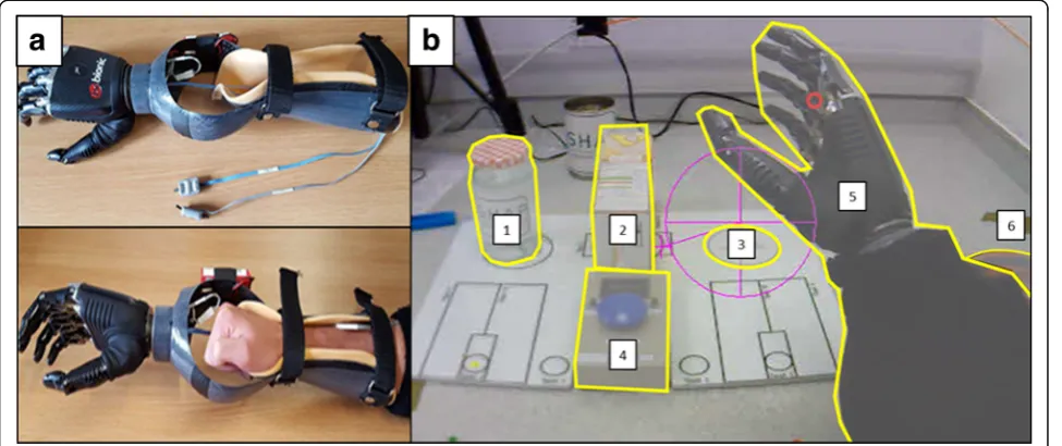
Participants wore the BebionicTM (Otto Bock Health-Care, Duderstadt, Germany) fully articulating myoelec-tric prosthetic hand simulator [7]. To fit able-bodied participants, the simulator was attached to the end of a carbon fibre trough in which the participants’ forearm and fist was positioned and fastened with Velcro straps (Fig. 1a). The prosthetic hand is controlled by muscle contraction detected by two electrodes placed on the extensor and flexor muscles in the forearm. These elec-trodes measure electrical changes on the skin covering
the control muscles. Activation of the extensors
triggered the opening of the hand whereas activation of the flexors triggered the closing of the hand.
The jar task
This task was taken from the Southampton Hand Assessment Procedure (SHAP) [29] which is a clinical tool used to measure hand dexterity. For this experi-ment, we chose the SHAP “lifting a heavy object” task. This required participants to lift a water-filled jar from the left side of the board over an empty carton and onto a designated area on the right side of the board as quickly and accurately as possible (Fig. 1b). Participants were required to begin each trial with their hand on a specified hand mat before (at a time of their own
choosing) initiating the trial with the press of a button located centrally on the board. Following the successful placement of the jar, the task was terminated by a second button press.
Mobile eye-tracker
Gaze behaviour was measured with an Applied Science Laboratories (ASL; Bedford, MA) Mobile Eye XG gaze registration system that measures eye line of gaze at 30 Hz. Data were recorded directly onto a laptop (Dell Inspiron 6400) with‘Eye-vision’software installed. Video data from the eye-tracker were analysed offline using Quiet Eye Solutions software (Quiet Eye Solutions Inc.) which enables detailed frame-by-frame coding of the motor action and gaze behaviour of the performer. For each frame, gaze was manually determined to be lying within one area of interest (AOI) by the researcher,
de-fined in Fig. 1. On occasions where two AOIs
over-lapped, priority was given to the AOI that was initially fixated upon so long as the obscuring AOI did not cause the position of this fixation to change. If gaze shifted from its position following AOI overlap then priority was given to the now obscuring AOI. Fixations made outside of AOIs were collectively labelled as“Other”. To understand the disruptions to gaze throughout the different phases of the task, the task was broken down into two distinct movement phases; reach for the jar (Reach), and lift the jar (Lift).
EEG
During the testing period, 64 active electrodes were positioned on the scalp according to the 10–20 system.
Four additional electrodes were also placed above and below the left eye, and on the outer canthi of both eyes, to record the vertical electrooculogram (VEOG) and horizontal electrooculogram (HEOG). The signal was amplified and digitized at 512 Hz using the ActiveTwo recording system (Biosemi, the Netherlands). This system replaces the ground electrode used in conven-tional systems with common mode sense (CMS) and driven right leg (DRL) electrodes to enhance the com-mon mode rejection ratio of the signal. Offline, signals were separately epoched from−1250 ms to + 250 ms for theReachphase and from −250 ms to + 1250 ms for the Liftphase relative to the time the jar was first lifted from the table in each individual trial. We chose to segment signals this way as (a) it allowed a standardised move-ment phase across hand conditions despite differences in performance time and (b) it allowed an examination of two distinct movement phases that demand relatively low (the Reach phase) and high (Lift phase) dependence on vision [7]. This therefore offered the best opportunity to analyse the relationship between the dependence on vision and neural efficiency. The timing of these events was indicated via the recorded gaze videos derived from the eye-tracker, and were manually inputted into the EEG data as triggers offline following data collection.1 Signals were then band-pass filtered from 1 to 35 Hz (Fi-nite Infi(Fi-nite Response), and referenced to the average of all scalp electrodes. Data were then subject to Independ-ent ComponIndepend-ent Analysis (Runica Infomax algorithm [30],) to remove components accounting for blinks, eye movements, and other non-neural activity. At this stage, if epochs were deemed too noisy they were removed from further analysis. Although ICA was used for arte-fact rejection purposes, subsequent analyses were con-ducted on EEG channel data, as the most relevant literature within the psychomotor domain has tested the alpha-gating phenomenon via the mean regional activa-tion occurring across selected EEG channels [19,21,31]. The spatial information of the processed epochs was then enhanced by surface Laplacian estimation that acts as a spatial filter of EEG potential distribution to reduce head volume conductor effects and eliminate electrode reference influence [32].
Procedure
Upon arriving for testing, participants were informed of the purpose of the investigation and were sat comfortably on a chair so their elbows were in a 90 degree flexed pos-ition when resting on the table, as per SHAP instructions. They were then prepared for electrooculographic (EOG) and EEG measurements. The eye tracker was then fitted and calibrated by asking participants to direct their gaze to eight different points marked within the scene. Gaze behaviour was continuously monitored throughout testing
and recalibrated at least every 15 trials, or when calibra-tion had been lost. Participants first performed 30 trials of the task with their anatomic right hand before being intro-duced to the myoelectric prosthetic hand. This ensured that all prosthesis data reflected the difficulty in control-ling the device rather than reflecting any deficit in under-standing the task. Once fitted with the prosthetic hand simulator, participants were allowed to practice sending open and close signals. Once participants were able send five consecutive open and close signals, they were given one full practice trial before completing 30 full experimen-tal trials.
Measures
Performance
Performance was measured as the time (in seconds) taken to successfully complete the task, as indicated by the timer that was initiated and terminated by the performers first (before the trial started) and second but-ton press (after the trial ended).
Target locking strategy (TLS)
Previous research has shown that more proficient visuo-motor performance is indexed by a high TLS, with performers spending most of their time fixating the
to-be-manipulated target, whereas, less proficient
performance is indexed by a switching strategy, with performers shifting gaze between the hand/tool and the to-be-manipulated target [7, 33, 34]. TLS was computed by subtracting the percentage of time spent fixating the hand (either anatomic or prosthetic) from the time spent fixating the target (jar/target area). Positive scores reflect more time fixating relevant targets whereas negative scores reflect more time spent fixating on the hand. A score of ‘0’reflects equal time spent fixating on the hand and targets and represents a ‘switching strategy’. A fixation towards the target object of a current movement
phase was considered “target focused” but would
become “hand focused” as soon as the hand grasped or manipulated it. For example, during the Reach phase, fixations towards the jar were considered‘target focused’ but as soon as the hand grasped the jar fixations to the jar were then classified as‘hand-focused’.
Gaze shifting
the time taken to shift gaze to the jar after having pressed the start button (Reach), and for the time to shift gaze to the target location after having first lifted the jar from the board (Lift). This measure has previ-ously been shown to predict proficient prosthetic hand control with poorer performers slower to shift gaze to the next object in the task sequence [7].
EEG alpha power
Time-frequency decomposition was performed through short-time Fast Fourier Transform (FFT) on 9 overlap-ping segments (overlap of 87.5%), each of 500 ms duration and linearly spaced, with centre points ranging
from −1000 ms to 0 ms for Reach and from 0 ms to
1000 ms for Lift, relative to jar lift. Prior to FFT, data points within each segment were Hanning tapered and 0-padded to reach 2000 ms, providing complex-valued coefficients with a precision of 0.5 Hz for each channel and trial separately. Power was calculated for the entire alpha frequency band (8–12 Hz) as the squared ampli-tude of each signal, which was then averaged across the
nine overlapping segments obtained for both the Reach
and Lift phases. Seven regions of interest (ROI) were
chosen for further analysis; left temporal (T7, TP7, FT7), left central (C1, C3, CP1, CP3), frontal (F1, F3, Fz, F2, F4), right central (C2, C4, CP2, CP4), right temporal (T8, TP8, FT8), parietal (P1, P3, Pz, P2, P4) and occipital (O1, Oz, O2). Power was averaged across these channels to yield values for each region. As no neutral baseline could be identified, non-normal distributions and inter-individual differences were dealt with by employing a median-scaled log transformation (see [19]. This trans-formation is implemented by scaling all power values for each participant (across all electrodes, trials, segments and conditions) by the median power value within that participant, before then employing a 10 log10 transform-ation to all values. EEG signals were processed using the
EEGLAB toolbox [30] and custom MATLAB scripts
(Mathworks, Natick, MA).
Statistical analyses
Performance
A Shapiro-Wilk’s test revealed that performance data for the prosthesis condition were significantly non-normally distributed (p= .03). A Wilcoxon signed ranks test was therefore used to compare the time taken (in seconds) to complete the task between hand conditions.
TLS
To directly complement the EEG data, a 2 × 2 repeated measures analysis of variance (ANOVA) with the factors hand (anatomic, prosthetic) and phase (Reach, Lift) was performed on the TLS implemented by participants
during the second prior to (Reach phase) and the second after (Lift phase) lifting the jar from its position.
Gaze shifting
For gaze shifting, a 2 (hand) × 2 (phase) repeated mea-sures ANOVA was also conducted to compare the effect of hand condition on gaze shifting time for the Reach and Lift phases.
Alpha gating
A 2 × 2 × 7 repeated measures ANOVA with the factors hand, phase, and ROI (left temporal, left central, frontal, right central, right temporal, parietal, occipital) was per-formed on absolute alpha power to evaluate how the regional gating of alpha is altered across anatomic and prosthetic hand control. Furthermore, by comparing the Reach phase and the Lift phase, we evaluate how the extent of hand-related visual attention may influence the gating of alpha power.
Non-parametric effect sizes were calculated as,r¼Z= ffiffiffiffi
N p
[35], where Zis the test statistic and Nis the total sample size. For all ANOVAs, Greenhouse-Geisser cor-rections were applied when sphericity was violated and effect sizes were calculated using partial eta squared (ηp2). All pairwise comparisons were adjusted via Bon-ferroni corrections to counteract the problem of mul-tiple comparisons.
Results Performance
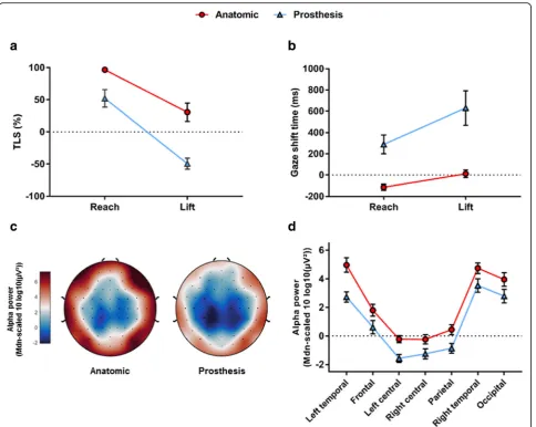
Participants performed significantly slower during the pros-thesis (Mdn = 6.35 s) compared to the anatomical (Mdn = 1.56 s) hand condition, Z =−3.92,p< .001, r =−0.87.
TLS
There was a significant main effect of hand, F(1, 18) = 144.746, p< .001, ηp2 = 0.89, and phase, F (1, 18) = 255.904,p< .001,ηp2 = 0.93. There was also a significant hand x phase interaction, F (1, 18) = 30.562, p < .001,
ηp2 = 0.63. Pairwise comparisons revealed that TLS was significantly lower during the prosthetic hand condition across both phases (ps< .01), and that for both anatomic and prosthetic hand conditions TLS was lowest during the Lift phase (p< .001; Fig.2a).
Gaze shifting
They also revealed that participants were slowest to shift their gaze during the Lift phase for both hand conditions (p< .001; Fig.2b).
Alpha gating
Results revealed a significant main effect of hand,F(1, 19) = 28.942,p< .001, p2= .604, indicating a global decrease in alpha power that occurred during prosthetic hand use. Results also revealed a main effect of ROI, F (6, 114) = 52.044, p< .001, p2= .733, in which alpha power was lowest over the central and parietal regions, higher over the frontal region, and highest over the temporal and occipital regions for both anatomic and prosthetic hand
control (Fig.2c, d). There was no significant main effect of phase,F(1, 19) = 0.765,p= .393, p2= .039. No significant interactions were present (Fig.2c, d).
Discussion
lower TLS (more hand-focused gaze) and significant delays in the time to disengage from hand movements in all phases of the task. This again supports the idea that novice prosthetic hand use is reflected by an increased de-pendence on vision to monitor hand movements [5–7] and the inability to fixate targets ahead of time [7]. As hypothesised, the phase of the task that required the high-est dependence on vision was the Lift phase [7]. During this phase, participants dedicated considerably more visual attention to the hand than the target (Mean TLS =−49%) and took ~ 600 ms to disengage gaze from the jar follow-ing its pick up (the first 30% of the entire Lift phase).
When examining regional alpha power, our results revealed a focal pattern in which neural resources were directed away from occipital and temporal regions (gen-erally highest alpha power) and diverted towards central and parietal regions (generally lowest alpha power), a pattern that was insensitive to both hand condition and movement phase. This pattern is in line with the
gating-by-inhibition hypothesis [17] and supports
research evidencing the bilateral activation of sensori-motor processes required to perform reaching and grasping movements [36]. It was surprising that this gat-ing pattern was insensitive to hand condition given pre-vious research has shown specific regional changes that occur as a function of expertise [14] and learning [31]. This is particularly the case for the left-temporal region that is thought to represent the conscious verbal pro-cesses present in the early stages of learning. However, such an effect may have been masked by the global de-crease in alpha power that occurred during the pros-thetic hand condition. Indeed, previous research has shown that novice performers exhibit a greater decrease in global alpha power compared to experts in visuo-motor tasks [16,25,26], reflecting the increased cortical activation and mental effort required to perform the task [25]. Our results therefore support the hypothesis that initial prosthetic hand is underpinned by decreased neural efficiency as well as an increased dependence on vision. Examination of global alpha power could there-fore provide a measure of skill development or cognitive effort to compliment measures of gaze in future studies.
However, contrary to our hypotheses, alpha power was consistent across both phases of our task despite these phases requiring distinctly target focused (Reach) and hand focused (Lift) visual strategies. This suggests that the cognitive processes behind visual attention are not straightforward, and raises questions concerning the validity of inferring the cognitive burden imposed during prosthetic hand control from overt visual attention alone [6]. It is also possible that alpha power may not be a suitable measure to detect more subtle changes in cogni-tive functioning that develop throughout a task. Indeed, the link between alpha power and neural efficiency in
motor tasks has primarily been based on expert-novice differences [14, 15, 25]. Based on these considerations, regional alpha power may be more suited to reflect more radical or long-term changes in the functional architec-ture of the brain.
While these results are exciting, and could be used to quantify the usability and embodiment of prosthetic devices, questions remain concerning whether this cog-nitive burden can ever be alleviated, and, if so, which training interventions would be best suited to facilitate this process. Here, we have established that initial pros-thetic hand control disrupts performance, increases the dependence on vision, and decreases neural efficiency. An interesting question going forward is whether train-ing a prosthesis user to use their eyes more effectively would increase neural efficiency and facilitate the acqui-sition of prosthetic hand control. In the next experi-ment, we attempt to answer these questions by examining the impact of a gaze training (GT) interven-tion on measures of neural efficiency, conscious control and prosthetic hand learning.
Experiment 2
While there are no evidence based guidelines for teach-ing prosthesis use, instructions are generally very explicit
in nature, focusing the patient’s attention on limb
movement [37]. Such instruction encourages the accrual of declarative knowledge and the conscious control of movement that can place high demands on attentional resources [38]. This type of movement control is indica-tive of the early stages of learning where cogniindica-tive demands are high, performance is error strewn and vision is the dominant sensory modality used to supervise on-going action [24]. In contrast, GT interven-tions use observational learning principles to guide novice performers to adopt eye-movement behaviours that are indicative of experts. Not only has GT been shown to expedite skill acquisition in novices learning surgical skills [11, 33, 39], in patients with movement coordination disorders [40–43] and in sports performers [44–46], but this learning has been found to be more implicit [34], and less cognitively demanding [39] when compared to technical instructions focused on limb movements. GT may therefore prove fruitful for pros-thetic hand rehabilitation by lowering demands on visual attention and potentially reducing conscious cognitive control.
A method of measuring conscious control is through
EEG connectivity; the phase synchrony or “
co-activa-tion” between two signals from the brain, with high
connectivity reflecting functional communication and low connectivity reflecting regional independence [47]. Increased conscious movement control can be reflected
between the motor planning (Fz) and verbal-analytical (T7) regions of the brain [48]. For example, T7-Fz con-nectivity has been shown to reduce as a function of ex-pertise [14, 20], and increase in individuals who are exposed to explicit rather than implicit training
in-structions [48, 49], whereas connectivity between
motor planning (Fz) and visuo-spatial (T8) regions are not as susceptible to change [50]. Indeed, these disparate connectivity patterns have been shown in various skills, including surgery [49], postural control [51], rifle shooting [14], and golf putting [20, 50].
As well as providing a novel method of testing the effi-cacy of GT, EEG connectivity can allow further investi-gation into the relationship between visual attention and neural efficiency. Whilst topographical alpha power may reveal more long term changes in the functional archi-tecture of the brain that arise via practice, evidence has shown T7-Fz connectivity to actively change in response to the ongoing context of practice; such as implicit vs explicit learning [49], internal vs external focus of atten-tion [52], and increased task difficulty [51]. In fact, Gha-semian et al. [53] showed direct evidence that changes in EEG connectivity are sensitive to both short-term (same day) and long-term (1 week) training, whereas changes in EEG power are more affected by long-term changes. Therefore, alpha connectivity may be better suited to reflect a more immediate link between visually guided and consciously controlled movement than alpha power.
In this second experiment, we examined the efficacy of a GT intervention on prosthetic hand skill learning and retention compared to movement-related instructions typical of rehabilitation settings. Using a coin lifting task, we specifically focussed on the cortical dynamics occur-ring duoccur-ring object manipulation when demands on vis-ual attention were highest. By doing so, we can clearly demonstrate how preventing learners from monitoring the prosthetic hand subsequently influences neural efficiency and learning. We also examined how effect-ively participants could transfer these skills to a more complex tea-making task. Accordingly, we make several hypotheses. First, we hypothesise that both interventions will facilitate performance improvements that should subsequently reduce the cognitive demands of the task. Second, we hypothesise that optimising gaze control (in-creased TLS & reduced gaze shifting) via GT will exped-ite learning and develop visuomotor strategies that are ultimately more neurally efficient (increased alpha) and less consciously controlled (reduced T7-Fz connectivity) compared to movement training (MT). As such, we ex-pect a relationship between visual attention and con-scious movement control to emerge. Finally, we hypothesise these benefits will be transferred to the more complex tea-making task.
Method Participants
Twenty-four participants (12 male and 12 female, M=
24.36 years, SD= 7.23) participated in the experiment. Minimum sample size estimates were based on effect sizes reported in previous work showing the influence of explicit vs implicit learning on high alpha connectivity [49]. To detect an effect size ofηp2= .285 with an alpha of .05, a sample size of at least 18 was required to yield 80% power. All participants were able-bodied, right-handed, had normal or corrected-to-normal vision, and had no prior experience with a prosthesis simulator. The study was approved by an institutional ethics committee and all participants provided written informed consent prior to testing.
Apparatus
Prosthesis
The present study utilised the same myoelectric pros-thesis used in Experiment 1.
Modified coin task
The task chosen for the present study was a modified
version of the picking up coins task derived from the
SHAP. This task was estimated to provide the best chance to examine a training effect, as Vasluain et al.
[54] showed the number of participants failing to
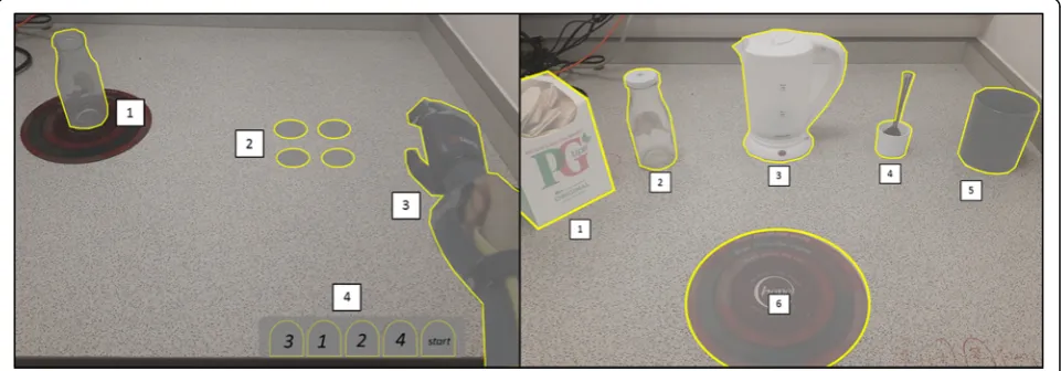
complete this task under the 35 s time limit reduced from 95 to 25% over the course of seven administrations of the entire SHAP protocol. This is in comparison to the much shorter and less complex jar task used in ex-periment 1, from which participants yielded an initial mean time of ~ 6 s. The task itself is made up of 4trial coins placed approximately 30 cm away from the partici-pant, 1startcoin positioned directly in front of the par-ticipant ready to grasp, and an empty glass jar in which all coins are to be placed. After successfully placing the start coin in the jar, participants were required to sequentially drag each trialcoin to a desired drag zone so it could subsequently be placed into the jar. Each trial was completed following the placement of the finaltrial coin in the jar.
Tea-making transfer task
To examine the transfer of learning, we included a tea-making task. This task was chosen as it requires par-ticipants to apply acquired myocontrol skills to novel task variants such as object size, object weight, and grasping angle. It also provided a novel comparison with
previous accounts of visuomotor control during
(Fig. 3). To complete the task, participants had to place the mug onto the place mat, add a teabag, a sugar cube, milk, water, and stir the contents twice with a spoon. Participants were told they could perform these tasks in an order of their choosing, as long as they started by pla-cing the mug onto the place mat, completed all steps and ended by stirring the spoon.
Mobile eye-tracker
Gaze behaviour was measured and analysed using the same equipment and analysis steps as experiment 1. For the modified coin task, four AOIs (jar, coin, drop zone, prosthesis) and three task phases (reach, grasp, and lift) were identified. For the more complextransfer tea task, a total of 19 AOIs and 17 task phases were identified (a complete breakdown can be seen in Additional file1).
EEG
EEG data from one participant was removed from ana-lysis due to excessive noise during the baseline record-ing. All data collection and pre-processing steps were identical to experiment 1, except here we used an array of 32 electrodes. This decision was made to decrease preparation time and data storage size to compensate for the increase in recording blocks. For the modified coin task, offline signals were specifically epoched to represent aLifttask phase. To do so, data were epoched from −1250 ms to + 250 ms relative to the instance the coins made contact with the bottom of the jar following
placement. This instance was detected using a
custom-made microphone placed behind the jar that automatically inserted digital triggers into the EEG re-cording when it detected sound > 70 dB. Time-frequency decomposition was performed through short-time FFT
on 9 overlapping segments (overlap of 87.5%), each of 500 ms duration and linearly spaced with centre points
ranging from −1000 ms to 0 ms. For the tea-making
task, manually inserted triggers were linearly spaced every 500 ms between the start and end of each trial – identified via previous calibration. EEG data were then epoched from−2000 ms to 0 ms relative to each trigger, resulting in 75% overlap to increase the signal to noise ratio during signal processing. Short-time FFT was per-formed on 17 overlapping segments (overlap of 87.5%), each of the duration of 500 ms and linearly spaced with centre points ranging from−1750 ms to−250 ms. Prior to FFT, data points within all segments (across both tasks) were Hanning tapered and 0-padded to reach 2 s.
Procedure
Participants were required to attend the laboratory on five consecutive days and a further day approximately one week later (M= 6.52 days, SD= 2.11) for a delayed retention and transfer test. On day 1, the experiment was explained, and participants were fitted with the EEG and eye-tracking equipment. Once participants were fit-ted with the prosthesis, and could demonstrate adequate control, thecoin taskwas explained and a demonstration was given by the researcher using the anatomic limb and via a video demonstration showing the task performed with the prosthesis. Participants were then given one full practice trial (5 coins) before completing 15 consecutive experimental trials (75 coins).
Participants were randomly allocated into GT and MT groups, with sex differences equally distributed. The training period lasted from days 2 (T1) to 4 (T3) and re-quired participants to perform 15 trials of the coin task on each visit. On day 2, the GT group was first shown a
video derived from the eye-tracker that depicted a per-former purposely adopting expert visual control whilst performing the task using the prosthesis. Audio com-mentary that overlaid the video highlighted the per-former’s target-focused gaze strategy, and the speed at which gaze was shifted to target locations following the completion of each task phase [40–42]. Participants were then fitted with the eye-tracker and advised to mimic the gaze strategy of ourexpertin the 15 subsequent at-tempts that followed. Eye movements were again re-corded on days 2 and 3 so participants could assess their attempts to mimic the expert model upon repeated viewing on days 3 and 4 [39].
For the MT group, a video of the sameexperttrial was shown on day 2 but from a third person perspective. This was done so participants could more easily be made aware of the smooth and direct manner in which the
ex-pert controlled the prosthesis – as was emphasised by
the audio commentary [40–42]. The video also gave par-ticipants a set of movement rules to help describe the expert’s performance, such as“drag the coin with the tip of the thumb”and“position the thumb beneath the coin before grasping”. Like the GT group, participants were then advised to mimic the movement style of the expert in the 15 subsequent experimental trials. No eye-tracker was worn, instead participants were recorded (Finepix S6500fd) from the same third person perspective as their training video so participants could assess their attempts to mimic the movements of the expert model on days 3
and 4 [39]. For both groups, EEG was not recorded
throughout training.
On day 5, no further instructions were given and par-ticipants were asked to perform a further 15 trails of the coin task whilst measures of EEG and eye-tracking were taken (i.e. non-delayed retention test). Before performing the tea-making transfer task, participants were provided with a demonstration by the researcher using the ana-tomic limb, and shown a video demonstration of the re-searcher performing a single trial using the prosthesis. Participants then repeated this procedure approximately 1 week later for delayed retention and transfer tests.
Measures
Performance time
For the coin task, performance time was measured as the time (in seconds) elapsed between the successful placement of thestart coin and the final trial coin into the jar, recorded by the researcher using a stopwatch (Casio, Japan). If a coin was dropped, time was contin-ued as participants were instructed to move on to the next coin in the sequence whilst the researcher replaced the dropped coin. In the instance that a participant dropped the final coin, time was paused until the re-searcher replaced the coin upon its starting position. For
the tea-making task, performance time was measured as the time elapsed (in seconds) between first grasping the mug and replacing the spoon following two stirs.
Performance error (coin drops)
To provide an indication of performance error within the coin task, we recorded the total number of coins that were dropped within each block of 15 trials.
Visual attention
Target locking strategy (TLS)
TLS was measured following the same procedure as ex-periment 1.
Gaze shifting
Gaze shifting was also measured following the same pro-cedure as experiment 1. However, for the tea-making task, gaze-shifting time was only recorded for the phases of the task that started with an object manipulation. This ensured the ensuing shift location (usually a drop loca-tion) was consistent across participants for each chosen phase, and did not reflect a more indecisive visual search
behaviour that occurred when participants were
in-between task phases.
EEG
Alpha power
As the actual alpha frequency band can show inter-subject variability, standardising alpha (8–12 Hz) across all participants might prevent the detection of more subtle changes in alpha activity. The individual alpha frequency (IAF) of each participant was therefore detected using the eyes-closed centre of gravity method [55] to enhance the ability to detect the differential ef-fects of training instruction. Power (μV2) was then aver-aged across overlapping FFT segments in the adjusted alpha frequency band (IAF-2 to IAF + 2) for each
channel and trial. Based on previous research [31]
seven regions of interest (ROI) were chosen; left tem-poral (T7, FC5, CP5), left central (C3, FC1, CP1), frontal (F3, Fz, F4), right central (C4, FC2, CP2), right temporal (T8, FC6, CP6), parietal (P3, Pz, P4) and oc-cipital (O1, Oz, O2). Power was averaged across these channels to yield values for each region following median-log scaling [19, 31].
High alpha connectivity
the coin task and the tea task, ISPC was calculated for each epoch using bespoke Matlab scripts as, ISPCðfÞ ¼jn−1Pn
w¼1eiðθxðw;fÞ−θyðw;fÞÞj, where i is the imaginary
operator;θxandθy are the phase angles of the recorded signal at two different scalp locations at FFT time win-doww and frequency f;ei(θx(w,f)−θy(w,f)) denotes a
com-plex vector with magnitude 1 and angle θx−θy;
n−1Pn
w¼1ðÞ denotes averaging across the overlapping
FFT time windows; and ∣·∣is the module of the aver-age vector [32]. ISPC values were then averaged over tri-als before being FisherZtransformed (inverse hypabolic
tangent), meaning values could range from 0 to ∞.
Values were then averaged across channel pairs and the high-alpha frequency band (IAF to IAF + 2 Hz). In line with previous research, we focused on left temporal frontal (T7-Fz) and right temporal frontal (T8-Fz) connectivity.
Data analyses
Performance
For the coin task, performance time and error were sub-ject to a 2 × 6 mixed-design ANOVAs, with group
(movement trained, gaze trained) as the
between-subjects factor and time (baseline, T1, T2, T3, retention, delayed retention) as the within-subjects fac-tor. For the tea-making transfer task, a Kruskal-Wallis tests was run to compare performance time between groups at retention and delayed retention due to viola-tions of Shapiro Wilk’s test of normality. Within group changes from retention to delayed retention were then analysed using Wilcoxon signed ranks tests.
Visual attention
To align with EEG data, only TLS and gaze shifting data specific to theLiftphase were included for the coin task. For the tea-task, both measures were averaged over task phases to derive an overall indication of visual control. Both measures were then subject to a 2 (group) × 3
(time; baseline, retention, delayed retention)
mixed-design ANOVA for the coin task, and a 2 (group) × 2 (time; retention, delayed retention) mixed design ANOVA for the tea-making task.
Alpha power
For the coin task, changes in regional alpha power were examined using a 2 (group) × 3 (time) × 7 (ROI) mixed design ANOVA. For the tea-making task, a 2 (group) × 2 (time) × 7 (ROI) mixed-design ANOVA was performed.
High alpha connectivity
For the coin task, changes in T7-Fz and T8-Fz connect-ivity over time were examined using a 2 (group) × 2 (hemisphere) × 3 (time) mixed-design ANOVA. To
provide direct between group comparisons unbiased from baseline levels of connectivity, we also examined the change (Δ) in ISPC values from baseline to retention (Ret Δ) and from baseline to delayed retention (Del Δ) using a 2 (group) × 2 (hemisphere) × 2 (time) mixed de-sign ANOVA. Finally, baseline ISPC values derived from the coin task were also used to allow the same between
group Δ ISPC comparisons in the transfer tea-making
task at retention and delayed retention.
Regression analyses
To directly explore the relationship between visual attention and conscious control, regression analyses were performed to determine if T7-Fz could be pre-dicted using our measures of visual attention (TLS & gaze shifting) for our coin task.
Results Coin task
Performance
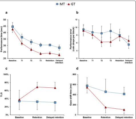
For performance time, results revealed a significant main effect of time, F (3.08, 67.712) = 48.19, p< .001, ηp2 = .687, a significant main effect of group, F (1, 22) = 6.94, p= .015, ηp2= .712, but no time x group inter-action, F (5, 110) = 0.772, p= .572, ηp2= .034. Pairwise comparisons showed that the MT group performed significantly faster at T3 compared to B1 (p< .001) and T1 (p= .020), after which no further improvements were made (p= 1.00). Similar results were found for the GT
group, who performed faster at T3 compared to B1 (p
< .001), T1 (p= .001), and T2 (p= .091), but subsequently plateaued at retention and delayed retention (p= 1.00). Importantly, comparisons also revealed that whilst there were no significant difference between groups at B1 (p
= .638) and T1 (p= .108), the GT group performed
significantly faster than the MT group on all subsequent visits (ps= .022).
For performance error, results failed to reveal a signifi-cant main effect of time,F(5, 110) = 2.101,p= .071,ηp2 = .087, suggesting the number of coin drops to be fairly insensitive to practice. There was also no main effect of group,F(1, 22) = 0.481,p= .495,ηp2= .021, and no time x group interaction, F (5, 110) = 0.745, p= .592, ηp2 = .033.
Target locking score (TLS)
Results revealed a significant main effect of time, F
(1.56, 34.24) = 9.97, p< .001, ηp2= .312, a main effect of group, F(1, 22) = 35.212,p< .001,ηp2= .410, and a sig-nificant time x group interaction, F (2, 44) = 13.481, p < .001, ηp2= .380. Post-hoc pairwise comparisons
revealed no difference between groups at baseline (p
delayed retention (p< .001). Participants in the MT group showed no significant improvement from baseline to retention (p= 1.00) or baseline to delayed retention (p= 1.00). Conversely, the GT group significantly in-creased their TLS from baseline to retention (p< .001) and delayed retention (p< .001).
Gaze shifting
Results revealed a significant main effect of time, F
(1.29, 28.42) = 34.269, p< .001, ηp2= .609, a main effect of group, F (1, 22) = 26.902, p< .001, ηp2= .550, and a significant time x group interaction,F(2, 44) = 8.361,p
= .001, ηp2= .279. Post-hoc pairwise comparisons
revealed no difference between groups at baseline (p
= .586), but the GT group to exhibit significantly faster gaze shifts than the MT group at retention (p= .001) and delayed retention (p< .001). They also revealed both the MT group (ps= .018) and the GT group (p< .001) shifted their gaze significantly faster from baseline to retention and delayed retention. Performance data and gaze data can be seen in Fig.4.
Alpha power
For the coin task, the ANOVA also showed a significant main effect of ROI, F (3.712, 70.530) = 87.703, p< .001,
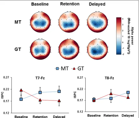
ηp2= .822, revealing a focal pattern in which alpha was lowest over central and parietal regions, higher over temporal and frontal regions, and highest over the oc-cipital region. There was also a significant main effect of time,F(2, 40) = 3.279,p= .049,ηp2= .049, and a signifi-cant time x ROI interaction, F (6.685, 127.022) = 2.819, p= .010, ηp2= .129. Pairwise comparisons revealed that both groups exhibited a significant decrease over the left-temporal (p= .001) and right temporal (p= .042) re-gions from baseline to delayed retention. All other inter-actions were non-significant (Fig.5).
High alpha connectivity
When examining cross hemispheric (T7 vs T8) changes in temporal-frontal (Fz) connectivity, results showed no overall main effect of time, F (2, 40) = 0.427, p= .655,
ηp2= .021, and no overall main effect of group, F(1, 20) = 0.156,p= .697,ηp2= .008. There was however a signifi-cant time x group interaction,F(2, 40) = 3.387,p= .044,
ηp2= .145, and a significant time x hemisphere x group interaction, F (2, 40) = 4.532, p= .017, ηp2= .185. Pair-wise comparisons revealed that participants in the GT group exhibited a significant reduction in T7-Fz con-nectivity from baseline to delayed retention (p= .043), and a marginally significant reduction from baseline to retention (p= .056). No changes were observed in the MT group (Fig.5).
Δhigh alpha connectivity
Results from ANOVA showed no effect of time,F(1, 20) = 0.260, p= .616, ηp2= .013, hemisphere, F (1, 20) = 3.333,p= .083,ηp2= .143, or group,F(1, 20) = 4.284,p = .052,ηp2= .176. There was however a significant hemi-sphere x group interaction, F (1, 20) = 7.934, p= .011,
ηp2= .284, in which a significant difference between groups was observed only for the change in T7-Fz con-nectivity (p= .003). Pairwise comparisons also showed an overall significant difference between hemispheric changes for the GT group (p= .003) which consisted of a decrease in T7-Fz connectivity and an increase in T8-Fz connectivity.
Regression analyses
At baseline, a non-significant regression equation was found when predicting T7-Fz connectivity based on TLS,F(1, 21) = 0.718,p= .406,r2= .033, and gaze shift-ing, F (1, 21) = .028, p= .868, r2= .001. At retention, however, both TLS,F (1, 21) = 4.532,p= .045, r2= .177, and gaze shifting, F (1, 21) = 8.056, p= .010, r2= .287, were significant predictors of T7-Fz connectivity. The same was true at delayed retention, with TLS,F(1, 21) = 7.238, p= .014, r2= .256, and gaze shifting, F (1, 21) = 5.004, p= .036, r2= .192, again significant predictors of T7-Fz connectivity (Fig.6).
Transfer tea-making task
Due to time-locking synchronisation errors, EEG data for three participants could not be analysed for the tea-task.
Performance
Results showed no significant difference between the
MT (Mdn= 73.20 s) and GT (Mdn= 64.55 s) groups’
performance time at retention (H (1) = 1.763, p= .184).
There was also no difference between the MT (Mdn=
57.70) and GT (Mdn= 57.39 s) at delayed retention (H (1) = .033,p= .564).
Target locking score (TLS)
No significant main effect of time F (1, 22) = 3.799, p = .065,ηp2= .147, but a significant main effect of group, F(1, 22) = 22.328,p< .001,ηp2= .504, was observed, re-vealing participants in the GT group to exhibit signifi-cantly lower TLS compared to participants in the MT group. There was no significant time x group inter-action,F(1, 22) = 0.009,p= .926,ηp2= 00.
Gaze shifting
Results revealed no main effect of time, F (1, 22) =
Alpha power
Results revealed no main effect of time,F(1, 18) = .257, p= .618, ηp2= .014, or group, F (1, 18) = .195, p= .664,
ηp2= .011, but a main effect of ROI,F (3.495, 62.917) = 27.837, p< .001, ηp2= .607, revealing alpha power to be lowest over central and parietal regions, and highest overall temporal, frontal and occipital regions. All other interactions were non-significant.
High alpha connectivity
When examining cross hemispheric (T7 vs T8) changes in frontal (Fz) connectivity, results from ANOVA showed no significant main effect of time, F (1, 17) = 3.693,p= .072,ηp2= .178, or group,F(1, 17) = 3.248,p = .089,ηp2= .160. There was however a significant main
effect of hemisphere, F (1, 17) = 11.694, p= .003, ηp2 = .408, showing overall higher T7-Fz connectivity com-pared to T8-Fz connectivity.
Δhigh alpha connectivity
When examining Δ cross hemispheric (T7 vs T8)
changes in frontal (Fz) connectivity, results from
ANOVA showed no effect of time, F(1, 17) = 1.054, p
= .318, ηp2= .055. There was however a main effect of hemisphere, F (1, 17) = 4.751, p= .041, ηp2= .232, and group, F (1, 17) = 4.977,p= .037, ηp2= .217, which was superseded by a significant hemisphere x group inter-action, F (1, 17) = 4.751, p= .041, ηp2= .209. Follow up pairwise comparisons showed a significant difference be-tween groups for T7-Fz connectivity (p= .022), in which Fig. 4Performance and gaze data before, during and after training Line plots representing mean (± s.e.m) performance time (a) and
the MT group exhibited a much greater increase from baseline compared to the GT group. The MT group also exhibited overall significant hemispheric asymmetry, with connectivity higher for T7-Fz compared to T8-Fz (p= .009), whereas the GT group did not (p= .906). Data for the transfer task can be seen in Fig.7.
Discussion
The aim of the second experiment was to determine the efficacy of GT in expediting prosthetic hand learning and alleviating the associated cognitive burden. We hypothesised that GT would optimise visual control, ex-pedite skill acquisition, and promote neural efficiency by reducing conscious control, compared to MT instruc-tions. We also hypothesised that these benefits would
carry over to our complex transfer task [33]. Finally, we hypothesised that an increased dependence on vision to monitor the prosthesis would be related to increases in conscious movement control.
group was ~ 20% faster than the MT group without be-ing any more errorful. Encouragbe-ingly, the improved vis-ual control adopted by the GT group also transferred to the more complex tea-making task, with participants using a higher TLS (~ 20%) compared to the MT group (Fig.7).
While GT optimised gaze behaviour and expedited learning, we found mixed results when determining whether this decreased dependence on vision enhanced
neural efficiency. For regional alpha power, we found a focal pattern consistent with Experiment 1, in which cognitive resources were primarily gated towards the central and parietal regions of the brain, regardless of training received. As such, our findings seemingly valid-ate the utility of measuring regional alpha power to examine the functional architecture of the brain during prosthetic hand control. Although this gating pattern was insensitive to change from baseline to retention, Fig. 6Relationship between gaze indices and conscious movement control Scatter plots displaying the relationship between TLS and T7-Fz (top row), and between gaze-shifting times and T7-Fz (bottom row), across three time points. Each plot displays the line of best fit (in red) with 95% confidence intervals (shaded in grey), the shared variance (r2) and the significance value (p) of each regression
there was a significant decrease in temporal alpha at delayed retention compared to baseline, regardless of which training was received. This increased excitability of the temporal regions is contrary to our predictions that increased skill would decrease (left) temporal activ-ity. However, it should be noted that our predictions
were primarily based upon research comparing
expert-novice differences (as was seen in Experiment 1), or longitudinal training (~ 15 weeks) in target sports. Given the dynamic nature and complexity of our task, it is likely that the putative link between motor-skill expertise and optimal cortical organisation, as indexed by alpha power, might flexibly depend on external de-mands and required performance rather than a rigid strategy (always reduced activity [56];). Future research could explore this by conducting longitudinal interven-tion studies or by examining expert vs. novice compari-sons of prosthesis users.
Our EEG results did however provide stronger
evidence to suggest that GT reduces conscious
verbal-analytical processes. Specifically, we showed that participants in the GT group exhibited a significant re-duction in T7-Fz connectivity from baseline to retention and delayed retention, whereas the MT group did not (Fig. 5). We also showed a significant difference in the baseline change in T7-Fz connectivity between groups, with GT showing a decrease and the MT group showing an increase. The change in temporal-frontal connectivity also showed significant hemispheric asymmetry for the GT group, showing decreased T7-Fz and increased T8-Fz connectivity. Encouragingly, similar results were observed in the transfer tea-making task, with the train-ing conditions again significantly altertrain-ing the change in T7-Fz connectivity. However here, participants in the GT group displayed similar levels to that seen at baseline coin task performance, whereas the MT group showed a large increase.
These findings strongly suggest that encouraging learners to engage visual attention on the target rather than object manipulation, discourages burdensome verbal-analytical control [34]. In fact, regression analyses provided direct support for this claim, revealing that re-duced T7-Fz connectivity was significantly predicted by increased TLS and faster gaze shifting times at retention and delayed retention. Conversely, our results also high-light how the provision of explicit instructions can ac-centuate the reliance on verbal processes, especially during complex tasks that are more reflective of the ac-tivities of daily living. Indeed, as these relationships were not present at baseline, the link between visual monitor-ing and conscious control appears to be highly dependent on the cognitive strategies encouraged through training rather than being inherent in prosthesis control. As conscious control processes require high
cognitive demands they can result in performance break-down under increasing task difficulty and fatigue [38] and should therefore be minimised in prosthesis rehabilitation.
These results provide evidence that GT alleviates con-scious control and promotes neural efficiency, reducing the non-essential interaction between the motor plan-ning and verbal-analytical regions of the brain. They also provide evidence that the provision of explicit instruc-tion via MT can have the opposite effect, increasing the functional communication between motor-planning and verbal-analytical regions – an effect that increased dur-ing the more complex transfer task. Indeed, these find-ings are in line with previous research in laparoscopic surgery [49], and should not only act to promote the benefits of implicit learning via GT, but also act as a warning against the provision of more explicit training methods.
General discussion
In this study, we report the first attempt to simultan-eously examine the visuomotor and cortical mechanisms that contribute to the cognitive burden experienced by upper-limb prosthesis users [4,13]. In both experiments, we provide further evidence that prosthetic hand control places high demands on visual attention and cognitive processes in order to guide and monitor movements, particularly during object manipulations [7]. Import-antly, we also show that individuals can be trained to reduce their reliance on vision via GT, which subse-quently expedites learning and encourages greater neural efficiency compared to more traditional explicit training methods. The findings of these experiments therefore have important theoretical and practical implications.
From a theoretical perspective, it is important to understand why prosthesis users appear to maintain these inefficient strategies despite skill improvements. In the development of eye-hand coordination, vision is initially utilised primarily as a feedback mechanism to monitor ongoing action as learners develop sensori-motor mapping rules between commands and
move-ments, and between vision and proprioception [9].
control. So how then does GT help to overcome such deficits? And why might this be beneficial to long-term prosthesis control?
There are a number of potential theoretical explana-tions for this. First, being trained to use vision in this more proactive manner and to“look at the right place at the right time”is thought to aid effective coordination of the visuomotor system [8, 9, 43]. Specifically, by adopt-ing early and accurate look-ahead fixations users are able to effectively pass visually acquired target-related infor-mation to the motor system so accurate movements can be planned and executed [8,9]. The faster performance times exhibited by the GT group support these predic-tions, and suggest increased proficiency of movements. Second, reducing the dependence on vision reduces con-scious movement control, supporting the idea that GT alleviates the reliance on these explicit and burdensome processes [34]. Third, it could be that case that GT forces the development of ‘new’ sensorimotor mapping rules using the remaining senses (e.g., proprioception, or auditory information from the prosthesis’ motors [13]) to enable vision to be used in a more proactive feed-forward manner.2Finally, the benefits of GT could also be attributable to encouraging learners to adopt an external focus of attention (FOA). Research has shown that focusing on the effect of movement (external FOA) rather than the mechanics of the movement itself (in-ternal FOA) promotes better performance in a variety of movement contexts [57]. Interestingly, an external FOA has also been shown to improve movement economy by reducing muscle stiffness and activity [58]. Reducing de-mands on muscle fibre recruitment may therefore mitigate the negative effects of fatigue upon
electromyo-graphic (EMG) signal quality [58] and improve
long-term myoelectric control.
From an applied perspective, the methods used in these experiments could be used to assess the usability of prosthetic hands from a design perspective. While the technological development of hand prosthesis is increas-ing rapidly, research examinincreas-ing the usability and inter-action between the user and the prosthesis is lacking. For example, while performance measurements are ad-equate in accessing the functionality of prosthesis hand devices, they are not sensitive enough to assess their us-ability. As our transfer task shows, both training groups performed similarly but the magnitude of mental re-sources needed to perform was significantly less in the GT group. So, just because a user canuse a hand pros-thesis does not mean that the hand prospros-thesis is
intui-tively useable. From technologies that provide
vibrotactile feedback [59] to hands that can actually ‘see’ for themselves [60], each will increase or lessen the cog-nitive resources needed to interact with the world. It is
this user-prosthesis-world interaction that needs
examining in future research, which to be effective, will depend on significant collaborations between applied psychologists, prosthesis engineers, occupational thera-pists and prosthesis users themselves.
Similarly, an examination of the cognitive demand experienced during prosthesis learning could also aid occupational therapists to assess a patient’s progress. However, the methods used in these studies are probably not cost effective given the expensive equipment required and the expertise needed to operate it. Researchers therefore should develop and validate a multidimensional workload measure specific to pros-thesis use. Such a measure has previously been
devel-oped for surgical skills (SURG-TLX; [61], and would
allow for more cost-effective and immediate clinical as-sessment of the cognitive demand experienced by pros-thesis users during the rehabilitation process.
Despite the important first steps presented here, sev-eral limitations should be noted. First, we are limited by our use of intact users of a simulator rather than patients with limb loss. However, evidence has shown that these populations display comparable kinematic profiles [1], visuomotor behaviours [6,7], and perceptual experiences [62], suggesting that using a simulator pro-vides a useful surrogate to examine the sensory-motor deficits that prosthesis users face. Yet, it is unclear how increasing the length of the operating arm when using the prosthesis simulator (approximately 7 cm when the hand is unclenched) independently influences visuo-motor and neurophysiological behaviours. Furthermore, the cortical reorganisation that occurs following amputa-tion can cause large-scale changes in neural networks, making direct transfer of our results to an amputee population potentially difficult. For example, evidence shows that neuroplasticity of the cortex following ampu-tation can promote an expansion of the residual limb segments into the former limb territory [63], and pro-mote a progressive disconnection of the missing hand cortex and the sensorimotor cortex [64]. Clearly, future work is needed to evaluate the cognitive burden in a clinical population and to explore if this can be allevi-ated in the same manner using a GT intervention.
The degree of ambiguity in the temporal accuracy of EEG data must also be highlighted. Here, data
were segmented through clearly defined epoch
lengths relative to a given manual action (i.e., jar lift in experiment 1). Whilst this method enabled mean-ingful comparisons to be made, it fails to guarantee that the segmented data represent the exact same
“portion” of movement on a trial-to-trial basis.
Finally, in these studies we limited our EEG analysis to the alpha frequency band in order to contextualise our findings with previous research on alpha gating [31] and connectivity during movement execution [20, 49]. In fu-ture, more exploratory research could benefit from in-vestigating multi-scale interactions across different frequencies in order to acknowledge the fact that changes in specific frequency bands do not occur in isolation [65]. Such analyses could help to attain a more holistic understanding of the cortical disrup-tions evident during initial hand use and this could help develop objective methods to assess training pro-grammes in the future.
Conclusions
We believe that these two experiments represent the most comprehensive evaluation of the visual and cor-tical mechanisms relating to the cognitive burden
as-sociated to prosthetic hand control. We also
demonstrate the efficacy of a GT intervention de-signed to alleviate this burden. This is important be-cause this intervention seems to promote better learning and transfer, increased neural efficiency and both of these factors are what prosthesis users actu-ally desire in a functional prosthetic hand [4]. This demonstrates that the problem of making prosthesis hands more useable is not necessarily a technological
issue – both groups used the same hand in our study
– but an issue relating to how the user interacts with this technology. Therefore, in future research and development we propose that a greater emphasis should be placed on understanding human factors alongside technological ones.
Endnotes
1
The sampling rate of the eye-tracker was 30 Hz, pro-viding temporal accuracy of approximately ±33 ms when determining the onset of the Lift phase. Although this level of precision may not be suitable to detect specific event-related changes in EEG activity, it is not necessary for the present investigation that instead examines the consistency of spectral power across the entirety of a long time-window (1500 ms).
2
We did explore this explanation in an additional con-dition that eliminated auditory feedback via in-ear white noise. This had no effect on performance, gaze or neural activity after training (see Additional file2).
Additional files
Additional file 1:A breakdown of the 17 task phases and 16 AOIs for the tea-making transfer task. (PDF 998 kb)
Additional file 2:Results from the additional“white noise”condition administered during Experiment 2. (PDF 589 kb)
Abbreviations
ANOVA:Analysis of variance; AOI: Area of interest; ASL: Applied Science Laboratories; CMS: Common mode sense; DRL: Driven right leg; EEG: Electroencephalography; EMG: Electromyographic;
EOG: Electrooculographic; FFT: Fast Fourier Transform; FOA: Focus of attention; GT: Gaze training; HEOG: Horizontal electrooculogram; IAF: Individual alpha frequency; ISPC: Inter site phase clustering; MT: Movement training; ROI: Region of interest; SHAP: Southampton Hand Assessment Procedure; TLS: Target locking strateg; VEOG: Vertical electrooculogram
Acknowledgements
The authors would like to thank Bruce Ratray and Tim Verrall (Steeper Ltd.) and the technicians at Aintree Hospital, Liverpool, for their assistance with the design and manufacture of the prosthetic hand simulator.
Funding
This research was supported by a Royal Society grant (RG140418) that was awarded to G. Wood and S. J. Vine. The views expressed in this article are those of the authors and do not necessarily reflect the position or policy of the sources which funded this research. All authors report no actual or potential conflicts of interest.
Availability of data and materials
The datasets supporting the conclusions of this article are available from http://e-space.mmu.ac.uk/id/eprint/621799
Authors’contributions
JP, GW, SV and NH conceived the study; JP collected and analysed all data; JP and GW drafted the paper; SV, NH and MW offered critical revisions; all authors reviewed the manuscript. All authors read and approved the final manuscript.
Ethics approval and consent to participate
The experimental procedure was designed and performed in accordance with the relevant guidelines and regulations, particularly those set out in the Declaration of Helsinki pertaining to the ethical treatment of human subjects. Participants signed informed consents, and were instructed on their rights as participants, including the right to withdraw from the experiment at any time without fear of negative consequences. The study protocol has been approved by the Liverpool Hope University Ethics Committee (approval code S 09-10-2017 DEL 018).
Consent for publication
Not applicable.
Competing interests
The authors declare that they have no competing interests.
Publisher’s Note
Springer Nature remains neutral with regard to jurisdictional claims in published maps and institutional affiliations.
Author details
1School of Health Sciences, Liverpool Hope University, Liverpool, UK.2College
of Life & Environmental Sciences, University of Exeter, Exeter, UK.
3Department of Psychology, Liverpool Hope University, Liverpool, UK. 4Research Centre for Musculoskeletal Science and Sports Medicine
Department of Sport and Exercise Science, Manchester Metropolitan University, Manchester, UK.
Received: 5 November 2018 Accepted: 16 April 2019
References
1. Bouwsema H, van der Sluis CK, Bongers RM. Changes in performance over time while learning to use a myoelectric prosthesis. J NeuroEngineering Rehabil. 2014;11:16.