Report
Neofunctionalization of Duplicated P450 Genes
Drives the Evolution of Insecticide Resistance in the
Brown Planthopper
Highlights
d
The cytochrome P450
CYP6ER1
is duplicated in imidacloprid
resistant
N. lugens
strains
d
Amino-acid alterations in certain CYP6ER1 variants confer
resistance to imidacloprid
d
Resistant hoppers have paralogs with and without the
gain-of-function mutations
d
The susceptible and mutant
CYP6ER1
copies show marked
divergence in their expression
Authors
Christoph T. Zimmer, William T.
Garrood, Kumar Saurabh Singh, ..., Ralf
Nauen, T.G. Emyr Davies, Chris Bass
Correspondence
In Brief
Zimmer et al. explore the functional
significance of genetic variation at the loci
encoding CYP6ER1, a cytochrome P450
enzyme, in field strains of the brown
planthopper. They show that duplication
of
CYP6ER1
provided opportunities for
functional and regulatory innovation,
leading to resistance to the insecticide
imidiacloprid.
Zimmer et al., 2018, Current Biology28, 268–274
Current Biology
Report
Neofunctionalization of Duplicated P450 Genes
Drives the Evolution of Insecticide Resistance
in the Brown Planthopper
Christoph T. Zimmer,1,4,5William T. Garrood,2,4Kumar Saurabh Singh,1Emma Randall,1Bettina Lueke,3Oliver Gutbrod,3
Svend Matthiesen,3Maxie Kohler,3Ralf Nauen,3T.G. Emyr Davies,2and Chris Bass1,6,*
1College of Life and Environmental Sciences, Biosciences, University of Exeter, Penryn Campus, Penryn, Cornwall TR10 9FE, UK
2Department of Biointeractions and Crop Protection, Rothamsted Research, Harpenden AL5 2JQ, UK
3Bayer AG, Crop Science Division, Alfred Nobel-Strasse 50, 40789 Monheim, Germany
4These authors contributed equally
5Present address: Syngenta Crop Protection, Werk Stein, Schaffhauserstrasse, Stein CH4332, Switzerland
6Lead Contact
*Correspondence:[email protected] https://doi.org/10.1016/j.cub.2017.11.060
SUMMARY
Gene duplication is a major source of genetic
variation that has been shown to underpin the
evo-lution of a wide range of adaptive traits [
1, 2
]. For
example, duplication or amplification of genes
en-coding detoxification enzymes has been shown to
play an important role in the evolution of insecticide
resistance [
3–5
]. In this context, gene duplication
performs an adaptive function as a result of its
effects on gene dosage and not as a source of
func-tional novelty [
3, 6–8
]. Here, we show that
dupli-cation and neofunctionalization of a cytochrome
P450, CYP6ER1, led to the evolution of insecticide
resistance in the brown planthopper. Considerable
genetic variation was observed in the coding
sequence of
CYP6ER1
in populations of brown
planthopper collected from across Asia, but just
two sequence variants are highly overexpressed in
resistant strains and metabolize imidacloprid. Both
variants are characterized by profound amino-acid
alterations in substrate recognition sites, and the
introduction of these mutations into a susceptible
P450 sequence is sufficient to confer resistance.
CYP6ER1
is duplicated in resistant strains with
indi-viduals carrying paralogs with and without the
gain-of-function mutations. Despite numerical parity in
the genome, the susceptible and mutant copies
exhibit marked asymmetry in their expression with
the resistant paralogs overexpressed. In the primary
resistance-conferring CYP6ER1 variant, this results
from an extended region of novel sequence
up-stream of the gene that provides enhanced
expres-sion. Our findings illustrate the versatility of gene
duplication in providing opportunities for functional
and regulatory innovation during the evolution of an
adaptive trait.
RESULTS
We previously demonstrated that resistance to the insecticide imidacloprid in the brown planthopper (BPH), Nilaparvata lugens, is associated with the overexpression of the cyto-chrome P450, CYP6ER1 [9]. To explore if qualitative changes in this P450 also play a role in resistance, we first sequenced the complete coding cDNA ofCYP6ER1in field populations of BPH collected from across Asia that all exhibit resistance to imidacloprid (Tables S1 and S2). Comparison of the se-quences obtained with a reference sequence (CYP6ER1vL) derived from a lab-susceptible strain (NLS,Table S1) identified a total of 114 polymorphic sites that result in 27 amino-acid alterations (Figures 1A and S1A). These nucleotide se-quences resolved to seven unique amino acid sequence vari-ants: CYP6ER1vA, CYP6ER1vB, CYP6ER1vC, CYP6ER1vD1, CYP6ER1vD2, CYP6ER1vE, and CYP6ER1vF (Figures 1A,
S1A, and S1B).
The relative expression of the differentCYP6ER1variants in field populations of BPH was assessed by cDNA cloning and sequencing and using variant-specific qPCR. This revealed that just two of theCYP6ER1variants are highly expressed in strains from field populations:CYP6ER1vAwas the major variant expressed in strains from Thailand, Vietnam, and Indonesia, whereasCYP6ER1vBwas expressed in strains originating from India (Figures 1B and 1C). The expression of one predominant sequence variant in imidacloprid resistant populations of BPH in these regions suggests that CYP6ER1vA and CYP6ER1vB
may play a primary role in resistance.
To test this, we expressed CYP6ER1vA, CYP6ER1vB, the lab susceptible variant CYP6ER1vL, and its closest relatives observed in the field, CYP6ER1vF and CYP6ER1vC, in vitro
and examined their capacity to metabolize imidacloprid. Liquid chromatography tandem mass spectrometry (LC-MS/MS) anal-ysis demonstrated that CYP6ER1vA and, to a lesser extent, CYP6ER1vB are effective metabolizers of imidacloprid, convert-ing it to 4/5-hydroxy imidacloprid and 6-chloronicotinic acid (6-CNA) (Figure 1D). In contrast, no significant metabolism of imidacloprid was observed in the case of CYP6ER1vC, CYP6ER1vL, or CYP6ER1vF (Figure 1D).
To explore which amino acid polymorphisms in CYP6ER1vA and CYP6ER1vB are responsible for imidacloprid metabolism, we first mapped polymorphisms in all CYP6ER1 variants to important known P450 domains (Figures 2A and S1A). This highlighted two features unique to these variants. First, both CYP6ER1vA and CYP6ER1vB share an amino-acid substitution in substrate recognition site (SRS) 4 at position 318, where a threonine in all other variants is replaced with a serine. Signifi-cantly, this occurs at a highly conserved position in a P450 signature sequence [A/G]GX[E/D]T[T/S] in helix I, known as the oxygen-binding motif. The second alteration unique to CYP6ER1vA/B is the deletion of an amino acid in SRS5 in both variants. In CYP6ER1vA, this occurs at Ala375 and is immediately followed by an alanine to glycine substitution, whereas in CYP6ER1vB, a proline is deleted at position 377. To predict the effect of these amino acid changes on imidaclo-prid binding, CYP6ER1 was computationally modeled and docking simulations of imidacloprid within the active site of CYP6ER1vL, CYP6ER1vA, and CYP6ER1vB performed ( Fig-ures 2B, 2C, and 2D). This revealed that T318 and A376, along with additional residues, create the hydrophobic interface of the binding cavity (Figure 2B). In CYP6ER1vA, T318S in
combi-nation with A376G increases the conformational space acces-sible to imidacloprid between these two positions (Figure 2C). Although less pronounced, this is also true for CYP6ER1vB, where the T318S mutation comes together with an alanine at position 376 (Figure 2D). In this instance, due to the proline deletion, the fold of CYP6ER1vB is shifted slightly away from the I-helix substitution with the consequence of a similar gap opening. These alterations are consistent with the hydrox-ylation capacity seen for individual CYP6ER1 variants when functionally expressed and are strong candidates for the gain-of-function observed.
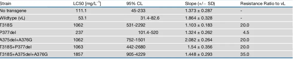
To validate the predictions made by homology modeling, we employed site-directed mutagenesis in combination with functional expression in vivo. For this, a series of amino-acid alterations were introduced into the susceptible CYP6ER1vL sequence as follows: T318S, P377del, A375del+A376G, T318S+P377del, and T318S+A375del+A376G. A series of trans-genicDrosophilalines were created that ubiquitously express each of these mutated P450s, or the wild-type variant CYP6ER1vL, and their sensitivity to imidacloprid was assessed (Table 1). The T318S substitution, shared by both variants, re-sulted in a marked (20-fold) and significant increase in resistance
A B
C D
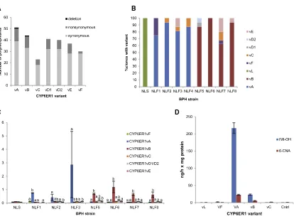
Figure 1. Characterization ofCYP6ER1Variants in BPH Populations
(A) Number and type of nucleotide polymorphisms in different CYP6ER1 variants relative to CYP6ER1vL, the variant observed in the lab-susceptible strain. (B and C) Relative expression of CYP6ER1 variants in imidacloprid-resistant BPH field strains (NLF1-8) and a susceptible strain (NLS), as determined by cDNA cloning and sequencing (B), and variant-specific QPCR (C). In (C), letters above bars are used to denote significant (p = < 0.01 in all cases) differences in expression between variants within each strain as assessed by one-way ANOVA with post-hoc Tukey HSD. Error bars in (C) indicate 95% confidence intervals (n = 4).
(D) Metabolism of imidacloprid by recombinantly expressed CYP6ER1 variants. NADPH-dependent conversion of imidacloprid to 4/5-hydroxy imidacloprid (IMI-OH) and 6-chloronicotinic acid (6-CNA) is shown. Error bars indicate 95% confidence intervals (n = 3).
See alsoFigure S1andTables S1andS2.
compared to the wild-type susceptible variant. Deletion of Pro377, as seen in CYP6ER1vB, provided a more moderate, but significant, 4.5-fold increase in resistance. In contrast, the A375del+A376G alteration observed in CYP6ER1vA conferred higher levels (20-fold) of resistance to imidacloprid. When T318S was combined with P377del (as seen in CYP6ER1vB), an epistatic interaction was observed, with the resistance conferred by the double mutation (20-fold) less than the sums of the effects of the component single mutations. In contrast, an additive interaction was observed when T318S was com-bined with A375del+A376P (as observed in CYP6ER1vA), with this combination exhibiting the highest resistance of all mutant lines (35-fold). These data are consistent with the relative efficiency of imidacloprid hydroxylation by CYP6ER1vA and CYP6ER1vB observed in vitro (Figure 2D) and convincingly demonstrate the adaptive nature of the genetic alterations observed in these isozymes.
To examine if the high levels of genetic variation inCYP6ER1in BPH field populations results, in part, from gene copy number variation (CNV), we resequenced the genomes of three BPH
strains that primarily expressCYP6ER1vA(NLF2),CYP6ER1vB
(NLF7), orCYP6ER1vL(NLS), respectively. Reads were mapped to the coding sequence ofCYP6ER1and two single-copy refer-ence genes in the genome of BPH: the P450 CYP6AY1 and the voltage-gated sodium channel (VGSC); and read coverage across each gene was compared between strains (Figures 3A–3F). No significant shift in coverage was observed be-tween the three strains across CYP6AY1or VGSC. However, an approximately 2-fold increase in read depth was observed over the coding sequence ofCYP6ER1between the two resis-tant strains NLF2 and NLF7 and the susceptible strain NLS, sug-gesting thatCYP6ER1is duplicated in the field strains. qPCR confirmed this finding, with the copy number of CYP6ER1
2.2-fold higher in NLF7 and 1.9-fold higher in NLF2 than in NLS. The mean cycle threshold values in qPCR of CYP6ER1
and the single-copy reference gene in NLS were essentially the same (23.50 [SEM = 0.04] and 23.62 [SEM = 0.08], respectively), indicating that CYP6ER1 is present as a single copy in the haploid genome of NLS and two copies in NLF2 and NLF7 ( Fig-ures 3G and 3H).
A
B C D
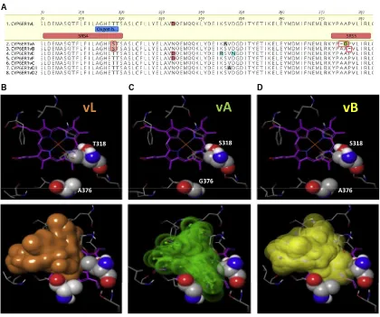
Figure 2. Modeling the Active Site of CYP6ER1 Reveals the Impact of Amino Acid Alterations on Imidacloprid Binding
(A) Amino-acid alignment of CYP6ER1 variants highlighting substitutions and deletions within substrate recognition sites four and five (boxed in red). (B–D) Protein homology modeling for 3 different CYP6ER1 variants (upper row), showing key residues surrounding the catalytic site. Amino-acid positions T318/A376 in CYP6ER1vL (B), S318/G376 in CYP6ER1vA (C), and S318/A376 in CYP6ER1vB (D) are in close proximity (spacefill representation). Imidacloprid docking into the active site is illustrated (lower row) by colored volumes, constituting an envelope around an ensemble of possible binding poses.
See alsoFigure S1.
To explore if one or both of the gene copies in resistant BPH individuals carry the resistance-conferring mutations,30 indi-viduals of the NLF2 and NLF7 strains were genotyped by PCR and direct sequencing. All individuals of the two strains scored as heterozygous for the diagnostic resistance mutations that characterizeCYP6ER1vA and CYP6ER1vB, respectively ( Fig-ures S2A and S2B). This observed excess of apparent heterozy-gosity is a characteristic signature of a duplicated gene and demonstrates that individuals of both strains carry CYP6ER1
copies with and without the resistance conferring mutations. To more precisely confirm which variants are present in these two strains, we cloned and sequenced amplified products from three individuals of each strain. For individuals of NLF2 we recov-ered two different sequences in equal abundance; the first car-ried the indels/SNPs that defineCYP6ER1vA, while the second carried SNPs diagnostic forCYP6ER1vC(Figures 3I,S2C, and S2D). Sequencing of colonies from NLF7 individuals again recovered two alternative sequences in equal abundance: the first corresponded toCYP6ER1vB, while the SNP profile of the second most closely matched CYP6ER1vF (Figures 3I, S2C, and S2D). Thus, taken together with functional analyses ( Fig-ure 1D), these data reveal that the genomes of individuals of both resistant strains haveCYP6ER1copies that encode the ca-pacity to metabolise imidacloprid and those that do not.
To examine the expression of the different gene copies observed in NLF2 and NLF7, we repeated these experiments but used RNA extracted from individuals of each strain as template in RT-PCR. Again, we only observed sequences representing CYP6ER1vA and CYP6ER1vC in individuals of NLF2; however,CYP6ER1vArepresented > 90% of the clones sequenced (Figures 3I andS2D). Similarly, in the case of NLF7, we only observed sequences representing CYP6ER1vB and
CYP6ER1vF, with the former representing > 90% of the se-quences recovered (Figures 3I andS2D). Thus, the duplicate genes exhibit marked divergence in their expression, with the copy with the gain-of-function mutations overexpressed.
To explore the molecular basis of this and simultaneously examine the genomic architecture of different CYP6ER1 vari-ants, we employed a gene capture approach. This allowed the close-to-complete gene sequence ofCYP6ER1vL,CYP6ER1vA,
CYP6ER1vB,CYP6ER1vC, and CYP6ER1vFto be assembled (Figure 3J). Considerable variation was seen in intron size and sequence between different CYP6ER1 variants (Figures 3J and S3). This was particularly pronounced in the case of
CYP6ER1vA and CYP6ER1vB. For example, in CYP6ER1vA, intron 1 is > 4.9 kb in size in contrast to4 kb in other variants,
and intron 4 is just 316 bp in size compared to an average size of5 kb in other variants. In the case ofCYP6ER1vB, intron 2 was much larger than in all other variants (5,478 bp compared to an average of 3,302 bp), with this expanded intron size result-ing from an internal duplication that included the last 123 bp of exon 3. This pseudo-exon was never observed inCYP6ER1vB
transcripts, likely due to the loss of the splice site consensus sequence upstream of the duplicated region.
Five putative promoter variants were identified by gene cap-ture, four of which could be linked toCYP6ER1vA,CYP6ER1vB,
CYP6ER1vC, andCYP6ER1vL(Figures 3K andS4). Surprisingly, only the promoter associated with CYP6ER1vA matched the sequence upstream of CYP6ER1 on scaffold KN153994.1 of the reference genome [10]. The remaining four promoter variants do not show sequence similarity with any other scaffold in the genome. Indeed, alignment of the promoter variants reveals a clear break point 104 bp upstream of the start codon in
CYP6ER1vA, with the sequence of this variant completely diverging from the other four variants after crossing this point (Figures 3K andS4). We used reporter gene assays to explore the effect of this on gene expression. No significant differences were seen in reporter gene expression driven by a1.8 kb region of the promoters ofCYP6ER1vB,CYP6ER1vC,and CYP6ER1vL
(Figure 3L). This finding suggests that the overexpression of
CYP6ER1vB seen in resistant BPH strains results from either
trans-acting factors orcis-acting elements outside of the region analyzed. In contrast, we observed a significant (up to 9.5-fold) increase in expression driven by the promoter ofCYP6ER1vAin comparison to all other promoter variants (Figure 3L). This suggests that cis-acting elements in the region upstream of
CYP6ER1vA, derived from the novel genomic sequence, are responsible for the high expression of this variant in BPH popula-tions across Southeast Asia.
DISCUSSION
Our data show that two CYP6ER1 sequence variants are highly expressed in imidacloprid resistant field populations of BPH from across Asia. Both variants are defined by the same or similar mutations that confer the capacity to metabolise imida-cloprid but, despite this, appear to have independent origins— CYP6ER1vA evolving in southeast Asia and CYP6ER1vB in India. Cases of parallel evolution can shed light on the repeat-ability of evolution while also providing insight into molecular constraints. In the case of CYP6ER1, the repeated acquisition of amino-acid alterations at the same or similar sites suggests Table 1. Log-Dose Probit-Mortality Data for Imidacloprid against Female Transgenic Drosophila-Expressing CYP6ER1 Mutants
Strain LC50 [mg/L-1] 95% CL Slope (+/ SD) Resistance Ratio to vL
No transgene 111.1 45-233 1.373 ± 0.287
-Wildtype (vL) 53.1 31.4-82.6 1.864 ± 0.328
-T318S 1062 531-2292 1.103 ± 0.183 20.0
P377del 237 101.4-520 1.324 ± 0.262 4.5
A375del+A376G 1062 752-1501 2.082 ± 0.264 20.0
T318S+P377del 1063 442-2680 1.54 ± 0.356 20.0
T318S+A375del+A376G 1857 905-4229 1.448 ± 0.293 35.0
The wildtype reference line expresses CYP6ER1vL. No transgene: Flies of the same genetic background but minus the transgene.
A B C
D E F
G H I
J
K L
(legend on next page)
that only modification of these sites bring about the func-tional change in imidacloprid sensitivity while satisfying other constraints.
We demonstrate that CYP6ER1 is duplicated in resistant strains and show that resistant individuals carry one copy with the gain-of-function mutations and one without. In most examples of gene duplication or amplification of detoxification, enzymes associated with metabolic resistance duplicates are identical and are retained because of the clear benefits of increased gene dosage [3, 4, 7, 8]. In contrast, in this example, copying of the ancestralCYP6ER1gene would not immediately provide a fitness benefit in the presence of insecticide, as it lacks the capacity to metabolise imidacloprid. Rather, the evolution of the novel, selectively beneficial function of the mutant copy of
CYP6ER1, which was not present in the ancestral gene, is a compelling example of neofunctionalization [1]. In the classical model outlined by Ohno, gene duplication creates redundancy, allowing the second gene copy, under relaxed constraint, to accrue mutations, which, if adaptive, are fixed under selection [11]. Several of our findings are consistent with this model and suggest that gene duplication was required to free CYP6ER1 from functional constraint and permit the acquisition of muta-tions that led to resistance. First, resistant BPH individuals retain a wild-type copy of CYP6ER1 despite the fact that it encodes an enzyme with no capacity to metabolise imidacloprid, suggesting that it is important for organismal fitness. Second, the genetic al-terations seen inCYP6ER1vAandCYP6ER1vBare profound, re-sulting in the substitution of a highly conserved residue in SRS4 and the deletion of amino acids in SRS5. Although the native substrate(s) of CYP6ER1 is unknown, it is reasonable to predict that the nature and location of these alterations would alter the binding and subsequent enzymatic conversion of the natural substrates of this enzyme. Finally, comparison of CYP6ER1
from BPH and its orthologs in white-backed planthopper (Sogatella furcifera) and small brown planthopper (Laodelphax striatellus) reveal that, while the two orthologous P450s diverge fromCYP6ER1at > 30% of amino acids, they are completely conserved at the site of the resistance mutations, where they all have the wild-type residues, suggesting that these sites are highly constrained.
Gene capture analyses revealed marked changes in the genomic architecture of CYP6ER1vA and CYP6ER1vB
compared to other variants, including the putative single-copy
ancestorCYP6ER1vL, strongly suggesting that the duplication of CYP6ER1 predates the introduction of imidacloprid in the early 1990s. Thus, gene duplication itself was not ade novo mu-tation occurring in response to insecticide use. Rather, our data are most parsimonious with a model of evolutionary opportunism with the exaptation of existing standing genetic variation (in this case, CNV), when the environmental conditions changed, facili-tating subsequent functional innovation.
A central question in evolutionary biology is the relative contri-bution of functional versus regulatory divergence during the evo-lution of new genes. In this regard, a significant finding of our study was that, although resistant BPH individuals carry copies of CYP6ER1with and without resistance mutations, only the mutant copy is highly expressed. We provide a molecular expla-nation for this in the case of CYP6ER1vA with gene capture revealing an extended region of novel sequence upstream of this variant in comparison to all other variants. The extent and size of this region of divergent sequence is consistent with a duplication breakpoint, although we cannot exclude a large indel upstream of this variant as an alternative possibility. Reporter genes assays demonstrated that the novel sequence provides
cis-acting elements that result in the increased expression ofCYP6ER1vA. In the presence of insecticide, this increased expression would be highly beneficial, likely explaining, in part, whyCYP6ER1vAis now the predominant variant expressed in resistant BPH populations in Asia.
In summary, we provide a novel example of the evolution of metabolic resistance by gene duplication and neofunctionali-zation. In this case study, the chromosomal rearrangements involved provided opportunities for functional and regulatory innovation, once again highlighting the remarkable capacity of gene duplication to drive the evolution of adaptive traits.
STAR+METHODS
Detailed methods are provided in the online version of this paper and include the following:
d KEY RESOURCES TABLE
d CONTACT FOR REAGENT AND RESOURCE SHARING
d EXPERIMENTAL MODEL AND SUBJECT DETAILS B Insect strains
B Insect cell lines
Figure 3. Genomic Analyses of theCYP6ER1Locus
(A–F) Coverage plots of DNA-seq reads from the NLS (imidacloprid susceptible) strain and NLF7 and NLF2 (imidacloprid resistant) strains mapped to the coding sequence of two reference single-copy genes: the voltage-gated sodium channel (VGSC) (A and D) andCYP6AY1(B and E), andCYP6ER1(C and F). (G and H) Copy number ofCYP6ER1in the NLF7 (G) and NLF2 (H) strains relative to NLS determined by qPCR. Error bars indicate 95% confidence intervals (n = 4). ***p < 0.001; one-way ANOVA with post hoc Tukey HSD.
(I) Number of sequenced colonies obtained of eachCYP6ER1variant after cloning and sequencing PCR products amplified from either genomic DNA or mRNA of individuals of the NLF2 and NLF7 strains. Error bars indicate 95% confidence limits (n = 3). NS, not significant. **p < 0.01; paired t test.
(J) Assembly of gene capture long reads reveals marked variation in intron size between different variants. The position of exons is shown by blue arrow heads, with the partially duplicated exon inCYP6ER1vBhighlighted in red. Gaps illustrate the position of assembly gaps.
(K) Alignment of different putative promoter variants ofCYP6ER1upstream of the translation start codon. Gray regions indicate similarity between sequences, and black regions indicate sequence differences. Indels are indicated by gaps in the sequences. The identity plot above the alignment displays the identity across all sequences for every position. Green indicates that the residue at the position is the same across all sequences. Yellow is for less than complete identity, and red refers to very low identity for the given position. The position of the breakpoint observed inCYP6ER1vAis illustrated with an arrow.
(L) Reporter gene activity (normalized to renilla fluorescence) ofCYP6ER1promoter variants. Letters above bars indicate significant differences, p < 0.001; one-way ANOVA with post-hoc Tukey HSD. Error bars indicate 95% confidence limits (n = 3).
See alsoFigures S2,S3, andS4.
d METHOD DETAILS
B Identification and expression analysis of CYP6ER1 sequence variants
B Heterologous expression of P450s
B Metabolism assays and LC-MS/MS analysis
B Homology modeling of CYP6ER1 and molecular dock-ing simulation
B Transgenic expression of candidate genes in
D. melanogaster
B Copy number analysis and sequencing of individual hoppers
B Gene capture
B Reporter gene assays
d QUANTIFICATION AND STATISTICAL ANALYSIS
d DATA AND SOFTWARE AVAILABILITY
SUPPLEMENTAL INFORMATION
Supplemental Information includes four figures and three tables and can be found with this article online athttps://doi.org/10.1016/j.cub.2017.11.060.
ACKNOWLEDGMENTS
This project has received funding from the European Research Council (ERC) under the European Union’s Horizon 2020 research and innovation pro-gramme (grant agreement n646625), the Biotechnology and Biological Sci-ences Research Council of the UK (BB/G023352/1), and Bayer Crop Science.
AUTHOR CONTRIBUTIONS
Conceptualization, C.T.Z., and C.B.; Investigation, C.T.Z., W.T.Z., K.S.S., E.R., B.T., O.G., S.V.M., M.K., R.N., C.B.; Writing – Original Draft, C.B.; Writing – Re-view & Editing, all authors; Visualization: C.T.Z., K.S.S., R.N., and C.B.; Super-vision, C.T.Z., R.N., T.G.E.D., and C.B.
Received: November 4, 2017 Revised: November 22, 2017 Accepted: November 23, 2017 Published: January 11, 2018
REFERENCES
1.Conant, G.C., and Wolfe, K.H. (2008). Turning a hobby into a job: how duplicated genes find new functions. Nat. Rev. Genet.9, 938–950. 2.Kondrashov, F.A. (2012). Gene duplication as a mechanism of genomic
adaptation to a changing environment. Proc. Biol. Sci.279, 5048–5057. 3.Bass, C., Zimmer, C.T., Riveron, J.M., Wilding, C.S., Wondji, C.S.,
Kaussmann, M., Field, L.M., Williamson, M.S., and Nauen, R. (2013). Gene amplification and microsatellite polymorphism underlie a recent in-sect host shift. Proc. Natl. Acad. Sci. USA110, 19460–19465.
4.Field, L.M., and Devonshire, A.L. (1997). Structure and organization of am-plicons containing the E4 esterase genes responsible for insecticide resis-tance in the aphidMyzus persicae(Sulzer). Biochem. J.322, 867–871. 5.Mouches, C., Pauplin, Y., Agarwal, M., Lemieux, L., Herzog, M., Abadon,
M., Beyssat-Arnaouty, V., Hyrien, O., de Saint Vincent, B.R., Georghiou, G.P., et al. (1990). Characterization of amplification core and esterase B1 gene responsible for insecticide resistance in Culex. Proc. Natl. Acad. Sci. USA87, 2574–2578.
6.Field, L.M., Blackman, R.L., Tyler-Smith, C., and Devonshire, A.L. (1999). Relationship between amount of esterase and gene copy number in insec-ticide-resistantMyzus persicae(Sulzer). Biochem. J.339, 737–742.
7.Puinean, A.M., Foster, S.P., Oliphant, L., Denholm, I., Field, L.M., Millar, N.S., Williamson, M.S., and Bass, C. (2010). Amplification of a cytochrome P450 gene is associated with resistance to neonicotinoid insecticides in the aphidMyzus persicae. PLoS Genet.6, e1000999.
8.Schmidt, J.M., Good, R.T., Appleton, B., Sherrard, J., Raymant, G.C., Bogwitz, M.R., Martin, J., Daborn, P.J., Goddard, M.E., Batterham, P., and Robin, C. (2010). Copy number variation and transposable elements feature in recent, ongoing adaptation at theCyp6g1locus. PLoS Genet.
6, e1000998.
9.Bass, C., Carvalho, R.A., Oliphant, L., Puinean, A.M., Field, L.M., Nauen, R., Williamson, M.S., Moores, G., and Gorman, K. (2011). Overexpression of a cytochrome P450 monooxygenase, CYP6ER1, is associated with resistance to imidacloprid in the brown planthopper,
Nilaparvata lugens. Insect Mol. Biol.20, 763–773.
10.Xue, J., Zhou, X., Zhang, C.X., Yu, L.L., Fan, H.W., Wang, Z., Xu, H.J., Xi, Y., Zhu, Z.R., Zhou, W.W., et al. (2014). Genomes of the rice pest brown planthopper and its endosymbionts reveal complex complementary con-tributions for host adaptation. Genome Biol.15, 521.
11.Ohno, S. (1970). Evolution by gene duplication (New York: Springer).
12.Garrood, W.T., Zimmer, C.T., Gorman, K.J., Nauen, R., Bass, C., and Davies, T.G.E. (2016). Field-evolved resistance to imidacloprid and ethi-prole in populations of brown planthopperNilaparvata lugenscollected from across South and East Asia. Pest Manag. Sci.72, 140–149.
13.Tamura, K., Stecher, G., Peterson, D., Filipski, A., and Kumar, S. (2013). MEGA6: Molecular Evolutionary Genetics Analysis version 6.0. Mol. Biol. Evol.30, 2725–2729.
14.Livak, K.J., and Schmittgen, T.D. (2001). Analysis of relative gene expres-sion data using real-time quantitative PCR and the 2(-D DC(T)) Method. Methods25, 402–408.
15.Phillips, I.R., and Shephard, E.A. (2006). Cytochrome P450 Protocols (Humana Press).
16. Schrodinger Release 2017-2. Schro¨dinger LLC, New York.
17. Biosolve, I.T. LeadIThttp://www.biosolveit.de/LeadIT.
18.Hindle, S.A., Rarey, M., Buning, C., and Lengaue, T. (2002). Flexible dock-ing under pharmacophore type constraints. J. Comput. Aided Mol. Des.
16, 129–149.
19. Andrews, S. (2010). FastQC: a quality control tool for high throughput sequence data. http://www.bioinformatics.babraham.ac.uk/projects/ fastqc.
20. Krueger, F. (2012) Trim Galore: A wrapper tool around Cutadapt and FastQC to consistently apply quality and adapter trimming to FastQ files, with some extra functionality for MspI-digested RRBS-type (Reduced Representation Bisufite-Seq) libraries. http://www. bioinformatics.babraham.ac.uk/projects/trim_galore/.
21. Li, H. (2013). Aligning sequence reads, clone sequences and assembly contigs with BWA-MEM. arXiv, arXiv:1303.3997v1 [q-bio.GN].
22.Xie, C., and Tammi, M.T. (2009). CNV-seq, a new method to detect copy number variation using high-throughput sequencing. BMC Bioinformatics10, 80.
23.Li, H., Handsaker, B., Wysoker, A., Fennell, T., Ruan, J., Homer, N., Marth, G., Abecasis, G., and Durbin, R.; 1000 Genome Project Data Processing Subgroup (2009). The Sequence Alignment/Map format and SAMtools. Bioinformatics25, 2078–2079.
24.Pfaffl, M.W. (2001). A new mathematical model for relative quantification in real-time RT-PCR. Nucleic Acids Res.29, e45.
25.Hackl, T., Hedrich, R., Schultz, J., and Fo¨rster, F. (2014). proovread: large-scale high-accuracy PacBio correction through iterative short read consensus. Bioinformatics30, 3004–3011.
STAR
+
METHODS
KEY RESOURCES TABLE
REAGENT or RESOURCE SOURCE IDENTIFIER
Chemicals, Peptides, and Recombinant Proteins
NutriFly premix food: SLS Cat# FLY1034
Phusion HF DNA polymerase Thermo Fisher Cat# 10024537
SYBR Green JumpStart Taq Readymix Sigma-Aldrich Cat# S4438-500RXN
Bradford reagent Sigma-Aldrich Cat# B6916-500ML
Insect GeneJuice Transfection Reagent Novagen Cat# 71259-3
Critical Commercial Assays
ISOLATE II RNA Mini Kit Bioline Cat# BIO-52073
SuperScript III Reverse Transcriptase kit Invitrogen Cat# 18080044
Strataclone Blunt PCR Cloning Kit Agilent Cat# 240207
Bac-to-Bac Baculovirus Expression System GIBCO Cat# 10359016
NADPH Regeneration system Promega Cat# V9510
Plant DNeasy Mini Kit QIAGEN Cat# 69104
Dual Luciferase Reporter assay system Promega Cat# E1910
Library Efficiency DHaCompetent cells Invitrogen Cat# 18263012
GeneJet Plasmid Miniprep kit Thermo Scientific Cat# K0502
Deposited Data
Genomic sequence https://www.ncbi.nlm.nih.gov/ genbank/
GenBank: MF970458
Genomic sequence https://www.ncbi.nlm.nih.gov/ genbank/
GenBank: MF970459
Genomic sequence https://www.ncbi.nlm.nih.gov/ genbank/
GenBank: MF970460
Genomic sequence https://www.ncbi.nlm.nih.gov/ genbank/
GenBank: MF970461
Genomic sequence https://www.ncbi.nlm.nih.gov/ genbank/
GenBank: MF970462
Genomic sequence https://www.ncbi.nlm.nih.gov/ genbank/
GenBank: MF970463
Experimental Models: Cell Lines
Sf9 GIBCO Cat# 11496015
High Five GIBCO Cat# B85502
Experimental Models: Organisms/Strains
Nilaparvata lugens:NLS strain Japan This paper
Nilaparvata lugens:NLF1 strain Thailand This paper
Nilaparvata lugens:NLF2 strain Tra` Vinh Province, Southern Vietnam This paper
Nilaparvata lugens:NLF3 strain Hau Giang, Vietnam This paper
Nilaparvata lugens:NLF4 strain Anjatan District, Indramayu, Indonesia
This paper
Nilaparvata lugens:NLF5 strain Raipur, Chhattisgarh, India This paper
Nilaparvata lugens:NLF6 strain Koppal District, Karnataka State, India
This paper
Nilaparvata lugens:NLF7 strain East Godavari District, Andhra Pradesh, India
This paper
Nilaparvata lugens:NLF8 strain Sidhikerra, Karnataka State, India This paper
(Continued on next page)
CONTACT FOR REAGENT AND RESOURCE SHARING
Further information and requests may be directed to and will be fulfilled by the Lead Contact, Chris Bass ([email protected]).
EXPERIMENTAL MODEL AND SUBJECT DETAILS Insect strains
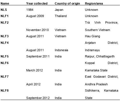
A laboratory-maintained strain ofN. lugensexhibiting susceptibility to imidacloprid (NLS) and eight field strains of BPH collected from Thailand (NLF1), Vietnam (NLF2, NLF3), Indonesia (NLF4) and India (NLF5, NLF6, NLF7, NLF8) were reared in the laboratory on whole rice plants (Oryza sativaL. ssp.) under controlled environmental conditions (26C/16h photoperiod). Year of collection is detailed in
Table S1. We have previously shown that all eight of the field strains exhibit resistance to imidacloprid (Table S2) [12].
TheDrosophila melanogasterstock 13-20 [‘‘y1w67c23; P attP40 25C6,’’ ‘‘1;2’’] obtained from the University of Cambridge was used to create all transgenic lines. Virgin females of this line were crossed to males of the Act5C-GAL4 strain [‘‘y[1] w[*]; P(Act5C-GAL4-w)E1/CyO,’’’’1;2’’] (Bloomington Stock Center) to activate transgene expression (see below for description of methods). All flies were reared on NutriFly food (NLS) at 24C. Only female flies 2-5 days post eclosion were used for insecticide bioassays.
Continued
REAGENT or RESOURCE SOURCE IDENTIFIER
Drosophila melanogaster, genotype: 13-20 [‘‘y1w67c23; P attP40 25C6,’’ ‘‘1;2’’]
University of Cambridge Stock 13-20
Drosophila melanogaster,genotype: Act5C-GAL4 strain ‘‘y[1] w[*]; P(Act5C-GAL4-w)E1/CyO,’’’’1;2’’]
Bloomington Stock Centre Act5C-GAL4
Oligonucleotides
SeeSupplemental Materials N/A SeeTable S3
Recombinant DNA
Cytochrome P450 variants GeneArt, CA, USA N/A
Cytochrome P450 reductase (CPR) GeneArt, CA, USA GenBank: Q07994
Gateway pDEST8 Vector Invitrogen Cat#11804010
pUASTattB40 Vector Bischof et al.,2007 Gift from Jacob Riveron, Liverpool School of Tropical Medicine
pGL3-basic luciferase reporter vector Promega Cat# E1751
pRL-CMVRenillaluciferase control reporter vector
Promega Cat# E2261
Software and Algorithms
Geneious v 9.1.8 Biomatters https://www.geneious.com/download/
MEGA v 6.0 [13] www.megasoftware.net
Schro¨dinger Software Suite Schro¨dinger https://www.schrodinger.com/downloads/ releases
LeadIT v 2.2.0 BioSolveIT https://www.biosolveit.de/LeadIT/
Genstat v 16 VSN International https://www.vsni.co.uk/software/genstat/
FastQC v 0.11.5 [19] https://www.bioinformatics.babraham.ac.uk/
projects/download.html#fastqc
Trim Galore! v 0.4.2 Babraham Institute https://www.bioinformatics.babraham.ac.uk/ projects/download.html#trim_galore
BWA-MEM (Burrows-Wheeler Aligner) [21] https://sourceforge.net/projects/bio-bwa/files/
R v 3.0 R Core Team, 2013 https://cran.r-project.org/bin/windows/base/old/
CNV-seq [22] http://tiger.dbs.nus.edu.sg/cnv-seq/
SAM tools [23] https://sourceforge.net/projects/samtools/files/
Proovread [25] https://github.com/BioInf-Wuerzburg/proovread
GraphPad Prism v 7 GraphPad Software https://www.graphpad.com/
Other
Nilaparvata lugensreference genome [10] GenBank: GCA_000757685.1
Insect cell lines
The Sf9 and High Five insect cell lines (ovarian cells fromSpodoptera frugiperdaandTrichoplusia nirespectively) were maintained in suspension culture under serum-free conditions at 27C containing 25mg/mL-1gentamycin in SF-900 II SFM (GIBCO) and Express
Five SFM (GIBCO), respectively.
METHOD DETAILS
Identification and expression analysis of CYP6ER1 sequence variants
Total RNA was extracted from 4 pools of 8 adult hoppers of each strain using the ISOLATE II RNA Mini Kit (Bioline) and reverse tran-scribed to cDNA using Superscript III reverse transcriptase (Invitrogen) following manufacturer protocols in both cases. Phusion DNA polymerase (Thermo) was used to amplify the full coding sequence of CYP6ER1 following the manufacturers protocol and using 10 ng of cDNA as template in 50ml reactions and the primers listed inTable S4. Thermocycling conditions consisted of an initial dena-turation step at 98C for 30 s, followed by 35 cycles of 98C for 10 s, 55C for 20 s, 72C for 1 min, and a final extension at 72C for 5 min. Products were either direct Sanger sequenced using the primers detailed inTable S4or cloned using the Strataclone Blunt PCR Cloning kit (Stratagene) and sequenced using T3/T7 primers. Variant calling was carried out in Geneious version 9 (Biomatters), and phylogeny performed in MEGA version six [13]. Expression ofCYP6ER1variants was initially assessed by PCR amplification of theCYP6ER1coding sequence followed by cloning and sequencing of 16 colonies per strain (as detailed above). Quantitative PCR analysis of the expression of differentCYP6ER1variants was performed using the primers detailed inTable S4. PCR reactions (20ml) contained 10 ng of cDNA, 10ml of SYBR Green JumpStart Taq Readymix (Sigma), and 0.25mm of each primer. Samples were run on a Rotor-Gene 6000 (Corbett Research) using temperature cycling conditions of: 2 min at 95C followed by 40 cycles of 95C for 15 s, 57C for 15 s and 72C for 20 s. A final melt-curve step was included post-PCR (ramping from 72C–95C by 1C every 5 s) to check for nonspecific amplification. The efficiency of PCR for each primer pair was assessed using a serial dilution of 100 ng to 0.01 ng of cDNA. Each quantitative RT-PCR experiment consisted of three independent biological replicates with two technical replicates for each. Data were analyzed in Microsoft Excel according to theDCTmethod [14], using the geometric mean of two reference genes
(actin anda2-tubulin) for normalization.
Heterologous expression of P450s
Natural and mutatedCYP6ER1variants andM. domesticaNADPH-dependent cytochrome P450 reductase (CPR) (GenBank acces-sion number Q07994) were codon optimized for expresacces-sion in lepidopteran cells and obtained by gene synthesis (Geneart, CA, USA) in the pDEST8 expression vector (Invitrogen). The PFastbac1 vector with no insert DNA was used to produce a control virus. The recombinant baculovirus DNA was constructed and transfected into Sf9 cells using the Bac-to-Bac baculovirus expression system (Invitrogen) according to the manufacturer’s instructions. The titer of the recombinant virus was determined following the protocols of the supplier. High Five cells grown to a density of 23106cells/mL-1were co-infected with recombinant baculoviruses containing P450 and CPR at various MOI (multiplicity of infection) ratios to identify the best conditions. Control cells were co-infected with the baculovirus containing vector with no insert (ctrl-virus) and the recombinant baculovirus expressing CPR using the same MOI ratios. Ferric citrate andd-aminolevulinic acid hydrochloride were added to a final concentration of 0.1 mM at the time of infection and 24 h after infection to compensate the low levels of endogenous heme in the insect cells. After 60 h, cells were harvested by centrifugation, washed with PBS, and microsomes of the membrane fraction prepared according to standard procedures [15]. Briefly, pellets were homogenized for 30 s in 0.1M Na/K-phosphate buffer, pH 7.4 containing 1mM EDTA and DTT and 200mM sucrose using a Fastprep (MP Biomedicals), filtered through miracloth and centrifuged for 10 min at 680 g at 4C. The supernantant was then centrifuged for 1 h at 100,000 g at 4C, with the pellet subsequently resuspended in 0.1M Na/K-phosphate buffer, pH 7.6 containing 1mM EDTA and DTT and 10% glycerol using a Dounce tissue grinder. P450 expression and functionality was estimated by measuring CO-difference spectra in reduced samples using a dual beam Cary 300 UV-Vis Spectrophotometer (Agilent) and scanning from 500 nm to 400 nm [15]. The protein content of samples was determined using Bradford reagent (Sigma) and bovine serum albumin as a reference following the manufacturer’s instructions.
Metabolism assays and LC-MS/MS analysis
Metabolism of imidacloprid was assayed by incubating recombinant P450/CPR (2 pmol P450 / assay) or ctrl-virus/CPR microsomes in 0.1 M potassium phosphate buffer pH 7.6 with an NADPH-regenerating system (Promega; 1.3 mM NADP+, 3.3 mM glucose-6-phosphate, 3.3 mM MgCl2, 0.4 U/mL-1glucose-6-phosphate dehydrogenase) and substrate (12.5mM) at 27C for 1 h. The total assay
volume was 200mL using three replicates for each data point. Microsomes incubated without NADPH served as a control. The assay was quenched by the addition of ice-cold acetonitrile (to 80% final concentration), centrifuged for 10 min at 3000 g and the super-natant subsequently analyzed by liquid chromatography-tandem mass spectrometry. Chromatography was performed using a Waters UPLC utilizing a Waters Acquity HSS T3 (2.1x50 mm, 1.8mm) column. Solvents were water/0.1% formic acid and acetonitrile/0.1% formic acid used in a 4 min gradient. The mass spectrometer used was a Sciex API4000 in positive ionization mode for imidacloprid and its hydroxylated metabolite (MRM transitions: 256 > 175, 272 > 191, respectively). 6-CNA was measured in negative ion mode using the ion transition 165 > 121. Quantification was performed by external calibration using reference compounds. Recovery rates of parent compounds using microsomal fractions without NADPH were close to 100%.
Homology modeling of CYP6ER1 and molecular docking simulation
3D models for CYP6ER1vA, CYP6ER1vB and CYP6ER1vL were generated by the advanced homology modeling tool within the Schrodinger software suite [16]. Following a BLAST search for the most suitable template-fold all three protein models were con-structed based on the crystal structure of human CYP3A4 (PDB-ID: 1TQN) and refined by an energy minimization step to remove conformational strains and contacts. For the docking studies a catalytic oxygen atom was manually added to the heme iron center. Imidacloprid was docked into the three CYP6ER1 variants using the virtual screening software package LeadIT 2.2.0 utilizing the FlexPharm option [17]. The pharmacophore constraint [18] required a non-hydrogen-atom within a distance of 2.5 A˚ from the catalytic oxygen. All resulting docking poses underwent hierarchical clustering using a script based on pairwise in-place RMSD values; for this only non-hydrogen atoms were taken into account. The cut height for the cluster generation was 2.0. For each resulting cluster the pose with the lowest FlexX docking rank was selected. This subset of cluster representatives was further reduced by removing all poses where not at least one of the five membered ring atoms was within a distance of 2.5 A˚ (or less) from the catalytic oxygen. For the models of vL, vA and vB the resulting pose spaces were then visually inspected and compared.
Transgenic expression of candidate genes inD. melanogaster
Wild-type (CYP6ER1vL) and mutantCYP6ER1variants were synthesized and provided in the pUASTattB40 plasmid (Geneart, CA, USA). Using the PhiC31 system, clones were transformed into the germline of aD. melanogasterstrain carrying the attP40 docking site on chromosome 2 [‘‘y1w67c23; P attP40 25C6,’’ ‘‘1;2’’]. The transgenic lines obtained were balanced and the integration of genes confirmed by PCR and sequencing using Phusion DNA polymerase (Thermo) as detailed above with the primers detailed inTable S4. Virgin females of the Act5C-GAL4 strain were crossed with UAS-gene-of-interest males. Bioassays were used to assess the suscep-tibility of adult female flies to imidacloprid. Several concentrations were overlaid onto 1.5% agar containing 1% sucrose in standard
Drosophilavials and allowed to dry overnight at room temperature. 10-15 adult flies (two to five days post eclosion) were then added to each vial and mortality assessed after 48 hr. Four replicates were carried out for each concentration. Control mortality was assessed using vials containing agar/sucrose minus insecticide. Lethal concentrations (LC50values) and 95% fiducial limits were
calculated by probit analysis using Genstat version 16 (VSN International).
Copy number analysis and sequencing of individual hoppers
Genomic DNA was extracted from multiple pools of 10 hoppers using the Plant DNeasy Mini kit (QIAGEN) and used to construct PCR-free libraries. Libraries of each strain were sequenced across a single lane of an Illumina HiSeq2500 using a 250 bp paired-end read metric. FastQC was used to check the quality of the raw reads obtained [19] and reads trimmed using Trim Galore [20]. In initial attempts to estimate gene copy number the reads of each strain were mapped to the reference genome (sequenced from an inbred line derived from a strain collected in Hangzhou, China, in 2008 [10], GenBank assembly accession number: GCA_000757685.1) us-ing BWA-MEM [21] and CNV estimated using CNVseq [22] with data of each field strain compared to the lab susceptible strain. This analysis failed to identify significant differences in copy number between the strains at the CYP6ER1 locus, likely because the single scaffold of the reference genome where CYP6ER1 is located (KN153994.1) fails to accurately represent the genetic diversity observed at this locus in different variants and strains resulting in, at best, only partial mapping ofCYP6ER1-related reads. Thus, in a second approach reads were mapped to the coding sequence ofCYP6ER1and two reference genes: the P450CYP6AY1
and the voltage-gated sodium channel (VGSC), both of which are single copy genes in BPH, using BWA-MEM [21]. Read coverage was then compared in 100bp non-overlapping windows across the coding sequence of the three genes using SAMtools [23]. Results were validated with qPCR using DNA as template and two sets of primers (designed in conserved regions (variant non-specific) of the gene) forCYP6ER1listed inTable S4and the conditions described above. Data were analyzed according to theDDCT method [24] using theVGSCas a reference gene for normalization with the expression values of the four biological replicates obtained for each of the twoCYP6ER1primer sets averaged.
For variant analysis on individual hoppers, DNA and RNA was extracted (as above) from individuals of the NLF2 and NLF7 strains and used as template in PCR using Phusion DNA polymerase (Thermo) (as detailed above) and the primers detailed inTable S4which amplify a region containing key SNPs that are diagnostic for differentCYP6ER1variants.
Gene capture
Approximately 2.5 ug of gDNA was extracted per strain from several pools of insects as detailed above and sent to Earlham Institute for processing according to Roche’s NimbleGen gene capture protocol. The gDNA was sheared with a Covaris tube and size selected within the range of 5-9 kb on a BluePippin. The pre-capture library was amplified for 6 rounds to increase the starting material for the capture set up. The library was incubated with the baits (fifteen 100 nt baits per transcript) designed to cover the entire 1.5 kb coding sequence of all knownCYP6ER1variants described in this study) at 47C for 22 hr. The post capture library was gener-ated through 19 cycles of amplification of the bait extracted fragments. Each library was sequenced on a single SMRT cell (P6C4). To minimize sequencing errors inherent in PacBio data only circular consensus (CCS) reads, generated from repeated passes of polymerase over a single molecule, or raw reads that were error corrected using the Illumina reads for NLS, NLF2 and NLF7 were used to assemble gene sequences. Assembly and bioinformatics analyses was conducted in Geneious version 9 (Biomatters). Error correction of long Pacbio reads with short-read Illumina data was performed using proovread [25].
Reporter gene assays
Promoter sequences were synthesized, subcloned into pGL3-Basic (Promega) and transformed into Library Efficiency DH5a
Competent Cells (Invitrogen). Plasmids were extracted with the GeneJet plasmid miniprep kit (Fermentas), sequenced and then adjusted to 400 ng/ml for use in dual luciferase assays using the Sf9 insect cell line. Approximately 13106cells per well were plated into 6-well plates 2 hr prior to transfection and allowed to reach 60%–70% confluency. Insect GeneJuice Transfection Reagent (Novagen) was used for transfection of constructs and the Dual-Luciferase Reporter Assay (Promega) used for promoter activity measurements according to the manufacturer’s protocols. 2mg of reporter constructs and pGL3 without insert (as a control) was co-transfected with 4 ng Renilla luciferase pRL-CMV using GeneJuice and incubated at 27C. 4 hr post-transfection, the transfection mixture was removed and replaced with supplemented Grace’s Insect Medium (GIBCO). Following further incubation at 27C for 48 h and washing of cells with PBS, cells from each well were harvested in 500mL passive lysis buffer (Promega) and luciferase activity measured on a GloMax 20/20 (Promega). Construct luciferase activity was normalized to Renilla luciferase activity as instructed in the manufacturer’s protocol.
QUANTIFICATION AND STATISTICAL ANALYSIS
All statistical analyses were performed in GraphPad Prism 7 (GraphPad Software). Significant differences in expression or copy number in all QPCR experiments were determined using one-way ANOVA with post hoc Tukey HSD. Significant differences in the number of colonies obtained for each variant for each strain in the cloning and sequencing of individuals was determined using a paired t test. Significant differences in normalized reporter gene expression between promoter variants was determined using one-way ANOVA with post hoc Tukey HSD. Statistical details of experiments (value of n, precision measures and definitions of significance) are provided in figure legends.
DATA AND SOFTWARE AVAILABILITY
The sequences reported in this paper have been deposited in the GenBank database (accession numbers GenBank: MF970458, GenBank: MF970459, GenBank: MF970460, GenBank: MF970461, GenBank: MF970462, and GenBank: MF970463).
Current Biology, Volume
28
Supplemental Information
Neofunctionalization of Duplicated P450 Genes
Drives the Evolution of Insecticide Resistance
in the Brown Planthopper
CYP6ER1 vD2
CYP6ER1 vD1
CYP6ER1 vE
CYP6ER1 vA
CYP6ER1 vB
CYP6ER1 vC
CYP6ER1 vL
CYP6ER1 vF 100
95 85
92 73
0.0050
A
A
B
C
0
5
10
15
20
25
30
No
. s
equ
enced
colo
n
ies
BPH strain and template used in PCR
vF
vB
vC
vA
0
5
10
15
20
25
30
35
N
u
m
b
e
r
o
f i
n
d
iv
id
u
al
s
Genotype
NLF2
NLF7
Figure S2.
CYP6ER1
genotyping of individuals of the NLF2 and NLF7 BPH strains using
DNA and RNA as template in (RT-)PCR. Related to Figure 3. (A) Representative sequence
traces obtained from direct sequencing of a diagnostic fragment of
CYP6ER1
encompassing the
resistance mutation sites in exon 6. DNA extracted from individuals of the NLF2 and NLF7
strains was used as template in PCR. Two representative sequences of each strain are aligned
to a representative sequence obtained from NLS (which has just one copy of
CYP6ER1
). Boxed
regions indicate the sites of the A375del+A376G mutations in
CYP6ER1v
A and P377del in
CYP6ER1vB
(see also part C of this figure) and highlight the heterozygosity observed at these
regions (overlapping chromatogram peaks). (B) Results of genotyping 27-30 individuals of the
NLF2 and NLF7 for the resistance mutations that define
CYP6ER1v
A and
CYP6ER1vB
. (C)
Alignment of a diagnostic
CYP6ER1
sequence fragment from BPH individuals of resistant
strains. Alignment shows representative sequence reads obtained from cloning and sequencing
an amplicon containing SNPs and INDELs diagnostic for each of the unique
CYP6ER1
variants
using DNA extracted from individuals of the NLF2 and NLF7 strains. For reference the
Figure S3. Sequence analysis of the introns of different
CYP6ER1
variants. Related to
Figure 3.
A
Figure S4. Alignment of
CYP6ER1
promoter variants. Related to Figure 3. A) Alignment of
1750bp of the putative promoter region upstream of different
CYP6ER1
variants. Contig 5 could
not be assigned to a known
CYP6ER1
variant. The position of the sequence breakpoint
Name
Year collected
Country of origin
Region/area
NLS
1984
Japan
Unknown
NLF1
August 2009
Thailand
Unknown
NLF2
November 2010
Vietnam
Trà
Vinh
Province,
Southern Vietnam
NLF3
August 2011
Vietnam
Hau Giang
NLF4
August 2011
Indonesia
Anjatan
District,
Indramayu
NLF5
September 2011
India
Raipur, Chhattisgarth
NLF6
March 2012
India
Koppal
District,
Karnataka State
NLF7
April 2012
India
East Godavari District,
Andhra Pradesh
NLF8
September 2012
India
Sidhikerra, Karnataka
State
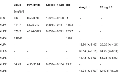
Strain
LC50-value
[mg/L
-1]
95% limits
Slope (+/- SD) RR
Mortality
4 mg L
-1Mortality
20 mg L
-1NLS
0.6
0.50-0.70
1.822+/- 0.158 1
-
-
NLF1
111.7
66.05-212
0.891+/- 0.11 186.2
-
-
NLF2
170.2
46.44-5095
0.655+/- 0.221 283.7
-
-
NLF3
>1000
-
-
1666
-
-
NLF4
-
-
-
-
16.50 (+/-8.42) 20.20 (+/-4.21)
NLF5
-
-
-
-
30.14 (+/-8.11) 34.25 (+/-8.14)
NLF6
-
-
-
-
15.13 (+/-5.67) 58.31 (+/-8.00)
NLF7
14.49
4.55-30.81
0.653+/- 0.134 24.2
-
-
NLF8
-
-
-
-
15.74 (+/-5.69) 42.42 (+/-8.02)
Table S2. Sensitivity of susceptible and resistant strains of
Nilaparvata lugens
to
imidacloprid topically applied to adult females. Related to Figure 1.
Oligo
Sequence
Purpose
ER1 CDS F1
ATGTGGGAAAACTCGTGGTTGG
PCR/sequencing of CYP6ER1 coding sequence
ER1 CDS F2
GGTTGGCCTAYCTTGTCACAGG
PCR/sequencing of CYP6ER1 coding sequence
ER1 CDS R1
CTAAGTATCTCTCGCTACCAGC
PCR/sequencing of CYP6ER1 coding sequence
ER1 CDS R2
GCTACCAGCTTCAGTGTGAGG
PCR/sequencing of CYP6ER1 coding sequence
CYP6ER1 vL/vF F
CATCCATGAGGTCTACGAAG
CYP6ER1 variant-specific QPCR
CYP6ER1 vL/vF R
GAGTGCTGAACAGATGGTGT
CYP6ER1 variant-specific QPCR
CYP6ER1 vA F
CTTTCTTCACCCCCGCCC
CYP6ER1 variant-specific QPCR
CYP6ER1 vA R
CCTGCATGGTCTCGAACATG
CYP6ER1 variant-specific QPCR
CYP6ER1 vB F
TCTTGTCACAATCCTGTTGCTG
CYP6ER1 variant-specific QPCR
CYP6ER1 vB R
TGGATGCATTTCTTGGACAATACG
CYP6ER1 variant-specific QPCR
CYP6ER1 vC F
GAGACTACTTCTGCATCTTTGT
CYP6ER1 variant-specific QPCR
CYP6ER1 vC R
GGAAACCATTGGGAAGAATGA
CYP6ER1 variant-specific QPCR
CYP6ER1 vD F
AGATCAAATCGGCGGATGGA
CYP6ER1 variant-specific QPCR
CYP6ER1 vD R
CGGAATCATCACTTGAGTTCC
CYP6ER1 variant-specific QPCR
CYP6ER1 vE R
CCGGAATCATTACTTGAGTTCC
CYP6ER1 variant-specific QPCR
CYP6ER1 vE F
GTATGATGAGATCAGATCTGTGA
CYP6ER1 variant-specific QPCR
D099 pUAST F
TCACTGGAACTAGGCTAGCA
Sequence validation of transgenic flies
D102 pUAST F
GGATCCAAGCTTGCATGCCTG
Sequence validation of transgenic flies
D100 pUAST R
AAAGGCATTCCACCACTGCT
Sequence validation of transgenic flies
D101 pUAST R
CCACCACTGCTCCCATTCAT
Sequence validation of transgenic flies
ER1 deletion F
GCAGAAATGTTGAGGAAATATCCC
Sequencing of exon 6 of BPH individuals
ER1 deletion R1
ACCAAAGGGTATGAAAGAGAAGG
Sequencing of exon 6 of BPH individuals
Ex3 qPCR F2
GAATGTGATTGCCTCCACGG
Copy number QPCR of CYP6ER1
Ex3 qPCR R2
AGCATCAGCAAGTGGGTTCT
Copy number QPCR of CYP6ER1
Ex4 qPCR F2
AACATGAGGTTCACGCCGAA
Copy number QPCR of CYP6ER1
Ex4 qPCR R2
TGCATGAAATCCTTCCTCACCA
Copy number QPCR of CYP6ER1
VGSC qPCR F2
CACCATTGTCACACAGCAGC
qPCR of reference gene (VGSC)
VGSC qPCR R2
CCCTGGAGTAGTGCTTGTCG
qPCR of reference gene (VGSC)
Nl_Actin_F
TAACGAGAGGTTCCGTTGCC
qPCR of reference gene (actin)
Nl_Actin_R
GACAGGACAGTGTTGGCGTA
qPCR of reference gene (actin)
Nl_α2_tubulin_F
CCACCCTGGAACACTCTGAC
qPCR of reference gene (α2_tubulin)
Nl_α2_tubulin_R
CGAAGCAGTGATCGAGGACA
qPCR of reference gene (α2_tubulin)
![Table S1. We have previously shown that all eight of the field strains exhibit resistance to imidacloprid (Table S2) [12].](https://thumb-us.123doks.com/thumbv2/123dok_us/1090413.1609617/10.603.62.551.115.544/table-previously-eld-strains-exhibit-resistance-imidacloprid-table.webp)