Vaccinated with
Mycobacterium bovis
BCG and Cattle Infected with
M
.
bovis
Shelley G. Rhodes,aLucy C. McKinna,b* Sabine Steinbach,aGilly S. Dean,aBernardo Villarreal-Ramos,aAdam O. Whelan,a* C. Pirson,a Gareth J. Jones,aDerek Clifford,aH. Martin Vordermeiera
TB Research Group, Animal Health and Veterinary Laboratories Agency, Surrey, United Kingdoma; Royal Veterinary College, London, United Kingdomb
We describe here the application of a novel bovine interleukin-2 (IL-2) enzyme-linked immunosorbent assay (ELISA) for the measurement of antigen-specific IL-2 in cattle naturally infected withMycobacterium bovisand in cattle vaccinated with Myco-bacterium bovisBCG and then experimentally challenged with pathogenicM. bovis. Supernatants from whole-blood cultures stimulated with mycobacterial antigen (bovine purified protein derivative [PPDB] or the peptide cocktail ESAT6-CFP10) were assessed using a sandwich ELISA consisting of a new recombinant monoclonal fragment capture antibody and a commercially available polyclonal anti-bovine-IL-2. The production of IL-2 was compared to the production of gamma interferon (IFN-␥) in the same antigen-stimulated whole-blood supernatants. The data show that cattle infected withM. bovisproduced quantifiable levels of antigen-specific IL-2, while IL-2 levels in cattle vaccinated withM. bovisBCG did not. Furthermore, cattle vaccinated withM. bovisBCG and then challenged with pathogenicM. bovisdisplayed a more rapid induction of IL-2 but ultimately had lower levels of infection-induced IL-2 than did unvaccinated challenge control cattle. These data suggest that IL-2 responses are not detectable post-BCG vaccination and that these responses may require infection with virulentM.bovisto develop. This may be useful to differentiate infected cattle from uninfected or BCG-vaccinated cattle, although the overall sensitivity is relatively low, particularly in single intradermal comparative cervical tuberculin (SICCT)-negative infected animals. Furthermore, the strength of the IL-2 response may correlate with pathology, which poses interesting questions on the immunobiology of bovine tuberculosis in contrast to human tuberculosis, which is discussed.
D
espite a compulsory test and slaughter regime since 1950, bo-vine tuberculosis (BTB) has reached epidemic status in focal areas of Great Britain, and the recently published “Bovine TB Eradi-cation Programme for England” (https://www.gov.uk/government /publications/bovine-tb-eradication-programme-for-england) par-ticularly prioritizes the development of a TB cattle vaccine with an associated test to differentiate between infected and vaccinated/pro-tected animals (DIVA).The gamma interferon (IFN-␥) test has been used in Great Britain since 2006 as an ancillary test to the single intradermal cervical comparative tuberculin skin test (SICCT) for the diagno-sis of preclinical BTB in Great Britain. Recent research in both human and bovine TB fields indicates that other cytokines pro-duced in response to TB antigen-specific stimulation may be uti-lized as potential diagnostic tools. For example, in BTB using both IFN-␥and interleukin-1(IL-1) responses to the peptide cock-tail ESAT6-CFP10 (EC) (present in pathogenic mycobacteria, but absent in BCG [1]) showed an increase in the sensitivity of detec-tion ofMycobacterium bovis-infected cattle without compromis-ing specificity (2).
In terms of IL-2, multifunctional CD4⫹ T cells producing more than one cytokine, including IFN-␥, tumor necrosis factor alpha (TNF-␣), and IL-2, have been described in murine models of protection against TB (3–5). Multifunctional T cells have been identified in human TB and in some reports as potential correlates of pathology and bacterial load (6–8), while Millington et al. (9) described kinetic changes in T cells producing different cytokines over time, and correlated IL-2-producing T cells with a protective outcome. In keeping with a protective role for IL-2, more recently reported studies have shown that IL-2-positive (multifunctional)
T cells are involved in subunit vaccine boosts of BCG-induced protective immunity both in humans (10) and in mice (11).
Recently, multifunctional T cells (IFN-␥, TNF-␣, and IL-2) have also been described in cattle naturally infected withM. bovis
(12), and previous work had demonstrated IL-2 activity in BTB, both in natural and experimental infections (13,14). However, the IL-2 in these early reports was assessed using a T cell bioassay, which lacks specificity.
Using a bespoke bovine IL-2-specific antibody identified using recombinant phage display technology, we investigated this cyto-kine more fully in BTB by generating a sandwich enzyme-linked immunosorbent assay (ELISA) that incorporates the recombinant monoclonal antibody with a commercially available polyclonal reagent for bovine IL-2. The aim of this study therefore was to apply this new IL-2 ELISA to the investigation of antigen-specific
Received9 August 2013 Returned for modification3 September 2013
Accepted23 October 2013
Published ahead of print30 October 2013
Editor:W. R. Waters
Address correspondence to Shelley G. Rhodes, shelley.rhodes@ahvla.gsi.gov.uk. * Present address: Lucy C. McKinna, Stocton Vets, Stocton Close, Guildford, Surrey, United Kingdom; Adam O. Whelan, Department of Biomedical Sciences, Defence Science and Technology Laboratory, Porton Down, Salisbury, Wiltshire, United Kingdom.
Supplemental material for this article may be found athttp://dx.doi.org/10.1128 /CVI.00522-13.
Copyright © 2014, American Society for Microbiology. All Rights Reserved.
IL-2 production in whole-blood stimulation cultures, as used for the assessment of IFN-␥in BTB. Results are shown for IL-2 and IFN-␥production in cattle naturally infected withM. bovis, cattle vaccinated withM. bovisBCG and challenged with pathogenicM. bovis, and uninfected controls.
MATERIALS AND METHODS
Animals. (i) Comparison of SICCT-positive/IFN-␥-positive naturally infected cattle withM. bovisBCG-vaccinated and uninfected control cattle.Included in this cohort were (i) 33 SICCT-positive, IFN-␥-positive naturally infected cattle; (ii) 31 cattle from Great Britain herds without a TB breakdown for at least 4 years that had been vaccinated subcutane-ously with approximately 106CFUM. bovisBCG-1331 in a 0.5-ml dose and sampled at 4 weeks (n⫽6), 8 weeks (n⫽20), or 10 months (n⫽5) postvaccination (3 separate experiments); and (iii) 31 uninfected control cattle from BTB-free herds also without a breakdown for at least 4 years (15).
(ii) Performance of IL-2 in comparison with the IFN-␥test in SICCT-negative surveillance cattle from Great Britain breakdown herds.Sixty-six SICCT-negative/IFN-␥-positive cattle were identified via routine blood testing of Great Britain breakdown herds at AHVLA Lud-dington between October and December 2012. These were compared with 64 uninfected control cattle from BTB-free herds with no history of breakdown in the past 4 years.
(iii) Longitudinal investigations ofM. bovis-BCG vaccination and challenge withM. bovis.Two separate BCG-vaccination/M. bovis -chal-lenge experiments were included as follows.
(a) Experiment 1. Twenty-four animals were recruited from BTB-free farms at 5 weeks of age. Sixteen animals were vaccinated subcutaneously within 6 weeks of age with a 0.5-ml volume dose of approximately 106 CFU of BCG Danish (Statens Serum Institute, Copenhagen, Denmark). All animals were challenged at approximately 7 months of age with 200 CFU ofM. bovisAF2122/97 (AHVLA Weybridge) by the endobronchial route, and blood samples were taken at 0, 2, 4, 6, and 8 weeks postchal-lenge (16).
(b) Experiment 2. Nine animals of 4 to 6 months of age were recruited from BTB-free farms. Five were vaccinated subcutaneously with a 0.5-ml-volume dose of approximately 106CFU of BCG Danish (Statens Serum Institute, Copenhagen, Denmark). Twelve weeks later these 5 animals plus a further 4 unvaccinated controls were challenged with 12,200 CFU
M. bovisAF2122/97 (AHVLA Weybridge) by the endobronchial route. Blood samples from this experiment were taken at 0, 3, 6, 8, and 12 weeks post-BCG and at 2, 4, 7, and 11 weeks post-M. bovischallenge.
TheM. bovisinoculum was prepared from a frozen seed stock at known concentrations and the administered doses were confirmed retro-spectively by culture as described previously (16).M. bovisBCG was pre-pared fresh from lyophilized stocks on the day of vaccination according to the manufacturer’s instructions and administered subcutaneously as de-scribed previously (16).
Whole-blood IFN-␥and IL-2 assays.Supernatants were generated from whole heparinized blood stimulatedin vitrowith (i) bovine tuber-culin (bovine purified protein derivative [PPDB]) (Lelystad, Nether-lands) at a 300-U/ml final concentration; (ii) peptide cocktails ESAT6-CFP10 and Rv3615c (Pepseuticals, Leicestershire, United Kingdom) at a final concentration of 5g/ml; and (iii) the mitogen (sample positive control) staphylococcal enterotoxin B (SEB) (Sigma, United Kingdom) or pokeweed mitogen (PWM) (Sigma, United Kingdom) at a final concen-tration of 2g/ml or 10g/ml, respectively. Unstimulated whole-blood cultures provided the background negative sample controls. We added 250l of whole blood, in duplicate wells of 96-well plates (Nunc, Thermo Fisher Scientific, United Kingdom) to 25l of 11⫻concentrated antigen, mitogen, or medium only. Cultures were incubated for 16 to 24 h at 37°C, 5% CO2. Plates were then centrifuged at 300⫻g for 5 min at room temperature (RT) to pellet the cellular fraction, and cell-free supernatants were harvested and duplicates pooled and stored at⫺20°C.
(i) IFN-␥ELISA.IFN-␥content of the supernatants was measured using the BOVIGAM ELISA kit (Prionics, Switzerland) following the manufacturer’s instructions. Samples were tested in duplicate and the⌬ optical density (OD) at 450 nm was calculated for each stimulated super-natant for each animal (i.e., the mean OD of sample duplicate in the presence of antigen/mitogen minus the mean OD in the absence of anti-gen/mitogen).
(ii) IL-2 ELISA.Flat-bottomed 96-well ELISA plates (Nunc Maxisorp, Thermo Fisher Scientific, United Kingdom) were coated with 50l/well of human recombinant monoclonal fragment capture antibody (HuCAL bivalent boIL-2 Fab antibody fragments containing a heavy-chain C-ter-minal dHLX-dimerization domain followed by myc and 6⫻His tag [Fab-dHLX-MH, AbD14385] AbD [Serotec, United Kingdom]) diluted to 4 g/ml in carbonate coating buffer (Sigma, United Kingdom) (pH 9.6) and incubated overnight at 4°C. Plates were blocked with 200l/well of 4% bovine serum albumin (BSA)-phosphate buffered saline (PBS) (BSA; Sigma, United Kingdom) for 1 h at RT, and washed three times with PBS-0.05% Tween 20. Supernatants were added in duplicate, 50l/well. A 1% concentration of BSA-PBS (50l/well) was used as a negative control, while 50l/well of recombinant bovine IL-2 (R&D Systems, United King-dom) diluted to 10 ng/ml in 1% BSA-PBS was used as a positive control. Plates were incubated for 2 h at RT (or overnight at 4°C) and washed 3 times with PBS-0.05% Tween 20 and 50l/well of biotinylated goat-anti-bovine IL-2 antibody (R&D Systems, United Kingdom), diluted to 1 g/ml in 1% BSA/PBS, was added for 1 h at RT. Wells were washed as above, and 50l/well of streptavidin-horseradish peroxidase (HRP) (GE Healthcare, Amersham, United Kingdom) diluted 1:2,000 in 1% BSA-PBS was added for 1 h at RT. Wells were then washed six times and 100 l/well of tetramethylbenzidine (TMB) substrate (Sigma, United King-dom) was added. Plates were allowed to develop for 20 min before adding 100l/well of 0.5 M H2S04. Plates were read immediately on an ELISA reader at a 450-nm wavelength. Results are expressed as the⌬OD 450 nm (i.e., mean OD of duplicates in the presence of antigen/mitogen minus the mean OD of duplicates in the absence of antigen/mitogen).
Statistics.Prism software was used for group comparisons and re-ceiver operating characteristic (ROC) analysis of data (GraphPad, San Diego, CA). Group comparisons were made using a 2-tailed Mann-Whit-ney nonparametric statistical test.
RESULTS
There were no positive IFN-␥responses to EC in the vaccinated group (Fig. 1e), as expected, but four control animals did show low positive responses to this peptide cocktail (Fig. 1f). Responses to EC may occur due to the presence of these antigens in the environmental mycobacteriumMycobacterium kansasii(17–20).
The data for IL-2 suggest that PPDB-stimulated IL-2 could provide an alternative tool for differentiating vaccinated from in-fected cattle. ROC analysis of the IL-2 data in this small study suggested that using a positive/negative OD test cutoff of 0.1 (450 nm) would provide a 100% specificity (95% confidence interval [CI], 88.78 to 100) and sensitivities of 93.9% (95% CI, 79.77 to 99.26) and 66.7% (95% CI, 48.17 to 82.04) for PPDB and EC, respectively. None of the BCG-vaccinated cattle results would have been positive using these cutoff criteria.
The sensitivity of EC-specific (but not PPDB-specific) IL-2 in the reactor cohort could be increased without reducing the 100% specificity (in the negative-control cohort) by lowering the cutoff, e.g., an OD cutoff of 0.05 provided a sensitivity of 75.7% (95% CI, 57.74 to 88.91). However, lowering the cutoff generated positivity in the BCG-vaccinated group, e.g., 3/31 (9.7%) positives at this lower cutoff.
Low IL-2 positivity in SICCT-negative-IFN-␥-positive field reactor cattle.We next investigated PPDB-specific IL-2 responses in a cohort of SICCT⫺/IFN-␥⫹animals that had been identified via routine blood testing of breakdown herds in Great Britain. Sixty-six SICCT⫺/IFN-␥⫹animals were compared with a further 64 TB-free control cattle. Since blood testing in Great Britain is applied in parallel with the SICCT (i.e., SICCT⫹animals were removed and then the remaining animals were blood tested), we were interested to know how an IL-2 test would compare against the IFN-␥test using the above 0.1 OD cutoff for optimum sensi-tivity and specificity. The results inFig. 2show that 30.3% (20/66)
of the IFN-␥-positive cattle also produced a PPDB-specific IL-2 response. The specificity was 100% in that none of the control animals produced positive IL-2 responses. These data suggest that in animals that would be routinely blood tested in the event of a confirmed TB breakdown, an IL-2 test would not be as sensitive as the IFN-␥test. This was confirmed when we compared IL-2 pos-FIG 1IL-2 (a through c) and IFN-␥(d through f) responses to PPDB, ESAT6-CFP10 (EC), and a mitogen positive control (PWM or SEB). Each spot represents a single animal.
itivity to the postmortem (PM) status of the SICCT⫺/IFN-␥⫹ an-imals that were slaughtered as IFN-␥reactors. Of the 66 animals in this group, we obtained PM data for 64 (37 with visible lesions [VL] and 27 with nonvisible lesions [NVL]). We found that just 14 (37.8%) of the 37 VL cattle were IL-2⫹, while 5 (18.5%) of the 27 NVL cattle were IL-2⫹. However, this meant that of the IL-2⫹ cattle for which we had PM data (n⫽19), 14 (73.7%) were VL, while 5 (26.3%) were NVL, suggesting a higher VL capture rate in IFN-␥⫹animals with a positive IL-2 test outcome than in those with a negative IL-2 test outcome. Note that in Great Britain the IFN-␥surveillance test is applied only to herds undergoing a con-firmed breakdown (identified by anM. bovisculture-positive in-dex case). Additional cultures on further test-positive animals from that breakdown are not required unless there is a specific reason for doing so. Therefore, mycobacterial culture data for this group of cattle are not available.
In terms of IL-2 test quality control criteria, we found that the application of sample-positive (PWM stimulation) and negative-control (unstimulated) cutoffs used for the IFN-␥test would be equally viable for the IL-2 test, with only 3/130 (2.3%) animals failing the positive control and 2/130 (1.5%) failing the negative control. These data are shown as Fig. S1 in the supplemental ma-terial, and they compare favorably with fail rates for routine IFN-␥ testing. For example, in 2012 there was a 1.5% positive-control fail rate and a 1.8% negative-control fail rate.
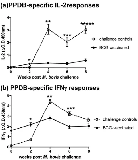
Vaccine-induced IL-2 response following challenge with pathogenicM. bovis. (i) Experiment 1.Fig. 3shows the PPDB-specific IL-2 (Fig. 3a) and IFN-␥(Fig. 3b) responses from BCG
vaccination/M. bovischallenge experiment 1. This experiment in-cluded 16 vaccinated animals plus 8 unvaccinated challenge con-trols. Whole-blood supernatants were tested post-BCG/pre-M. bovischallenge (week 0) and then 2, 4, 6, and 8 weeks postchal-lenge.
IL-2 responses (Fig. 3a) in the BCG-vaccinated group were negligible beforeM. bovischallenge (day 0) but were positive and significantly higher than those of the challenge control group at 2 weeks postinfection (P⫽0.002). While the IL-2 response of this vaccinated group increased over time, the levels remained signif-icantly lower throughout than those of the unvaccinated challenge control group (P⬍0.005 at all time points). Levels of IL-2 in the challenge control group were detected by 4 weeks postchallenge and levels remained high thereafter to 8 weeks postchallenge.
On challenge withM. bovis, the IFN-␥response (Fig. 3b) of vaccinated animals, already displaying a BCG-induced positive response to PPDB on day 0 (the day of challenge), was boosted by 2 weeks postinfection and was significantly higher than the re-sponse of the unvaccinated challenge controls at this time point (P⫽0.0011). The response of unvaccinated challenge controls was detected by 2 weeks postchallenge and thereafter increased, significantly exceeding the levels of IFN-␥in the challenge control group at 4 (P⫽0.002) and 6 (P⫽0.0011) weeks postinfection.
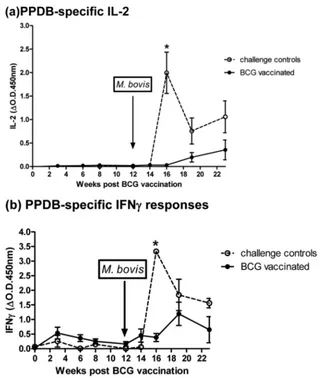
(ii) Experiment 2.We next investigated longitudinal responses to BCG prior toM. bovischallenge, primarily to see if BCG alone, at any point postvaccination, would induce an IL-2 response. Fig-ure 4shows the IL-2 (Fig. 4a) and IFN-␥(Fig. 4b) results from vaccination and challenge experiment 2. While consisting of fewer animals than experiment 1, the results demonstrated that IL-2 was not induced during any time point post-BCG vaccination and suggested that infection with pathogenicM. bovisis required to stimulate a detectable PPDB-induced IL-2 response.
In contrast to the results of experiment 1, IL-2 responses in the BCG-vaccinated group remained negligible until 4 weeks post-M. bovischallenge. But in agreement with experiment 1, the levels of IL-2 generated in unvaccinated control animals following chal-lenge withM. boviswere significantly higher (*P⫽0.0159 at 4 weeks) than those induced in BCG-vaccinated and -challenged cattle.
The IFN-␥results for experiment 2 (Fig. 4b) reflected those of experiment 1 in that followingM. bovischallenge, BCG-vacci-nated animals produced a rapid IFN-␥response, observed at 2 weeks postchallenge, which was absent in challenge control cattle (though not quite statistically significant in this experiment,P⫽
0.0635). Once established, the IFN-␥response of the challenge control group significantly exceeded the IFN-␥response of the BCG-vaccinated group (*P⫽0.0159 at 4 weeks postchallenge).
The results of these two separate experiments suggest that the priming of IL-2 by BCG vaccination may occur (experiment 1, containing 24 cattle, showed statistically higher IL-2 in BCG-vac-cinated cattle at 2 weeks post-M. bovischallenge). However, this increase was not consistent across the two experiments. All of the BCG-vaccinated animals in experiment 1 were protected follow-ing challenge (pathology score for this group, mean⫾standard error of the mean [SEM] of 0.833⫾0.386 compared to 15.0⫾ 2.171 [16]), while just 2/5 BCG-vaccinated cattle in experiment 2 had a medium pathology score (9 and 11, respectively) (group mean⫾SEM of 4.8⫾2.27 compared to 18.5⫾5.84 in the chal-lenge control group). A full description of the pathology scoring method is described in Vordermeier et al. (21). Interestingly, the FIG 3IL-2 (a) and IFN-␥(b) responses to PPDB followingM. bovischallenge
of BCG-vaccinated and unvaccinated animals (experiment 1). Data show the group mean and standard error for each group at each time point (n⫽8 unvaccinated;n⫽16 BCG vaccinated). Significant differences in cytokine levels between vaccinated and control groups are indicated.
vaccinated animal with the highest pathology score of 11 in exper-iment 2 also had the highest IL-2 response. These data collectively suggest that IL-2, as well as IFN-␥, when measured postinfection may correlate with the degree of pathology rather than protection.
DISCUSSION
This presence of PPDB-specific IL-2 responses in naturally in-fected field reactor cattle when none were apparent in eitherM. bovisBCG-vaccinated or uninfected controls provides the possi-bility that IL-2 could make a useful addition to the DIVA test toolbox to distinguish vaccinated fromM. bovis-infected individ-uals. Interestingly, it is the crude PPDB antigen that would be required for this, since IL-2 stimulation by peptide cocktails (es-sential for an IFN-␥DIVA test) had a much lower sensitivity in infected cattle. PPDB-specific IL-2 responses, in contrast, could be detected at a high sensitivity (93.9%) in SICCT⫹/IFN-␥⫹cattle, at a somewhat lower sensitivity in SICCT⫺/IFN-␥⫹cattle (30.3%), and crucially within 4 weeks following experimental infection. This study demonstrated that a high specificity (100%) of PPDB antigen alone (i.e., without using PPDA) was possible.
That a positive IL-2 response may reflect the stage or degree of
infection was suggested by a higher VL identification rate in SICCT⫺/IFN-␥⫹cattle that were IL-2 positive; 73.7% of IL-2⫹ cattle were VL, while 26.3% were NVL. Therefore, an IFN-␥⫹/ IL-2⫹animal could be almost three time more likely to be VL than NVL. Although, compared to the IFN-␥test, the IL-2 test was not as sensitive in SICCT-negative animals compared to skin test-positive animals, identifying just 30.3% of all the IFN-␥⫹animals, and 37.8% of the IFN-␥⫹VL cattle. However, it is possible that future improvement in the sensitivity to detect IL-2, for example using enzyme-linked immunosorbent spot (ELISPOT) assay read-outs (as we are currently investigating in our laboratory [data not shown]) or a more sensitive system, such as Meso Scale Discovery technology (which we already use in our laboratory to detect other low-level cytokines, like IL-10 [22]) to quantify IL-2 in supernatants could enhance its overall test sensitivity.
were significantly lower than the IL-2 responses of unvaccinated challenge controls. This suggests a relative modulation of antigen-specific responses (IFN-␥and IL-2) in vaccinated/protected cat-tle, and potential correlation of these responses with the degree of pathology. In this way, and similar to IFN-␥(21), IL-2 could pres-ent a marker of pathology, or advanced infection.
While some reports support this view in humanM. tuberculosis
infection (i.e., that T cells from people with advanced or active TB only, but not latent TB, secrete IL-2 after specific stimulation [23, 24]), other reports suggest that the reverse may be true. Sester et al. (25), using flow cytometry, and Casey et al. (26), using a dual-purpose IFN-␥/IL-2 ELISPOT assay (both reports from the same group), showed increases in T cells producing both IFN-␥and IL-2, or IL-2 only in protected (BCG vaccinated, treated, or la-tently infected) individuals and not in people with active TB who had more T cells producing IFN-␥.
That BCG alone was unable to induce detectable specific IL-2 responses in our experiments is supported by other work in both human TB and in mouse models. Peptides of the antigen Rv1986, absent from commonly used BCG strains, stimulated strong IL-2-positive T cell responses inM. tuberculosis-infected patients, and deletion of this region from the M. tuberculosisbacillus led to reduced IL-2 responsiveness (27). In mice, vaccination with a re-combinant vaccine (Ag85B-ESAT6 plus CAF01) that gave better protection againstM. tuberculosisthan BCG was found to induce IL-2-positive multifunctional (TNF-␣⫹and/or IFN-␥⫹) T cells, while BCG induced mainly IL-2-negative (TNF-␣⫹/IFN-␥⫹) T cells (5). Furthermore, the time-dependent loss of BCG-mediated protection againstM. tuberculosisin this same mouse model that was coincident with a decrease in IL-2-positive T cells was re-versed by booster vaccination using the recombinant vaccine. The authors suggest that the new vaccine stimulated central memory T cells while BCG did not (11). If true, then this lends support to the persistence of BCG in the host, rather than its generation of central memory being responsible for any protection afforded by this vac-cine.
In summary, we provide the first data for an IL-2 ELISA in BTB, which suggests its use as a potential tool that could be opti-mized and further developed for DIVA assays for the differentia-tion ofM. bovis infection following BCG vaccination and also suggests that, as IL-2 productionin vitroappeared more likely to identify animals with visible gross pathology, an IL-2 test could potentially provide useful information in addition to that of the current BTB IFN-␥test.
ACKNOWLEDGMENTS
We thank the dedicated staff of the Animal Services Unit at AHVLA Wey-bridge. We also acknowledge the excellent support of Team Gamma rou-tine testing (Laboratory Services Department) at AHVLA-Luddington for the provision of field samples and test data, and also Madeleine McCor-mick (ITU, AHVLA) for data trawls.
REFERENCES
1.Cockle PJ, Gordon SV, Lalvani A, Buddle BM, Vordermeier HM.2002. Identification of novelMycobacterium tuberculosisantigens with potential as diagnostic reagents or subunit vaccine candidates by comparative genomics. Infect. Immun.70:6996 –7003.http://dx.doi.org/10.1128/IAI .70.12.6996-7003.2002.
2.Jones GJ, Pirson C, Jones RG, Vordermeier HM.2010. Simultaneous measurement of antigen stimulated interleukin-1and gamma interferon production enhances test sensitivity for the detection ofMycobacterium
bovisinfection in cattle. Clin. Vaccine Immunol.17:1946 –1951.http://dx .doi.org/10.1128/CVI.00377-10.
3.Kaveh DA, Bachy VS, Hewinson RG, Hogarth PJ.2011. Systemic BCG immunization induces persistent lung mucosal multifunctional CD4 T(EM) cells which expand following virulent mycobacterial challenge. PLoS One6:e21566.http://dx.doi.org/10.1371/journal.pone.0021566. 4.Derrick SC, Yabe IM, Yang A, Morris SL.2011. Vaccine-induced
anti-tuberculosis protective immunity in mice correlates with the magnitude and quality of multifunctional CD4 T cells. Vaccine29:2902–2909.http: //dx.doi.org/10.1016/j.vaccine.2011.02.010.
5.Lindenstrom T, Agger EM, Korsholm KS, Darrah PA, Aagaard C, Seder RA, Rosenkrands I, Andersen P.2009. Tuberculosis subunit vaccination provides long-term protective immunity characterized by multifunc-tional CD4 memory T cells. J. Immunol.182:8047– 8055.http://dx.doi .org/10.4049/jimmunol.0801592.
6.Li L, Qiao D, Li Q, Zhang X, Lao S, Wu C.2012. Distinct polyfunctional CD4⫹T cell responses to BCG, ESAT-6 and CFP-10 in tuberculous pleurisy. Tuberculosis (Edinb.) 92:63–71. http://dx.doi.org/10.1016/j.tube.2011.11 .004.
7.Caccamo N, Guggino G, Joosten SA, Gelsomino G, Di Carlo P, Titone L, Galati D, Bocchino M, Matarese A, Salerno A, Sanduzzi A, Franken WP, Ottenhoff TH, Dieli F.2010. Multifunctional CD4(⫹)T cells
corre-late with activeMycobacterium tuberculosisinfection. Eur. J. Immunol.
40:2211–2220.http://dx.doi.org/10.1002/eji.201040455.
8.Mueller H, Detjen AK, Schuck SD, Gutschmidt A, Wahn U, Magdorf K, Kaufmann SH, Jacobsen M. 2008.Mycobacterium tuberculosis-specific CD4⫹IFNgamma⫹and TNFalpha⫹multifunctional memory T cells co-express GM-CSF. Cytokine43:143–148.http://dx.doi.org/10.1016/j.cyto .2008.05.002.
9.Millington KA, Innes JA, Hackforth S, Hinks TS, Deeks JJ, Dosanjh DP, Guyot-Revol V, Gunatheesan R, Klenerman PA, Lalvani A.2007. Dy-namic relationship between IFN-gamma and IL-2 profile of Mycobacte-rium tuberculosis-specific T cells and antigen load. J. Immunol.178:5217– 5226.
10. Billeskov R, Elvang TT, Andersen PL, Dietrich J.2012. The HyVac4 subunit vaccine efficiently boosts BCG-primed anti-mycobacterial pro-tective immunity. PLoS One7:e39909.http://dx.doi.org/10.1371/journal .pone.0039909.
11. Lindenstrom T, Knudsen NPH, Agger EM, Andersen P.2013. Control of chronicMycobacterium tuberculosisinfection by CD4 KLRG1-IL-2-secreting central memory cells. J. Immunol.190:6311– 6319.http://dx.doi .org/10.4049/jimmunol.1300248.
12. Whelan AO, Villarreal-Ramos B, Vordermeier HM, Hogarth PJ.2011. Development of an antibody to bovine IL-2 reveals multifunctional CD4 T(EM) cells in cattle naturally infected with bovine tuberculosis. PLoS One6:e29194.http://dx.doi.org/10.1371/journal.pone.0029194. 13. Rhodes SG, Buddle BM, Hewinson RG, Vordermeier HM.2000. Bovine
tuberculosis: immune responses in the peripheral blood and at the site of active infection. Immunology 99:195–202. http://dx.doi.org/10.1046/j .1365-2567.2000.00944.x.
14. Rhodes SG, Gaver-Widen D, Buddle BM, Whelan AO, Singh M, Hewinson RG, Vordermeier HM.2000. Antigen specificity in experi-mental bovine tuberculosis. Infect. Immun.68:2573–2578.http://dx.doi .org/10.1128/IAI.68.5.2573-2578.2000.
15. Pirson C, Vipond J, Hall Y, Williams A, Vordermeier HM. 2011. Vaccines designed to protect againstMycobacterium tuberculosisinfection may aid the identification of novel vaccine constructs and diagnostic an-tigens for bovine tuberculosis. Vet. Microbiol.148:232–237.http://dx.doi .org/10.1016/j.vetmic.2010.08.019.
16. Whelan AO, Coad M, Upadhyay BL, Clifford DJ, Hewinson RG, Vor-dermeier HM.2011. Lack of correlation between BCG-induced tubercu-lin skin test sensitisation and protective immunity in cattle. Vaccine 29: 5453–5458.http://dx.doi.org/10.1016/j.vaccine.2011.05.057.
17. Waters WR, Nonnecke BJ, Palmer MV, Robbe-Austermann S, Banna-tine JP, Stabel JR, Whipple DL, Payeur JB, Estes DM, Pitzer JE, Minion FC.2004. Use of recombinant ESAT-6:CFP-10 fusion protein for differ-entiation of infections of cattle byMycobacterium bovisand byM. avium
subsp.aviumandM. aviumsubsp.paratuberculosis. Clin. Diagn. Lab. Immunol. 11:729 –735. http://dx.doi.org/10.1128/CDLI.11.4.729-735 .2004.
sasii. Clin. Vaccine Immunol.17:247–252.http://dx.doi.org/10.1128/CVI .00442-09.
19. Arend SM, de Haas P, Leyten E, Rosenkrands I, Rigouts L, Andersen P, Mijs M, van Dissel JT, van Soolingen D.2005. ESAT-6 and CFP-10 in clinical versus environmental isolates ofMycobacterium kansasii. J. Infect. Dis.191:1301–1310.http://dx.doi.org/10.1086/428950.
20. Vordermeier HM, Brown J, Cockle PJ, Franken WP, Drijfhout JW, Arend SM, Ottenhoff TH, Jahans K, Hewinson RG.2007. Assessment of the cross-reacticity betweenMycobacterium bovisandM. kansasiiESAT-6 and CFP-10 at the T-cell epitope level. Clin. Vaccine Immunol.14:1203– 1209.http://dx.doi.org/10.1128/CVI.00116-07.
21. Vordermeier HM, Chambers MA, Cockle PJ, Whelan AO, Simmons J, Hewinson RG.2002. Correlation of ESAT6-specific gamma-interferon production with pathology in cattle followingMycobacterium bovis-BCG vaccination against experimental bovine tuberculosis. Infect. Immun.70:
3026 –3032.http://dx.doi.org/10.1128/IAI.70.6.3026-3032.2002. 22. Coad M, Clifford D, Rhodes SG, Hewinson RG, Vordermeier HM,
Whelan AO.2010. Repeat tuberculin skin testing leads to desensitization in naturally infected tuberculous cattle which is associated with elevated interleukin-10 and decreased interleukin-1-beta responses. Vet. Res.41:
14 –26.http://dx.doi.org/10.1051/vetres/2009062.
23. Biselli R, Mariotti S, Sargentini V, Sauzullo I, Lastilla M, Mengoni F, Vanini V, Girardi E, Goletti D, D’Amelio R, Nisini R.2010. Detection of interleukin-2 in addition to interferon-gamma discriminates active
tu-berculosis patients, latently infected individuals, and controls. Clin. Mi-crobiol. Infect. 16:1282–1284. http://dx.doi.org/10.1111/j.1469-0691 .2009.03104.x.
24. Chiappini E, Della Bella C, Bonsignori F, Sollai S, Amedei A, Galli L, Niccolai E, Del Prete G, Singh M, D’Elios MM, de Martino M.2012. Potential role ofM. tuberculosisspecific IFN-␥and IL-2 ELISPOT assays in discriminating children with active or latent tuberculosis. PLoS One
7:e46041.http://dx.doi.org/10.1371/journal.pone.0046041.
25. Sester U, Fousse M, Dirks J, Mack U, Prasse A, Singh M, Lalvani A, Sester M.2011. Whole-blood flow-cytometric analysis of antigen-specific CD4 T-cell cytokine profiles distinguishes active tuberculosis from non-active states. PLoS One6:e17813.http://dx.doi.org/10.1371/journal.pone .0017813.
26. Casey R, Blumenkrantz D, Millington K, Montamat-Sicotte D, Kon OM, Wickremasinghe M, Bremang S, Magtoto M, Sridhar S, Connell D, Lalvani A.2010. Enumeration of functional T-cell subsets by fluores-cence-immunospot defines signatures of pathogen burden in tuberculo-sis. PLoS One5:e15619.http://dx.doi.org/10.1371/journal.pone.0015619. 27. Gideon HP, Wilkinson KA, Rustad TR, Oni T, Guio H, Kozak RA, Sherman DR, Meintjes G, Behr MA, Vordermeier HM, Young Wilkin-son DB RJ.2010. Hypoxia induces an immunodominant target of tuber-culosis specific T cells absent from common BCG vaccines. PLoS Pathog.