O R I G I N A L A R T I C L E
Analysing the effect of dietary prebiotics and probiotics on
gut bacterial richness and diversity of Asian snakehead
fingerlings using T
‐
RFLP method
Mohammad Bodrul Munir
1|
Terence L. Marsh
2|
Aimeric Blaud
3|
Roshada Hashim
4|
Wizilla Janti Anak Joshua
1|
Siti Azizah Mohd Nor
51
Aquatic Resource Science and
Management, Faculty of Resource Science and Technology, Universiti Malaysia Sarawak, Kota Samarahan, Sarawak, Malaysia
2
Department of Microbiology and Molecular Genetics, Biomedical & Physical Sciences, Michigan State University, East Lansing, Michigan,
3
Department of Sustainable Agriculture Science, Rothamsted Research, Harpenden, UK
4
Faculty of Science and Technology, Universiti Sains Islamic Malaysia, Bandar Baru Bangi, Malaysia
5
Institute of Marine Biotechnology, Universiti Malaysia Terengganu, Kuala Terengganu, Malaysia
Correspondence
Mohammad Bodrul Munir, Aquatic Resource Science and Management, Faculty of Resource Science and Technology, Universiti Malaysia Sarawak, 94300 Kota Samarahan, Sarawak.
Email: hsjewel730@yahoo.com
Funding information
FRGS, Grant/Award Number: 203/ PBIOLOGI/6711308; USM Global Fellowship
Abstract
This study was designed to evaluate the effects of three prebiotics (
β
‐
glucan,
galacto
‐
oligosaccharide [GOS], mannan
‐
oligosaccharide [MOS]) and two probiotics
(
Saccharomyces cerevisiae
,
Lactobacillus acidophilus
) on the microbiome of snakehead
during growth of fingerlings. In addition, the experiment evaluated the capacity of
Channa striata
fingerlings to retain the benefits derived from these supplements
after withdrawal. Throughout the study, it was observed that supplementation with
dietary prebiotics and probiotics led to significant (
p
<
0.05) change in gut bacterial
profile and improvement in gut morphology. Terminal restriction fragment length
polymorphism (T
‐
RFLP) was used for the comparative analysis of gut communities
and all 46 of the T
‐
RFLP detected phylotypes were present in the
Lactobacillus
sup-plemented fish, while significantly fewer were detected in controls and other
experi-mental supplement regimes. Histological studies and electron microscopy revealed
that both the prebiotic and probiotic treated fish had significantly longer and wider
villi and deeper crypts compared to the controls. The microvilli length, as evaluated
with electron microscopy, was also longer in all treated fish compared to controls.
Furthermore, this study is the first to report the absence of differences in sustaining
the efficacies attained after intake of
β
‐
glucan, GOS, MOS and live yeast upon post
‐
feeding with an unsupplemented feed, over a prolonged period.
K E Y W O R D S
gut bacterial richness and diversity, prebiotics, probiotics, snakehead fingerlings, terminal
restriction fragment length polymorphism
1
|
I N T R O D U C T I O N
Analysing the intestinal bacterial profile is one of the important
fac-tors in fish nutrition studies (Daniel et al., 2014; Lara‐Flores,
Olvera‐Novoa, Guzmán‐Méndez, & López‐Madrid, 2003; Moen,
Saeed, Mohammad, & Faranaz, 2011). The gastrointestinal (GI) tract
of fish is known as the ecological nich (Austin & Austin, 1987;
Cahill, 1990; Denev, Staykov, Moutafchieva, & Beev, 2009; Holben
et al., 2002; Kim, Brunt, & Austin, 2007; Llewellyn, Boutin,
Hoseinifar, & Derome, 2015; Ringø et al., 2000; Ringø, Sperstad,
Myklebust, Mayhew, & Olsen, 2006; Ringø, Strøm, & Tabachek.,
1995) for a group of diverse selected beneficial bacteria derived
from the surrounding aquatic environment, including water
sedi-ment and feed. The abundance of beneficial bacteria in the fish
intestine is expected to influence fish growth and health (Cahill,
1990; Hoseinifar Khalili, & Sun, 2016; Hoseinifar, Ahmadi, et al.,
2016; MacFarlane, McLaughlin, & G. Bullock., 1986). Moreover, a
diverse microbial community has been correlated with a well‐
func-tioning GI system (Nayak, 2010).
The Asian snakehead (Channa striata, Bloch, 1793) is one of the
most economically important freshwater fish in Asia‐Pacific region
(Hossain, Latifa, & Rahman, 2008; Jamsari, Tan, & Siti‐Azizah, 2011;
Wee, 1982). It contains higher protein (16.2 g in 100 g muscle)
com-pare to other freshwater fish such as gold fish orCarassius auratus
and eel orAnguilla anguilla(Annasari, Aris, & W., Yohanes, K., 2012)
and has a high market value due to the high quality of flesh with
low fat, less intramuscular spines and medicinal qualities (Haniffa &
Marimuthu, 2004). Extracts of its fins and scales are a good source
of albumin for the people who have a deficiency of albumin (Haniffa
& Marimuthu, 2004). Therefore, the aquaculture production trend
has increased 61.90% from 2000 to 2012 (FAO, 2014) through
semi‐intensive, intensive culture and cage aquaculture practices.
Fre-quently, when stocked at high density of snakehead fingerlings, the
resulting accumulation of organic matter leads to deterioration of
water quality and increases in diseases (Sinh & Pomeroy, 2010).
Biologically, the fish grows slowly but growth can be accelerated
using high‐quality feed supplements (Hossain et al., 2008; Wee,
1982). Antibiotics can no longer be used to manage infection and
fish health, to indirectly promote growth because of international
food security regulations imposed by the Food and Agriculture
Orga-nization in 2006 (FAO, 2007). The FAO authority permits
alterna-tives such as dietary prebiotics, probiotics, symbiotics, phytobiotics
and other natural dietary supplements (Denev, 2008). Dietary
prebi-otics and probiprebi-otics can improve the growth performance, feed
uti-lization, body indices (Hoseinifar, Safari, & Dadar, 2017; Munir,
Hashim, Abdul Manaf, & Nor, 2016; Talpur, Munir, Marry, & Hashim,
2014) nutrient protein digestibility, the expression of immune
regula-tory genes (Hoseinifar, Ahmadi, et al., 2016; Munir, Roshada, Yam,
Terence, & Azizah, 2016) and health status (Talpur et al., 2014) of
snakehead. In fact, dietary prebiotics and probiotics provide direct
beneficial effects on growth by improving intestinal microbial
bal-ance (Al‐Dohail, Hashim, & Aliyu, 2009; Dhanaraj et al., 2010) and
by modifying the structure and function of the GI tract in the fish
(Akter, Sutriana, Talpur, & Hashim, 2015; Amalia, Roshada, Nahid, &
Siti‐Azizah, 2018; Carly et al., 2010; Jian et al., 2012; Ringø,
Mykle-bust, Mayhew, & Olsen, 2007). This study analysed the effect of
dietary prebiotics and probiotics on gut bacterial richness and
diver-sity of snakehead using terminal restriction fragment length
polymor-phism (T‐RFLP) method. The T‐RFLP is a technique for profiling
microbial communities based on the position of a restriction site
closest to a labelled end of an amplified gene (Christensen, Reynolds,
Shukla, & Reed, 2004; Coolen, Post, Davis, & Forney, 2005; Davis
et al., 2010; Nieminen et al., 2011).
2
|
M A T E R I A L S A N D M E T H O D S
2.1
|
Experimental fish and husbandry conditions
The study was conducted in Universiti Sains Malaysia (USM)
Aquaculture Research Complex. A total of 4,800 C. striata
fingerlings (av. wt. 22.40 ± 0.06 g) were selected from 10,000
master stocked snakehead fries and distributed equally (400 fish/
tank) in 12 outdoor rectangular cement tanks (2 m×1 m×0.5 m).
The fish were maintained in optimum condition with a natural
photoperiod where the mean water temperature, pH and dissolved
oxygen were 27.54 ± 0.30°C, 7.1 ± 0.08 and 6.1 ± 0.18 mg/L
respectively.
2.2
|
Experimental diets and feeding trial
Six experimental diets including the control contained 40% protein
and 12% lipid (Table 1). Fish were fed the experimental diets in two
phases. Phase 1 involved feeding six cohorts of fish experimental
diets for 16 weeks while in Phase 2, all experimented fish were fed
the non‐supplemented control diet for 8 weeks. This was done to
evaluate the efficacy of prebiotics and probiotic intake in Phase 1
and the reversibility of the supplementation. In both phases, fish
were fed to satiation three times daily (i.e., early morning at 6.30
a.m., noon at 1 p.m. and late afternoon at 7 p.m.).
2.3
|
Viability of
Lactobacillus acidophilus
in the LBA
diet
The viability of L. acidophilus in LBA diet was performed three
times, that is immediately after LBA diet preparation, two times (8
and 16 weeks) during storing at −20°C temperature followed by
the method described by Al‐Dohail (2010); Wang, Yu, and Chou
(2004) and Ishibashi, Tatematsu, Shimamura, Tomita, and Okonogi
(1985).
2.4
|
Gut bacterial profile
The gut bacterial profile inC. striata was conducted using T‐RFLP,
which was designed according to the method Cancilla, Powell, Hillier,
and Davidson (1992), refined by Brunk's laboratory (Avaniss‐Aghajani
et al., 1996; Avaniss‐Aghajani, Jones, Chapman, & Brunk, 1994)
described by Bruce (1997), Liu, Marsh, Cheng, and Forney (1997),
Clement, Kehl, Bord, and Kitts (1998) and Marsh (2005). Twelve fish
were randomly collected from each replicate tank making three
groups of equal number of fish and were stocked in three black
plas-tic tanks for 24 hr without feeding. After 24 hr, the fish were
sacri-ficed individually using ice block. The fish were dissected, removing
undesired gut tissues such as liver, spleen and stomach. The
intes-tine was carefully separated from the stomach by first tying it off at
the junction of the stomach and intestine, cutting at the end of
stomach before the tied area, thereby isolating materials in the
intes-tine from contamination with stomach content. The exterior of the
intestines was washed several times with sterile PBS buffer (pH 7.4)
and blotted to remove the buffer. The intestines were cut into small
pieces and mixed together. One replicate consisted of intestines of
three fish. Cold environment was strictly maintained during this
pro-cesses using ice blocks to protect the bacterial DNA from
2.5
|
Genomic DNA extraction and PCR
amplification
The genomic DNA was extracted using PowerSoil®DNA isolation kit
(Catalog No: 12888‐S; MO BIO Laboratories Inc., CA, USA)
accord-ing to the recommended vendor's protocol. The PCR amplification
was performed using a conventional PCR machine (Model: ABI
VER-ITI, Manufactured by Applied Biosystem, USA). The universal
bacte-rial primers 63F (5'‐CAG GCC TAA CAC ATG CAA GTC‐3') (5'FAM‐
labelled) and 1389R (5'‐ACG GGC GGT GTG TAC AAG‐3')
(unla-belled) (Aburto et al., 2009; Masanori, James, Kim, & Marsh, 2013;
Quaak & Kuiper, 2011) were used to perform the 16S rDNA
amplifi-cation. The PCR reaction was carried out using i‐TaqTMplus DNA
polymerase (Cat No. 25152; iNtRON Biotechnology, Inc.
Manufac-ture) in a 50 µl reaction volume containing 1.5 µl DNTPs (2.5 mMol),
5 µl 10× Buffer, 1 µl (1 µg) universal primers each, 0.5 µl (2.5 U)
iTaq plus, 1 µl diluted DNA extracted template and 40 µl ddH2O
under the following cycle conditions: an initial denaturing step at
94°C for 3 min, 30 cycles of denaturing step at 94°C for 30 s,
annealing at 55°C for 30 s and extension at 72°C for 1.5 min. The
final extension was performed at 72°C for 5 min. The PCR mixture
was prepared under freezing temperature using ice block chamber.
After PCR, the amplification size was verified by gel electrophoresis
using 1 kb ladder.
2.6
|
Purification of PCR products and digestion
The PCR products were purified using MEGAquick‐spin™Total
Frag-ment DNA Purification Kit (iNtRON Biotechnology Manufacture)
according to the Manufacturer's guideline. The purified PCR
prod-ucts were digested withHhal (Product code R0139S; New England
Biolabs) followed by the Manufacturer's guideline. The products
were checked using gel electrophoresis.
2.7
|
T
‐
RFLP fragment sequencing
After gel electrophoresis, 15 µl of digested DNA samples each was
sent to Macrogen Inc., South Korea for T‐RFLP sequencing. The
ser-vice provider separated the DNA fragment (size standard 500LIZ)
using ABI 3730XL Genetic Analyzer automated sequencer (Applied
Biosystem). Each feeding treatment had four biological replicates and
each biological replicate had two technical replicates. The data were
from samples taken at the end of 8 and 16 weeks in Phase 1 and at
the end of Phase 2.
2.8
|
Comparative analysis of gut bacterial
community profile
The sequence data obtained from the service provider were
retrieved using PEAKSCAN 3.1 software with the 500LIZ size
stan-dards. Therefore, the terminal restriction fragments (T‐RFs) were
binned with a 0.5 bp interval and the T‐RFs from 50 to 500 bp were
included in the analysis. T‐Align software was used for the
align-ments of the fragment size.
2.9
|
Morphological measurement of intestine
The villus length, width and crypt depth were determined using
nor-mal histological procedure following the method of Davenport in
1969, whereas the transmission electron microscopy (TEM) was used
to determine the microvillus length following the method described
by Lewis and Knight (1977).
2.10
|
Statistical analysis
The bacterial richness in the gut ofC. striatafingerlings for inclusion
of dietary prebiotics and probiotics was estimated by counting the
T A B L E 1 Feed ingredients of the six experimental diet (g/kg, dry matter)
Ingredients Control β‐Glucana0.2% GOSb1% MOSc0.5% Live yeastd1%
L. acidophiluse0.01%
Danish Fish Mealf 534 534 534 534 534 534
Korean Corn Starch 340 340 340 340 340 340
Fish oil 5 5 5 5 5 5
Soybean oil 60 60 60 60 60 60
Cellulose 11 9 1 6 1 10.9
CMC 10 10 10 10 10 10
Vitamins mixg 20 20 20 20 20 20
Minerals mixh 20 20 20 20 20 20
Supplement 0 2 10 5 10 0.1
Note. CMC = carboxymethyl cellulose..aβ‐Glucan = Macrogard®..bGOS = Galactooligosaccharides of Vivinal®GOS syrup, Friesland Campina Domo, the
Netherland..cMOS = Mannan‐oligosaccharides of Alltech®, Actigen 1, USA..dLive Yeast =Saccharomyces cerevisiaeof Alltech®, YEA‐SACC 1026, USA..
eL. acidophilus=Lactobacillus acidophiluspowder (Sigma®LBA)..fDanish Fish Meal/kg = Crude Protein 746.6 and Crude Lipid 101.6..gVitamin Mix/kg =
Rovimix 6288, Roche Vitamins Ltd. Switzerland; VitA50 million i.u., VitD310 million i.u., VitE130 g, VitB110 g, VitB225 g, VitB616 g, VitB12100 mg,
Biotin 500 mg, Pantothenic acid 56 g, Folic Acid 8 g, Niacin 200 g, Anticake 20 g, Antioxidant 200 mg, VitK310 g and VitC35 g..hMineral Mix/kg =
T‐RFs number in each T‐RFLP profile. The diversity of the bacterial
communities took into account both species richness and species
evenness. The species evenness indicates how close in population
size of each bacterial species is in a community, or in this case,
indi-vidual feeding trial and it was done using the Shannon index (H′,
Shannon, 1948). This involved three steps. (a) cluster analysis
(illus-trated through dendrogram) and data ordination using non‐metric
multi‐dimensional scaling (nMDS) to visualize the result showing the
difference between bacterial communities in the fish gut
correspond-ing to different feedcorrespond-ing treatments environment in the studied
per-iod; (b) test of significance difference using two‐way ANOISM and
two‐way PERMANOVA; (c) finally, correlation between the bacterial
community composition and the gut morphology correspond to the
six feeding treatments over time.
To remove background noise from each T‐RFLP profile for
sub-sequent statistical analysis, the T‐RFs that were below 0.5% (i.e.,
0.005) were removed by setting a logical formula (Blaud, Diouf,
Her-rmann, & Lerch, 2015). The normalized data were then square root
transformed and a Bray–Curtis matrix was used for analysing
similar-ities between samples and displaying the result using dendrogram
(Culman, Gauch, Blackwood, & Thies, 2008; Ramette, 2007) and
nMDS plots. The significant difference between clusters of the
den-drogram was tested using similarity profile (SIMPROF) analysis (999
permutations). The significant differences between feeding
treat-ments with the time period were tested using two‐way analysis of
similarities (ANOSIM; 9,999 permutations). The significance levels,
that is p value, andRvalue, that is the strength of the factors on
samples were determined.Rvalues close to 1 indicated high
separa-tion between groups (e.g., between two feeding treatments or
between two time period), while R values close to 0 indicated no
separation between groups (Clarke & Gorley, 2001). Two‐way
per-mutational multivariate analysis of variance (PERMANOVA) was also
used to test for difference in T‐RFLP profiles between feeding
treat-ments and time. All these analysis were carried out using PRIMER
V6 statistical software (PRIMER‐E Ltd., Plymouth, UK).
The gut morphological particulars (i.e., villus length, villus width,
crypt depth and microvillus length) of C. striatawere analysed using
ANOVA to detect significant changes (p<0.05) corresponding to six
supplemented diets over time. Finally, the correlation between bacterial
community composition (T‐RFs or phylotypes) and the gut
morphologi-cal particulars were analysed using Pearson correlation in SPSS.
3
|
R E S U L T S
3.1
|
Gut bacterial profile
3.1.1
|
Bacterial community richness and evenness
Supplementation with dietary prebiotics and probiotics enhanced the
richness and evenness of the bacterial communities in the fish gut
compared to the fish fed with the control (Figure 1). The restricted
enzyme digestion generated different T‐RFs (size standard 500LIZ)
which representing distinct bacterial phylotypes found from the six
feeding treatments. A total 49 different phylotypes were found
(Fig-ure 1). The T‐RFs richness and evenness were significantly and
sys-tematically lower in the fish guts fed with the control (richness‐
31 ± 4, evenness = 3.41 ± 0.13, 8 weeks) than with any
supple-mented diet. The fish gut which was fed with the L. acidophilus
showed the highest T‐RFs richness and evenness (richness = 45±2;
evenness = 3.80 ± 0.04, 8 weeks) in comparison to the fish fed with
β‐glucan, galacto‐oligosaccharide (GOS) and mannan‐oligosaccharide
(MOS), regardless the feeding period, but was only slightly higher
than fish fed with live yeast at 16 weeks. No significant differences
in T‐RFs richness and evenness were found at 8 weeks betweenβ‐
glucan, GOS, MOS and live yeast (average richness = 41±2,
even-ness = 3.70 ± 0.03) but at 16 weeks, live yeast was significantly
(p<0.05) higher than those three prebiotics. At the end of post‐
feeding treatments (i.e., 24 weeks), gut microbiomes fed with GOS
were lower diversity compared to all the supplemented diets, and
not significantly different from the control in richness and evenness.
3.1.2
|
Gut bacterial community composition
The mean richness (i.e., the number of detected phylotypes) data of
different bacterial community composition is presented in Figure 2.
Fish fed withL. acidophilus supplemented diets carried the greatest
number of distinct terminal fragments (49). A total of 11 T‐RFs (i.e.,
131.2, 137.34, 146.48, 157.79, 171, 199.69, 250.14, 327.03, 433.22,
455.39, 489.22 bp) were absent in the control treated fish guts; and
all of only four phylotypes (171, 199.69, 250.14 and 327.03 bp)
were absent in other feeding treatments (β‐glucan, GOS, MOS, live
yeast) compared to the fish gut treated with theL. acidophilus
probi-otic.
The cluster data analysis (Figure 3) revealed that the bacterial
community compositions generated by T‐RFLP were strongly
affected by the inclusion of supplemented diets over the time
per-iod. According to the analysis, the bacterial community from the fish
fed with the control diet shown the lowest similarity to the other
treatments (~66%–75% similarity). The control fish at 8 weeks
tended to group separately from the other weeks. The fish fed with
L. acidophilus probiotic significantly (p<0.05) clustered separately
from the other supplemented diets (three prebiotics and probiotic‐
live yeast), regardless the week of treatment, and showed ~83% of
similarity with these treatments. The remaining prebiotics treatments
(i.e., β‐glucan, GOS, MOS) and probiotic (i.e., live yeast) grouped
together with high percentage of similarity (~90%) and showed
inconsistent differences between the weeks of treatments. The
nMDS plot (Figure 4) also showed similar community differences
with the cluster analysis, where based on bacterial community
com-position, there were three groups: the control, LBA treated and all
other treatments.
3.1.3
|
ANOISM and PERMANOVA analysis
Two‐way analysis of similarity (ANOISM) and PERMANOVA were
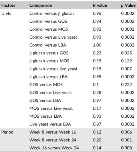
and periods. Two‐way ANOSIM test revealed that diets had a strong
effect on the bacterial community composition (R≤1). The data
demonstrated that the highest significance in comparison to the
con-trol feed was observed in the fish gut treated with LBA feed
supple-ments, followed β‐glucan, GOS, MOS and live yeast (Table 2).
Between the two prebiotics (i.e.,β‐glucan to GOS; GOS to MOS and
MOS to β‐glucan), there was no pronounced difference (Table 2).
Similar values were seen between beta‐glucan and yeast; GOS and
live yeast; and MOS and live yeast. A strong difference was
observed between live yeast and LBA (R= 0.87). In this study, time
period did not have a strong effect on the bacterial community
structure (R≤0.2); 8 and 24 weeks showed the highest R values
(R= 0.2), and 8 versus 16 and 16 versus 24 showed similarRvalues,
0.12 and 0.14 respectively.
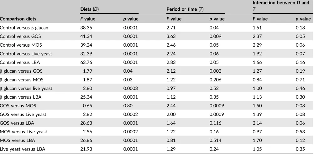
Similar results were also found in PERMANOVA analysis.
How-ever, it also revealed the significance analysis result in three ways,
that is between the diets, periods and the interaction of diets and
periods. Table 3 represents the PERMANOVA analysis result of
bac-terial community composition affected by the comparison between
diets, periods and their interactions. The result obtained from the
PERMANOVA demonstrated that the highest significant difference
between the control and supplemented diets was observed in the
fish gut treated with LBA feed supplements, followed GOS, beta‐
glu-can and live yeast. In comparisons with ANOISM, fish fed LBA
con-tained the highest significant value for bacterial communities
composition. Among the three prebiotics, there was not a strong
effect. Although no strong effect was found between the three
pre-biotics and live yeast (probiotic), there was a significant difference
between two probiotics amended fish (Table 3). The comparison
among the rearing periods (week 8, 16 in the Phase 1 and the Phase
2 or post‐feeding treatments period) and the interaction between
the comparison of diets and rearing period did not show strong
sig-nificant difference on the bacterial communities composition.
3.2
|
Relation between bacterial communities
structure and gut morphology
The study has revealed that inclusion of dietary prebiotics and
probi-otics profoundly influenced the gut microbiome. The villus length,
villus width and crypt depth of the fish gut measured using light
microscopic analysis were significantly increased (p<0.05) in
sup-plemented feeding treatments compared to control. Highest
varia-tion was observed in the fish fed with LBA (Table 4). There was no
significant difference (p<0.05) among three prebiotics (β‐glucan,
GOS and MOS) feeding treatments but in several instances live yeast
feeding treatments was very near to these three prebiotics. The
microvillus lengths studied under TEM were found similar to villus
length. The pearson revealed the correlations between the mean
value of gut morphology and mean value bacterial communities
rich-ness in different feeding treatments over rearing periods. A positive
correlation (Table 5) between the gut morphology and bacterial
com-munities structure were found. There was very little significant
F I G U R E 1 Variation in the number of
T‐RFs and the Shannon index from the
bacterial community structure generated
by T‐RFLP from fish gut feed with six
different diets over a 8, 16 (Phase 1) and 24 weeks (Phase 2) period. Mean
values ± standard errors (n= 3) are shown.
Superscripts represent significant
(p<0.05) differences among the
change in gut morphology between Phase 1 (at the end of
16 weeks) and Phase 2.
4
|
D I S C U S S I O N
In this study, the application of dietary prebiotics and probiotics over
a prolonged period significantly changed the bacterial community
composition. The cluster and nMDS analysis showed clear separation
between all supplemented feed treatments and the control, which
was further confirmed by SIMPROF, two‐way ANOISM and two‐
way PERMANOVA analysis. Hence the supplemented feeding
treat-ments likely affected the composition of bacterial communities either
via direct or indirect effects of dietary probiotics or the dietary
pre-biotics respectively. This result agrees with the previous studies that
8 8 8 8 16 24 24 16 16 24 16 24 24 8 16 24 24 24 8 8 16 8 8 24 16 16 8 8 16 8 8 16 24 8 8 8 16 24 24 8 24 16 16 16 24 24 16 24 24 8 24 16 16 16 8 8 16 24 16 24 24 8 24 16 8 8 16 8 24 24 16 16
100 90 80 70 60
S
im
ila
rit
y
(%
)
Feed
Control
Beta-glucan
GOS
MOS
Live yeast
LBA
F I G U R E 3 Dendrogram of the bacterial community composition from fish's gut that were feed with six different diets over a 8 and
16 weeks in Phase 1 and 24 weeks (following 8 weeks in Phase 2 or at the end of post feeding period). The numbers 8, 16, 24 indicate the period in week the fish are being feed. The dendrogram was produced using the group average linking method based on square root
transformed data and Bray‐Curtis similarity matrix. Red lines indicate clusters that are not significantly different (p<0.05) using SIMPROF
analysis (999 permutations) [Colour figure can be viewed at wileyonlinelibrary.com]
F I G U R E 2 Mean abundance of T‐RFs (size standard 500LIZ; size range 50–500 base pair) represents as phylotypes by six feeding
treatments including control. Theβ‐glucan, GOS, MOS, Live Yeast and LBA represent feed with beta‐glucan, galactooligosaccharides, mannan‐
oligosaccharides,Saccharomyces cerevisiae, Lactobacillus acodophilusfeed supplements, respectively [Colour figure can be viewed at wileyonline
evaluated the bacterial community composition of channel catfish,
Pangasius hypophthalmus (Nahid, 2015) and small killifish, Fundulus
heteroclitus(Givens, 2012).
The performance trend of six experimental diets on bacterial
community composition was LBA>live yeast>β‐glucan≥ MOS≥
GOS>control at the end of week 8 and 16 in Phase 1. The reasons
for increased performance with the experimental diets include the
direct manipulation of intestinal microbial communities, suppression
of pathogens, immunomodulation, stimulation of epithelial cell
prolif-eration, differentiation, fortification of the intestinal barrier (Amalia
et al., 2018; Hoseinifar, Mirvaghefi, Amoozegar, Merrifield, & Ringø,
2015; Hoseinifar, Sharifian, Vesaghi, Khalili, & Esteban, 2014;
Thomas & Versalovic, 2010; Yarahmadi, Kolangi Miandare, &
Hoseinifar, 2016) as well as meeting dietary requirements. The
speci-fic combination of positive effects at the molecular level may differ
with the nature of each supplement. Nevertheless, the mode of
action in each case led to increased performance with the greatest
seen with LBA. This statement is supported by Nahid (2015) who
conducted studies on the effect of prebiotic, MOS and probiotics,
L. acidophilus at different doses on striped cat fish, Pangasianodon
hypopthalmus. Earlier studies by Noh, Han, Won, and Choi (1994)
and Bogut, Milaković, Bukvić, Brkić, and Zimmer (1998) on common
carp fingerlings showed similar results. The study also evaluated the
viability of L. acidophilusin LBA diet during frozen (−20°C) storage
(Table 6). This study also indicated that there were no significant
differences among the three prebiotics on bacterial community
com-position in the intestine over the time period. This is likely due to
the unique stimulating and immunomodulatory characteristic of the
three prebiotics tested that facilitated the growth of beneficial
bac-teria in the GI tract. Furthermore, these two attributes make the
pre-biotics a gut microflora management tool of fish (Gibson, 2008).
Prebiotics are non‐digestible dietary fibre comprising of non‐starch
polysaccharides such as cellulose and many other plant components
such as pectins, dextrins, lignins,β‐glucans (Philippe & Sylvie, 2010).
Numerous reports in the literature have observed that diets
contain-ing prebiotics have a stimulation effect of species belongcontain-ing to the
genera Bacteroides, Bifidobacterium, Ruminococcus, Eubacterium and
Lactobacillus (Nino, 2013; Snart, Bibiloni, & Grayson, 2006).
Prebi-otics can avert the attachment and colonization of harmful bacteria
in the digestive tract (Gültepe, Salnur, Hossu, & Hisar, 2011;
Feed
Control Beta-glucan GOS MOS Live yeast LBA 8 8 8 8 8 8 8 8 8 8 8 8 8 8 8 8 8 8 8 8 8 8 8 8 16 16 16 16 16 1616 16 16 16 16 16 16 16 16 16 1616 16 16 16 16 16 16 24 24 24 24 24 24 24 24 24 24 24 24 24 2424 24 24 24 24 24 24 24 24 242D Stress: 0.14
F I G U R E 4 nMDS ordination of
bacterial community composition from fish's gut that were feed with six different diets over a 8, 16 in Phase 1 and
24 weeks (following 8 weeks in Phase 2 or at the end of post feeding period). The numbers 8, 16, 24 indicate the period in week the fish are being feed. The nMDS was constructed from the data generated
by T‐RFLP that were square root
transformed data and Bray‐Curtis similarity
matrix [Colour figure can be viewed at wile yonlinelibrary.com]
T A B L E 2 Two‐way analysis of similarities of the bacterial
community structure generated by T‐RFLP, testing differences
between different fish diet and feeding period
Factors Comparison Rvalue pValue
Diets Control versusβglucan 0.96 0.0002
Control versus GOS 0.94 0.0002
Control versus MOS 0.93 0.0002
Control versus Live yeast 0.93 0.0002
Control versus LBA 1.00 0.0002
βglucan versus GOS 0.23 0.025
βglucan versus MOS 0.19 0.129
βglucan versus live yeast 0.19 0.007
βglucan versus LBA 0.95 0.0002
GOS versus MOS 0.1 0.222
GOS versus Live yeast 0.28 0.0002
GOS versus LBA 0.97 0.0002
MOS versus Live yeast 0.17 0.0002
MOS versus LBA 0.93 0.0002
Live yeast versus LBA 0.87 0.0002
Period Week 8 versus Week 16 0.12 0.006
Week 8 versus Week 24 0.20 0.001
Week 16 versus Week 24 0.14 0.008
Note. Weeks 8 and 16 in the Phase 1 and Week 24 was in Phase 2 or
at the end of post‐feeding treatments period of the study.. GOS =
galac-tooligosaccharides; LBA =Lactobacillus acidophilus; live yeast =
Rodriguez‐Estrada, Satoh, Haga, Fushimi, & Sweetman, 2013) and
create an opportunity for attachment of the beneficial bacteria in
the GI tract of fish which consequently promotes the selective
colo-nization of beneficial bacteria such as lactic acid bacteria,
bifidobac-teria (Wu et al., 2014) and exclude that of harmful bacbifidobac-teria. Similar
conclusions were drawn by Menne, Guggenbuhl, and Roberfroid
(2000); Buddington, Williams, Chen, and Witherly (1996); Gibson,
Beatty, Wang, and Cummings (1995); and Mitsuoka, Hidaka, and
Eida (1987).
In contrast, dietary probiotics (live bacteria or yeast) may have
the ability to adhere to intestinal cells and mucus directly
immedi-ately after consumption, resulting in rapid colonization of the GI
tract (FAO, 2007; Gibson & Wang, 1994; Kruger & Mann, 2003;
Mäyrä‐Mäkien & Bigret, 1993). This study indicated that the
probiotics made the greatest difference in performance compared to
the prebiotics. This is likely if the probiotic bacteria successfully
col-onized the intestine and then integrated into the host microbiome to
exert a long‐term effect (Waché et al., 2006). On the other hand,
the effects of prebiotics are more indirect and dependent on a
posi-tive response of populations already present in the gut (Bouhnik
et al., 1997; Gibson et al., 1995; Kleesen, Sykura, Zunft, & Blaut,
1997). In this study, both performance and bacterial diversity were
better with the probiotics treatment compared to the control and
that of prebiotics. A total of 46 different T‐RFLP phylotypes were
obtained in this study. The LBA amended fish intestine carried all 46
phylotypes and four phylotypes were absent from the other four
supplemented diets (three prebiotics—βglucan, GOS and MOS; and
one probiotic‐live yeast). Eleven phylotypes were absent from the
T A B L E 3 PERMANOVA analysis (Fvalue andpvalue) of bacterial composition in fish gut with control (PermutationN= 9,999). Three
times, that is week 8, week 16 and the post‐feeding or week 24 were togetherly analysed by PERMANOVA
Comparison diets
Diets (D) Period or time (T)
Interaction betweenDand
T
Fvalue pvalue Fvalue pvalue Fvalue pvalue
Control versusβglucan 38.35 0.0001 2.71 0.04 1.51 0.18
Control versus GOS 41.34 0.0001 3.63 0.009 2.37 0.05
Control versus MOS 39.24 0.0001 2.46 0.05 2.29 0.06
Control versus Live yeast 32.39 0.0001 2.24 0.06 1.92 0.07
Control versus LBA 63.76 0.0001 2.83 0.05 1.66 0.16
βglucan versus GOS 1.79 0.04 2.12 0.002 1.27 0.19
βglucan versus MOS 1.87 0.03 1.22 0.206 0.84 0.71
βglucan versus live yeast 2.80 0.0003 0.97 0.52 1.00 0.46
βglucan versus LBA 25.34 0.0001 1.12 0.35 1.13 0.30
GOS versus MOS 0.65 0.80 2.44 0.0009 1.50 0.08
GOS versus Live yeast 2.82 0.0002 2.00 0.0009 1.39 0.08
GOS versus LBA 28.63 0.0001 1.64 0.116 2.14 0.06
MOS versus Live yeast 2.56 0.0002 1.22 0.16 0.97 0.53
MOS versus LBA 26.86 0.0001 0.81 0.514 1.70 0.12
Live yeast versus LBA 21.93 0.0001 1.29 0.24 1.05 0.35
Note. GOS = galactooligosaccharides; LBA =Lactobacillus acidophilus; live yeast =Saccharomyces cerevisiae; MOS = mannan‐oligosaccharides.
T A B L E 4 Gut morphology (mean ±SD) changed for inclusion of dietary prebiotics and probiotics
Gut particulars Phase Control β‐glucan GOS MOS Live yeast LBA
Villus length in m P 1* 344.23 ± 2.4a 548.86 ± 2.3b 542.37 ± 10.8b 540.09 ± 6.1b 652.27 ± 2.0c 709.74 ± 8.0d
P 2† 415.92 ± 6.1a 635.58 ± 1.1c 619.76 ± 8.7b 523.14 ± 8.8b 725.66 ± 2.8d 908.30 ± 9.6e
Villus width in m P 1* 72.54 ± 1.5a 114.66 ± 7.6b 110.73 ± 5.4b 108.25 ± 3b 123.81 ± 2.1c 144.97 ± 8.2d
P 2† 87.62 ± 0.9a 132.76 ± 8.5cd 113.58 ± 4.6c 124.90 ± 3.6c 137.21 ± 7.1d 146.73 ± 9.1e
Crypt depth in m P 1* 45.71 ± 3.3a 60.75 ± 0.2b 60.06 ± 1.9b 59.94 ± 1.4b 61.90 ± 2.0b 74.07 ± 2.7c
P 2† 55.31 ± 5.0a 69.92 ± 2.6bc 68.63 ± 4.4b 68.65 ± 2.6b 74.94 ± 2.4c 74.98 ± 3.4c
Microvillus length in m P 1* 0.96 ± 0.034a 1.40 ± 0.010b 1.39 ± 0.012b 1.40 ± 0.012b 1.42 ± 0.024b 1.49 ± 0.026c
P 2† 1.16 ± 0.07a 1.62 ± 0.02bc 1.58 ± 0.3b 1.59 ± 0.02b 1.67 ± 0.03cd 1.71 ± 0.03d
Note. Superscripts represent significant (p<0.05) differences among the treatments tested.. GOS = galactooligosaccharides; LBA =Lactobacillus
aci-dophilus; live yeast =Saccharomyces cerevisiae; MOS = mannan‐oligosaccharides. *P 1 = Phase 1 where fish were fed with dietary prebiotics and
unsupplemented diet or control. As the control diet did not contain
any supplementation, the study assumed that control fish intestine
carried indigenous bacteria. The absence of phylotypes in control
treated intestine proves that the supplemented diets had an ability
to manipulate the indigenous bacteria living in the intestine.
Changes of the intestine bacterial community composition and
enhanced growth (Munir, Hashim, et al., 2016; Munir, Roshada,
et al., 2016) may be linked the morphology of intestine. Colonization
mechanism of the epithelial tissues allows the harmful bacteria to
colonize, is considered an essential step in the infection of fish
(Spring, Wenk, Dawson, & Newman, 2000). The application of
diet-ary prebiotics and probiotics can effectively reduce the colonization
of these pathogenic bacteria (Kim, Seo, Kim, & Paik, 2011; Ng, Hart,
Kamm, Stagg, & Knight, 2009). This study suggests that with a
con-trol diet there were stresses on the epithelial tissue that resulted in
reduced villi and microvilli size. The addition of pre‐ or probiotics
alleviated these stressors and produced significantly more robust villi
and microvilli. The precise mechanism for this is unknown but it is
consistent with reducing access of pathogens to intestinal
epithe-lium. The enhanced intestinal morphology increased the absorptive
surface area leading to better growth performance in fish (Hoseinifar
et al., 2015, 2014 ; Munir, Hashim, et al., 2016; Munir, Roshada,
et al., 2016; Yarahmadi et al., 2016). The results were very similar to
T‐RFLP data analysis where the performance of LBA supplemented
diets produced the greatest difference in epithelial morphology.
At the end of Phase 2 of the present study, the performance
trend of bacterial community composition changed slightly, but the
result derived from the supplemented diets was still higher than the
control treated intestine. It is now well documented that the
taxo-nomic composition of vertebrate gut microflora is affected positively
by the host dietary intake (Muegge et al., 2011; Wu et al., 2011).
Consistently, the present investigation also demonstrated a
concur-rent increase in intestine bacterial richness and evenness
corre-sponding to the dietary prebiotics and probiotics at the end of
Phase 1. Consistently, the present investigation also demonstrated a
concurrent increase in intestine bacterial richness and evenness
cor-responding to the dietary prebiotics and probiotics at the end of
Phase 1. During this period, the fish were fed regularly with the
for-mulated diets. The effects of dietary prebiotics and probiotics on
community diversity and evenness were maintained during the post‐
feeding trial period where the treated fish were fed with the control
diet only. The details of this extended effect of pre‐and probiotics
require additional studies.
A C K N O W L E D G M E N T S
The authors would like to express the thanks to FRGS (Ref: 203/
PBIO-LOGI/6711308) as well as the USM Global Fellowship for the financial
support to conduct the research. Special thanks go to FRI Pulau Sayak,
Kedah for proving the facility of experimental diets preparation,
All-Tech®for providing free of cost (for research) of the Bioactin and Yaa‐
Sac, as well as to similar to FriedlandCampina Domo®for Vivinal GOS
Syrup and Bio‐Origin for Macroguard®β‐glucan.
O R C I D
Mohammad Bodrul Munir http://orcid.org/0000-0003-1162-1702
R E F E R E N C E S
Aburto, A., Fahy, A., Coulon, F., Lethbridge, G., Timmis, K. N., Ball, A. S., & McGenity, T. J. (2009). Mixed aerobic and anaerobic microbial
communities in benzene‐contaminated groundwater. Journal of
Applied Microbiology,106, 317–328.
T A B L E 5 Pearson correlation (near to 1 = positive correlation; near to−1 = negative correlation) between gut morphology and bacterial
communities
16 Weeks T‐RF VL VW CD MVL 24 Weeks T‐RF VL VW CD MVL
Pearson Correlation T‐RF T‐RF
VL 0.985* VL 0.908*
VW 0.966* 0.987* VW 0.976* 0.869*
CD 0.941* 0.953* 0.986* CD 0.931* 0.856* 0.958*
MVL 0.857* 0.912* 0.916* 0.898* MVL 0.889* 0.798* 0.943* 0.982*
Sig. (1‐tailed) T‐RF T‐RF
VL 0.000 VL 0.006
VW 0.001 0.000 VW 0.000 0.012
CD 0.003 0.002 0.000 . CD 0.003 0.015 0.001
MVL 0.015 0.006 0.005 0.008 MVL 0.009 0.028 0.002 0.000
Note. CD = crypt depth; MVL = microvillus length; T‐RF = terminal restriction fragment as phylotype; VL = villus length; VW = villus width.. *Correlation
is significant at the 0.05 level.
T A B L E 6 Viability ofLactobacillus acodophilusin LBA
supplemented diet
Validation period CFU 108
Initial day 8.71 ± 0.07
After 8 weeks 8.35 ± 0.12
Akter, N. M., Sutriana, A., Talpur, A. D., & Hashim, R. (2015). Dietary sup-plementation with mannan oligosaccharide influences growth, diges-tive enzymes, gut morphology, and microbiota in juvenile striped
catfish. Pangasianodon Hypophthalmus. Aquaculture International, 24
(1), 127–144. https://doi.org/10.1007/s10499-015-9913-8
Al‐Dohail, M. A., Hashim, R., & Aliyu, P. M. (2009). Effects of the
probi-otics,Lactobacillus acidophilus, on the growth performance,
haematol-ogy parameters and immunoglobulin concentration in African Catfish (Clarias gariepinus, Burchell 1822) fingerling.Aquaculture Research,40,
1542–1652.
Al‐Dohail, M. A. (2010). Effect of probiotic, Lactobacillus acidophilus on
pathogenic bacteria, growth, hematological parameters and
histopathology of African catfish, Clarias gariepinus. A PhD thesis
under School of Biological Science in Universiti Sains Malaysia, pp.
1–201.
Amalia, S., Roshada, H., Nahid, A., & Siti‐Azizah, N. (2018).
Galac-tooligosaccharide and a combination of yeast and β‐glucan
supple-ments enhance growth and improve intestinal condition in striped
catfishPangasianodon hypophthalmusfed soybean meal diets.Fisheries
Science Fisheries Science, 84(3), 523–533. https://doi.org/10.1007/
s12562-018-1195-4
Annasari, M., Aris, W. M., & Yohanes, K. (2012). Albumin and zinc
con-tent of snakehead fish (Channa striata) extract and its role in health..
IEESE International Journal of Science and Technology (IJSTE),1(2), 1–8.
Austin, B., & Austin, D. A. (1987). Bacterial fish pathogens: Disease in
farmed and wild fish. New York, NY: E. Horwood; Halsted Press.
Avaniss‐Aghajani, E., Jones, K. A. H., Aronson, T., Glover, N., Boian, M.,
Froman, S., & Brunk, C. F. (1996). Molecular technique for rapid
iden-tification of Mycobacteria. Journal of Clinical Microbiology, 34, 98–
102.
Avaniss‐Aghajani, E., Jones, K., Chapman, D., & Brunk, C. (1994). A
molecular technique for identification of bacteria using small subunit
ribosomal RNA sequences.BioTechniques,17, 144–149.
Blaud, A., Diouf, F., Herrmann, A. M., & Lerch, T. Z. (2015). Analysing the
effect of soil organic matter on bacterial communities using T‐RFLP
fin-gerprinting: Different methods, different stories?Biology and Fertility of
Soils,51(8), 959–971. https://doi.org/10.1007/s00374-015-1041-0
Bloch, M. E. (1793). Naturgeschichte der Ausländischen fische, 7. Berlin,
Germany: Morino and Co. 144 pp.
Bogut, I., Milaković, Z., Bukvić, Z., Brkić, S., & Zimmer, R. (1998).
Influ-ence of probiotic (Streptococcus faeciumM74) on growth and content
of intestinal microflora in carp (Cyprinus carpio).Czech Journal of
Ani-mal Science,43, 231–235.
Bouhnik, Y., Flourie, B., D'Agay‐Abensour, L., Pochart, P., Gramet, G.,
Durand, M., & Rambaud, J. C. (1997). Administration of transgalacto‐
oligosaccharides increases faecal bifidobacteria and modifies colonic
fermentation metabolism in healthy humans.Journal of Nutrition,127,
444–448.
Bruce, K. D. (1997). Analysis of mer gene subclasses within bacterial
communities in soils and sediments resolved by fluorescent‐PCR‐
re-striction fragment length polymorphism profiling.Applied and
Environ-ment Microbiology,63, 4914–4919.
Buddington, R. K., Williams, C. H., Chen, S. C., & Witherly, S. A. (1996). Dietary supplement of neosugar alters the fecal flora and decreases
activities of some reductive enzymes in human subjects.The
Ameri-can Journal of Clinical Nutrition, 63, 709–716. https://doi.org/10.
1093/ajcn/63.5.709
Cahill, M. M. (1990). Bacterial flora of fishes: A review.Microbial Ecology,
19, 21–41.
Cancilla, M. R., Powell, I. B., Hillier, A. J., & Davidson, B. E. (1992). Rapid
genomic fingerprinting of Lactococcus lactis strains by arbitrarily
primed polymerase chain reaction with 32P and fluorescent labels.
Applied and Environment Microbiology,58, 1772–1775.
Carly, L. D., Daniel, L. M., Dominic, P. B., Simon, J. D., Jan, R. F., & Katie,
E. A. (2010). Effect of dietary Bacillus spp. and mannan
oligosaccharides (MOS) on European lobster (Homarus gammarusL.)
larvae growth performance, gut morphology and gut microbiota.
Aquaculture,304(1–4), 49–57.
Christensen, J. E., Reynolds, C. E., Shukla, S. K., & Reed, K. D. (2004).
Rapid molecular diagnosis of Lactobacillus bacteremia by terminal
restriction fragment length polymorphism analysis of the 16S rRNA
gene.Clinical Medicine & Research,2, 37–45.
Clarke, K. R., & Gorley, R. N. (2001).Primer v5: User manual/tutorial.
Ply-mouth, UK: Primer‐E Ltd.
Clement, B. G., Kehl, L. E., De Bord, K. L., & Kitts, C. L. (1998). Terminal
restriction fragment pattern (TRFPs), a rapid, PCR‐based method for
the comparison of complex bacterial communities.Journal of Microbiol
Methods,31, 135–142.
Coolen, M. J., Post, E., Davis, C. C., & Forney, L. J. (2005). Characteriza-tion of microbial communities found in the human vagina by analysis of terminal restriction fragment length polymorphisms of 16S rRNA
genes.Applied and Environmental Microbiology,71, 8729–8737.
Culman, S. W., Gauch, H. G., Blackwood, C. B., & Thies, J. E. (2008). Analy-sis of TRFLP data using analyAnaly-sis of variance and ordination methods: A
comparative study.Journal of Microbiological Methods,75, 55–63.
Daniel, I. B., Lisa, K. S., Philipp, E. H., Christian, L. L., Rob, K., Gregory, J.
C., & Richard, S. (2014). Individuals’ diet diversity influences gut
microbial diversity in two freshwater fish (threespine stickleback and
Eurasian perch).Ecology Letters,17, 979–987.
Davis, E., Rehberger, J., King, M., Brown, D. C., Maxwell, C. V., & Rehber-ger, T. (2010). Characterization of gastrointestinal microbial and
immune populations post‐weaning in conventionally‐reared and
seg-regated early weaned pigs.Livestock Science,133, 92–94.
Denev, S. A. (2008).Ecological alternatives of antibiotic growth promoters
in the animal husbandry and aquaculture. DSc thesis, Department of
Biochemistry Microbiology, Trakia University Stara Zagora, Bulgaria (pp. 294).
Denev, S., Staykov, Y., Moutafchieva, R., & Beev, G. (2009). Microbial ecology of the gastrointestinal tract of fish and the potential
applica-tion of probiotics and prebiotics in finfish aquaculture. Aquaculture
Research,1, 1–29.
Dhanaraj, M., Haniffa, M. A., Arun, S., Jesu, A. A., Muthu, R. C., Seethara-man, S., & Arthimanju, R. (2010). Effect of probiotics on growth
per-formance of koi carp (Cyprinus carpio).Journal of Applied Aquaculture,
22, 202–209.
FAO Technical Meeting Report (2007). FAO Technical Meeting on
Prebi-otics, September, 15–16, 2007.
FAO. Food and Agriculture Organization (2014). The state of world
fish-eries and aquaculture 2014(pp. 223). Rome, Italy: FAO.
Gibson, G. R. (2008). Prebiotics as gut microflora management tools.
Journal of Clinical Gastroenterology,42(Suppl. 2), S75–S79. https://doi. org/10.1097/MCG.0b013e31815ed097
Gibson, G. R., Beatty, E. R., Wang, X., & Cummings, J. H. (1995). Selective stimulation of bifidobacteria in the human colon by oligofructose and
inulin. Gastroenterology, 108, 975–982. https://doi.org/10.1016/
0016-5085(95)90192-2
Gibson, G. R., & Wang, X. (1994). Bifidogenic properties of different
types of fructo‐oligosaccharides.Food Microbiology,11, 491–498.
Givens, E. C. (2012).Altering the balance: The effects of modified diet on
the gut microflora ofFundulus heterocutusandLagodon rhomboides.
A doctoral thesis of Graduate School in the University of Georgia. Gültepe, N., Salnur, S., Hossu, B., & Hisar, O. (2011). Dietary
supplemen-tation with mannanoligosaccharides (MOS) from Bio‐MOS enhances
growth parameters and digestive capacity of gilthead sea bream (Sparus aurata).Aquaculture Nutrition,17(5), 482–487. https://doi.org/ 10.1111/j.1365-2095.2010.00824.x
Haniffa, M. A., & Marimuthu, K. (2004). Seed production and culture of
snakehead.INFOFISH International,2, 16–18.
microflora indicates a novel Mycoplasma phylotype in farmed and
wild salmon. Microbial Ecology, 44, 175–185. https://doi.org/10.
1007/s00248-002-1011-6
Hoseinifar, S. H., Ahmadi, A., Raeisi, M., Hoseini, S. M., Khalili, M., & Behnampour, N. (2016). Comparative study on immunomodulatory and growth enhancing effects of three prebiotics
(galactooligosaccha-ride, fructooligosaccharide and inulin) in common carp (Cyprinus
car-pio).Aquaculture Research,48(7), 3298–3307.
Hoseinifar, S. H., Khalili, M., & Sun, Y.‐Z. (2016). Intestinal
histomorphol-ogy, autochthonous microbiota and growth performance of the oscar
(Astronotus ocellatus Agassiz, 1831) following dietary administration
of xylooligosaccharide.Applied Ichthyology,32(6), 1137–1141.
Hoseinifar, S. H., Mirvaghefi, A., Amoozegar, M. A., Merrifield, D., & Ringø, E. (2015). In vitro selection of a symbiotic and in vivo evalua-tion on intestinal microbiota, performance and physiological response
of rainbow trout (Oncorhynchus mykiss) fingerlings.Aquaculture
Nutri-tion,23, 111–118. https://doi.org/10.1111/anu.12373
Hoseinifar, S. H., Safari, R., & Dadar, M. (2017). Dietary sodium propi-onate affects mucosal immune parameters, growth and appetite
related genes expression: Insights from zebrafish model.General and
Comparative Endocrinology,243, 78–83.
Hoseinifar, S. H., Sharifian, M., Vesaghi, M. J., Khalili, M., & Esteban, M. Á. (2014). The effects of dietary xylooligosaccharide on mucosal parame-ters, intestinal microbiota and morphology and growth performance of
Caspian white fish (Rutilus frisii kutum) fry.Fish & Shellfish Immunology,
39(2), 231–236. https://doi.org/10.1016/j.fsi.2014.05.009
Hossain, M. K., Latifa, G. A., & Rahman, M. M. (2008). Observations on
induced breeding of snakehead murrel,Channa striatus (Bloch, 1793).
International Journal of Sustainable Crop Production,3, 65–68. Ishibashi, N., Tatematsu, T., Shimamura, S., Tomita, M., & Okonogi, S.
(1985).Effect of water activity on the viability of freeze‐dried
bifidobac-teria. Fundamentals and applications of freeze‐drying to biology
materi-als, drugs and foodstuffs (pp. 227–234). Paris, France: International
Institute of Refrigeration.
Jamsari, A. F. J., Tan, M. P., & Siti‐Azizah, M. N. (2011). Genetic structure
of the snakehead murrel, Channa striata (channidae) based on the
cytochrome c oxidase subunit I gene: Influence of historical and
geo-morphological factors.Genetics and Molecular Biology,34(1), 152–160.
https://doi.org/10.1590/S1415-47572011000100026
Jian, Z., Yongjian, L., Lixia, T., Huijun, Y., Guiying, L., & Donghui, X. (2012). Effects of dietary mannan oligosaccharide on growth perfor-mance, gut morphology and stress tolerance of juvenile Pacific white
shrimp, Litopenaeus vannamei. Fish and Shell Fish Immunology, 33,
1027–1032.
Kim, D. H., Brunt, J., & Austin, B. (2007). Microbial diversity of intestinal
contents and mucus in rainbow trout (Oncorhynchus mykiss).Journal
Applied Microbiology, 102, 1654–1664. https://doi.org/10.1111/j.
1365-2672.2006.03185.x
Kim, G. B., Seo, Y. M., Kim, C. H., & Paik, I. K. (2011). Effect of dietary prebiotic supplementation on the performance, intestinal microflora,
and immune response of broilers. Poultry Science, 90(1), 75–82.
https://doi.org/10.3382/ps.2010-00732
Kleesen, B., Sykura, B., Zunft, H.‐J., & Blaut, M. (1997). Effects of inulin
and lactose on faecal microflora, microbial activity and bowel habit in
elderly constipated persons.American Journal of Clinical Nutrition,65,
1397–1402.
Kruger, C. L., & Mann, S. W. (2003). Safety evaluation of functional
ingredients.Food and Chemical Toxicology,41, 793–805.
Lara‐Flores, M., Olvera‐Novoa, M. A., Guzmán‐Méndez, B. E., & López‐
Madrid, W. (2003). Use of the bacteria Streptococcus faecium and
Lactobacillus acidophilus, and the yeast Saccharomyces cerevisiae as
growth promoters in Nile tilapia (Oreochromis nilotic). Aquaculture,
216, 193–201.
Lewis, P. R., & Knight, D. P. (1977). Staining methods for sectioned
mate-rial. In A. M. Glauert (Ed.), Practical methods in electron microscopy
(Vol. 5, pp. 43–44). Amsterdum, The Netherlands: Elsevier/ North
Holland Biomedical Press.
Liu, W., Marsh, T. L., Cheng, H., & Forney, L. (1997). Characterization of microbial diversity by determining terminal restriction fragment
length polymorphisms of genes encoding 16S rRNA.Applied and
Envi-ronmental Microbiology,63, 4516–4522.
Llewellyn, M. S., Boutin, S., Hoseinifar, S. H., & Derome, N. (2015). Tele-ost microbiomes: The state of the art in their characterization,
manip-ulation and importance in aquaculture and fisheries. Frontiers in
Microbiology,207, 1–18.
MacFarlane, R., McLaughlin, J., & Bullock, G. (1986). Quantitative and qualitative studies of gut flora in striped bass from estuarine and
coastal marine environments. Journal of Wildlife Diseases, 22, 344–
348.
Marsh, T. L. (2005). Culture‐independent microbial community analysis
with terminal restriction fragment length polymorphism. Methods in
Enzymology,397, 308–329.
Masanori, F., James, A. C., Kim, T. S., & Marsh, T. L. (2013). Microbial community assembly and succession on lake sturgeon egg surfaces
as a function of simulated spawning stream flow rate.Microbial
Ecol-ogy,66, 500–511. https://doi.org/10.1007/s00248-013-0256-6
Mäyrä‐Mäkien, A., & Bigret, M. (1993). Industrial use and production of
lactic acid bacteria. In S. Salminen, & A. Von Wright (Eds.),Lactic acid
bacteria(pp. 65–95). New York, NY: Marcel Dekker.
Menne, E., Guggenbuhl, N., & Roberfroid, M. B. (2000). Fn‐type chicory
inulin hydrolysate has a prebiotic effect in humans.Journal of
Nutri-tion,130, 1197–1199. https://doi.org/10.1093/jn/130.5.1197
Mitsuoka, T., Hidaka, H., & Eida, T. (1987). Effect of fructo‐
oligosaccha-rides on intestinal microflora. Food/Nahrung, 31(5–6), 427–436.
https://doi.org/10.1002/food.19870310528
Moen, F., Saeed, K., Mohammad, L., & Faranaz, I. (2011). The
investiga-tion ofLactobacillus acidophilusas probiotics on growth performance
and disease resistance of Rainbow Trout (Oncorhychus mykiss).
Ameri-can‐Euroasian Journal of Scientific Research,6(1), 32–38.
Muegge, B. D., Kuczynski, J., Knights, D., Clemente, J. C., Gonzalez, A., & Fontana, L. (2011). Diet drives convergence in gut microbiome
func-tions across mammalian phylogeny and within humans.Science,332,
970–974. https://doi.org/10.1126/science.1198719
Munir, M. B., Hashim, R., Abdul Manaf, M. S., & Nor, S. A. M. (2016). Dietary prebiotics and probiotics influence the growth performance,
feed utilisation, and body indices of snakehead (Channa striata)
fin-gerlings. Tropical Life Sciences Research, 27(2), 111–125. https://doi.
org/10.21315/tlsr2016.27.2.9
Munir, M. B., Roshada, H., Yam, H. C., Terence, L. M., & Azizah, S. (2016). Dietary prebiotics and probiotics influence growth perfor-mance, nutrient digestibility and the expression of immune regulatory
genes in snakehead (Channa striata) fingerlings.Aquaculture,460, 59–
68. https://doi.org/10.1016/j.aquaculture.2016.03.041
Nahid, A. M. (2015). Effect of Lactobacillus acidophilus and mannan
oligosaccharide on growth performance, intestinal morphology, diges-tive enzymes activities, haematology and resistance of striped catfish
(Pangasianodon hypophthalmus, Sauvage, 1878) juveniles against
Aero-monas hydrophila. A doctoral thesis of School of Biological Sciences
in Universiti Sains Malaysia.
Nayak, S. K. (2010). Role of gastrointestinal microbiota in fish.
Aquacul-ture Research,41, 1553–1573.
Ng, S. C., Hart, A. L., Kamm, M. A., Stagg, A. J., & Knight, S. C. (2009).
Mechanisms of action of probiotics: Recent advances. Inflammatory
Bowel Diseases,15(2), 300–310. https://doi.org/10.1002/ibd.20602
Nieminen, T. T., Vihavainen, E., Paloranta, A., Lehto, J., Paulin, L.,
Auvi-nen, P.,…Bjorkroth, K. J. (2011). Characterization of psychrotrophic
bacterial communities in modified atmosphere‐packed meat with
ter-minal restriction fragment length polymorphism.International Journal
of Food Microbiology, 144, 360–366. https://doi.org/10.1016/j.ijf
Nino, B. (2013). Probiotics, prebiotics and the gut microbiota. ILSI Europe Concise Monograph Series. ILSI Europe a.i.s.b.l. D/2013/10.996/36.
pp. 1–40, ISBN: 9789078637394 ISSN 2294‐5490.
Noh, H., Han, K. I., Won, T. H., & Choi, Y. J. (1994). Effect of antibiotics, enzymes, yeast culture and probiotics on the growth performance of
Israeli carp.Korean Journal of Animal Sciences,36, 480–486.
Philippe, G., & Sylvie, R. (2010). GI bacteria changes in animal models
due to prebiotics. In R. R. Watson, & R. V. Preedy (Eds.), Bioactive
foods in promoting health: Probiotics and prebiotics(Chapter 33, pp.
553–570). Elsevier Inc. Retrieved from www.elsevierdirect.com
Quaak, F. C. A., & Kuiper, I. (2011). Statistical data analysis of bacterial
T‐RFLP profiles in forensic soil comparisons.Forensic Science
Interna-tional,210, 96–101.
Ramette, A. (2007). Multivariate analyses in microbial ecology. FEMS
Microbiology Ecology,62, 142–160.
Ringø, E., Bendiksen, H. R., Wesmajervi, M. S., Olsen, R. E., Jansen, P. A., & Mikkelsen, H. (2000). Lactic acid bacteria associated with the
digestive tract of Atlantic salmon (Salmo salar L.). Journal Applied
Microbiology, 89, 317–322. https://doi.org/10.1046/j.1365-2672.
2000.01116.x
Ringø, E., Myklebust, R., Mayhew, T. M., & Olsen, R. E. (2007). Bacterial translocation and pathogenesis in the digestive tract of larvae and
fry. Aquaculture, 268, 251–264. https://doi.org/10.1016/j.aquacul
ture.2007.04.047
Ringø, E., Sperstad, S., Myklebust, R., Mayhew, T. M., & Olsen, R. E. (2006). The effect of dietary inulin on aerobic bacteria associated
with hindgut of Arctic charr (Salvelinus alpinus L.). Aquaculture
Research, 37, 891–897. https://doi.org/10.1111/j.1365-2109.2006.
01509.x
Ringø, E., Strøm, E., & Tabachek, J. A. (1995). Intestinal microflora of
sal-monids: A review.Aquaculture Research,26, 773–789.
Rodriguez‐Estrada, U., Satoh, S., Haga, Y., Fushimi, H., & Sweetman, J.
(2013). Effect of inactivated Enterococcus faecalis and mannan oligosaccharide and their combination on growth, immunity and
dis-ease protection in rainbow trout.North American Journal of
Aquacul-ture,75(3), 416–428.
Shannon, C. E. (1948). A mathematical theory of communication.Bell
Sys-tem Technical Journal,27, 379–423.
Sinh, L. X., & Pomeroy, P. S. (2010). Current situation and challenges for
the farming of Snakehead fish (Channa micropeltesandChanna
stria-tus) in the Mekong Delta. Vietnam Aquaculture.Asia,XV(4), 11–18.
Snart, J., Bibiloni, R., & Grayson, T. (2006). Supplementation of the diet
with high‐viscosity beta‐glucan results in enrichment for lactobacilli in
the rat cecum. Applied and Environmental Microbiology, 72, 1925–
1931. https://doi.org/10.1128/AEM.72.3.1925-1931.2006
Spring, P., Wenk, C., Dawson, K. A., & Newman, K. E. (2000). The effects of dietary mannanoligosaccharides on cecal parameters and the
concentrations of enteric bacteria in the ceca of Salmonella‐
chal-lenged broiler chicks.Poultry Sciences,79, 205–211.
Talpur, A. D., Munir, M. B., Marry, A., & Hashim, R. (2014). Dietary probi-otics and prebiprobi-otics improved food acceptability, growth perfor-mance, haematology and immunological parameters and disease
resistance againstAeromonas hydrophilain snakehead (Channa striata)
fingerlings.Aquaculture Journal,426–427, 14–20.
Thomas, C., & Versalovic, J. (2010). Probiotics‐host communication:
Modulation of signalling pathways in the intestine.Gut Microbes,1,
148–163.
Waché, Y., Auffray, F., Gatesoupe, F. J., Zambonino, J., Gayet, V., Labbé,
L., & Quentel, C. (2006). Cross effects of the strain of dietary
Saccha-romyces cerevisiaeand rearing conditions on the onset of intestinal
microbiota and digestive enzymes in rainbow trout, Oncorhynchus
mykiss, fry.Aquaculture,258(1–4), 470–478.
Wang, Y. C., Yu, R. C., & Chou, C. C. (2004). Viability of lactic acid
bacte-ria and bifidobacteriain fermented soymilk after drying, subsequent
rehydration and storage. International Journal of Food Microbiology,
93, 209–217.
Wee, K. L. (1982). Snakehead‐their biology and culture. In R. Muir (Ed.),
Recent advances in aquaculture (pp. 181–213). Boulder, CO:
West-view.
Wu, G. D., Chen, J., Hoffmann, C., Bittinger, K., Chen, Y. Y., & Keilbaugh,
S. A. (2011). Linking long‐term dietary patterns with gut microbial
enterotypes.Science,334, 105–108. https://doi.org/10.1126/science.
1208344
Wu, Z. X., Yu, Y. M., Chen, X., Liu, H., Yuan, J. F., Shi, Y., & Chen, X. X.
(2014). Effect of prebiotic konjac mannan‐oligosaccharides on growth
performance, intestinal microflora, and digestive enzyme activities in
yellow catfish. Pelteobagrus Fulvidraco. Fish Physiology and
Biochem-istry,40(3), 763–771.
Yarahmadi, P., Kolangi Miandare, H., & Hoseinifar, S. H. (2016).
Hae-mato‐immunological and serum biochemical parameters, intestinal
his-tomorphology and growth performance of rainbow trout
(Oncorhynchus mykiss) fed dietary fermentable fibre (Vitacel®).
Aqua-culture Nutrition, 22(5), 1134–1142. https://doi.org/10.1111/anu.
12388
How to cite this article:Munir MB, Marsh TL, Blaud A, et al. Analysing the effect of dietary prebiotics and probiotics on
gut bacterial richness and diversity of Asian snakehead
fingerlings using T‐RFLP method.Aquac Res. 2018;49:3350–


![FIGURE 2Mean abundance of Ttreatments including control. Theoligosaccharides,‐RFs (size standard 500LIZ; size range 50–500 base pair) represents as phylotypes by six feeding β‐glucan, GOS, MOS, Live Yeast and LBA represent feed with beta‐glucan, galactooligosaccharides, mannan‐ Saccharomyces cerevisiae, Lactobacillus acodophilus feed supplements, respectively [Colour figure can be viewed at wileyonlinelibrary.com]](https://thumb-us.123doks.com/thumbv2/123dok_us/1081842.1608922/6.595.50.546.291.570/ttreatments-theoligosaccharides-galactooligosaccharides-saccharomyces-lactobacillus-supplements-respectively-wileyonlinelibrary.webp)