The
“
Ram Effect
”
: A
“
Non-Classical
”
Mechanism for Inducing LH Surges in Sheep
Claude Fabre-Nys1*, Audrey Chanvallon1, Joëlle Dupont1, Lionel Lardic1, Didier Lomet1, Stéphanie Martinet1, Rex J. Scaramuzzi2
1UMR 7247 Physiologie de la Reproduction et des Comportements, CNRS, INRA, Université de Tours, Institut français du cheval et de l’équitation, 37380 Nouzilly, France,2Department of Comparative Biomedical Sciences, Royal Veterinary College, Hawkshead Lane South Mimms, Hertfordshire AL9 7TA, United Kingdom
Abstract
During spring sheep do not normally ovulate but exposure to a ram can induce ovulation. In some ewes an LH surge is induced immediately after exposure to a ram thus raising ques-tions about the control of this precocious LH surge. Our first aim was to determine the plasma concentrations of oestradiol (E2) E2 in anoestrous ewes before and after the“ram effect”in ewes that had a“precocious”LH surge (starting within 6 hours), a“normal”surge (between 6 and 28h) and“late» surge (not detected by 56h). In another experiment we tested if a small increase in circulating E2 could induce an LH surge in anoestrus ewes. The concentration of E2 significantly was not different at the time of ram introduction among ewes with the three types of LH surge.“Precocious”LH surges were not preceded by a large increase in E2 unlike“normal”surges and small elevations of circulating E2 alone were unable to induce LH surges. These results show that the“precocious”LH surge was not the result of E2 positive feedback. Our second aim was to test if noradrenaline (NA) is involved in the LH response to the“ram effect”. Using double labelling for Fos and tyrosine hydroxylase (TH) we showed that exposure of anoestrous ewes to a ram induced a higher density of cells positive for both in the A1 nucleus and the Locus Coeruleus complex com-pared to unstimulated controls. Finally, the administration by retrodialysis into the preoptic area, of NA increased the proportion of ewes with an LH response to ram odor whereas treatment with theα1 antagonist Prazosin decreased the LH pulse frequency and amplitude induced by a sexually active ram. Collectively these results suggest that in anoestrous ewes NA is involved in ram-induced LH secretion as observed in other induced ovulators.
Introduction
In all mammalian species that have been studied, ovulation is caused by the secretion from the adenohypophysis, of a large quantity of luteinizing hormone over a relatively short period: the LH surge. The LH surge is induced by an increase in secretion of the hypothalamic neuropep-tide, gonadotrophin releasing hormone (GnRH). In species such as the sheep, the rat, the
a11111
OPEN ACCESS
Citation:Fabre-Nys C, Chanvallon A, Dupont J, Lardic L, Lomet D, Martinet S, et al. (2016) The“Ram Effect”: A“Non-Classical”Mechanism for Inducing LH Surges in Sheep. PLoS ONE 11(7): e0158530. doi:10.1371/journal.pone.0158530
Editor:Peter J. Hansen, University of Florida, UNITED STATES
Received:March 9, 2015
Accepted:June 17, 2016
Published:July 6, 2016
Copyright:© 2016 Fabre-Nys et al. This is an open access article distributed under the terms of the Creative Commons Attribution License, which permits unrestricted use, distribution, and reproduction in any medium, provided the original author and source are credited.
Data Availability Statement:All relevant data are within the paper.
Funding:The research was supported by Grants from the Région Centre (Durarep N° 2008 00030333 & Durarep 2 N°2011 00064290) and the European Union Framework 6 funding program (MEXC-CT-2006-042499). RJS was the recipient of an EU Marie Curie Chair of Excellence (MEXC-CT-2006-042499). AC was recipient of a PhD grant funded by the
rhesus monkey and the human, the preovulatory secretion of GnRH and LH is tightly con-trolled by the oestradiol secreted from dominant follicle(s). In these species, the circulating concentration of oestradiol is elevated for several hours before a LH surge is first detected [1– 5]. In ovariectomized ewes, rats and rhesus monkeys, the LH surge commences between 10 and 24 hours after exogenous oestradiol and independently of the mode of its administration: intramuscular [6], intravenous [7] or by sub-cutaneous implant [8–10].
In“induced ovulators”, such as the rabbit, the cat and the ferret the increases in GnRH and LH are also preceded by a pre-ovulatory increase in the concentration of oestradiol [11–14] but this rise alone is not sufficient to induce normal LH surges and ovulations [15–17]. These surges are only induced if the females are mated [18] although in some species (e.g. mink) pair-ing with a male is a sufficient stimulus [19–20]. The noradrenergic system has a central role in this phenomenon. In the rabbit and ferret, mating activates noradrenergic neurons [21–22]; and the extracellular concentration of noradrenaline in the mediobasal hypothalamus increases rapidly just prior to GnRH [23]. This increase can be reduced by aα1 antagonist administered into the arcuate nucleus [24]. Conversely, mating alone has a very limited effect on the induc-tion of a LH surge without the sensitizing effect of oestradiol. For example mating alone induced an LH surge in only in 1 of 10 ovariectomized does [25].
In sheep, reproduction is seasonal and ewes stop cycling as day length increases and the ewes enter a season of anoestrus. During anoestrus their ovaries secrete very little oestradiol, there are no spontaneous pre-ovulatory LH surges and thus, the ewes do not ovulate. The introduction of a sexually active ram into a group of seasonally anoestrous ewes will induce an immediate increase in the pulsatile secretion of LH in close to 100% of ewes [26] and in a vari-able proportion of ewes, initiate a sequence of physiological events that culminate in a LH surge and ovulation [26–28]. This socio-sexual stimulation is often referred to as the“ram effect”. In most ewes this male-induced LH surge is preceded by a sustained increase in the plasma concentration of oestradiol lasting between 8 and 56 hours [29] and is similar to the reproductive neuroendocrine events seen in cyclic ewes during the breeding season [3,4].
However, there have been consistent reports that small numbers of anoestrous ewes stimu-lated by the“ram effect”have "precocious" LH surges, defined as a LH surge starting between 0 and 8 hours after the introduction of rams [30–31]. The causes of these early LH surges have not been studied. In our laboratory precocious LH surges were seen in about 15% Ile-de-France and Mérinos d’Arles ewes [26,29] and as happens with“normal”LH surges, they induced ovu-lation [26]. The time interval 0–8 hours is much shorter than that observed when the LH surge is induced with exogenous oestradiol [6,9]; these LH surges occur too soon to be explained by the normal oestradiol-induced positive feedback mechanism.
One explanation is that although these ewes had low concentrations of progesterone and were considered anoestrous, they were on the verge of ovulating and that the precocious LH surge was simply a spontaneous LH surge that had been induced by a normal oestradiol-induced positive feedback just before or at the time of the“ram effect”. If this is so, then the cir-culating concentrations of oestradiol in these ewes should already be elevated at the time of introduction of the rams. An alternative explanation is that in these animals, the LH surge was induced by the contact with the ram, by a mechanism different from the classic oestradiol-induced positive feedback mechanism and perhaps closer to the mechanism responsible for the LH surge in induced ovulators. In fact several authors have suggested that the“dualistic” con-cept of ovulation as either spontaneous or induced is an over simplification and that the neural circuitry underlying induced ovulation also exists in species that ovulate“spontaneously”[32–
33]. Some authors even suggest that induced ovulation is the ancestral mode of pre-ovulatory LH secretion [34]. In sheep contact with a sexual partner is known to have profound effects on the timing of reproductive events at all stages of reproductive life; it hastens puberty [35],
and the“Association Nationale de la Recherche et de la Technologie”.
induces ovulation during seasonal anoestrus [28] or lactational anoestrus [36] and modifies the latency of the LH surge during the breeding season [37]. In one study this effect was observed in oestradiol-treated ovariectomized ewes and is therefore the result of a direct stimulation of the hypothalamo-hypophyseal complex that does not involve ovarian feedback [38]. The path-way involved is not known but, increases in the extracellular concentrations of noradrenaline were detected in the posterior part of the preoptic area of ewes exposed to a sexually active ram and to a lesser extent, to his odor [39]. This supported the possibility of a role of noradrenaline in male-induced LH secretion in anoestrous sheep.
The aim of this study was to determine the concentrations of oestradiol in jugular venous plasma of anoestrous ewes immediately before and after the“ram effect”and to compare these patterns of oestradiol secretion in ewes with precocious and normal LH surges in response to the“ram effect”. In a second part we aimed to determine if noradrenergic neurons were acti-vated during the“ram effect”and if by modulating this system, we could modify the LH response of ewes to the“ram effect”or to ram odor.
Materials and Methods
The ewes were housed indoor on straw bedding under natural lighting in an enclosed building situated 800m from the rams building were the rams kept and were isolated from all contact with rams until the“ram effect”or the beginning of the experiments. They were always handled by staff that had no contact with the rams or their odours. They were fed a maintenance diet of hay supplemented with concentrate and with free access to water. The experiment was carried out in accordance with French and European regulations on the care and welfare of animals in research and with the authorization of the French Ministry of Agriculture (permit N° 006259) and the approval of the local ethics committee (permit N° 2012-01-2, comité d'éthique en expérimentation animale Val de Loire", N°19).
The Relationship between the ram-induced LH surge and oestradiol
Experiment 1. The plasma samples analyzed in experiment 1 (the“precocious LH surge” study) were selected from a set of samples from another study the results of which have already been published [26]. In the published experiment we studied the pattern of plasma LH after the
“ram effect”in adult sexually experienced Ile de France (IF) and Mérinos d’Arles (M) ewes some of which had“precocious LH surges”. Ewes were only used once and rams were always introduced at 10.00 am. In experiment 1, sets of samples from ewes with precocious LH surges (“precocious”n = 6 for each breed) were compared with sets of samples from ewes with a nor-mal a LH surge (“normal”IF; n = 7 and M; n = 8). A“precocious”LH surge was defined as one starting between 0 and 6 hours after the introduction of rams and a“normal”surge was defined as one starting between 16 and 28 hours after the introduction of rams. The characteristics of the LH surge in these two groups are presented inTable 1. A third group consisting of ewes that had no evidence of an LH surge starting before the end of the experiment at 56h (“late LH surge”IF; n = 4 and M; n = 1) was also included. The concentration of oestradiol was deter-mined in plasma samples collected 24, 22, 20, 1.5 and 0h before the introduction of rams and at 2, 4, and 8h after the introduction of rams as summarized inFig 1. Blood samples were also collected once a week for 2 weeks before the“ram effect”for progesterone (P4) analysis to ensure that all experimental ewes were anoestrus [26] and then once a day after the ram effect to detect ovulation [26]. Details of the blood specimen and processing procedures have been published [26].
was more than 3 standard deviations above the baseline that preceded a sustained increase in LH of at least 4 hours and with at least one value above 10ng/mL [9]. The surge was considered to finish when the concentrations fell to below 10% of the maximum concentration. The latency of P4 increase was defined as the time of first observed concentration of P4 that was more than 3 standard deviations above the baseline.
Experiment 2. A second experiment (the“follow-up”study) was designedde novoto con-firm and extend the results of Experiment 1. The experiment used 30 mature anoestrous Ile de
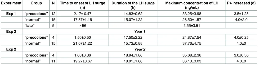
Table 1. Characteristics of the LH surge in the animals selected for study.
Experiment Group N Time to onset of LH surge (h)
Duration of the LH surge (h)
Maximum concentration of LH (ng/mL)
P4 increased (d)
Exp 1 “precocious” 12 2.17±0.47 14.83±0.62 33.25±3.98 3.5±1.25
“normal” 15 17.87±1.16 15.07±1.22 28.50±1.57 4.0±2.0
“late” 5 >56 5.55±3.51
Exp 2 Year 1
“precocious” 4 1.50±0.50 17.50±2.22 24.87±7.54 4.0±0.25
“normal” 15 21.07±1.22 15.73±0.88 37.76±4.75 4.0±0
Exp 2 Year 2
“precocious” 4 1.06±0.36 18.94±1.86 35.68±2.36 3.0±0.50
“normal” 11 19.27±0.67 18.91±1.86 36.13±3.03 4.0±0
doi:10.1371/journal.pone.0158530.t001
Fig 1. Schematic representation of the experimental design showing the time of collection of the samples that were used in the different experiments to measure oestradiol concentrations (Exp 1 and 2), and LH concentration (All experiments).Double time lines indicate that the samples were collected every 15 min.
France ewes. They were subjected to the“ram effect”at the end of anoestrus (July). The experi-ment was repeated the following year with the 28 ewes that were still alive. Two days before the experiment a catheter was introduced in the jugular vein for blood sampling and ewes were randomly distributed into small pens containing 5 ewes per pen. For the ram effect, we intro-duced into each pen, one ram from a group of 10 sexually active Ile de France rams from the UEPAO breeding flock that was used for the whole experiment. To make the stimulation as homogenous as possible in the different years, to minimize individual effects of rams and to maintain their levels of sexual activity they were changed regularly. After the 56h period of serial bleeding the ewes were reunited as a single group with 2 rams that were changed every week for 4 weeks. Ewes and rams were then separated until the next year. Blood samples col-lected between 0 and 56h after the introduction of the rams were analyzed for LH. These data were then used to identify and select ewes with a“precocious”LH surge (n = 4 each year, 2 of which had“precocious”surges in both years) or ewes with a“normal”LH (n = 15 year 1 and n = 11 year 2, 7 were the same on both years) as defined for experiment 1. Characteristics of the LH surges and progesterone increases in these ewes are shown inTable 1. The samples from these ewes were then analyzed for oestradiol as summarized inFig 1: Year 1; 21h and -19h before the introduction of rams and Year 2; -24h, -20h, -16h, -12h and then for both years every 2 hours between -6h and 8h relative to the introduction of rams. As in experiment 1, blood samples were also collected to measure progesterone and detect ovulation [26].
Experiment 3. A third experiment (the“low E2”experiment”) was done to determine if a small increase in the circulating concentrations of E2 lasting three hours could by itself induce a LH surge in anoestrous ewes. Eleven intact anoestrus ewes were implanted subcutaneously for 3 hours with a 3 cm sealed silastic tube containing oestradiol a technique known to produce increases in oestradiol concentrations in plasma in the range of those observed in experiment 2 [40]. This experiment was carried out at the end of anoestrus using ewes from experiment 2 that had had at least one“precocious”LH surge in the previous years and were therefore pre-sumably more sensitive to oestradiol (7 ewes) or that had had only“normal”surges (4 ewes). These ewes were kept without contact with rams for a minimum of 3 months.
Samples were collected at hourly intervals and just before oestradiol implantation (-2, -1 and 0 hours) then every 1.5 hours for the first 6 hours after implantation and finally every 3 hours until 36 hours as illustrated inFig 1to measure LH concentrations. E2 concentrations were measured in samples collected on 5 ewes (2 that had never had a precocious LH surge and 3 that had) 2 hours before and just before implantation (time 0), during implantation (time 1.5 and 3) and after implant withdrawal (time 4.5h). Details of the blood sampling and processing procedures have been published [26].
The role of the noradrenergic system in the male-induced LH Surge
Experiment 4. In a fourth study (the“histological”study), noradrenergic neurons acti-vated during the“ram effect”were identified using a double labelling procedure to detect pres-ence of cFos a known marker of neuronal activation and tyrosine hydroxylase (TH) the rate limiting enzyme for synthesis of noradrenaline.
(a) Sample collection
continuing in isolation from rams (control group, n = 5). The 2 groups were handled by differ-ent staff to avoid any contamination of the control group with male odour. Ninety minutes after the introduction of the ram the ewes were terminated by decapitation by a licensed slaughter man following a protocol agreed by the ethics committee. Blood samples were col-lected every 15 min during the last hour to determine the endocrine response to stimulation. The heads were then immediately perfused through both carotid arteries with 2L of 1% sodium nitrite in phosphate buffer (0.1 M, pH 7.6) and 4L of cold 4% paraformaldehyde (in phosphate buffer). The brains were then removed intact, post-fixed for 24h in 4% paraformaldehyde and then left in phosphate buffer containing 30% sucrose and 0.1% sodium azide. The fixed brains were cut transversely into three approximately equal blocks. Free floating sections of the poste-rior block (40μm) were cut on a freezing microtome (Leica, Paris, France) and stored in cryo-protectant (NaCl 9%, polyvinyl pyrrolidone 10%, saccharose 30%, ethylene glycol 30% in phosphate buffer 0.1 M pH 7.4) at 4°C. Every 10th section was stained with Cresyl violet to allow identification and delineation of noradrenergic brain areas according to Tillet and Thi-bault [41].
(b) Immunohistochemistry
For each animal, the noradrenergic nuclei were identified and sections selected to contain comparable structures among ewes (5 sections per ewe for A1, A2, A6, A7 and 3 for A5 nuclei). The sections were first stained for Fos [42] using an affinity-purified rabbit polyclonal antibody raised against the Fos protein (Ab-2, PC38, Oncogene Research Products, Calbiochem, San Diego, CA, USA, diluted 1/60,000 in PBS-TA-BSA, 2 days, 4°C) and peroxidase-anti-peroxi-dase complex solution (Jackson Immunoresearch, West Grove, PA, USA), diluted in 1/1,000 in PBS-BSA, 4°C and visualized using 3-3’diaminobenzidine tetrahydrochloride (DAB, Sigma Chemical, St Louis, MO, USA) intensified with 0.3% nickel ammonium sulphate. The sections were then rinsed in phosphate-buffered saline (PBS, 10% phosphate buffer 0.1 M, pH 7.4, 0.9% NaCl in distilled water) and stained for TH [43] using a mouse monoclonal antibody raised against TH (Chemicon International) followed by diluted 1/1,000 and a peroxidase-antiperoxi-dase conjugated to sheep anti-mouse antibody (Jackson Immunoresearch) diluted 1/500 and then visualized with 3-3’diaminobenzidine tetrahydrochloride (Sigma Chemical, St Louis, MO, USA).
(c) Quantification of immuno-labelling
Quantification of Fos positive cells (Fos-IR) was performed using an image analysis system equipped with software to analyze cell-count data (Mercator, Explora Nova, and La Rochelle, France). To count Fos positive cells a microscope with a motorized stage and a video camera was connected to a computer with a color monitor. Using the program we established parame-ters of size, shape and threshold for grey scale to characterize Fos-IR positive cells [42]. The average background grey scale was automatically estimated for every section and subtracted from the original image before the software identified cells meeting the established parameters as Fos-IR positive cells. Each section was also examined visually and any suspect objects (e.g. dust) were erased manually. For counting TH-IR and Fos-IR/TH-IR positive cells, a manual system (Biocom, Paris, France) was used. Images were viewed on a monitor by an observer blind to the treatment used and who identified each immunoreactive cell on the screen. Because A6 and A7 overlap they were counted together as LC-A7.
short-term LH response evoked by exposure to a sexually active ram with the local administra-tion of theα1 adrenergic antagonist Prazosin (experiment 5B).
Two to 4 weeks before the start of the experiment, 15 mature Ile-de-France ewes were fitted bilaterally, with guide cannulae directed at the posterior preoptic area using a technique which combined a stereotaxic method with lateral and frontal radiography [44]. The procedures were carried out under general anesthesia induced by the intravenous injection of thiopental (1g per ewe; Nesdonal, Specia Rhone Poulenc, Paris, France) and atropine sulfate (20mg per ewe; Lavoisier, Paris, France) and maintained by closed-circuit halothane (Bélamont, Neuilly, France). Full aseptic precautions were taken throughout. After surgery, ewes were injected with 5ml of Dexamethasone per ewe (Diurizone, Vetoquinol, Lure, France) daily for 3 days. The ewes were allowed a minimum of 2 weeks recovery time and during their recovery the ewes were habituated to handling and the presence of humans.
The day before the experiment, noradrenaline (Research Biochemical international N-112 Natick USA) was dissolved in 1 mL HClO40.25M. On the day of the experiment noradrena-line, and crystalline Prazosin (Sigma P7791 Saint Quentin Fallavier France) were dissolved in ringer’s solution (pH6.5) to a final concentration of 100ng/mL for noradrenaline and 100μg/ mL for Prazosin. The dose of noradrenaline was chosen to produce local concentrations in the range of those measured in a previous experiment [39] and the dose of Prazosin was chosen from preliminary trials. Noradrenalin, Prazosin or their solvent were infused by retrodialysis at a rate of 2μl/min using microdialysis probes (Mab 6 Microbiotech Sweden, 5mm membrane) inserted into the guide cannulae. Infusion started half an hour before exposure to a handful of ram fleece that will increase LH increase in some but not all ewes (experiment 5A), or a sexu-ally active ram that will increase LH in all ewes (experiment 5B). The fleece used was a mixture of fleeces from 10 rams (Ile de France and Romanov) different from those used in the experi-ment and that had been collected during the breeding season and stored at -20°C. The rams used in this experiment were from a group of sexually active rams from the station breeding flock and were changed regularly among pens during the 3 hours to minimize individual effects of the rams and maintain their level of sexual activity. A delay of half an hour was allowed for the test reagents to be transferred from the syringe to the dialysis membrane at the brain site. Experiment 5A took place in March and April and experiment 5B in July. In each experiment ewes acted as their own controls and the order of infusion of the treatment solution and the control solution was random at least one week apart. Any ewe that showed evidence of luteal activity (a progesterone concentration above 0.5 ng/mL) was not used. Plasma samples were collected every 15 min for 3 hours before and 3 hours after treatment (Fig 1).
The concentrations of progesterone in plasma were monitored weekly during the experi-ment and infusions were carried out only on ewes confirmed as anoestrus that is with proges-terone concentrations<1ng/mL for a minimum of 2 weeks. In both experiments (5A & 5B) 11 anoestrous ewes were available, 7 ewes were used in both experiments and 4 ewes were used in only 1 experiment.
At the end of experiment all the females were killed. Free-floating frontal sections (40μm thick) were cut and stained with cresyl violet to facilitate histological identification of probes location as described for experiment 4.
Assay of Oestradiol
intra-assay and inter assay coefficients of variation were: 16.6% and 15.3% (at 0.46 pg/mL), 11.9% and 11.7% (at 0.80 pg/mL) and 4.6% and 4.7% (at 5.13 pg/mL).
Assay of Progesterone
The concentration of progesterone was measure using an ELISA [26]. The sensitivity of the assay was 0.2 ng/mL and the intra-assay and inter-assay coefficient of variation were 6.8% and 8.1% for a reference at 1.5 ng/mL and 6.6% and 10.3% for a reference at 2.5ng/mL.
Assay of LH
The concentrations of LH in jugular venous plasma were determined using a radioimmunoas-say [45]. The assay sensitivity was 0.16 ng/mL standard 1051-CY-LH (equivalent to 0.31 ng/ mL NIH LH-S1). The intra-assay and inter assay coefficients of variation were: 4.4% and 10.3% respectively.
The onset of the LH surge was defined as the time of the first observed concentration of LH that was more than 3 standard deviations above the baseline and preceding an increase in LH of at least 4 hours and with at least one value above 10ng/mL [9,46]. LH pulses were identified as an increase in LH concentrations>3SD above the baseline defined as the mean concentra-tion before ram introducconcentra-tion. An animal was classified as having a short-term LH response if the number of LH pulses during the 3 hours after stimulation was superior to the number of pulses during the 3 hours before stimulation. The amplitude of a LH pulse was calculated as the difference between the maximum concentration of LH in the pulse and the concentration of LH in the sample before the start of the pulse.
Statistical analyses
Statistical tests were carried out using Statistica version 10 (Statsoft Inc.). The data involving oestradiol measured over time were analyzed using a mixed model ANOVA run under the gen-eral linear model with time as a repeated measure. Data from each year in experiment 2 were analyzed separately. Paired comparisons within treatments were carried when appropriate, using the Bonferroni correction. The data involving LH measured over time were also analyzed using an ANOVA with repeated measures.
The mean density of immunoreactive cells was calculated for each region of each animal and the overall median and inter-quartile values calculated for the two groups. Because the mean densities were not normally distributed, statistical comparisons were carried out using nonparametric tests: The Kruskal and Wallis test followed by the Mann Whitney U test to compare groups or the Friedman tests to compare the density among nuclei in each group.
A ewe was considered as responding if the frequency of LH pulse was at least 1 pulse per 3 hours greater after stimulation than before. The proportions of ewes showing increased pulsa-tile LH activity were compared byχ² tests. The LH pulse frequency and the amplitudes of LH pulses before and after stimulation and during the treated versus control session were com-pared using Wilcoxon test.
Differences were taken as statistically significant at p<0.05 and as a trend at p>0.05 but
<0.10.
Results
The
“
precocious
”
surge study
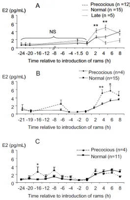
concentrations and the latency of this increase did not differ (Table 1). However, the concen-trations of oestradiol in ewes with "precocious", "normal" or "late" LH surges did differ (Fig 2A). There was a significant effect of time (p<0.0001) and of the type of response (p = 0.029), but no effect of breed (p = 0.890) so the data of the 2 breeds were pooled. There was a trend towards a significant interaction between the type of response and time (p = 0.064). Paired comparisons showed that the concentrations of oestradiol in the samples taken at 2h, 4h and 8h after the introduction of the rams differed significantly from of those taken before the intro-duction of rams (all comparisons p<0.001). Before the introduction of rams, there were no sig-nificant differences in the mean concentration of oestradiol among the types of response ("precocious”, 1.15 ± 0.15 pg/mL; "normal", 0.82 ± 0.08 pg/mL and "late" 1.06 ± 0.31 pg/mL).
Fig 2. Changes in oestradiol concentrations before and after ram introduction in anoestrous ewes who presented LH surges at different time after ram introduction."precocious": ewes who presented a LH surge within 4 hours after male introduction; "normal": ewes presented a LH surge between 16 and 28 hours after the introduction of rams; "late": ewes who did not present a surge before 56h. A exp. 1, B exp. 2 year 1, C exp. 2 year 2. Data are expressed as Mean±SEM.*p<0.03,**p<0.01 compared to“normal”at the same time.
Following the introduction of rams the concentrations of oestradiol at 2h and 4h, in the "preco-cious" group (2h: 4.26 ± 0.27 pg/mL and 4h: 5.03 ± 0.65 pg/mL) were significantly greater than those in "normal" (2h: 2.60 ± 0.27 pg/mL, p<0.005 and 4h: 2.65 ± 0.32 pg/mL, p<0.001) and at 4 h than the "late" responders (1.17 ± 0.36 pg/mL, p<0.002).
The
“
follow-up
”
study
The concentrations of oestradiol in ewes with "precocious" LH surges compared to ewes with
“normal”LH surges are illustrated inFig 2B and 2C. In both years, the concentrations of oes-tradiol at the time of ram introduction (time 0) were not significantly different in ewes with "precocious" and“normal”LH surges and increased after the introduction of the ram.
The global analysis showed a significant effect of time for each year (p<0.0001) and an interaction between time and type of response (year 1 p = 0.002, year 2 p = 0.0006) but no sig-nificant effect of type of response (p = 0.21 and 0.22 for years 1 and 2).
When the periods before and after the“ram effect”were analyzed separately, a significant effect of the type of response was observed. During year 1 type of response had no significant (P = 0.28) effect before the "ram effect" (Mean oestradiol concentrations = 1.01±0.28 and 0.75 ±0.11pg/mL for“normal”and“precocious”). But type of response had a significant effect after the "ram effect" (p<0.047). Paired comparisons showed significant differences at 4 and 6 hours after the ram effect between the“precocious”and“normal”groups (Fig 2B.; +4h: 5.1±0.87 ver-sus 2.43±0.32 pg/mL, p<0.006 and +6h: 5.66±1.25 versus 3.24±0.39 pg/mL p<0.027).
In the second year a significant effect of the type of response was observed before the“ram effect”(p<0.03) and paired comparison showed that oestradiol concentration at -16h before the“ram effect”was significantly greater in the“precocious”group (Fig 2C; 2.61±1.27 versus 0.82±0.16 pg/mL; p<0.03) and approached significance at -8h (3.02±0.81 versus 1.30±0.35 pg/ mL; p = 0.056). The mean concentrations of oestradiol after ram introduction were higher than before but, did not vary between type of response (“precocious”: 2.20±0.10,“normal”: 2.49±0.37 pg/mL).
The
“
small E2
”
study
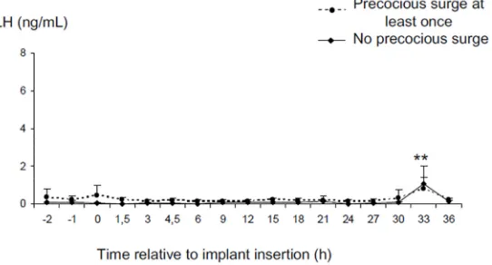
Data from one ewe was discarded because the oestradiol implant could not be retrieved. Inser-tion of the oestradiol implant for 3 hours increased the plasma concentraInser-tions of oestradiol from the detection limit of the assay (0.51±0.3 and 0.65±0.25 respectively at time -2 and 0) to 1.0± 0.24, 2.29±0.96 and 5.91±4.53 pg/mL respectively 1.5h 3h and 4.5h after implantation. These implants did not stimulate a LH surge in any ewe (Fig 3). However, LH concentrations changed with time (p<0.001). The mean E2 concentration fell from 0.25± 0.06 ng/mL before implantation to 0.13±0.03 ng/mL while the implants were in place (p>0.1) and to 0.11±0.03 ng/mL in the following 3 hours (p>0.1) and then increased at 33h after insertion of the implant (0.88± 0.35 ng/mL p<0.01 compared to the concentrations before during and just after E2 implantation). There was no significant difference between groups with a history of“ preco-cious”LH surges compared to those with no history of“precocious”LH surges. By contrast the ewe with an implant present for the whole experiment, had a LH surge that started 18 hours after E2, lasted for 15 hours and had a maximum LH concentration of 13.6 ng/mL (Fig 3).
The
“
histological
”
study
day before (Ram: 1.15±1.6 ng/mL vs 0.55±0.03, p<0.01; control: 0.40±0.09 vs 0.37± 0.09 ng/ mL, p = 0.64).
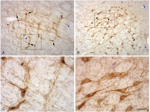
The TH immunoreactive cells were characterized by a brown precipitate in the cytoplasm and could be identified in all the locations where noradrenergic neurons have been described in sheep [41].The fos immunoreactive cells were characterized by a dense black precipitate in their nuclei that appeared as a round black structure easily detected by our image analysis sys-tem and very similar to the labelling we had previously observed in hypothalamic and olfactory structures [42]. Double labelled cells could be identified by the presence of a dark nucleus in a brown cytoplasm (Fig 4).
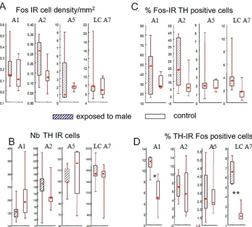
The density of Fos immunoreactive cells was significantly different between monoaminergic nuclei (p<0.001 in both groups) and was higher in LC-A7 complex than in the other nuclei (p<0.005) but did not differ in the control or treated ewes (Fig 5A). Similarly, the number of TH immunoreactive cells did not differ between females exposed to ram and to the control sit-uation (Fig 5B). The proportion of Fos immunoreactive cells that were also TH immunoreac-tive did not differ between groups of females although in the LC A7 nucleus it tended to be higher in ewes exposed to rams (p = 0.060,Fig 5C). By contrast the proportion of TH immuno-reactive cells that were also immunoimmuno-reactive for Fos differed between groups and the propor-tion of double-labelled cells in A1ventrolateral medulla nucleus (p<0.02) and the locus coeruleus complex (p<0.01) was higher in ewes exposed to a ram than in the controls (Fig 5D).
The
“
pharmacological
”
study
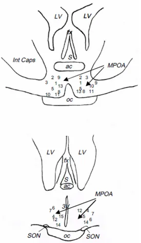
As shown inFig 6, the ends of the guide cannulae were all located in the posterior preoptic area (Anteroposterior coordinates = 32 from the Richard’s atlas [47]).
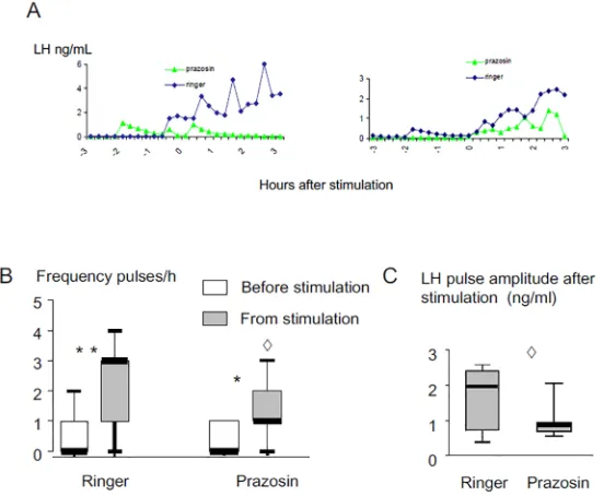
Examples of LH profiles in response to ram fleece in ewes infused with noradrenaline or ringers are shownFig 7A. The infusion of noradrenaline into this region increased the propor-tion of ewes responding to ram odor (9/11 versus 5/11; p = 0.03). The frequency of LH pulses before stimulation was not different between ewes infused with noradrenaline or ringers (p>0.1). But after exposure to ram odor the frequency of LH pulse was higher than before in ewes infused with noradrenaline (Fig 7B; p<0.003) and higher than after in ewes infused with ringers (p<0.02). By contrast exposure to ram odor for 3 hours had no significant effect in con-trol ewes. Noradrenaline treatment did not affect the amplitude of LH pulses after exposure to ram odor (p = 0.12;Fig 7C).
Fig 3. Changes in LH concentrations before and after insertion of a 3 cm silastic oestradiol implant for 3 hours. (small E2 study).Data are expressed as Mean±SEM**p<0.01 compared to other times.
Examples of LH profiles in response to exposure to a ram in ewes infused with prazosin or ringer are shownFig 8A. Prazosin did not affect the proportion of ewes responding to the“ram effect”(7/11 versus 8/11) and introduction of a ram increased LH pulse frequency in both ring-ers (p<0.003) and Prazosin-treated sessions (p<0.02;Fig 8B). However, the frequency
(p<0.03;Fig 8B) and the amplitude (p<0.04;Fig 8C) of LH pulses following the“ram effect” were significantly lower after Prazosin treatment compared to the ringers sessions.
Discussion
Mammals are traditionally considered as either spontaneous or induced ovulators. Sheep are spontaneous ovulators and the LH surge in cyclic ewes is driven by a large and sustained pre-ovulatory rise of oestradiol [3,5,48]. During anoestrus there are no spontaneous LH surges. However, anoestrous sheep can be induced to ovulate by the sudden exposure to a ram. In the present study we show that some ewes have LH surges occurring within 4 h of the introduction
Fig 4. Photographs illustrating neurons expressing Fos and TH proteins in the A1 (A, C) and A7 (B, D) nucleus of a female exposed to a ram. C and D are details of the area shown in the rectangle.Small blue arrows: Fos alone; black arrows: Fos protein in a TH neuron; arrow heads: TH without Fos protein. Scale bar = 160μm for A and B and 40μm for C and D.
of rams ("precocious" LH surge) that are not preceded by large increased concentrations of oes-tradiol. We also show that in anoestrous ewes, exposure to rams activated the noradrenaline neuronal populations of the locus coeruleus complex and the A1 nucleus and that we can mod-ify the LH response to male socio-sexual cues by locally manipulating the noradrenergic system in the preoptic area. Together these results suggest that in anoestrous ewes two partly different neural circuits are involved in the ram-induced LH surge, the neural circuitry involved in the mechanism of the ram-induced“precocious”LH surge in anoestrous ewes being different from that for the LH surge induced by the classic oestradiol positive feedback mechanism. They also suggest that the noradrenergic system is involved in the LH response to male socio-sexual cues and extrapolating further, that“precocious”LH surges are a result of higher activation of the noradrenergic system possibly using a neuronal circuit partly similar to that in the mating-induced LH surge of mating-induced ovulators.
In anoestrous ewes LH pulsatility is very low, ovaries secrete little oestradiol and no proges-terone because ewes do no ovulate and do not have corpora lutea. However introduction of a sexually active ram will induce an immediate increase in the pulsatile secretion of LH in close to 100% of ewes, and in a lower and variable proportion of ewes a LH surge [26]. In most ewes this LH surge occurs 12 to 56 hours after the introduction of a ram [26–28,30] and is preceded by a sustained increase in the plasma concentration of oestradiol lasting between 8 and 56 hours with concentrations above 2.5 pg/mL 4 hours before the surge [29]. This is similar to what happens in cyclic ewes during the breeding season; when the LH surge is preceded by an increase in the plasma concentration of oestradiol that normally lasts more than 12 hours [3,
4]. The present study showed that this was not the case in ewes with a“precocious”LH surge
Fig 5. Box Plot representations of the density of Fos-IR neurons (A), the number of TH-IR cells counted (B), the proportion of Fos-IR cells also containing TH-IR (C) and proportion of TH-IR cells also containing Fos-IR (D) in the different noradrenergic nuclei (A1, A2, A5, LC-A7) in adult Ile de France ewes exposed to a ram (hatched boxes) or to a control situation (empty boxes).The bottom and top of the boxes are the first and third quartiles, the red square inside the boxes is the median and the end of the whiskers are the minimum and maximum of all the data;*p<0.02,**p<0.01 compared to the control group. Because of the differences in the number, density and proportions among the different nuclei the scales are different for the different nuclei.
although the durations and peak concentrations of these“precocious”LH surges are not differ-ent from“normal”LH surges and they were followed by similar increases in progesterone con-centration indicating that the ewes ovulated. In our study these“precocious”LH surges were not preceded by a large increase in oestradiol concentrations and oestradiol concentrations at the time of ram introduction when the“precocious”surges were starting were still low. So these ewes probably were not on the verge of a spontaneous ovulation at the time of exposure
Fig 6. Schematic representation of the localization of the tip of dialysis probes used in the
“pharmacological study”.Numbers correspond to the different animals. ac: anterior commissure, fx: fornix, Int Caps: Internal Capsule, LV: lateral ventricle, MPOA: medial preoptic area, oc: optic chiasma, S: septum, SON: supraoptique nucleus, 3V: 3rdventricle.
to rams. In all ewes, the concentration of oestradiol increased within 2 hours of the“ram effect”. This increase is undoubtedly the result of increased pulsatile secretion of LH, each pulse of LH stimulating the release of a pulse of oestradiol by the ovary [49]. In our study the immediate increase in oestradiol concentration was greater in ewes with“precocious”LH surge compared to those with a“normal”surge in exp. 1 and exp. 2A but surprisingly not in exp 2B.
Fig 7. Effect of infusion with noradrenaline (100 ng/mL) on LH secretion induced in anoestrous Ile de France ewes by exposure to ram fleece.A: Representative LH profiles in 2 ewes; B: Box Plot representations of the effect on pulse frequency, C: Box Plot representations of the effect on pulse amplitude. The thick horizontal lines depict the median.**Different from noradrenaline before the stimulation p<0.003,^different from ringers after stimulation p<0.02.
doi:10.1371/journal.pone.0158530.g007
Fig 8. Effect of infusion with Prazosin (100μg/mL) on LH secretion induced in anoestrous Ile de France ewes by exposure to a ram.A: Representative LH profiles in 2 ewes; B: Box Plot representations of the effect on pulse frequency, C: Box Plot representations of the effect on pulse amplitude.*Different from before the stimulation p<0.02,**different from before the stimulation p<0.003,^Prazosin different from ringer p<0.04.
The fact that we did not see this difference in all 3 experiments suggests that this is not a pre-requisite for a“precocious”LH surge.
Another possibility is that, in these“precocious”ewes, LH surges were induced by small and short increases in oestradiol concentrations that we could not detect. Indeed, in the second year of experiment 2 when samples were collected over a longer interval some ewes had epi-sodic increases in oestradiol secretion resulting in significantly higher concentration of oestra-diol 16 hours before ram introduction compared to ewes with a normal LH surge. Studies on ovariectomized ewes treated with oestradiol implants during the breeding season have shown that LH surges can be induced by a much shorter period of elevated oestradiol than normally occurs in cycling ewes. A LH surge was observed in 1/12 Suffolk ewes exposed to oestradiol for 7 hours [50] and in 3/10 Ile de France ewes exposed to oestradiol for 3 hours [51]. However in these studies the reported concentrations of oestradiol were higher than in our study (8pg/ mLversus 2.6 and 3pg/mL, 16h and 8h before the“ram effect”). In our third experiment we showed that an increase in oestradiol concentration in the range of the increase observed in experiments 1 & 2 was not enough, by itself, to induce a LH surge although it did slightly mod-ify LH secretion. So it is very unlikely that the small increases in oestradiol observed in our study were alone, sufficient to induce“precocious”LH surges using the same mechanism as for the "normal" surges. It is theoretically possible that long exposure to undetectable level of E2 may be sufficient to produce precocious surge but in all of our data we found no evidence that this is the case. We suggest that in our study the LH surge was induced by a combination of oestradiol priming and male stimulation in a similar way as occurs in induced ovulators.
In induced ovulators an increase in oestradiol induces receptive behavior and allows mating and vaginal stimulation produced by mating, to induce a LH surge [18]. The noradrenergic sys-tem has a central role in this male induced LH surge [14,18]. In rabbits and ferrets mating sti-muli activate noradrenergic neurons in the locus coeruleus and the brainstem [21,22]. This leads to a rapid increase in the extracellular concentrations of noradrenaline in the mediobasal hypothalamus just prior to the increase in GnRH [23] and this increase can be reduced byα1 antagonists [24].
Anoestrous ewes are not sexually receptive and refuse all male courtship behavior so there can be no vaginal stimulation. But in a previous study we have shown that the extracellular concentrations of noradrenaline increased in the posterior preoptic area of anoestrous ewes exposed to a ram and to a lesser extent, to ram odor [39]. Here we show that as in induced ovu-lators, noradrenergic neurons from the locus coeruleus complex and from the brainstem were activated when anoestrous ewes were exposed to a sexual stimulus but without vaginal stimula-tion. In sheep as in other species, noradrenergic neurons project fibers to the preoptic area [52,
53]. So these neurons are the probable origin of the increase in noradrenaline concentration in the preoptic area caused by exposure to a ram. Interestingly the A1 nucleus that was activated in our study is also activated in ovariectomized ewes after treatment with oestradiol at a dose that will induce a LH surge [54] suggesting that the neural networks involved in spontaneous and ram-induced LH surges overlap to some extent. The relatively low proportion of TH cells that were Fos positive probably reflects the functional heterogeneity of these cell groups and the fact that noradrenaline is in involved in many aspects of brain function.
In our study we were able to increase the effectiveness of male odor by the local administra-tion of noradrenaline into the preoptic area of anoestrous ewes or to reduce the pulse frequency and amplitude of the short-term LH response to the“ram effect”with aα1antagonist. These results suggest that in this context, noradrenaline has a facilitatory action on LH secretion in response to male socio-sexual cues. The effect of noradrenaline however was not total and we did not reduce the proportion of ewes showing a response to the ram. Noradrenergic receptors are present in high density in the preoptic area [57,58]. The most likely explanation is that our treatment did not block all the noradrenergic receptors and that enough noradrenergic recep-tors were active to evoke a slight increase in LH pulse frequency in response to the ram.
Because of the sampling duration relative to ram or fleece exposure, the stimulatory effects of noradrenaline we observed are most likely on LH pulsatility. Although noradrenaline has been implicated in the control of LH secretion for several decades [59] its role in the pulsatile activity of GnRH neurons is complex and not completely understood [60]. In rodents some studies have shown that the administration of noradrenaline or of aα1 adrenergic agonist inhibits LH pulsatility [61] but the same effects have been observed by others after administra-tion of aα1 antagonist [62]. The same results have been observed in sheep where the effects appear to vary with the steroid milieu. [63–64]. This has led authors to suggest that the role of noradrenaline is permissive and depends on the relative contribution of other neurotransmit-ters and neuropeptides [60]. However, LH pulsatility depends on a“pulse generator”situated in the mediobasal hypothalamus [65] that is 3mm from our infusion site, a distance that is too far away to be to be directly affected by our infusions. The LH surge depends at least partly on the preoptic area and can be stimulated by noradrenergic inputs [54,60,66,67] so it could have been affected by our treatment. In any case, it is difficult in ewes with“precocious LH surges”to dissociate increases in LH pulsatility and onset of the LH surge as can be done in the cyclic ewe at the end of the follicular phase [46,68]. The mechanism involved in the switch between the pulsatile and surge mode of secretion is still not clear [60]. It could depend on dif-ferent populations of GnRH neurons but this has never been clearly demonstrated or on the relative contribution of other neurotransmitters and neuropeptides [60,69] that are modulated by many different factors: steroids, nutrition, stress, photoperiod and the circadian clock to name but a few This would allow very broad control of reproduction by environmental factors. We suggest that the“ram effect”stimulates a pathway that allows the mating partner to directly influence the secretion of GnRH and that this involves noradrenaline and has similarities with the pathways involved in the secretion of GnRH for spontaneous ovulation. In most ewes as in other“spontaneous”ovulators, the influence of this pathway is modulatory because the LH surge is driven by increasing concentration of ovarian oestradiol [70]. But in some ewes, because of a higher noradrenergic tone or perhaps a higher sensitivity to noradrenaline, the ram-induced pathway is prematurely activated in ewes with“precocious”LH surges.
the time of the LH surge as has been reported for the sow [76], cow [77] and ewe [78] and even if the females have been ovariectomized and treated with exogenous oestradiol [38]. Some authors have suggested that induced ovulation is the ancestral mode of preovulatory LH secre-tion [18,34]. The presence of the neural circuits underlying the male-induced LH surge in aged rats or in some anoestrus ewes could be the evolutionary traces of an ancestral mode of LH secretion. Our speculation is that because the network for oestradiol positive feedback is domi-nant, the involvement of an ancestral circuit is restricted to conditions where the network of oestradiol positive feedback is not functional for example, in the aged rat, the hypogonadal mouse, and ovariectomized ewes [38] or as in our experiment, in anoestrous ewes with a high degree of responsiveness to a sexual partner.
Noradrenaline is involved in both the neural circuits for spontaneous and induced ovula-tion. The questions now are why one circuit is activated in preference to the other and how are these two circuits inter-connected.
Acknowledgments
We wish to thank the staff from INRA experimental stations in Nouzilly (UEPAO), Bourges (Domaine de la Sapinière) and Frejorgues for the care of the animals and for their help with the experiments. We wish also to thank several MSc students, S Bertaux, C Buron, M David, A Dherbe, A Durand, J Gauthier, C Journault, A Pelletier and O Robineau, who participated in various aspects of these experiments and L Dufourny who helped withFig 4.
Author Contributions
Conceived and designed the experiments: CFN RJS. Performed the experiments: CFN LL DL SM. Analyzed the data: CFN RJS. Contributed reagents/materials/analysis tools: AC JD. Wrote the paper: CFN JD AC RS.
References
1. Corker CS, Naftolin F, Exley D. Interrelationship between plasma luteinizing hormone and oestradiol in the human menstrual cycle. Nature 1969; 222:1063. PMID:5815071
2. Smith MS, Freeman ME, Neill JD. The control of progesterone secretion during the estrous cycle and early pseudopregnancy in the rat: prolactin, gonadotropin and steroid levels associated with rescue of the corpus luteum of pseudopregnancy. Endocrinology 1975; 96: 219–226. PMID:1167352 3. Hauger RL, Karsch FJ, Foster DL. A new concept for control of the estrous cycle of the ewe based on
the temporal relationships between luteinizing hormone, estradiol and progesterone in peripheral serum and evidence that progesterone inhibits tonic LH secretion. Endocrinology. 1977; 101:807–817. PMID:560959
4. Karsch FJ, Weick RF, Butler WR, Dierschke DJ, Krey LC, Weiss G et al. Induced LH surges in the rhe-sus monkey: strength-duration characteristics of the estrogen stimulus. Endocrinology. 1973; 92: 1740–1747. PMID:4196431
5. Karsch FJ, Foster DL, Legan SJ, Ryan KD, Peter GK. Control of the preovulatory endocrine events in the ewe: interrelationship of estradiol, progesterone, and luteinizing hormone. Endocrinology. 1979; 105: 421–426. PMID:572293
6. Scaramuzzi RJ, Tillson SA, Thorneycroft IH, Caldwell BV. Action of exogenous progesterone and estro-gen on behavioral estrus and luteinizing hormone levels in the ovariectomized ewe. Endocrinology. 1971; 88: 1184–1189. PMID:5102119
7. Banks JA, Mick C, Freeman ME. A possible cause for the differing responses of the luteinizing hormone surge mechanism of ovariectomized rats to short term exposure to estradiol. Endocrinology. 1980; 106: 1677–1681. PMID:7189459
9. Caraty A, Locatelli A, Martin GB. Biphasic response in the secretion of gonadotrophin-releasing hor-mone in ovariectomized ewes injected with oestradiol. J Endocrinol. 1989; 123: 375–382. PMID: 2691622
10. Xia L, Van Vugt D, Alston EJ, Luckhaus J, Ferin M. A surge of gonadotropin-releasing hormone accom-panies the estradiol-induced gonadotropin surge in the rhesus monkey. Endocrinology. 1992; 131: 2812–2820. PMID:1446619
11. Wu CH, Blasco L, Flickinger GL, Mikhail G. Ovarian function in the preovulatory rabbit. Biol. Reprod. 1977; 17: 304–308. PMID:889998
12. Wildt DE, Chan SY, Seager SW, Chakraborty PK. Ovarian activity, circulating hormones, and sexual behavior in the cat. I. Relationships during the coitus-induced luteal phase and the estrous period with-out mating. Biol. Reprod. 1981; 25: 15–28. PMID:7197173
13. Elias E, Bedrak E, Yagil R. Estradiol concentration in the serum of the one-humped camel (Camelus dromedarius) during the various reproductive stages. Gen. Comp. Endocrinol. 1984; 56: 258–264. PMID:6510688
14. Ramirez VD, Soufi Wl. The neuroendocrine control of the rabbit ovarian cycle. In: Knobil E, Neill JD edi-tors. The Physiology of Reproduction. 2nd ed., Vol. 2. New York: Raven Press; 1994. pp. 585–611. 15. Dufy-Barbe L, Dufy B, Vincent JD. Serum gonadotropin levels in the ovariectomized rabbit: effect of
acute and chronic administration of estradiol. Biol Reprod. 1978; 18: 118–124. PMID:626762 16. Milligan SR. The feedback of exogenous steroids on LH release and ovulation in the intact female vole
(Microtus agrestis). J Reprod Fertil. 1978; 54: 309–311. PMID:364048
17. Baum MJ, Carroll RS, Cherrv JA, Tobet SA. Steroidal control of behavioural, neuroendocrine and brain sexual differentiation: studies in a carnivore, the ferret J Neuroendocrinol. 1990; 2: 401–418. doi:10.
1111/j.1365-2826.1990.tb00425.xPMID:19215366
18. Bakker J, Baum MJ. Neuroendocrine regulation of GnRH release in induced ovulators. Front Neuroen-docrinol. 2000; 21: 220–262. PMID:10882541
19. Adams CE. The reproductive status of female mink,Mustela vision, recorded as 'failed to mate'. J Reprod Fertil. 1973; 33: 527–529. PMID:4736560
20. Adams CE. Observations on the induction of ovulation and expulsion of uterine eggs in the mink,
Mus-tela vison. J Reprod Fertil. 1981; 63: 241–248. PMID:7277325
21. Wersinger SR, Baum MJ. Sexually dimorphic activation of midbrain tyrosine hydroxylase neurons after mating or exposure to chemosensory cues in the ferret. Biol Reprod. 1997; 56:1407–1414. PMID: 9166692
22. Yang SP, Pau KY, Spies HG. Tyrosine hydroxylase and norepinephrine transporter mRNA levels increase in locus coeruleus after coitus in rabbits. J Mol Endocrinol. 1997; 19: 311–319. PMID: 9460652
23. Yang SP, Pau KYF, Hess DL, Spies HG. Sexual dimorphism in secretion of hypothalamic gonadotro-pin-releasing hormone and norepinephrine after coitus in rabbits. Endocrinology. 1996; 137: 2683– 2693. PMID:8770887
24. Yang SP, Pau KY, Airhart N, Spies HG. Attenuation of gonadotropin-releasing hormone reflex to coitus by alpha1-adrenergic receptor blockade in the rabbit. Proc. Soc. Exp. Biol. Med. 1998; 218: 204–209. PMID:9648937
25. Goodman AL, Neill JD. Ovarian regulation of postcoital gonadotropin release in the rabbit: reexamina-tion of a funcreexamina-tional role for 20 alphadihydroprogesterone. Endocrinology. 1976; 99: 852–860. PMID: 986294
26. Chanvallon A, Sagot L, Pottier E, Debus N, François D, Fassier T et al. New insights into the influence of breed and time of the year on the response of ewes to the 'ram effect'. Animal 2011; 5: 1594–1604.
doi:10.1017/S1751731111000668PMID:22440351
27. Martin GB, Oldham CM, Cognié Y and Pearce DL. The physiological responses of anovulatory ewes to the introduction of rams–a review. Livestock Production Science. 1986; 15: 219–247.
28. Ungerfeld R. Socio-sexual signalling and gonadal function: opportunities for reproductive management in domestic ruminants. Soc Reprod Fertil Suppl. 2007; 64: 207–221. PMID:17491149
29. Fabre-Nys C, Chanvallon A, Debus N, François D, Bouvier F, Dupont J et al. Plasma and ovarian oes-tradiol and the variability in the LH surge induced in ewes by the ram effect. Reproduction. 2015; 149: 511–521. doi:10.1530/REP-14-0587PMID:25823459
31. Pearce DT, Martin GB, Oldham CM. Corpora lutea with a short life-span induced by rams in seasonally anovulatory ewes are prevented by progesterone delaying the preovulatory surge of LH. J Reprod Fert. 1985; 75: 79–84.
32. Zarrow MX, Clark JH. Ovulation following vaginal stimulation in a spontaneous ovulator and its implica-tions. J Endocrinol. 1968; 40: 343–352. PMID:5640407
33. Jöchle W. Current research in coitus-induced ovulation: a review. J Reprod Fertil Suppl. 1975; 22: 165–207. PMID:810583
34. Conaway CH. Ecological adaptation and mammalian reproduction. Biol Reprod. 1971; 4: 239–247. PMID:5000279
35. Dyrmundsson OR, Lees JL. Effect of rams on the onset breeding activity in the clun forest ewe lamb. J. agric. Sci. Camb. 1972; 79: 269–271.
36. Mauleon P, Dauzier L. Variations de durée de l'anoestrus de lactation chez les brebis de race Ile-de-France. Ann. Biol. Anim. Bioch. Biophys. 1965; 5: 131–143
37. Signoret JP. Effet de la présence du male sur les mécanismes de reproduction chez la femelle des mammifères. Reprod. Nutr. Dev. 1980; 20: 457–468 PMID:6891479
38. Signoret JP. Influence of the presence of rams on the luteinizing hormone surge after oestradiol benzo-ate injection in ovariectomized ewes. J Endocr. 1975; 64: 589–590. PMID:1169283
39. Fabre-Nys C, Kendrick KM. Noradrenaline concentrations in the hypothalamus of anoestrus ewes fol-lowing the ram-induced luteinizing hormone release NeuroReport. 2015; 26: 438–443. doi:10.1097/
WNR.0000000000000367PMID:25839177
40. Goodman RL, Legan SJ, Ryan KD, Foster DL, Karsch FJ. Two effects of estradiol that normally contrib-ute to the control of tonic LH secretion in the ewe. Biol Reprod. 1980; 23: 415–422. PMID:7191344 41. Tillet Y, Thibault J. Catecholamine-containing neurons in the sheep brainstem and diencephalon,
immunohistochemical study with tyrosine hydroxylase (TH) and dopamine-beta-hydroxylase (DBH) antibodies. J. Comp. Neurol. 1989; 290: 69–104. PMID:2574197
42. Gelez H, Fabre-Nys C. Neural pathways involved in the endocrine response of anestrous ewes to the male or its odor. Neuroscience. 2006; 140, 791–800. PMID:16650943
43. Chanvallon A, Fabre-Nys C. In sexually naive anestrous ewes, male odour is unable to induce a com-plete activation of olfactory systems. Behav. Brain Res. 2009; 205: 272–279. doi:10.1016/j.bbr.2009.
08.013PMID:19695291
44. Fabre-Nys C, Blache D, Lavenet C. A method for accurate implantation in the sheep brain. In Green-stein B editor. Neuroendocrine Research Methods, Implantation and Transfection Procedures. Har-wood, Chur; 1991. pp. 295–314
45. Montgomery GW, Martin GB, Pelletier J. Changes in pulsatile LH secretion after ovariectomy in Ile-de-France ewes in two seasons. J Reprod Fertil. 1985; 73: 173–183. PMID:4038516
46. Moenter SM, Caraty A, Karsch FJ. The estradiol-induced surge of gonadotropin-releasing hormone in the ewe. Endocrinology. 1990; 127: 1375–1384. PMID:2201536
47. Richard P. Atlas Stéréotaxique du Cerveau de Brebis. Paris: INRA. 1967.
48. Goodman RL. Neuroendocrine control of the ovine estrous cycle. In: Knobil E, Neill JD editors. The Physiology of Reproduction, 2nd Edition, Vol. 2. New York: Raven Press; 1994. pp. 659–709. 49. Baird DT, Swanston I, Scaramuzzi RJ (1976) Pulsatile release of LH and secretion of ovarian steroids
in sheep during the luteal phase of the estrous cycle. Endocrinology 98: 1490–1496. PMID:945154 50. Evans NP, Dahl GE, Padmanabhan V, Thrun LA, Karsch FJ. Estradiol requirements for induction and
maintenance of the gonadotropin-releasing hormone surge: implications for neuroendocrine process-ing of the estradiol signal. Endocrinology. 1997; 138: 5408–5414. PMID:9389526
51. Ben Saïd S, Lomet D, Chesneau D, Lardic L, Canepa S, Guillaume D et al. Differential estradiol require-ment for the induction of estrus behavior and the luteinizing hormone surge in two breeds of sheep. Biol Reprod. 2007; 76: 673–680. PMID:17202388
52. Tillet Y, Batailler M, Thibault J. Neuronal projections to the medial preoptic area of the sheep, with spe-cial reference to monoaminergic afferents, immunohistochemical and retrograde tract tracing studies. J. Comp. Neurol. 1993; 330: 195–220. PMID:8491868
53. Wright DE, Jennes L. Origin of noradrenergic projections to GnRH perikarya-containing areas in the medial septum-diagonal band and preoptic area. Brain Res. 1993; 621: 272–278. PMID:7694764 54. Rawson JA, Scott CJ, Pereira A, Jakubowska A, Clarke IJ. Noradrenergic projections from the A1 field
55. Fabre-Nys C, Ohkura S, Kendrick KM. Male faces and odours evoke differential patterns of neurochem-ical release in the mediobasal hypothalamus of the ewe during oestrus: an insight into sexual motiva-tion? Eur J Neurosci. 1997; 9: 1666–1677. PMID:9283821
56. Etgen AM, Morales JC. Somatosensory stimuli evoke norepinephrine release in the anterior ventrome-dial hypothalamus of sexually receptive female rats. J. Neuroendocrinol. 2002; 14, 213–218. PMID: 11999721
57. Acosta-Martinez M, Fiber JM, Brown RD, Etgen AM. Localization of alpha1B-adrenergic receptor in female rat brain regions involved in stress and neuroendocrine function. Neurochem Int. 1999; 35: 383–391. PMID:10517699
58. Papay R, Gaivin R, Jha A, McCune DF, McGrath JC, Rodrigo MC et al. Localization of the mouse alpha1A-adrenergic receptor (AR) in the brain: alpha1AAR is expressed in neurons, GABAergic inter-neurons, and NG2oligodendrocyte progenitors. J Comp Neurol. 2006; 497: 209–222. PMID:16705673 59. Barraclough CA, Wise PM. The role of catecholamines in the regulation of pituitary luteinizing hormone
and follicle-stimulating hormone secretion. Endocr Rev. 1982; 3: 91–119. PMID:6120832
60. Herbison AE. Physiology of the Gonadotropin-Releasing Hormone neuronal network. In: Neill JD editor. Knobil and Neill’s Physiology of Reproduction, 3nd Edition, New York: Raven Press; 2006. pp. 1415– 1482.
61. Yamada S, Uenoyama Y, Maeda K, Tsukamura H. Role of noradrenergic receptors in the bed nucleus of the stria terminalis in regulating pulsatile luteinizing hormone secretion in female rats. J Reprod Dev. 2006; 52: 115–121. PMID:16340166
62. Gore AC, Terasawa E. Neural circuits regulating pulsatile luteinizing hormone release in the female guinea-pig: opioid, adrenergic and serotonergic interactions. J Neuroendocrinol. 2001; 13: 239–248. PMID:11207938
63. Scott CJ, Cummins JT, Clarke IJ. Effects on plasma luteinizing hormone levels of microinjection of nor-adrenaline and nor-adrenaline into the septo-preoptic area of the brain of the ovariectomized ewe: changes with season and chronic oestrogen treatment. J. Neuroendocr. 1992; 4: 131–141.
64. Goodman RL, Gibson M, Skinner DC, Lehman MN. Neuroendocrine control of pulsatile GnRH secre-tion during the ovarian cycle: evidence from the ewe. Reprod. Suppl. 2002; 59, 41–56 PMID: 12698972
65. Maeda K, Ohkura S, Uenoyama Y, Wakabayashi Y, Oka Y, Tsukamura H, et al. Neurobiological mech-anisms underlying GnRH pulse generation by the hypothalamus. Brain Res. 2010; 1364: 103–115.
doi:10.1016/j.brainres.2010.10.026PMID:20951683
66. Clarke IJ, Scott CJ, Pereira A, Pompolo S. The role of noradrenaline in the generation of the preovula-tory LH surge in the ewe. Domest Anim Endocrinol. 2006; 30: 260–275. PMID:16139986
67. Szawka RE, Poletini MO, Leite CM, Bernuci MP, Kalil B, Mendonça LB et al. Release of norepinephrine in the preoptic area activates anteroventral periventricular nucleus neurons and stimulates the surge of luteinizing hormone. Endocrinology. 2013; 154: 363–374. doi:10.1210/en.2012-1302PMID: 23150494
68. Caraty A, Antoine C, Delaleu B, Locatelli A, Bouchard P, Gautron J.P et al. Nature and bioactivity of gonadotropin-releasing hormone (GnRH) secreted during the GnRH surge. Endocrinology. 1995; 136: 3452–3460. PMID:7628381
69. Li Q, Millar RP, Clarke IJ, Smith JT. Evidence that Neurokinin B controls basal Gonadotropin-Releasing Hormone secretion but is Not critical for Estrogen-Positive Feedback in Sheep. Neuroendocrinology. 2015; 101: 161–74. doi:10.1159/000377702PMID:25677216
70. Spies HG, Pau KY, Yang SP. Coital and estrogen signals: a contrast in the preovulatory neuroendo-crine networks of rabbits and rhesus monkeys. Biol Reprod. 1997; 56: 310–319. PMID:9116126 71. Milligan SR. The feedback of exogenous steroids on LH release and ovulation in the intact female vole
(Microtus agrestis). J. Reprod. Fertil. 1978; 54, 309–311 PMID:364048
72. Hardy DF. The effect of constant light on the estrous cycle and behaviour of the female rat. Physiol Behav 1970; 5: 421–425. PMID:5535493
73. Brown-Grant K, Davidson JM, Greig F. Induced ovulation in albino rats exposed to constant light. J Endocrinol 1973; 57: 7–22. PMID:4735633
74. Gibson MJ, Wu TJ, Miller GM, Silverman AJ. What nature's knockout teaches us about GnRH activity, hypogonadal mice and neuronal grafts. Horm Behav. 1997; 31: 212–220. PMID:9213135
75. Day JR, Morales TH, Lu JK. Male stimulation of luteinizing hormone surge, progesterone secretion and ovulation in spontaneously persistent-estrous, aging rats. Biol Reprod. 1988; 38: 1019–26. PMID: 3408770
77. Randel RD, Short RE, Christensen DS, Bellows RA. Effects of various mating stimuli on the LH surge and ovulation time following synchronization of estrus in the bovine. J Anim Sci. 1973; 37:128–130. PMID:4737208