Factors determining the development of mammalian sensory
neurons and cutaneous innervation in vivo
Alana Jane Jackman
A thesis submitted for the degree of
PhD at University College London
Department of Anatomy and Developmental Biology University College London
ProQuest Number: 10106534
All rights reserved
INFORMATION TO ALL USERS
The quality of this reproduction is dependent upon the quality of the copy submitted.
In the unlikely event that the author did not send a complete manuscript
and there are missing pages, th ese will be noted. Also, if material had to be removed, a note will indicate the deletion.
uest.
ProQuest 10106534
Published by ProQuest LLC(2016). Copyright of the Dissertation is held by the Author.
All rights reserved.
This work is protected against unauthorized copying under Title 17, United States Code. Microform Edition © ProQuest LLC.
ProQuest LLC
789 East Eisenhower Parkway P.O. Box 1346
Abstract
The developing innervation of rat hindlimb skin and spinal cord was investigated from
E13-E21 using general and phenotype-specific markers. The timing, pattern and density
of axon bundles were elucidated using PGP 9.5, GAP 43 and peripherin as pan-neuronal
markers, RT97 for A-fibres and CGRP, trkA and IB4 for small A and C-fibres.
RT97 and trkA positive fibres innervated skin at E14, RT97 was also found in the dorsal
horn at E14 but no trkA labelling was detected until E l 8. CGRP expression appears
simultaneously in the both targets at E l9, suggesting it is expressed subsequent to target
innervation. The developmental series implies that large and small diameter fibres
innervate the peripheral target together while the central target is innervated by A-fibres
prior to C-fibres.
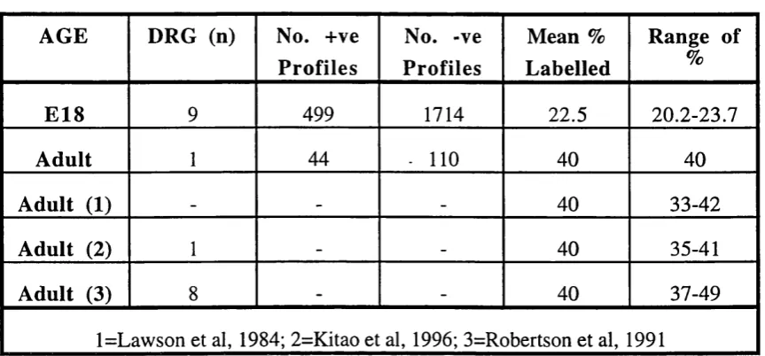
The selectivity of RT97 as an A-fibre label was confirmed by construction of
size-frequency histograms at E l 8. Using in situ hybridisation temporal expression of
CGRP mRNA was consistent with peptide expression. Quantitative analysis of epidermal
innervation shows all subpopulations transiently grow to the skin surface at E l 7-El 8
before retraction of fibres to a sub-epidermal plexus.
Administration of anti-NGF in utero results in a selective loss of small diameter DRG
cells (Ruit et al, 1992). This treatment was used to examine the consequences upon the
remaining DRG subpopulation and the pattern of cutaneous innervation.
DRG counts at E21 showed a cell loss of 43% restricted to the small cell population yet
this treatment resulted in total abolition of all epidermal innervation by E21, while
increase in the number and size of larger cells, with a population of cells sized
600-800|im^.
These results indicate that DRG subpopulations have different developmental patterns of
innervation but that anti-NGF treatment renders the epidermis non-permissive to
Acknowledgements
I would like to acknowledge my supervisor, Maria Fitzgerald for all her guidance during
my PhD. I wish to express my thanks for her encouragement and motivation, and for
providing the direction in my work.
There are some people I would firstly like to thank in our lab: Jacqueta Middleton,
Andrew Allchome and Penny Ainsworth, not only for technical assistance for their
multifaceted skills and wealth of information and advice. I would also like to thank
Richard Mannion for preparation of the CGRP probe and for his help in conducting in situ hybridisations.
I was fortunate in obtaining assistance outside our lab from Greg Michael at St. Thomas’s
hospital who carried out isotopic in situ hybridisations. I would like to additionally express gratitude to Paul Martin (UCL) for generously sharing his photography and
scanning facilities.
During my PhD, all colleagues in the lab were always available and willing to discuss,
assist and advice on a great many things, I would like them all to know how much I
appreciated their friendship.
Debts are not supposed to be happy things, but those incurred in the preparation of this
thesis are all owed to some special friends who showed such generosity, immense
kindness and one of the most valuable commodities, time, that I could not have wished for more - although maybe in different circumstances next time. To Simon Beggs, Herve
Bester, Jacqueta Middleton, Janie McCluskey and Ana Oliveira - Go Raibh Maith Agat (Thank-you).
Finally, the people who have supported me unfailingly and have always been there when I needed them even though far away, my parents, my brother Johnny, and Anthony for
Contents
T itle 1
A bstract 2
A cknow ledgem ents 4
List of Figures 11
List of Tables 14
List of Abbreviations 16
Chapter 1: General Introduction 18
1.1 Aim o f thesis 18
1.2 The development o f primary sensory neurons - birth o f dorsal root
ganglia (DRG) and axon outgrowth 20
1.3 Gangliogenesis 21
1.4 Neurogenesis 21
1.5 Prelude to axonal outgrowth: polarity and processes 22
1.6 Axon Outgrowth 23
Chapter 2: Materials and Methods 45
2.1 General Methods 46
2.2 Methods fo r Chapters 3, 4 49
2.3 Methods fo r Chapter 5 52
2.4 Methods fo r Chapter 6 54
2.5 Methods fo r Chapter 7 55
Chapter 3: Size-frequencv distribution of E18 and E21 dorsal root ganglia (DRG) neurons and distribution of RT97 in the E18 DRG 63
3.1 Growth and Development o f DRG Cells 64
3.2 RT97 - A Selective Marker o f Large Light DRG Cells 72
RESU LTS
3.3 The Size-Frequency Distribution o f E l 8 and E21 Dorsal Root Ganglia
(DRG) Neurons 77
D IS C U S S IO N
3.5 Size-frequency Distribution in the Dorsal Root Ganglia (DRG) 91
3.6 RT97 Distribution in the E l8 Dorsal Root Ganglia 94
Chapter 4: The development of subpopulations of cutaneous sen so ry neurons in the DRG. and the developmental pattern of innervation o f
hindlimb and spinal cord in embryonic rat 9 5
4.1 Growth Associated Protein (GAP 43) 99
4.2 Protein Gene Product (PGP) 9.5 108
4.3 Peripherin 111
4.4 TrkA 119
4 .5 1-B4 120
4.6 Intermediate Filaments (IF) 123
R E SU LTS
4.7 Aim 134
4.8 Information about selected markers 134
4.9 Information about experimental protocol 135
4.10 DRG : Description o f Innervation 136
4.11 Developmental Summary of Immunolabelling in the DRG 139
4.12 Periphery: Description o f Innervation 141
4.13 Developmental Summary o f Immunolabelling in the Periphery 153
4.14 Spinal Cord : Description of innervation 155
4.15 Developmental Summary o f Immunolabelling in the Spinal Cord 158
4.16 Analysis o f peripheral innervation according to A-fibre and C-fibre
phenotype 162
4.17 Analysis o f central innervation according to A-fibre and C-fibre
phenotype 168
4.18 Correlation between peripheral and central innervation at each age 169
D ISC U SSIO N
4.19 Do A-fibres reach the peripheral target prior to C-fibres ? 224
4.21 Are peripheral and central events correlated? 230
4.22 Why do C-fibres exhibit longer waiting periods? I f C-fibres are present
in the peripheral target, why is there a delay in afferent entry to the central
target? 233
4.23 What are the general features revealed by each o f the antibodies in this
series and how do they compare to previous studies? 237
4.24 Why does labelling o f the intermediate filaments peripherin and RT97,
precede that o f GAP 43 ? 246
Chapter 5: Quantitative analysis of cutaneous innervation density in the
developing hindlim b 2 4 7
5.1 Aim 248
5.2 Previous Studies 248
5.3 Parameters o f Study 249
RESULTS
5.4 Epidermal innervation o f a defined region - change with age 253
5.3 Epidermal innervation in relation to the comeal su fa c e o f the skin 262
5.4 Summary 263
D ISC U SSIO N
5.5 Why is it important to differentiate between the innervation density at a
particular age, the total volume o f the tissue innervated and the actual
amount o f innervation ? 280
5.6 How does epidermal innervation o f a defined region change with age? 280
5.7 Is it significant that changes are not detected between regions? 282
5.8 What are the differences between subpopulations? 283
5.9 What evidence supports changes in target structure as an influence on
innervation levels? 284
5.10 Why are changes in innervation anticipated and what evidence is there
Chapter 6: Ontogeny of CGRP mRNA in lumbar spinal cord and DRG 2 8 9
6.1 Aim 290
6.2 Neuropeptides 290
6.3 Calcitonin Gene Related Peptide (CGRP) 291
6.4 Substance P (SP) 299
RESU LTS
6.5 Ontogeny o f CGRP mRNA expression in the DRG and Spinal Cord 302
6.6 Comparison o f CGRP mRNA and peptide onset and expression 303
D IS C U S S IO N
6.7 Does the in situ data correlate with the immunohistochemical data on
embryonic CGRP expression ? 315
6.8 Why is CGRP expressed late in DRG development? 315
6.9 What is the signal fo r the onset o f DRG expression? 316
6.10 Does the existence o f intrinsic peptidergic neurons undermine the data
suggesting peptidergic expression is influenced by peripheral
target-derived NGF? 319
Chapter 7: Effect of foetal anti-NGF treatment on DRG cells and innervation of the skin
7.7 Aim .^20
7.2 Background 320
7.3 Neurotrophic Hypothesis ■ 320
7.4 NGF and trkA 322
7.5 BDNF and NT3 - additional roles in development 328
7.6NT3 andtrkC 329
7.7 BDNF, NT4 and trkB 333
7.8 Neurotrophin requirement o f central nervous system neurons ( CNS) 334
7.9 The low-affinity neurotrophin receptor p75 334
R E SU LTS
7.10 Information about experimental protocol 339
7.12 Examination o f sections show larger cells in treated DRG 341
7.13 Cell counts reveal 43% loss o f DRG cells in the anti-NGF treated
embryos 341
7.14 Effects o f anti-NGF treatment on skin innervation 342
7.15 Quantitative Analysis o f Dermal Innervation 342
7.16 Changes in the target - 344
D ISC U SSIO N
7.17 Do the size-frequency histograms and cell counts reflect selective loss of
small diameter neurons ? 351
7.18 Why is the DRG cell loss less than previous reports? 352
7.19 Why are the surviving DRG neurons hypertrophied compared to those
in controls? 353
7.20 What is the stimulus fo r the abolition o f epidermal innervation? 363
7.21 Why should A-fibres affected by this treatment? 365
7.22 Is the presence of CGRP-IR neurons an indication that all C-fibres were
not eliminated? 365
7.23 How is the dermal innervation density unaffected after 43% cell
death? 366
7.24 Are the structural changes in epidermal depth a primary consequence of
trophic factor deprivation 369
Concluding Remarks 371
Reference List 3 7 4
List of Figures
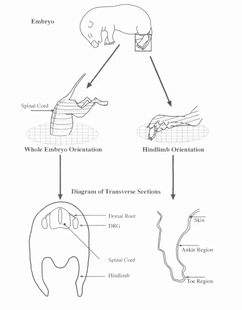
Fig. 2.1: Fig. 2.2: Fig. 2.3: Fig. 2.4: Fig. 3.1: Fig. 3.2: Fig. 3.3: Fig. 3.4: Fig. 3.5: Fig. 3.6: Fig. 4.1: Fig. 4.2: Fig. 4.3: Fig. 4.4: Fig. 4.5: Fig. 4.6: Fig. 4.7: Fig. 4.8: Fig. 4.9:Orientation of tissue sectioned on the freezing microtome.
Orientation of tissue sectioned on a cryostat.
The hindlimb regions selected for innervation density analysis.
Anti-NGF injection procedure and selection of tissue for
analysis.
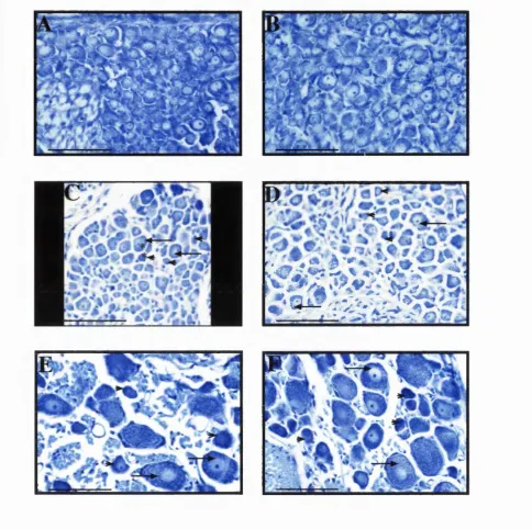
E18, E21 and adult DRG counterstained with Toluidine Blue.
Size-frequency distributions of E l 8 and E21 DRG.
Size-frequency and RT97 distribution in adult DRG.
RT97 immunohistochemistry on E l 8 and adult DRG.
RT97 immunohistochemistry on E l 8 and adult DRG
counterstained with Toluidine Blue.
RT97 antibody distribution in E l 8 DRG.
GAP 43 labelling in spinal cord and DRG of embryos aged E13-
E20.
GAP 43 labelling in the hindlimb of embryos aged E13-E16.
GAP 43 labelling in regions A-D of the hindlimb at E17 (top) and
E l 8 (bottom).
GAP 43 labelling in regions A-D of the hindlimb at E19 (top) and
E20 (bottom).
PGP 9.5 labelling in spinal cord and DRG of embryos aged E 13-
E l 8 and E20.
PGP 9.5 labelling in the hindlimb of embryos aged E 14-El7.
PGP 9.5 labelling in regions A-D of the hindlimb at E17 (top) and
E l 8 (bottom).
PGP 9.5 labelling in regions A-D of the hindlimb at E20.
Peripherin labelling in spinal cord and DRG of embryos aged E13-
Fig. 4.10: Fig. 4.11: Fig. 4.12: Fig. 4.13: Fig. 4.14: Fig. 4.15: Fig. 4.16: Fig. 4.17: Fig. 4.18: Fig. 4.19: Fig. 4.20: Fig. 4.21: Fig. 4.22: Fig. 4.23: Fig. 5.1: Fig. 5.2: Fig. 5.3: Fig. 5.4: Fig. 5.5: Fig. 5.6: Fig. 5.7:
Peripherin labelling in the hindlimb of embryos aged E l 3-El 6.
Peripherin labelling in the hindlimb of embryos aged E 17-El9.
Peripherin labelling in regions A-D of the hindlimb at E17 (top)
and E l 8 (bottom).
Peripherin labelling in regions A-D of the hindlimb at E19 (top)
and E20 (bottom).
RT97 labelling in spinal cord and DRG of embryos aged E13-E20.
RT97 labelling in the hindlimb of embryos aged E l 4-El 7.
RT97 labelling in regions A-D of the hindlimb at E17 (top) and
E l 8 (bottom).
RT97 labelling in regions A-D of the hindlimb at E l 9.
TrkA labelling in spinal cord and DRG of embryos aged E13-E16,
E19 and E20.
TrkA labelling in the hindlimb of embryos aged E l 3-El 6.
TrkA labelling in regions A-D of the hindlimb at E17 (top) and
E19 (bottom).
CGRP labelling in spinal cord and DRG of embryos aged E14-
E19.
CGRP labelling in the hindlimb of embryos aged E l 8 and E l 9.
CGRP labelling in regions A-D of-the hindlimb at E l 9.
PGP 9.5 innervation density in region A from E17-E20.
Area of region A from E17-E20 with PGP 9.5.
CGRP innervation density in region A from E19-P0.
Area of region A from E19-P0 with CGRP.
The relative thickness of the epidermis changes during
development.
Innervation density in regions A-D from E19-P0 with CGRP.
Fig. 5.9: Innervation density by sensory neuron subpopulations in the
E l 5 hindlimb.
Fig. 5.10: Area of skin in the E l 5 hindpaw.
Fig. 5.11 : Innervation density by sensory neuron subpopulations in the
E l 8 hindpaw.
Fig. 5.12: Area of skin in the E l 8 hindpaw.
Fig. 5.13: The projection ratio of epidermal fibres to epidermal depth
with age.
Fig. 6.1: CGRP mRNA expression in the E 14 DRG and spinal cord.
Fig. 6.2: CGRP mRNA expression in the E15 DRG and spinal cord.
Fig. 6.3: CGRP mRNA expression in the E16 DRG and spinal cord.
Fig. 6.4: CGRP mRNA expression in the E 17 DRG and spinal cord.
Fig. 6.5: CGRP mRNA expression in the E18 DRG and spinal cord.
Fig. 6.6: Digoxygenin-labelled CGRP mRNA expression in the E l 8 DRG
and spinal cord.
Fig. 6.7: Comparison of CGRP mRNA and peptide onset and localisation,
Fig. 7.1 : Size-frequency distributions of control and anti-NGF treated E21
DRG.
Fig. 7.2: Control and anti-NGF treated E21 DRG sections counterstained
with Toluidine Blue.
Fig. 7.3: Localisation of immunoreactive (IR) nerve terminals in the E21
glabrous foot pad (Region A) of control and anti-NGF treated
embryos.
Fig. 7.4: Camera Lucida drawings of innervation in the E21 glabrous foot
pad of control and anti-NGF treated embryos.
Fig. 7.5: Sub-epidermal innervation density in control and anti-NGF treated
List of Tables
Table 3.1: Table 3.2: Table 3.3: Table 4.1:
Parameters of the E18 and E21 Size-frequency distributions.
Parameters of RT97 Positive and Negative Populations.
Expression of RT97 in E l 8 and Adult DRG.
Onset and Intensity of Immunolabelling in the DRG at each
Developmental Age.
Table 4.2: Onset and Extent of Immunolabelled Fibres in the Hindlimb at each Developmental Age.
Table 4.3a: Onset and Location of Immunolabelling in the Spinal Cord at each Developmental Age.
Table 4.3b: Onset and Location of Immunolabelling in the Spinal Cord at each Developmental Age.
Table 4.3c: Onset and Location of Immunolabelling in the Spinal Cord at each Developmental Age.
Table 5.1: Innervation Density in Region A from E17-E20 with PGP 9.5.
Table 5.1a: ANOVA Factorial Analysis of Innervation Density in Region A from E17-E20 with PGP 9.5.
Table 5.2: Innervation Density in Region A from E19-P0 with CGRP. Table 5.3: Innervation Density in Regions A-D from E19-P0 with
CGRP.
Table 5.4: Effect of Age on CGRP Innervation Density from Combined Regions.
Table 5.4a: ANOVA Factorial Analysis of Innervation Density from E19- PO in Combined Regions with CGRP.
Table 5.5: Innervation Density by Sensory Neuron Subpopulations in the E15 Hindlimb.
Table 5.5a: ANOVA Factorial Analysis of Innervation Density in the E15 Hindlimb.
Table 5.6: Innervation Density by Sensory Neuron Subpopulations in Regions A-D of the E18 Hindlimb.
Table 5.7: Contribution of Different Sensory Neuron Populations to Innervation Density in the E l 8 Hindpaw.
Table 5.7a: ANOVA Factorial Analysis of Innervation Density by Sub populations of Sensory Neurons in the E18 Hindpaw.
Table 5.7b: ANOVA Factorial Analysis of Area Size for Different Sensory Neuron Subpopulations in the E l 8 Hindpaw.
Table 6.1: Comparison of m Hybridisation and Immunohistochemical Expression Results.
Abbreviation List
Ach Acetylcholine
AHP Afterhyperpolarization
BDNF Brain-Derived-Neurotrophic-Factor
BrdU B romodeoxyuridine
CAMs Cell Adhesion Molecules
CB B subunit of cholera toxin
CGRP Calcitonin Gene Related Peptide
ChAT, Choline Acetyl Transferase
CNS Central Nervous System
DIG Digoxygenin
DNES Diffuse Neuroendocrine System
DREZ Dorsal Root Entry Zone
DRG Dorsal Root Ganglion
E Embryonic
ECMs Extracellular Matrix Molecule
Eph Erythropoietin-producing hepatocellular
GAL Galanin
GAP 43 Growth Associated Protein 43
HE Hair Follicle
HRP Horseradish Peroxidase
HTM High Threshold Mechanoreceptors
ICE Interleukin-1 -p-converting enzyme
IF Intermediate Filament
IHC Immunohistochemistry
KB Kilobase
LDCV Low-Density Carrier Vessicle
LTMR Low Threshold Mechanoreceptor
LTP Long-Term Potentiation
NF Neurofilament
NF-H High Molecular Weight Neurofilament
NF-L, Low Molecular Weight Neurofilament
NF-M, Medium Molecular Weight Neurofilament
NFH-P Phosphorylated High Molecular Weight Neurofilament
NGF Nerve Growth Factor
nIF Neuronal Intermediate Filament
nm Nanometers
NMJ Neuromuscular Junction
NSA Netrin Synergising Activity
NSE, Neural Specific Enolase
NT-3 Neurotrophin-3
P Postnatal
PCD Programmed Cell Death
PDGF Platelet Derived Growth Factor
PGP Protein Gene Product
PKC Protein Kinase C
PNS Peripheral Nervous System
PPT Preprotachykinin
PSA Polysialic Acid
RAGS Repulsive Axonal Guidance System
RTKs Receptor Tyrosine Kinase
SA Slow Adapting
SCG Superior Cervical Ganglion
SG Substantia Gelatinosa ^
SP Substance P
STV Small Transport Vesicle
TH Tyrosine Hydroxylase
TM Transmembrane
TMP Thiamine monophosphate
TNF Tumour Necrosis Factor
GENERAL INTRODUCTION
1.1 Aim of thesis
The aims of this thesis are to describe the developmental timecourse of rat
hindlimb skin innervation by sub-populations of DRG neurons, to examine the
developmental interactions between the DRG sub-populations and to investigate the
interactions between DRG sensory neurons and target.
A study of central and peripheral axonal projections during development has been
undertaken (Mimics & Koerber, 1995a; 1995b). However, the technique of Dil labelling
means that the phenotype of axons cannot be determined. In the present study, a series of
general and phenotype-specific markers were used. The morphology, density, and
development of stereotypical patterns of axon bundles was examined using
immunohistochemical markers for sub-populations of neurons in skin and dorsal horn.
Protein Gene Product (PGP) 9.5, Growth Associated Protein (GAP) 43 and peripherin
were used as pan-neuronal markers, RT97 as an A-fibre marker and Calcitonin Gene
Related Peptide (CGRP), trkA and IB4 for small A and C-fibres.
Specific questions to be considered are whether A-fibres and C-fibres reach their
peripheral target at different times and with different developmental patterns. Target
innervation in the epidermis was quantified over a specific time period and compared
between the identified subpopulations of sensory axons.
In order to characterise developing DRG cells into different subpopulations with
confidence, several aspects of DRG cell development were investigated. The light
microscope characteristics of DRG cells were examined, to determine if cells at embryonic
day (E)18 or E21 could be characterised as large light and small dark. Size-frequency
SP Substance P
STV Small Transport Vesicle
TH Tyrosine Hydroxylase
TM Transmembrane
TMP Thiamine monophosphate
TNF Tumour Necrosis Factor
1.2 The development of primary sensory neurons - birth o f
dorsal root ganglia (DRG) and axon outgrowth
The general processes underlying the first stages of development in the
somatosensory system are described in this introduction. There are eight headings under
which these parameters are explained, in logical order pertaining to the development of the
system. The first four concern the formation of the DRG and the process of axon
outgrowth and are discussed below. The last four are described later as part of chapters
containing specific information about the background to each of the experiments.
Since this thesis is concerned with the relationship between the DRG
subpopulations and the interactions with their target, it is appropriate to outline how these
cells are generated and by what process they reach their targets. It is important for the
correct functioning of the somatosensory system that primary sensory neurons are
specialised to perform different tasks and make appropriate connections to particular
peripheral receptor or target tissues. Each DRG neuron innervates two targets, one
peripheral and one central, therefore there must be some correlation between them so that
in the adult the central pathways/effector cells receive the correct peripheral sensory
information with which they are best qualified to process. The targets of the lumbar DRG
neuron are the spinal cord and the hindlimb, which consists of both muscular and
cutaneous tissue. The function of the DRG is to facilitate the transfer of peripheral
sensory information to the central nervous system (CNS), hence its location near the
spinal cord. Its role requires that axon outgrowth is accurate to both targets and that
specification of different cell types is achieved.
The research in this thesis concerns the interaction between axonal subpopulations
and target, hence the nature of the targets and the specific phenotypes of DRG cells will
period of cell death is observed, after which the stereotypical patterns of connections are
established. These aspects are also briefly covered in those experimental chapters.
1) Gangliogenesis
2) Neurogenesis
3) Prelude to axonal outgrowth
4) Axon outgrowth
5) Nature of targets see Chapter 4
6) Cell death see Chapter 7
7) Somatotopy see Chapter 4
8) Characterisation of subpopulations see Chapter 3
1.3 Gangliogenesis
Dorsal root ganglia (DRG) are located in the peripheral nervous system and
contain the cell bodies of vertebrate primary somatic sensory neurons. They appear as
oval swellings of the dorsal roots. DRGs are derived from the unsegmented neural crest
cells. Following somitic dissociation into dermomyotome and sclerotome, neural crest
cells migrate into position in the rostral half of the sclerotome where they condense to
form a DRG (Verbout, 1985; Teillet et al, 1987; Lallier et al, 1988). The neural crest cells
segregate within each ganglion in a distinct topographical arrangement which reflects their
original craniocaudal position on the neural primordium (Teillet et al, 1987).
1.4 Neurogenesis
The lumbar DRG cells of the rat are generated in three successive waves
corresponding to different subpopulations. The large light cells are bom before the small
dark cells over the period E11-E15 - approximately midway through gestation, (Lawson
et al, 1974; Altman & Bayer, 1984) but recently a third small population born E 14-El5
has been reported (Kitao et al, 1996). Further details of this process are found in
1.5 Prelude to axonal outgrowth: polarity and processes
Once DRG cells are fully differentiated they begin to extend processes or neurites.
This is apparent in vitro after addition of neurotrophic factors, which results in a radial
pattern of short neurites emanating from the DRG. In vivo these processes initially grow
at the same rate and any one of the minor processes is capable of becoming the axon, but
eventually only one accelerates and becomes the axon (Goslin & Banker, 1989). The
axon is specialised to transmit information over long distances, i.e. from the cell body to
the target, and cell processes gradually acquire the biochemical properties unique to
axons.
The first evidence of axonal characteristics is the accumulation of microtubules
especially the low molecular weight tau proteins (Black & Baas, 1989). Preceding the
onset of axonal outgrowth, all minor processes show GAP 43-like immunoreactivity (see
section 4.1). GAP 43 becomes highly concentrated in the axonal growth cone and largely
disappears from the other processes, hence it is selectively transported into the neurite that
becomes the cell’s axon (Sargent, 1989). Axons then become polarised with a positive
end where new subunits are added to the growing tip. Additional membrane is added to
growth cones to enable advancement towards the target. The main constituents of
extension are microtubules, intermediate filaments and neurofilaments (see Chapter 4).
Antibodies to these growth cone/axon scaffolding components allow visualisation of the
developing axon and it is the principle exploited in this thesis (Chapter 4).
DRG cells are at first bipolar then undergo morphological changes to become
pseudounipolar early in development (Takahashi & Ninomiya, 1987). The transition to
unipolar cells is caused by the elongation of the cell body cytoplasm. Adult primary
sensory neurons are classically described as pseudounipolar, referring to the number of
axonal processes (Ranvier, 1875; Ramon y Cajal, 1909). The cell body is located in the
DRG, and while one axon originates from the cell body, it divides after a few hundred
microns, giving rise to one central axon to the spinal qord and one peripheral axon to the
spinal and peripheral nerve. These early reports proposed that a 1:1 ratio existed between
was challenged with the advent of the electron microscope as it was not possible to obtain
this ratio (Coggeshall, 1980; Langford & Coggeshall, 1979; 1980). Initially all studies
reported an excess of fibres compared to cells, leading to the conclusion that fibres branch
inside the ganglion. Recently, this 1:1 ratio has been obtained using new stereological
techniques and indicates that at least the cranial side of the ganglion can be considered
pseudounipolar, since the peripheral side of the ganglion is more complex
(Tandrup, 1995).
1.6 Axon Outgrowth
During development neurons initiate and extend axonal projections to a specific
target. Through this growth process, neurons acquire the distinctive and stereotyped
pattern of connections characteristic of the adult nervous system. The complexity of this
exercise has been highlighted in recent years by the discovery of many of the underlying
molecular mechanisms.
Essentially, there are five stages to this process. The first is the stimulation of
neurite outgrowth, under the influence of neurotrophins. Pioneering neurites then
navigate through many different tissues in a reproducible and identical pattern to find their
way to a specific target. This stage is regulated by interaction of the growth cone with
environmental guidance cues: signals from molecules on the surface of other neuronal and
non-neuronal cells, and within the extracellular matrix in the pathway of the axon.
Simultaneously, target-derived guidance factors then attract the axons while inhibitory
factors repulse axons away from inappropriate areas.
These extrinsic factors modulate the cellular mechanisms influencing the rate of
extension, orientation and control of the growth cone. Intrinsically, the response of the
axon to these external cues has also been investigated, since intracellular machinery
integrates the myriad extracellular cues to co-ordinate and direct growth cone navigation.
Once at the target site, the neuron must recognise and select a postsynaptic target among
covering many aspects of this developmental process in detail (Tanaka & Sabry, 1995;
Keynes & Cook, 1995; Garrity & Zipursky, 1995; Kennedy & Tessier-Lavigne, 1995),
so a brief overview of these stages will be described, focusing on the peripheral and
central targets of the lumbar DRG neuron. It is clear however, that the full relationship
between each of the stages is not understood although the effect of activity on pathway
guidance cues has been examined. Further research is therefore needed to understand the
interactions between the various mechanisms controlling neurite outgrowth.
1.6.1 S tim ulation o f neurite outgrowth
Developing DRG neurons are dependent on neurotrophins for survival after they
have reached their target, however, evidence is emerging that they may be dependent, not
for survival but for stimulation of outgrowth prior to reaching the peripheral target.
In vitro this has been apparent because without inclusion of the appropriate neurotrophic
factors in the culture media, neurite outgrowth would not occur (See Chapter 7).
1.6.2 P ath w ay gu id a n ce cues
The guidance of migrating cells depends on cues from the local environment. This
was initially thought to be mediated through a physical adhesion mechanism but now has
been superseded by the discovery that many cues guiding growth cones are not even
adhesive in nature and many are in fact inhibitory. The lack of correlation between
direction of migration and adhesive strength also contributed to the dismissal of the simple
haptotaxis mechanism, where growth cones followed adhesive gradients. It is now
realised that the role of cell adhesion is multifaceted utilizing a combination of haptotactic
and inhibitory mechanisms. The functional units of cell adhesion are multi-dimensional
complexes comprising of three general classes of proteins: cell adhesion molecules and
receptors, extracellular matrix proteins and the cytoplasmic plaque/peripheral membrane
proteins.
1.6.3 A d h e sio n M o lec u les c
The cell adhesion molecules (CAMs) have been implicated in axon guidance
macromolecules controlling cell-cell interactions via the regulation of processes such as
neural adhesion and migration, neurite outgrowth, axon fasciculation, synaptogenesis and
intracellular signalling.
A comprehensive role for these molecules has not yet been defined for two
reasons. Firstly, they show paradoxical activity. They can increase cell adhesion and axon
fasciculation but are conversely capable of increasing cell motility and neurite outgrowth.
Secondly, there is no direct in vivo evidence of their actions due to the absence of
abnormality associated with gene ablations of the CAMs in mice. Research on individual
CAMs and their homologues in different species has provided evidence supportive of
their necessity to axonal guidance. The role of individual pathfinding cues has been
extensively examined in the zebrafish retinotectal system. Large-scale genetic screening in
this vertebrate model revealed mutations relating to over 30 genes. A review of the mutant
phenotypes (Karlstrom et al, 1997) shows that individual genes can be mutated to give
clear axon-guidance defects, therefore some guidance systems are unique and do not have
completely redundant or overlapping functions.
The remaining mutant zebrafish phenotypes show a reduction in the number of
axons that arrive at the correct target but not a total elimination of accurate axon guidance.
Similarly with drosophila mutants (reviewed in Keynes & Cook, 1995) many growth
cones reach their targets but at later stages of development, implying that other cues may
ultimately allow errors to be corrected even after loss of contact with pioneer axons.
These studies suggest that multiple guidance cues are responsible for the extremely
high-fidelity axon guidance in vivo and the prevalence of these mutations with partial
defects may explain why mouse knockouts show no clearly interpretable axon-guidance
defects. The major drawback with the zebrafish mutants is that the nature of the
phenotypic aberrations are not yet characterised. It is not known whether the mutations
were the result of disruption of the environment or the ability of the growth cone itself to
respond to the guidance cues, yielding no significant clues as to how they exert their
Do CAMs possess the correct expression patterns and range of interactions to
fulfil the criteria of axon guidance molecules? The purpose of CAMs in neurite guidance
is not immediately clear, nor is the putative yet paradoxical roles of cell motility and cell
adhesion described for these molecules. Clearly neurites must be motile and fasciculated
to reach the target but it is vital that they interact with the pathway substrate via a
haptotactic mechanism to provide directional cues to the growth cone. On reaching the
target area, axons must defasciculate and adhere to the target matrix to transform the
growth cone into a synapse. The remainder of this account, will focus on how this may
be achieved for different types of neurites by adhesion molecules. Current proposals for
achievement of their antagonistic effects are combinations of changes in the expression
levels of specific CAMs, their molecular isoforms and also by post-translational
modification at different developmental stages.
The CAM family is functionally classified into calcium-dependent and
calcium-independent groups. Cadherins are the calcium-dependent group and have a
highly conserved cytoplasmic domain that is the site of association with the cytoplasmic
plaque proteins. The specific cytoplasmic plaque proteins that bind to cadherins are called
catenins. These mediate the interactions between the cadherins, cytoskeletal proteins and
signal-transduction pathways to regulate cell adhesion, a-catenin has actin-binding
activity and probably serves as the link between Ca^^-dependent CAMs and the
cytoskeleton (Rimm et al, 1995). This association is further regulated during development
and by phosphorylation to modulate adhesion, proliferation and morphogenesis.
The calcium-independent group of CAMs is largely composed of the
immunoglobulin superfamily. These molecules share Ig motifs and fibronectin-III repeats
but show multiple isoforms, each with distinct functional effects and expression patterns.
Examples of mammalian CAMs from the Ig superfamily include PO, MAG, NCAM (180,
140, 120), LI and TAG-1. Each of these have chick/drosophila homologues except for
the PO and MAG molecules. Novel non-mammalian CAMs already discovered are
NrCAM and neurofascin. The cytoplasmic domains of some Ig family members associate
cytoplasmic surface and facilitate management of cytoskeletal dynamics (Davis &
Bennett, 1994).
CAMs bind through homophilic interactions with like molecules on adjacent
membranes stimulating activation of several intracellular signalling cascades. Intracellular
signalling cascades alter the levels of calcium which is an important second messenger
regulating growth cone motility. LI and cadherin substrates have been shown to directly
influence actin and microtubules, but not neurofilaments to produce changes in growth
cone morphology of neurites grown in vitro (Burden-Gulley & Lemmon, 1996).
Heterophilic binding between different CAMs has also been established with LI and
NCAM combining to influence long term potentiation (LTP). In this example, binding
occurs between the oligomannosidic carbohydrate on LI and the Ig domain on NCAM, if
binding is abolished by soluble mannosides, LTP is strongly inhibited.
Integrins are a family of heterodimeric proteins composed of a and p subunits
that, like CAMs, mediate cell-cell interactions and cell-actin cytoskeleton interactions
through the intermediate proteins talin, a-actinin, tensin and vinculin, thus are often
described as CAMs. However, they also mediate cell-extracellular matrix adhesive
connections and are crucial in linking the CAMs, ECMs and the cytoskeleton. Key
intracellular signalling pathways involving integrins are beginning to be identified,
including activation of transcription factors and induction of gene expression after binding
of CAMs (for review see Lafrenie & Yamada, 1996).
The inclusion of integrins as components in mediating axon guidance means they
too must fulfil necessary criteria. A detailed review of the nature of integrin involvement
in neural crest-cell migration (Perris, 1997) ascertains that four putative integrin receptor
polypeptides are expressed by undifferentiated neural crest (NC)-cells and after in vitro
and in vivo integrin inactivation experiments, some identified subunits show involvement
with ECMs. At the same time, results from genetic deletions of single or double subunits
dismiss a role for integrins since no overt deficiencies in NC-cell migrations are observed.
present study, increased integrin expression is connected with the aggregation of NC-cells
and is a possible determining factor in the condensation of NC-cells into peripheral
nervous ganglia. Subunits ag and pg have been identified on peripheral sensory neurons
(Bossy et al, 1991; Venstrom & Reichardt, 1995; Beauvais et al, 1995) and six
heterodimers have been detected in chick/rodent DRG (Song et al, 1992; Sheppard et al,
1994; Tomaselli et al, 1993; Wu & Sontoro, 1994). Perturbation of the gene alters the
formation of skin in mice (Georges-Labousse et al, 1996) and in chick, is one of the
prevalent integrin receptors on neurons and axons in the developing spinal cord (Kil &
Bronner-Fraser, 1996).
Expression levels of CAMs that dictate the degree of axon fasciculation are also
modified at different stages of outgrowth. During cell motility periods, NCAM and
cadherins are downregulated, for example during formation of the mesoblast or on axons
at the start of myelination. This downregulation correlates with high motility of
myelinating Schwann cells (Edelman & Crossin, 1991). Conversely, outgrowth and
fasciculation is promoted by a subgroup of CAMs expressed predominantly on axon
pathways in the CNS and PNS during synaptogenesis (Goodman, 1996). Fasciculation is
maintained after reaching the target in drosophila overexpressing the NCAM homologue,
while in loss of function mutations defasciculation occurs in three CNS pathways prior to
reaching the target. Defects of fasciculation are generally dose and stage dependent, with
increases in fasciculation leading to subsequent errors m axon pathfinding, yet loss of
fasciculation does not appear to prevent growth cones from turning at critical regions and
occasionally may facilitate turning during normal development.
Post-translational modifications are a major mediator of CAM function, the best
characterised of such is glycosylation. This is regulated by changes in activity of the
enzyme that regulates the carbohydrate polysialic acid (PSA). PSA residues are bound by
various CAMs, their presence reducing binding rates three-four fold (Boisseau et al,
1991). Binding of PSA to NCAM has been localised to the fourth and fifth Ig-like
domains, forming a PSA-acceptor structure (Nelson et al, 1995). The components of the
binding in Aplysia and Drosophila NCAM-related molecules. Their effect is to inhibit
binding, effectively making axons less sticky and more dynamic and therefore attenuating
cellular interactions.
The level of polysialylation of NCAM has been implicated in regulating motor
nerve branching and fasciculation, since highest levels of PSA are detected when axons
are sorting in the plexus region and branching in the muscle (Landmesser et al, 1990;
Tang et al, 1992; 1994). The levels of PSA residues on NCAM have been shown to be
developmentally regulated, changing from 30% of mass in embryonic NCAM to only
10% in the adult (Edelman & Crossin 1991). Higher levels during the early period of cell
migration, is consistent with less adhesion and greater plasticity, whereas in the retina the
developmental shift to PSA-poor forms of NCAM is coincident with increased
morphological stability. This regulation is clearly important for neural plasticity, though
most of the spinal cord research has focused on the effect of PSA on motoneurons, the
detection of PSA during the timeframe of axon defasciculation in the plexus region also
infers this may be of importance to sensory neurons. It is also known that PSA enzymatic
activity can be regulated both by interaction with the peripheral target and by electrical
activity (Bruses et al, 1995).
In fact all of the strategies used by CAMs to affect axon guidance can be
modulated by neuronal electrical activity. Levels of the NCAM drosophila homologue,
which normally promotes fasciculation, shows decreased presynaptic expression in a
hyperactive mutant, with accompanying increases in presynaptic sprouting. Recent
in vitro models have found an association between CAMs and the defasciculation of
mouse DRG nerve terminals (Itoh et al, 1995). Neurite outgrowth is arrested in culture
under the influence of action potentials (Fields et al, 1990), consistent with the nerve
terminals of DRG neurons defasciculating upon arrival at the sub-epidermis which is
coincident with the commencement of low-frequency spontaneous electrical activity
(Fitzgerald & Fulton, 1992). Later, terminals refasciculate as neural impulse activity
activity-dependent mechanisms would be a powerful mechanism for linking the
developing structure of the nervous system to the functional activity of the developing
neural network, however the nature of this aspect remains to be established.
1.6.4 E xtracellular M atrix M olecules (ECMs)
The extracellular matrix contains an assortment of glycoproteins that have been
identified and are known to have important regulatory roles during development (Adams
& Watt, 1993). These are now referred to as extracellular matrix molecules (ECMs) and
have been designated into three subgroups according to their actions. The first group are
permissive molecules that promote cell motility, these include fibronectins, laminins and
collagen I, IV and VI. The second group are non-permissive, are mainly absent from the
migratory pathway and ineffective on cell motility. These include tenascins (reviewed in
Bartsch, 1996), proteoglycans and most collagens. Only one group of ECMs, the
aggrecans, belong to the third inhibitory group which are restricted to areas
non-permissive to migration and could directly/indirectly impede cell motility.
Cells attach to the ECM directly via components of the collagen-rich interstitial
matrix or the basement membrane. The basement membrane is composed of two layers,
the basal lamina - immediately adjacent to the cells, and the reticular layer in the
underlying connective tissue. The cell binding is mediated by two groups of receptors:
syndecans, which are a family of cell surface proteoglycans, and integrins which are
discussed above (Gumbiner, 1996).
ECMs provide cues that impose directionality upon the migrating cells as
long-range migrations usually occur along basement membranes, so motility can be
regulated by the spatio-temporal distribution of ECMs and the response of the cell at
definitive time-points. It has been shown that the phenotype and stage of commitment of
the cell is important since different cells react differently to the same ECM guidance cue.
In addition, certain cell types will only interact with some ECMs.
Apart from roles in migration and motility, therrelation of the ECM laminin to the
diffusible chemoattractant netrin, raises the possibility that other ECMs may also function
likely occur under particular conditions such as in the embryonic nervous system.
Evidence supportive of this theory, is that laminin loosely bound to the interstitial ECM in
neonatal peripheral nerve can be released in diffusible form in physiological buffer
(Kucherer-Ehret et al, 1990), in contrast to the adult insoluble form in basement
membranes. Another novel role for ECM proteins recently reviewed, is in apoptosis
(Meredith & Schwartz, 1997). In the absence of appropriate ECM contacts, cells undergo
apoptosis/programmed cell death (PCD). Apoptosis is considered to be a default pathway
for cells without extracellular signals to prevent it (Raff, 1992).
It would not be surprising if cell adhesion is required for cell survival, since
molecules that control cell location should be able to delete cells accidentally localised to
an improper environment. The assumption is based on the process of normal
morphogenesis, where death of cells lacking proper adhesive contacts occurs. An
example of this is the process of cavitation during tube formation, cells in the interior of
the cylinder die due to lack of contact with the basement membrane, leaving a hollow tube
(Coucouvanis & Martin, 1995). Secretion of matrix-degrading proteases is also an early
step in tissue regression and is advantageous to the process since ECMs are cell-type
specific and all cell types within the tissue can be affected simultaneously by loss of the
ECM (Boudreau et al, 1995). The cellular pathway facilitating this role has been partially
established, ECMs signal through integrin receptors which have been shown to regulate a
number of components of the death pathway: interleukin-1- p-converting enzyme (ICE),
Bcl-2 and P53 (for review see Meredith & Schwartz, 1997). Detailed examination of two
ECMs, proteoglycans and laminins, serve to highlight representative non-permissive and
permissive roles.
i) Proteoglycans
Proteoglycans belong to the group of non-permissive ECMs and are associated
with inhibitory roles. They are the candidate ECM involved in the guidance of sensory
neurites innervating the skin. In chick, this guidance is thought to be inhibitory since they
avoid chick epidermis in vitro but contact the dermis (Verna, 1985; Verna et al, 1986).
This avoidance response in neurites can be reproduced in vitro by the addition of
proteoglycans and glycosaminoglycans (Verna et al, 1989; Snow et al, 1990a) and can be
abolished when treated with a glycoprotein synthesis inhibitor (Fichard et al, 1990). Rat
skin also contains two populations of galactsaminoglycan-containing proteoglycans
whose concentration and ratio changes dramatically from E l 8 (Habuchi et al, 1986),
which coincides with the major period of terminal formation in the epidermis.
The in vitro inhibition of neurite growth after contact with proteoglycans also
confirmed that proteoglycans fulfilled the criteria of localisation and functional inhibition,
for their proposed role as an axon barrier in the roof plate (Snow et al, 1990b). These
molecules are also associated with a barrier to axonal growth in the dorsal root entry zone
(DREZ) after a critical period corresponding to postnatal days 2-3 in the rat (Pindzola et
al, 1993). Proteoglycans in association with tenascin, increase on the CNS side of the
DREZ after the critical period when axons cannot enter the spinal cord but are expressed
in relatively small amounts prior to the critical period when axons firstly grow into the
spinal cord during normal development and secondly, can regenerate. At E l 8, their
expression is restricted to the roof plate but by P2 they spread laterally to the DREZ and
into the dorsal columns. This band of expression clearly defines the DREZ and separates
the central and peripheral nervous systems.
Proteoglycan expression levels are also increased at the dermal-epidermal
junctional zone during the terminal phases of gangliogenesis and their role in the
preceding NC-cell migration has been described (Perris, 1997).
The inhibitory réponse to proteoglycans is concentration dependent and can be
modified by addition of growth-promoting molecules such as laminin, so the pattern of
neurite outgrowth may be regulated by the ratio of growth-promoting to growth-inhibiting
molecules (Snow & Letoumeau, 1992). An important deciding factor in the type of
response may be the combination of integrin receptor on the axon and the composition of
ii) Laminins
Laminins are a group of heterotrimers composed of a, p and y chains (Burgeson
et al, 1994). At least seven different heterotrimeric forms of laminin exist (Timpl &
Brown, 1994), with five a, three p and two y chains described. Chains containing the
recently discovered « 4 and a 5 (livanainen et al, 1995; Miner et al, 1995) have not yet
been characterised. Laminins are localised in the basement membrane separating epithelial
cells from underlying stroma and in the basement membrane surrounding fat, muscle and
peripheral nerve cells. They are the first ECMs detected in the developing embryo and
their primary role is cell-matrix attachment. Additionally, they have potent biological
activity on cell growth and migration, tumour growth and métastasés, neurite outgrowth
and wound repair (reviewed in Malinda & Kleinman, 1996). These different functions
may arise from the various chains binding different integrins.
The best characterised and first isolated laminin is referred to as laminin-1,
composed of a i p i y l . Laminin-1 has principal roles in three aspects of neuronal
development. It, and other isoforms are located along the migratory paths of NC-cells
with the appropriate integrin receptors, and are upregulated at stages preceding
gangliogenesis (reviewed in Perris, 1997), while perturbation of laminin or its receptors
disrupt migration (Bronner-Fraser, 1986; Bronner-Fraser & Lallier, 1988;
Bronner-Fraser, 1993a; 1993b). It is expressed by cells associated with the peripheral
nerve and is implicated in Schwann cell migration, differentiation and myelination
(Cornbrooks et al, 1983; McGarvey et al, 1984; Hsiao et al, 1993; Anton et al, 1994;
Fernandez-Valle et al, 1993). Importantly, it promotes neurite outgrowth from virtually all
classes of developing neurons both from the CNS and PNS (Manthorpe et al, 1983;
Rogers et al, 1983; Liesi et al, 1984; Unsicker et al, 1985; Engvall et al, 1986). This
global effect implies that it is a prime candidate for an in vivo guidance molecule, further
supported by the widespread distribution of integrin heterodimers responsive to laminin in
The developmental onset and distribution of all ten known laminin chain mRNAs
in the pathway and targets of DRG sensory neurons has recently been examined in the
mouse (Lentz et al, 1997). At E l l . 5, a l, a2, a4, pi, yl are expressed in the non-neuronal
cells surrounding the DRG, and a2, a4, pi, yl are found along the peripheral nerve and in
the ventral and dorsal roots at all embryonic stages examined. These are downregulated
by adulthood except for low levels of yl. Therefore, laminins may promote peripheral
axonal outgrowth and be of importance in maintenance of cells along the mature
peripheral nerve. By E l 3.5, DRG cells express laminin, which becomes progressively
more intense as a2, a4, pi and yl chain expression is initiated. Although at P I, DRG
neurons are labelled, it is not possible to ascertain at earlier developmental stages whether
expression is neuronal. In the limb-bud at E l 3, pi and yl are widely expressed, a2 and
a4 are found in muscle, while a3, a5, P2 and y2 are restricted to skin. The preferential
target distribution of the a laminin chains, suggests a contributory mechanism via which
cutaneous and muscle sensory axons identify their target.
In contrast to the above regions, laminins were absent from the grey matter at all
ages although they were expressed in discrete regions of the spinal cord, in accordance
with previous immunohistochemical data, and in addition were not found in cartilage or
along the optic nerve.
Laminins are expressed in the DRG, sensory and motor neuron pathways and
differentially in their respective targets. If laminins are responsible for different subsets of
neurons distinguishing their target, these axons would have to express appropriate
integrins to enable a response to the different laminin isoforms expressed in each of these
tissues. For example, muscle afferents should express a l and/or a4 specific integrins
while skin afferents should express integrins responsive to a3 and/or a5. From the
information currently available about integrin expression and ligand specificity, Lentz and
colleagues, speculate that the a3pl integrin may be the^pequired receptor for skin afferents,
1.6.5 T a rget-derived gu idan ce cues
Guidance cues emanating from the target are chemotropic in nature.
Chemotropism is defined as guidance directed by a gradient of a soluble factor diffusing
from a target. This mechanism of axon guidance was previously thought to be
chemoattractant only, however, since the early ‘80s the emergence of chemorepulsive
mechanisms has grown more prominent, demonstrating how non-target regions secrete
diffusible factors that prevent inappropriate innervation. Chemorepulsion now occupies a
position of equal importance with adhesion and chemoattraction in the guidance of
developing axons (Dodd & Schuchardt, 1995).
Three identified protein families have become prominent molecules in the search
for target-derived guidance cues. These are the netrins, the semaphorins and the
Eph-related receptor family and ligands.
i) netrins
The netrins are a small family of secreted proteins capable of guiding cell and
growth cone movements in vivo. There are two vertebrate members, netrin-1 and
netrin-2, whose receptor is DCC, one nematode member, unc-6, whose putative receptors
are unc-5 and unc-40. In Drosophila D-netrinA and D-netrinB are the two netrins with the
vtcQpiOT frazzled. The netrins appear to be members of a highly structurally-conserved
family of axon guidance molecules; further investigations have revealed that they are also
conserved in function.
Commissural neurons are bom in the dorsal spinal cord and extend axons along a
ventral trajectory to their intermediate target, the floorplate, at the ventral midline of the
spinal cord. The floorplate has been shown to secrete a long-range chemoattractant (for
review see Colamarino & Tessier-Lavigne, 1995a) and additionally has
outgrowth-promoting effects (Tessier-Lavigne et al, 1988). It was exploitation of its latter
function that led to the identification of netrin-1, but it has subsequently been confirmed
as the much sought after floor-plate derived chemoattractant. Netrin-1 (78 kDa) was
the cDNA into COS cells resulted in the release of a diffusible activity, that when
co-cultured with a dorsal spinal cord explant, led commissural axons to project towards
COS cells expressing either netrin in vitro (Kennedy et al, 1994) reproducing their growth
pattern towards floor plate explants (Placzek et al, 1990).
Netrin-1 and netrin-2 are differentially distributed in the spinal cord. Netrin-1 is
detected in the notocord prior to neural tube closure, then after closure is restricted to the
floor plate. Netrin-2 is expressed in the same timeframe and occupies the ventral
two-thirds of the spinal cord, exclusive of the floor plate. This suggests a model in which
there is a steep gradient of netrin-1 diffusing from the floor plate, superimposed on a
lower level of netrin-2 emanating from the ventral cord, which may modulate the netrin-1
gradient by extending it further into the dorsal spinal cord. The commissural neurons
would encounter an increasing concentration of netrin protein as they migrate ventrally
(Kennedy et al, 1994). At a recent axon guidance meeting (EMBL, Heidelberg, 1996) the
cloning of netrin-l and netrin-2 genes in the mouse was revealed (Tessier-Lavigne). In
contrast to the chick, netrin-2 is not expressed in the spinal cord although netrin-l
expression pattern is compatible to the sum of the two chick genes, it was suggested that
in rodents, netrin-l may additionally perform the functions of chick netrin-2.
Receptors for netrins in Drosophila and C. elegans were identified prior to that of
the chick netrins. These receptors are evolutionary conserved which helped lead to the
characterisation of the chick netrin receptor/s, DCC and neogenin (Keino-Masu et al,
1996). Together DCC and neogenin form a subgroup of the Ig superfamily. DCC was
previously cloned as a putative tumour suppressor gene deleted in colorectal cancer. It is
expressed in commissural axons and its binding to netrin has been visualised using a
specific antibody to netrin. Further confirmation of DCC - netrin interaction was provided
by blocking the extracellular domain of DCC in vitro, this inhibited netrin-l and floor
plate-evoked outgrowth of commissural axons. Neogenin also binds netrin-l but is
expressed in a complementary pattern to DCC in the chick spinal cord. The authors have
In the absence of in vivo perturbation data the chemoattractive function of netrin
was initially surmised after netrins were determined to be vertebrate homologues of the
Celegans unc-6 gene and 50% identical (Ishii et al, 1992). The unc-6 gene and protein has
been well characterised from its distribution, to mutant analysis and putative receptors.
These aspects have been the subject of several reviews, but briefly it regulates neuronal
outgrowth and cell migration along the dorsoventral axis of the nematode body wall
(Kennedy & Tessier-Lavigne, 1995; Keynes & Cook, 1995; 1996; Culotti & Kolodkin,
1996). Drosophila also express two netrins: D-netrinA and D-netrinB in the midline of the
developing CNS and mutants show these are required for guidance of commissural
neurons and peripheral motor axons (Harris et al, 1996, Mitchell et al, 1996). The
chemoattractant functions of netrin have now been confirmed in vertebrate mutants.
The phenotype of null mutants is consistent with the proposed roles of netrin.
Homozygotes fail to move their forelimbs independently and are neonate lethal due to
defective suckling. The commissural axons deviate from their normal projection towards
the floorplate. Analysis of the netrin-l mutant mice floor plate in vitro shows the
knockout is unable to promote the outgrowth of commissural axons in a collagen gel
assay, indicating that netrin-l is responsible for this activity of the floor plate (Serafini et
al, 1996). Netrin-l has previously also been attributed with a role rostral to the spinal
cord, in attracting cerebellofugal axons to the floor plate (Shirasaki et al, 1995). The
mutant confirms this and additionally raises the possibility that netrin-l is a
chemoattractant for migrating cells as well as axonal growth cones.
It is significant that netrin was identified as a putative diffusible factor but in COS
cells, more than 80% of netrin protein remains associated with cell surface (Kennedy et
al, 1994). The diffusion of the remaining netrin, may be subject to buffering by cell
surface or ECM components to establish both soluble and substrate-bound protein
gradients (Kennedy & Tessier-Lavigne, 1995). During netrin purification another
molecule with separate activity was isolated in the brain extract. This was termed NSA