Open Access
Review
Histological variants of cutaneous Kaposi sarcoma
Wayne Grayson
1and Liron Pantanowitz*
2Address: 1Histopathology Department, Ampath National Laboratory Support Services, Johannesburg, South Africa and 2Department of Pathology, Baystate Medical Center, Tufts University School of Medicine, Springfield, Massachusetts, USA
Email: Wayne Grayson - wayne.grayson@live.com; Liron Pantanowitz* - Liron.pantanowitz@bhs.org * Corresponding author
Abstract
This review provides a comprehensive overview of the broad clinicopathologic spectrum of cutaneous Kaposi sarcoma (KS) lesions. Variants discussed include: usual KS lesions associated with disease progression (i.e. patch, plaque and nodular stage); morphologic subtypes alluded to in the older literature such as anaplastic and telangiectatic KS, as well as several lymphedematous variants; and numerous recently described variants including hyperkeratotic, keloidal, micronodular, pyogenic granuloma-like, ecchymotic, and intravascular KS. Involuting lesions as a result of treatment related regression are also presented.
Introduction
Kaposi sarcoma (KS) is a vascular lesion of low-grade malignant potential that presents most frequently with skin lesions. Most histopathologists are au fait with the histologic picture of usual (typical) cutaneous KS as it progresses from patch, to plaque and finally nodular stages [1-4]. This morphologic spectrum of "usual" KS is common to classic, African endemic, transplant-associ-ated, and acquired immune deficiency syndrome (AIDS)-associated KS [4]. In recent decades, however, there has been an increasing awareness of a wider histologic spec-trum [5,6]. This has resulted in a growing number of reported clinical and/or histologic variants of KS. Failure to identify a given lesion as KS could lead to delayed diag-nosis or inappropriate management. It has also been sug-gested that certain variants, such as anaplastic KS and possibly lymphangioma-like KS, might have prognostic relevance [7-9].
Taking these factors into account, we have elected to divide KS skin lesions into four broad groups: (1) KS lesions which encompass usual variants related to disease
progression, (2) KS variants alluded to in the older litera-ture, (3) more recently described KS variants, and (4) KS lesions as a consequence of therapy. Although the appar-ent histogenesis of some of these morphologic variants is known, the pathogenesis of others is uncertain or subject to speculation. Hyperkeratotic KS, by way of example, fre-quently occurs as a result of KS-associated chronic lymph-edema of the lower extremities [10]. Intravascular KS, on the other hand, could either originate primarily as an intravascular proliferation, or alternatively develop as a consequence of intravascular extension of a lesion breach-ing the vessel wall [11]. KS lesions arisbreach-ing in extracutane-ous locations often differ histologically from their counterparts in the skin, including the recent description of "in situ" KS involving mediastinal lymphatic vessels [12].
Usual variants related to progression
Patch stage
Patch stage KS, which represents the earliest phase in the evolution of cutaneous KS, is perhaps the histologic vari-ant with the greatest propensity to cause diagnostic diffi-Published: 25 July 2008
Diagnostic Pathology 2008, 3:31 doi:10.1186/1746-1596-3-31
Received: 23 July 2008 Accepted: 25 July 2008
This article is available from: http://www.diagnosticpathology.org/content/3/1/31
© 2008 Grayson and Pantanowitz; licensee BioMed Central Ltd.
culties for the unwary. The initial low-power impressions are those of a "busy" dermis, or perhaps some form of mild inflammatory dermatosis [1,13]. On closer examina-tion, however, there are signs of a subtle vasoformative process composed of newly formed slit-like or somewhat jagged vascular spaces, which tend to be more conspicu-ous in the immediate vicinity of native dermal vessels and cutaneous appendages [1-4]. The protrusion of these native microscopic vascular structures into the lumens of more ectatic neoplastic channels results in the characteris-tic promontory sign (Figure 1). The intervening dermis frequently reveals dissection of its collagen bundles by slit-like vascular spaces lined by a monolayer of relatively banal, flattened endothelial cells, with a variable degree of erythrocyte extravasation. The newly formed channels often contain red blood cells. There is also a noticeable mild background inflammatory cell infiltrate comprising lymphocytes and plasma cells, often accompanied by a contingent of hemosiderin-laden macropahges. The aforementioned mononuclear cells tend to be concen-trated around the native vessels and skin adnexal struc-tures [1-4].
Plaque stage
In plaque stage lesions of KS, the histologic picture is char-acterized by a more diffuse dermal vascular infiltrate, accompanied by greater cellularity and occasional exten-sion of this process into the underlying subcutaneous
adi-pose tissue. The lesional cells tend to be more spindled and arranged in short, sometimes haphazard fascicles [1-4]. Fascicles cut in cross section demonstrate a sieve-like appearance. Mitotic figures are sparse and there is no sig-nificant nuclear or cytological pleomorphism. Intra- and extracellular hyaline globules, probably representing effete erythrocytes, are often seen. Careful scrutiny will fre-quently reveal "autolumination", whereby an erythrocyte is contained within a clear paranuclear vacuole in the cytoplasm of a spindled endothelial cell observed in cross section (Figure 2). Numerous dissecting vascular channels containing erythrocytes occupy the intervening dermis, and once again there is evidence of a background plasma cell-rich contingent of chronic inflammatory cells with admixed siderophages and free-lying hemosiderin pig-ment. The promontory sign, as described above, may also be encountered [1-4]. The histologic differential diagnosis includes tufted angioma, targetoid hemosiderotic heman-gioma, microvenular hemangioma and acroangioderma-titis ("pseudo-Kaposi's sarcoma") [1,2,4].
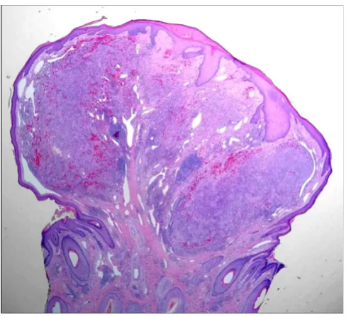
Nodular stage
The nodular form of KS usually poses no diagnostic diffi-culties. Occasionally, however, a small ulcerated nodular KS lesion may be mistaken for a pyogenic granuloma [1]. Nodular KS exhibits dermal expansion by a relatively cir-cumscribed, variable cellular proliferation of neoplastic spindled cells arranged in fascicles (Figure 4) [1-4]. Eryth-rocytes are contained within slit-like channels between the individual spindled cells. Although careful inspection may reveal occasional mitoses, the lesional cells are
rela-Patch stage Kaposi sarcoma showing newly formed vessels protruding into a larger vascular space characteristic of the promontory sign (H&E stain)
Figure 1
Patch stage Kaposi sarcoma showing newly formed vessels protruding into a larger vascular space char-acteristic of the promontory sign (H&E stain).
Plaque stage Kaposi sarcoma
Figure 2
tively monomorphic. Hyaline globules are seen more readily, as is the phenomenon of autolumination. In larger punch biopsy or excision biopsy specimens, the dermis away from the tumor nodule frequently exhibits changes associated with plaque stage KS, thus supporting the notion that patch, plaque and nodular stage lesions form part of a morphologic continuum. The periphery of some nodular KS lesions may show more dilated vascular
spaces, imparting a pattern that is strikingly reminiscent of a cavernous hemangioma (Figure 4) [2]. These larger, congested channels are an integral part of the lesion, as confirmed by positive immunohistochemical staining of the lining endothelial nuclei for HHV-8 latent nuclear antigen 1 (LNA-1).
Large cutaneous nodules may frequently undergo ulcera-tion. Superficial shave biopsies of such lesions may be diagnostically challenging to the histopathologist, as most of the specimen may contain only an inflammatory exudate with underlying granulation tissue; this may be misinterpreted as a pyogenic granuloma [1]. Distinguish-ing between spindle cells from granulation tissue and lesional KS cells from the uppermost portion of an under-lying KS nodule can be difficult, if not impossible without the aid of immunohistochemistry. The commercial anti-body to HHV-8 LNA-1 and the lymphatic endothelial cell marker D2-40 may prove very useful in this context. Stain-ing with these markers is preferable to less specific vascu-lar markers such as CD31 or CD34, as these do not facilitate recognition of the lesional and non-lesional endothelial cell populations. Rare instances of acquired immune deficiency syndrome (AIDS)-associated KS har-boring a concomitant opportunistic pathogen (e.g. cryp-tococcosis) may also go undiagnosed in superficial biopsy material [14,15]. Superficial shave biopsies, therefore, should be discouraged.
Lesions that may potentially be confused histologically with nodular KS include bacillary angiomatosis, other vascular tumors (e.g. spindle cell hemangioma and Kaposiform hemangioendothelioma), fibrohistiocytic tumors (e.g. cellular, angiomatoid and atypical variants of fibrous histiocytoma, and dermatofibrosarcoma protu-berans), resolving dermal fasciitis, spindle cell melanoma, and several other spindle cell mesenchymal neoplasms (e.g. cutaneous leiomyosarcoma) [1,2,4].
Variants reported in the older literature
Anaplastic Kaposi sarcoma
Anaplastic KS, sometimes referred to as pleomorphic KS, is poorly documented in the literature, possibly because of its rarity. Malignant transformation of KS, characterized by an increase in the number of mitoses and marked cel-lular pleomorphism, was first described in 1959 by Cox and Helwig [16]. A "monomorphic" variant was identi-fied by Templeton in several cases of African KS [17]. In a review of KS cases from Uganda (in 1971), investigators distinguished KS with a "monocellular pattern" (resem-bling anaplastic KS) from a so-called "anaplastic variant pattern" (resembling angiosarcoma) [18]. Anaplastic his-tology has been described in the context of classic, African, and AIDS-associated KS [7,8,19-24]. We are unaware of a
Nodular Kaposi sarcoma
Figure 3
Nodular Kaposi sarcoma.A. The dermis is expanded by a solid tumor nodule (H&E stain). B. Fascicles of relatively monomorphic spindled cells, with slit-like vascular channels containing erythrocytes (H&E stain). C. The nuclei of the tumor cells demonstrate immunoreactivity for HHV-8 (LNA-1 immunohistochemical stain).
Nodular Kaposi sarcoma showing a peripheral component at higher magnification reminiscent of a cavernous hemangioma (H&E stain)
Figure 4
report of this rare variant following iatrogenic immuno-suppression.
Anaplastic KS is clinically notable for its high local aggres-siveness, propensity for deep invasion, and increased met-astatic capacity. Progressive histologic dedifferentiation in otherwise typical cases of KS has been noted (Figure pend-ing permission) [22,25]. AIDS-associated tumors with anaplastic histology appear to have a propensity to occur in acral locations. In anaplastic KS, there is inherent potential for misdiagnosis as the vasoformative nature of the solid and frequently fascicular spindle cell prolifera-tion is not readily apparent. This variant displays a signif-icantly greater degree of nuclear and cellular pleomorphism than conventional nodular KS (Figure 5). In addition, there is an increased mitotic index (e.g. 5–20 mitoses per 10 high-power fields), and atypical mitoses may be encountered. Necrosis is occasionally observed.
It is easy to appreciate why a host of other malignant spin-dle cell neoplasms might be entertained in the histologic differential diagnosis, including certain sarcomas (e.g. lei-omyosarcoma, spindle cell rhabdlei-omyosarcoma, malig-nant peripheral nerve sheath tumor, fibrosarcoma), amelanotic spindle cell melanoma, and spindle cell carci-noma [14,15,20]. Angiosarcoma might also be consid-ered, particularly if erythrocytes are identified between the markedly atypical spindled cells. A comprehensive panel of immunohistochemical stains is often required to rule
out the aforementioned entities and confirm the presence of KS [15]. It is plausible that a proportion of anaplastic KS cases reported in the earlier literature, prior to the advent of immunohistochemistry, might not have been true cases of KS after all.
Lymphedematous variants
There are several variants of lymphedematous KS, all of which can present clinically with a deceptive bulla-like appearance (Figures pending permission). The inter-changeable terminology that has been used in the litera-ture for these variants is confusing. An attempt to classify this issue has been made [26], and includes variants asso-ciated with ectatic lymphatics such as lymphangioma-like and lymphangiectactic KS, and/or due to the accumula-tion of superficial dermal edema such as the subepider-mal and intraepidersubepider-mal (lymphatic) bullous KS variants. Most of these variants usually contain an admixture of more stereotypical KS lesions. In those cases where these lymphedematous variants form the predominant or sole histological pattern, the diagnosis of KS may be problem-atic.
Lymphangioma-like KS
Lymphangioma-like KS (LLKS), also referred to as "lym-pangiomatous" KS, is an uncommon variant that may be encountered in all four major clinicopathologic groups of KS patients [27-33]. Furthermore, lymphangioma-like morphology can occur in patch, plaque or nodular stage lesions [26]. This variant is said to account for less than 5% of KS cases [29]. Although Ronchese and Kern in 1957 are often credited with first describing the condition, the first reported case actually appears to date back to 1923, noted in a 66-year-old woman with clinical bullous KS lesions, the histology of which was described as being analogous to lymphangioma circumscriptum [27,34]. LLKS is most likely related to lymphedematous KS, bul-lous KS and hyperkeratotic (or verrucous) KS, as many of the reported patients with these clinical variants of KS have shown histopathologic features of LLKS on skin biopsy [10,26,27,34-39]. In some case reports patients with LLKS manifested with widespread and pronounced lymphedema [40] as well as effusions [41].
LLKS appears to exist (or co-exist) in two forms micro-scopically. The first comprises a patch or plaque stage lesion in which irregular, ectatic, interanastomosing vas-cular channels dissect dermal collagen bundles, resulting in a striking histological resemblance to a lymphatic tumor, such as a benign lymphangioendothelioma/ acquired progressive lymphangioma (Figure 6) [31,32,42]. In these cases the promontory sign tends to be particularly conspicuous (Figure 7). Erythrocytes are usu-ally absent from these channels. Slender papillae may project into vessels. In the second form, much larger, well
Anaplastic Kaposi sarcoma
Figure 5
formed endothelial lined spaces occupy the papillary der-mis and the upper reticular derder-mis (Figure 8). These chan-nels may closely abut on the overlying epidermis, in a pattern somewhat analogous to lymphangioma circum-scriptum. It is the latter pattern which may give rise to the
clinical appearance of "bullous" cutaneous lesions. Fea-tures of usual plaque stage KS are often encountered sub-jacent to these large channels; this useful diagnostic clue may be absent from biopsies that are too superficial, espe-cially shave biopsies. The endothelial cells lining the ectatic, lymphangioma-like channels in both forms are immunoreactive for HHV-8 LNA-1, as well as the lym-phatic endothelial marker D2-40 (Figure 8).
Lymphangiectatic Kaposi sarcoma
In lymphangiectactic KS there are large intratumoral and peritumoral dilated thin-walled lymphatic vessels (Figure 9). These ectatic lymphatics are much larger than those seen in LLKS, and less irregular and anastomosing [26]. They appear to be far less "compressible". Marked lym-phangiectasia present in the superficial dermis may result in a bullous appearing lesion (pseudoblister).
Bullous Kaposi sarcoma
The first published descriptions of bullous cutaneous lesions in patients with KS appeared in the early part of the twentieth century. These bullous lesions were ascribed to lymphangiectases [34]. Bullous lesions are observed most frequently in the context of lymphedematous KS, but this is not always the case [26,34]. In most instances, the term "bullous" is clinical rather than pathologic, since
Lymphangioma-like Kaposi sarcoma
Figure 6
Lymphangioma-like Kaposi sarcoma. In this florid example, the dermis is replaced by a haphazard proliferation of gaping, interanastomosing channels splaying apart the der-mal collagen (H&E stain).
Lymphangioma-like Kaposi sarcoma seen at higher magnifica-tion in which the promontory sign is well demonstrated (H&E stain)
Figure 7
Lymphangioma-like Kaposi sarcoma seen at higher magnification in which the promontory sign is well demonstrated (H&E stain).
Lymphangioma-like Kaposi sarcoma
Figure 8
pseudoblisters also occur as a consequence of lym-phangiectasia and/or LLKS involving the superficial der-mis in these patients (Figure 10) [26]. On other occasions, however, true subepidermal or intraepidermal bullae may arise in concert with KS. In the former, tense bullae are observed clinically due to peritumoral edema in the superficial dermis, while the latter may evolve either as a result of progression of a subepidermal bulla or due to resorption of lymphedema and re-epithelialization of a subepidermal blister [26].
Telangiectatic KS
There is a single case report of telangiectatic KS, which occurred in a man with thymoma and myasthenia gravis receiving long-term immunosuppressive therapy [43]. The term "telangiectatic" referred to the significant tel-angiectasia associated with the multiple cutaneous nod-ules, and not the histopathologic features thereof. The histopathology in this case report showed usual features of nodular KS, with no conspicuous background vascular ectasia [43]. The authors have encountered rare histologi-cal examples of telangiectatic KS in which nodular KS lesions contained large, intensely congested, ectatic vascu-lar spaces (Figure 11). Since these vascu-large spaces are lined by endothelial cells (Figure 12) whose nuclei are immunore-active for LNA-1, it must be assumed that they are an inte-gral part of the KS and not merely native dermal vessels
Lymphangiectactic Kaposi sarcoma
Figure 9
Lymphangiectactic Kaposi sarcoma. Large ecstatic lym-phatics can be seen within and around this KS tumor nodule (H&E stain).
Bullous Kaposi sarcoma
Figure 10
Bullous Kaposi sarcoma. In this patient with African endemic KS there is an intraepidermal bullous overlying sub-epidermal lymphedema associated with an underlying KS tumor nodule (not shown in this field) (H&E stain).
Telangiectatic Kaposi sarcoma is characterized by intensely congested, ectatic vascular spaces lined by lesional cells (H&E stain)
Figure 11
that have undergone telangiectasia as a consequence of compression by the dermal tumor.
Contemporary variants
Hyperkeratotic (Verrucous) Kaposi sarcoma
Hyperkeratotic KS is a rarely described clinicopathalogic variant of KS, which appears to be closely linked to severe KS-associated lymphedema in patients with AIDS [10,35,44]. There is verrucous epidermal acanthosis and hyperkeratosis overlying an often fibrotic epidermis (Fig-ure 13). In view of the latter feat(Fig-ure, diagnostic KS lesional tissue may be located at a relatively deeper level in the der-mis, further emphasizing the potential inadequacy of
superficial shave biopsies. On occasion, verrucoid epider-mal changes may occur with LLKS histology (Figure 14). Infrequently, such changes may involve the entire lower extremity manifesting as elephantiasis nostras verrucosa [45]. Chronic lymphedema may itself give rise to verruci-form epidermal hyperplasia and hyperkeratosis, with increased fibroblastic activity, blood vessels and thick-walled lymphatic vessels throughout the dermis [39]. Lympedematous AIDS-associated KS may also be associ-ated with exophytic fibroma-like nodules, characterized by dermal fibrosis, a loose arrangement of fibroblasts and collagen bundles, and dilated blood vessels and lym-phatic channels [10,39,44].
Keloidal Kaposi sarcoma
The description of this exceedingly uncommon KS variant is limited to a 1994 report of three cases [46]. Lesions are firm and rubbery, and may be linear [6]. Histologically, there is notable dermal expansion by dense, hyalinized collagen with a distinct resemblance to a keloid (Figure 15). In such lesions the spindled KS proliferation may be obscured by these keloidal alterations. The histologic dif-ferential diagnosis includes a dermal scar at the site of a previous skin biopsy of a KS lesion. It is postulated that cytokines play a key role in the evolution of the keloidal stromal changes in this unusual variant [46].
Micronodular KS
Micronodular KS (Figure 16) is a recently described vari-ant of nodular KS, which is characterized histologically by a small, unencapsulated, circumscribed spindle cell prolif-eration in the reticular dermis [47]. Although the paper by Kempf et al described micronodular cutaneous lesions in a patient with classic KS [47], similar lesions are occasion-ally encountered in the context of AIDS-associated KS, and are often removed in their entirety by a punch biopsy.
A CD31 immunostain highlights the many dilated vascular spaces seen in telangiectatic Kaposi sarcoma
Figure 12
A CD31 immunostain highlights the many dilated vascular spaces seen in telangiectatic Kaposi sar-coma.
Hyperkeratotic (verrucous) Kaposi sarcoma
Figure 13
Hyperkeratotic (verrucous) Kaposi sarcoma. A plaque stage lesion from the lower leg is surfaced by an epidermis showing verruciform acanthosis and hyperkeratosis, with fibrosis of the upper dermis (H&E stain).
Kaposi sarcoma with hyperkeratotic and lymphangioma-like histologic features (H&E stain)
Figure 14
Pyogenic granuloma-like Kaposi sarcoma
Small, superficially located nodular or micronodular KS lesions may be protuberant and thereby elicit the develop-ment of a peripheral epidermal collarette (Figure 17). Such lesions have been referred to as pyogenic granuloma (PG)-like KS [48]. Traumatized lesions may undergo ulceration and become inflamed, and potentially misdi-agnosed as a true PG (lobular capillary hemangioma). To further complicate matters true PGs may themselves har-bor kaposiform areas. PG-like KS must also be distin-guished from bacillary angiomatosis, as some examples of the latter can adopt striking PG-like low-power architec-ture [14,15].
Ecchymotic Kaposi sarcoma
In the variant referred to as ecchymotic KS, the intrader-mal KS proliferation is accompanied by extensive red blood cell extravasation (Figure 18) [49]. The marked pur-pura often obscures the underlying histologic features of KS. The differential diagnosis includes intralesional hem-orrhage brought about the biopsy procedure itself. Clini-cally, this variant of AIDS-associated KS manifests with ecchymotic or pityriasis-like patches [49]. Ecchymotic plaque lesions may clinically also resemble a bruise or port-wine stain. A rare case of clinical "hemorrhagic" KS has been reported [50]. However, it is unclear if the appearance in this case was attributed to extensive eryth-rocyte extravassation.
Intravascular Kaposi sarcoma
The only description of intravascular KS is limited to a report of six cases, including four patients with classic KS
and two with AIDS-associated KS [11]. Histologic exami-nation in this small series showed an exclusively intravas-cular solid spindle cell KS proliferation. Immunohistochemical stains for desmin and smooth muscle actin (SMA) confirmed that this proliferation was indeed intravenous. The histologic differential diagnosis includes intravascular papillary endothelial hyperplasia, intravenous PG, intravascular fasciitis, papillary intralym-phatic angioendothelioma (Dabska tumor) and intravas-cular myopericytoma [51].
Keloidal Kaposi sarcoma
Figure 15
Keloidal Kaposi sarcoma.A. Spindled cells from the edge of the Kaposi sarcoma plaque lesion (upper left) are flanked by an expanse of keloid-like collagen (lower right) (H&E stain). B. Masson's trichrome stain highlighting the keloidal collagen bundles. Note the many extravasated erythrocytes in the background.
Micronodular Kaposi sarcoma
Figure 16
Variants related to therapy
Therapy may result in KS regression, and less likely exac-erbation (so-called KS flare) [52]. The histopathology of regression in KS has been previously described and is dis-cussed below. KS exacerbation (flare or recrudescence) can occur following therapy with corticosteroids, after treatment with rituximab, or as part of the immune recon-stitution inflammatory syndrome (IRIS) seen with
antiret-roviral therapy in HIV-infected persons [52]. The histomorphology of KS flare lesions has yet to be described.
Regressing Kaposi sarcoma
The introduction of highly active antiretroviral therapy (HAART) for patients with human immunodeficiency virus infection (HIV) may lead to complete regression of established AIDS-associated KS lesions [53,54]. Clinical features of regression include flattening of lesions, reduc-tion in lesion size, and change from a purple-red appear-ance to an orange-brown macule. Following antiretroviral therapy, investigators have observed improved circum-scription of nodular lesions, which appear less cellular and are enveloped by a densely sclerotic stroma [55]. In some cases the only significant abnormalities are an increase in dermal capillary density around native dermal vessels and appendages (Figure 19), and an accompany-ing perivascular infiltrate of largely plasma cells (Figure 20). Partial or complete regression of KS lesions may also be brought about following the administration of chemo-therapeutic agents [56,57]. Histologic examination of such partially regressed lesions reveals residual spindled cells around native vessels in the mid- and upper dermis, and a significant reduction in the number of spindled lesional cells in the intervening dermis [56]. Lesions that have undergone complete regression, however, show an absence of these spindled cells, and a modest increase in microvessels (Figure 21) in relation to the superficial vas-cular plexus [56]. Other findings include the presence of hemosiderin-laden dermal macrophages and a conspicu-ous superficial perivascular lymphocytic infiltrate [56].
Pyogenic granuloma-like Kaposi sarcoma manifesting as an exophytic mass enveloped by an epidermal collarette (H&E stain)
Figure 17
Pyogenic granuloma-like Kaposi sarcoma manifest-ing as an exophytic mass enveloped by an epidermal collarette (H&E stain).
Ecchymotic Kaposi sarcoma
Figure 18
Ecchymotic Kaposi sarcoma. The spindled cell prolifera-tion in this example is somewhat obscured by the extensive purpura.
Regressed Kaposi sarcoma lesion
Figure 19
Conclusion
KS clearly has the ability to develop into lesions of varying morphologic appearance. It is important to be able recog-nize these variants in order to avoid potential misdiagno-sis and improper management of afflicted patients. KS has been shown to be of lymphatic origin [58]. This may explain the intimate association of abnormal lymphatics observed in several of these variants, such as lymphangi-oma-like and lymphangiectatic KS. KS is also intimately associated with lymphedema. Chronic lymphedema may
even precede KS lesions. Some authors believe that chronic lymphedema may promote KS development due to a combination of collateral vessel formation, lympahn-giogenesis and immune impairment [59]. Hyperkeratotic and bullous KS variants can be attributed to the long standing effects of tumor-associated lymphedema on the overlying epidermis. Deep dermal fibroma-like nodules seen in cases associated with marked lymphedema could explain the origin of micronodular KS. The clinical signif-icance of most of these KS variants has not been studied. Lymphedematous variants have been postulated to por-tend a poor prognosis. This is certainly plausible given the fact that significant KS-related edema carries a grave prog-nosis [60]. Anaplastic KS is perhaps the only variant asso-ciated with aggressive behaviour. The reason for progressive histological dedifferentiation in some cases of KS is unknown. Studies looking at the role of HHV-8 and the host (e.g. immunity) in anaplastic cases may provide some answers.
Competing interests
The authors declare that they have no competing interests.
Authors' contributions
LP and WG contributed equally to the literature review, photography of cases, and writing of this manuscript. Both authors read and approved the final manuscript.
References
1. Ackerman AB: Histologic features of Kaposi's sarcoma and simulators of it. In Kaposi's sarcoma Edited by: Cerimele D. Spec-trum Publications, Inc., New York; 1985:71-79.
2. Templeton AC: Pathology. In Kaposi's sarcoma: Pathophysiology and clinical management Edited by: Ziegler JL, Dorfman RF. Marcel Dekker, Inc., New York; 1988:23-70.
3. Ackerman AB, Gottlieb GJ: Atlas of the gross and microscopic features. In Kaposi's sarcoma: A text and atlas Edited by: Gottlieb GJ, Ackerman AB. Lea & Febiger, Philadelphia; 1988:29-72.
4. Sangüeza OP, Requena L: Malignant neoplasms. Kaposi's sar-coma. In Pathology of vascular skin lesions. Clinicopathologic correlations
Edited by: Sangüeza OP, Requena L. Humana Press, New Jersey; 2003:217-235.
5. Schwartz RA: Kaposi's sarcoma: an update. J Surg Oncol 2004, 87:146-151.
6. Jessop S: HIV-associated Kaposi's sarcoma. Dermatol Clin
2006:509-520.
7. Cerimele D, Carlesimo F, Fadda G, Rotoli M, Cavalieri S: Anaplastic progression of classic Kaposi's sarcoma. Dermatology 1997, 194:287-289.
8. Rwomushana RJ, Bailey IC, Kyalwazi SK: Kaposi's sarcoma of the brain. A case report with necropsy findings. Cancer 1975, 36:1127-1131.
9. Liebowitz MR, Dagliotti M, Smith E, Murray JF: Rapidly fatal lym-phangioma-like Kaposi's sarcoma. Histopathology 1975, 4(5):559-566.
10. Hengge UR, Stocks K, Goos M: Acquired immune deficiency syn-drome-related hyperkeratotic Kaposi's sarcoma with severe lymphoedema: report of five cases. Br J Dermatol 2000, 142:501-505.
11. Luzar B, Antony F, Ramdial PK, Calonje E: Intravascular Kaposi's sarcoma – a hitherto unrecognised phenomenon. J Cuta Pathol
2007, 34(11):861-864.
12. Konstantinopoulos PA, Dezube BJ, Pantanowitz L: Morphologic and immunophenotypic evidence of in-situ Kaposi's sar-coma. BMC Clin Pathol 2006, 6:7.
Regressed Kaposi sarcoma lesion seen at higher magnifica-tion showing a perivascular infiltrate comprised mainly of plasma cells (H&E stain)
Figure 20
Regressed Kaposi sarcoma lesion seen at higher mag-nification showing a perivascular infiltrate comprised mainly of plasma cells (H&E stain).
A completely regressed Kaposi sarcoma lesion still retains a modest amount of abnormal dermal microvessels, as evi-denced by this D2-40 immunostain
Figure 21
13. Ackerman AB: Subtle clues to diagnosis by light microscopy. The patch stage of Kaposi's sarcoma. Am J Dermatopathol 1979, 1:165-172.
14. Grayson W: A re-appraisal of vascular proliferations in human immunodeficiency virus infected patients. S Afr Dermatol Rev
2006, 6:48-57.
15. Grayson W: My approach to: The HIV-positive skin biopsy. J Clin Pathol 2008, 61:802-817.
16. Cox FH, Helwig EB: Kaposi's sarcoma. Cancer 1959, 12:289-298. 17. Templeton AC: Kaposi's sarcoma. Pathol Annu 1981, 16:315-336. 18. Taylor JF, Templeton AC, Vogel CL, Ziegler JL, Kyalwazi SK: Kaposi's sarcoma in Uganda: a clinico-pathological study. Int J Cancer
1971, 8:122-135.
19. O'Connell KM: Kaposi's sarcoma: histopathological study of 159 cases from Malawi. J Clin Pathol 1977, 30:687-695.
20. Owor R: Conventional Kaposi's sarcoma in Africa. In Kaposi's sarcoma: A text and atlas Edited by: Gottlieb GJ, Ackerman AB. Lea & Febiger, Philadelphia; 1988:143-149.
21. Ngendahayo P, Mets T, Bugingo G, Parkin DM: Kaposi's sarcoma in Rwanda: clinico-pathological and epidemiological aspects.
Bull Cancer 1989, 76:383-394.
22. Satta R, Cossu S, Massarelli G, Cottoni F: Anaplastic transforma-tion of classic Kaposi's sarcoma: clinicopathological study of five cases. Br J Dermatol 2001, 145:847-849.
23. Nayler SJ, Goetsch S, Grayson W, Taylor L: Pleomorphic Kaposi's sarcoma: Characterisation of an under-recognised variant of Kaposi's sarcoma. Mod Pathol 2000, 13:13A.
24. Craddock KJ, Labonte S, Ghazarian D: Anaplastic Kaposi sarcoma resembling epithelioid angiosarcoma in an HIV-positive man. Eur J Dermatol 2008, 18:358-359.
25. Weiss SW, Goldblum JR: Enzinger and Weiss's soft tissue tumors. 5th edition. Mosby, Philadelphia; 2008:721-732.
26. Pantanowitz L, Duke WH: Lymphoedematous variants of Kaposi's sarcoma. J Eur Acad Dermatol Venereol 2008, 22:118-120. 27. Ronchese F, Kern AB: Lymphangioma-like tumors in Kaposi's
sarcoma. AMA Arch Derm 1957, 75:418-427.
28. Gange RW, Jones EW: Lymphangioma-like variant of Kaposi's sarcoma. A report of three cases. Br J Dermatol 1979, 100:327-334.
29. Cossu S, Satta R, Cottoni F, Massarelli G: Lymphangioma-like var-iant of Kaposi's sarcoma: clinicopathologic study of seven cases with review of the literature. Am J Dermatopathol 1997, 19:16-22.
30. Davis DA, Scott DM: Lymphangioma-like Kaposi sarcoma: Eti-ology and literature review. J Am Acad Dermatol 2000, 43:123-127.
31. Ramirez JA, Laskin WB, Guirart J: Lymphangioma-like Kaposi sarcoma. J Cutan Pathol 2005, 32:286-292.
32. Mohanna S, Sanjez J, Ferrufino JC, Bravo F, Gotuzzo E: Lymphangi-oma-like Kaposi's sarcoma: report of four cases and review.
J Eur Acad Dermatol Venereol 2006, 20:999-1032.
33. Kalambokis G, Kitsanou M, Stergiopoulou C, Zioga A, Dimou S, Tsianos EV: Lymphangioma-like Kaposi's sarcoma with gastric involvement in a patient with lung cancer. J Eur Acad Dermatol Venereol 2005, 19:653-654.
34. Borroni G, Brazzelli V, Vignoli GP, Gaviglio MR: Bullous lesions of Kaposi's sarcoma: case report. Am J Dermatol 1997, 19:379-383. 35. Allen JA, Gillespie DL, Redfield RR, Gomez ER: Lower extremity lymphedema caused by acquired immune deficiency syn-drome-related Kaposi's sarcoma: case report and review of the literature. J Vasc Surg 1995, 22:178-181.
36. Recht B, Nickoloff BJ, Wood GS: A bullous variant of Kaposi's sarcoma in an elderly female. J Dermatol Surg Oncol 1986, 12:1192-1197.
37. Ruocco V, Astarita C, Guerra V, Lo Schiavo A, Moscariello CG, Satri-ano RA, et al.: Kaposi's sarcoma on a lymphedematous immu-nocompromised limb. Int. J Dermatol 1984, 23:56-60.
38. Atillasoy ES, Santoro A, Weinberg JM: Lymphedema associated with Kaposi's sarcoma. J Eur Acad Dermatol 2001, 15:364-365. 39. Ramdial PK, Chetty R, Singh B, Singh R, Aboobaker J:
Lymphedema-tous HIV-associated Kaposi's sarcoma. J Cutan Pathol 2006, 33:474-481.
40. Bossuyt L, Oord JJ Van den, Degreef H: Lymphangioma-like vari-ant of AIDS-associated Kaposi's sarcoma with pronounced edema formation. Dermatology 1995, 190:324-326.
41. Miller RM, Leibowitz MR: The radiological features of lym-phangioma-like Kaposi's sarcoma. A case report. S Afr Med J
1979, 56:146-148.
42. Guillou L, Fletcher CD: Benign lymphangioendothelioma (acquired progressive lymphangioma): a lesion not to be confused with well-differentiated angiosarcoma and patch stage Kaposi's sarcoma: clinicopathologic analysis of a series.
Am J Surg Pathol 2000, 24:1047-1057.
43. Snyder RA, Schwartz RA: Telangiectatic Kaposi's sarcoma. Occurrence in a patient with thymoma and myasthenia gravis receiving long-term immunosuppressive therapy. Arch Dermatol 1982, 118:1020-1021.
44. Caputo R, Gianotti R, Grimalt R, Monti M, Alessi E: Soft fibroma-like lesions on the legs of a patient with Kaposi's sarcoma and lymphedema. Am J Dermatopathol 1991, 13:493-496.
45. Sathyakumar S, Suh JS, Sharp VL, Polsky B: Images in HIV/AIDS. Elephantiasis nostras verrucosa secondary to Kaposi sar-coma: a rare case. AIDS Read 2008, 18:81-82.
46. Schwartz RA, Spicer MS, Janninger CK, Cohen PJ, Melczer MM, Lam-bert WC: Keloidal Kaposi's sarcoma: Report of three patients. Dermatology 1994, 189:271-274.
47. Kempf W, Cathomas G, Burg G, Trueb RM: Micronodular Kaposi's sarcoma – A new variant of classic-sporadic Kaposi's sar-coma. Dermatology 2004, 208:255-258.
48. Urquhart JL, Uzieblo A, Kohler S: Detection of HHV-8 in pyo-genic granuloma-like Kaposi sarcoma. Am J Dermatopathol
2006, 28:317-321.
49. Schwartz RA, Spicer MS, Thomas I, Janninger CK, Lambert WC: Ecchymotic Kaposi's sarcoma. Cutis 1995, 56:104-106. 50. Bassioukas K, Zioga A, Hantschke M, Klouvas G, Hatzis J:
Hemor-rhagic Kaposi sarcoma. Successful treatment with IFN-alpha. Eur J Dermatol 2004, 14:37-40.
51. Pantanowitz L, Duke WH: Intravascular lesions of the hand.
Diagn Pathol 2008, 3:24.
52. Pantanowitz L, Dezube BJ: Kaposi's sarcoma: progression, exac-erbation and regression. Cancer Research Journal 2008 in press. 53. Parra R, Leal M, Delgado J, Macias J, Rubio A, Gomez F, Soriano V,
Sanchez-Quijano A, Pineda JA, Lissen E: Regression of invasive AIDS-related Kaposi's sarcoma following antiretroviral ther-apy. Clin Infect Dis 1998, 26:218-219.
54. Wit FW, Sol CJ, Renwick N, Roos MT, Pals ST, van Leeuwen R, Goudsmit J, Reiss P: Regression of AIDS-related Kaposi's sar-coma associated with clearance of human herpesvirus-8 from peripheral blood mononuclear cells following initiation of antiretroviral therapy. AIDS 1998, 12:218-219.
55. Eng W, Cockerell CJ: Histological features of kaposi sarcoma in a patient receiving highly active antiviral therapy. Am J Der-matopathol 2004, 26:127-32.
56. Pantanowitz L, Dezube BJ, Pinkus GS, Tahan SR: Histological char-acterization of regression is acquired immunodeficiency syn-drome-related Kaposi's sarcoma. J Cutan Pathol 2004, 31:26-34. 57. Pantanowitz L, Dezube BJ, Hernandez-Barrantes S, Tahan SR, Dab-bous MK: Matrix metalloproteinases in the progression and regression of Kaposi's sarcoma. J Cutan Pathol 2006, 33:793-8. 58. Cheung L, Rockson SG: The lymphatic biology of Kaposi's
sar-coma. Lymphat Res Biol 2005, 3:25-35.
59. Ruocco V, Schwartz RA, Ruocco E: Lymphedema: an immuno-logically vulnerable site for development of neoplasms. J Am Acad Dermatol 2002, 47:124-127.