Nanotechnology, Science and Applications
Dove
press
O r i g i N A l r e S e A r c h
open access to scientific and medical research
Open Access Full Text Article
hybrid structures based on gold nanoparticles
and semiconductor quantum dots for biosensor
applications
Margarita Kurochkina1
elena Konshina1
Aleksandr Oseev2
Soeren hirsch3
1centre of information Optical Technologies, iTMO University, Saint Petersburg, russia; 2institute of Micro and Sensor Systems, Otto-von-guericke-University Magdeburg, Magdeburg, germany; 3Department of engineering, University of Applied Sciences Brandenburg, Brandenburg an der havel, germany
Background: The luminescence amplification of semiconductor quantum dots (QD) in the presence of self-assembled gold nanoparticles (Au NPs) is one of way for creating biosensors with highly efficient transduction.
Aims: The objective of this study was to fabricate the hybrid structures based on semiconductor CdSe/ZnS QDs and Au NP arrays and to use them as biosensors of protein.
Methods: In this paper, the hybrid structures based on CdSe/ZnS QDs and Au NP arrays were fabricated using spin coating processes. Au NP arrays deposited on a glass wafer were investigated by optical microscopy and absorption spectroscopy depending on numbers of spin coating layers and their baking temperature. Bovine serum albumin (BSA) was used as the target protein analyte in a phosphate buffer. A confocal laser scanning microscope was used to study the luminescent properties of Au NP/QD hybrid structures and to test BSA.
Results: The dimensions of Au NP aggregates increased and the space between them decreased with increasing processing temperature. At the same time, a blue shift of the plasmon resonance peak in the absorption spectra of Au NP arrays was observed. The deposition of CdSe/ZnS QDs with a core diameter of 5 nm on the surface of the Au NP arrays caused an increase in absorption and a red shift of the plasmon peak in the spectra. The exciton–plasmon enhancement of the QDs’ photoluminescence intensity has been obtained at room temperature for hybrid structures with Au NPs array pretreated at temperatures of 100°C and 150°C. It has been found that an increase in the weight content of BSA increases the photoluminescence intensity of such hybrid structures.
Conclusion: The ability of the qualitative and quantitative determination of protein content in solution using the Au NP/QD structures as an optical biosensor has been shown experimentally.
Keywords: quantum dots, hybrid structures, photoluminescence, exciton–plasmon interaction, optical biosensor, proteins
Introduction
The creation of optical biosensors with high sensitivity and resolution is required for the control of biological processes. The combination of different nanoparticles (NPs) with their specific features is one way for creating biosensors with highly efficient
methods of transduction.1 NPs of noble metals have unique optical properties in the
visible range of the spectrum due to the excitation of localized surface plasmons. An excitation of plasmon resonance is possible if the radius of an NP is greater than the wavelength. The concepts of bulk and localized surface plasmons and the working
principles of both sensing techniques for biosensors were developed.2 Currently,
gold nanoparticles (Au NPs) have become important component that are widely used in optical bioassays due to their chemical and physical properties as well as correspondence: elena Konshina
centre of information Optical Technologies, iTMO University, 49 Kronverksky Pr, 197101 Saint Petersburg, russia
Tel +7 812 457 1830 email [email protected]
Journal name: Nanotechnology, Science and Applications Article Designation: Original Research
Year: 2018 Volume: 11
Running head verso: Kurochkina et al
Running head recto: Hybrid structures for biosensor applications DOI: http://dx.doi.org/10.2147/NSA.S155045
Nanotechnology, Science and Applications downloaded from https://www.dovepress.com/ by 118.70.13.36 on 24-Aug-2020
For personal use only.
Dovepress
Kurochkina et al
good biocompatibility.3–7 The effect of the exciton–plasmon
interaction (an analog of the Purcell effect) that appears as an increase in the rate of spontaneous recombination of excitons in semiconductor materials near metallic structures has a practical interest. This is due to the acceleration of emission in the propagation mode of the surface plasmon polariton along the metal–semiconductor interface or the mode of the surface plasmon localized in a metallic NP. The practical significance of the effect consists of increas-ing the quantum efficiency of semiconductor light-emittincreas-ing heterostructures with sufficiently large internal nonradiative
losses.8,9 The increase of local fields in metal NPs and on
their surfaces leads to the improvement of the absorption and luminescence of semiconductor NPs and organic
mol-ecules on their surface.10–15 The tuning of plasmon resonance
is carried out by changing the size, shape and variation of dielectric properties of the metal NPs’ surrounding as well
as the distance between metal structures.16 The
interac-tion between metal NPs and semiconductor quantum dots (QDs) continues to attract a considerable attention due to its wide-ranging potential applications from sensing to quantum information processing. The basis of their interac-tion is the coupling between excitons in QDs and plasmons
localized in metal NPs.17–21 The decay control of radiative
and nonradiative rates as well as the emission intensity in a hybrid structure of metal/QDs using spectrally and spatially tuned plasmonic sources or nano-antennas are crucial in nanophotonics. These processes were used in the creation
of solar cells22–26 and the light-emitting diode
heterostruc-tures.27 Enhanced fluorescence II–VI semiconductor QDs
in the presence of a self-assembled Au NPs monolayer was
observed.28 Semiconductor QDs have found numerous
appli-cations in bioanalysis and bioimaging.29 The dependence
of fluorescence enhancement of CdSe/ZnS QDs by locales surface plasmon resonance on the amount of Au NPs and
pH value of solution was shown.30 Colloidal semiconductor
QDs assembled between layers of metal NPs created a highly
strong exciton–plasmon interaction.31,32
In this work, we investigated the enhancement of photolu-minescence (PL) as a result of exciton–plasmon interactions of semiconductor CdSe/ZnS QDs with an Au NP array on a glass wafer and used the resulting hybrid structures for testing proteins. With the help of optical microscopy, the structure of the Au NPs array surface was studied, depend-ing on the number of the spin coatdepend-ing layers and their bakdepend-ing temperature. The absorption spectra of both the Au NP arrays and their hybrid structures with QDs were recorded and com-pared. The PL spectra of QDs in such hybrid structures were
studied. The influence of the processing temperature of Au NP layers on the PL spectra of QDs in the hybrid structures is discussed. Changes in the spectra of PL hybrid structures during protein testing have been shown.
Materials and methods
NPs and reagents
The solution of Au single particles with a size range of 3–5 nm, purchased from UT Dots, Inc. (Champaign, IL, USA), in p-Xylol solvent (99.5%, Sigma-Aldrich) were used to obtain NP arrays. Hydrophobic spherical CdSe/ZnS QDs of core/shell type with a core diameter of 5.0 nm were obtained from Belarusian State University (Minsk). The surface of the CdSe core was covered with two monolayers of ZnS and a layer of surface-active trioctylphosphine oxide molecules. We used bovine serum albumin (BSA) lyophilized with a purity of greater than 98% in a phosphate buffer 20 times, with a preservative (0.1% sodium azide) with a pH of 6.8 purchased from BioloT (St. Petersburg, Russia) as a target protein analyte. All the reagents were used without any further purification process.
Preparation of hybrid NP structures
The Au NP arrays were fabricated on a glass wafer using a spin coating process with a rotation speed of 1,000 rpm. To obtain an even distribution of Au NPs in the layer, we used a low-concentration solution of NPs in p-Xylol. Each Au NPs layer after deposition was baked using a hotplate to remove the solvent in 1 or 2 minutes. The several samples of Au NP arrays on a glass wafer were fabricated
with different baking temperatures of 50°C, 100°C, and
150°C. CdSe/ZnS QDs were deposited on the Au NPs
array surface from toluene solution by the spin coating method. To investigate the interaction of hybrid structures with proteins, BSA in PBS was deposited on their surface using the spin layer method.
characterization of the prepared
structures
The absorption spectrum of Au NPs in p-Xylol was recorded using a spectrophotometer (U200, Jena, Germany), as shown in Figure 1A. The intensive peak at 542 nm is associated with plasmon resonance in Au NPs. Weak peaks at 892 nm, 924 nm, and 1032 nm correspond to p-Xylol (Figure 1A). The absorption spectra of Au NP arrays on a glass wafer obtained at different baking temperatures as well as the hybrid struc-tures with QDs were recorded using a spectrophotometer
Nanotechnology, Science and Applications downloaded from https://www.dovepress.com/ by 118.70.13.36 on 24-Aug-2020
Dovepress hybrid structures for biosensor applications
(Shimadzu, Duisburg, Germany). Figure 1B shows the absorption spectra of Au NP arrays, consisting of 5 and 10
layers, prepared with a baking temperature of 100°C for 1
and 2 minutes, respectively.
Images of the Au NPs structure on a glass wafer surface were obtained using an optical microscope (Carl Zeiss, Oberkochen, Germany). The gold particles’ sizes and their area were analyzed using the ToupView software. The
aver-age NPs aggregate area (mathematical expectation) was
defined as
S S
k i i k
=
∑
=1 ,where Si – the area of ith NPs aggregate, k – the number of
NP agglomerates.
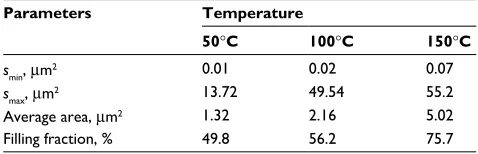
The filling fraction was calculated as the ratio of the sum of particles areas to the sample area. The results of statistical analysis of particles in the Au NP arrays are given in Table 1. To study the luminescent properties of Au NPs/QDs hybrid structures and to test proteins, a confocal laser scanning microscope LSM 710 (Carl Zeiss) with the excitation light wavelength of 405 nm at room temperature was used.
Results and discussion
Optical properties of Au NP arrays
The three main plasmonic structures, as nanohole arrays, diffraction gratings, and nanoslit arrays, that can provide transmission surface plasmon resonance were utilized to improve the detection limits, sensitivity, selectivity,
and dynamic range of biosensors.33 We used the Au NP
arrays on a glass wafer, obtained by sequentially deposit-ing several layers of NPs with a spin coatdeposit-ing process and baking each layer to remove the solvent. With an increase in the numbers of deposited Au NP layers from 5 to 10, the red shift of the maximum of the plasmon peak in the absorption spectra was observed, as shown in Figure 1B. The spectral shift to longer wavelengths is explained by the modification of the resonance frequencies of plasmon oscillations in Au NPs.
The images of Au NP aggregates on a glass surface are shown in Figure 2. An increase in the area of the particles and the filling fraction was observed with increasing baking temperature. The average area of particles increased from
1.32 µm2 to 5.02 µm2 with an increase in temperature from
50°C to 150°C, respectively (Table 1). The filling fraction
increased from 49.8% to 75.7% at these temperatures. This indicates a decrease in the distance between Au NP aggregates.
To obtain the maximum red shift of the plasmon peak, three Au NP arrays on a glass wafer were fabricated at
dif-ferent baking temperatures of 50°C, 100°C, and 150°C. The
A B
0.6
0.5
0.4
0.3
0.2
0.1
0.0
0.02 0.03 0.04 0.05 0.06 0.07
400 500 600
600 0.045 0.060
892 nm 924 nm
1032 nm 0.075
700
700
892 nm
5 layers 10 layers 542 nm
924 nm 1,032 nm
800
Wavelength (nm) Wavelength (nm)
Optical density Optical density
Optical density (au)
Wavelength (nm) 800
400 500 600 700 800
900
900
1,000
1,000
1,100
1,100
Figure 1 (A) Absorption spectra of a solution of Au NPs with a size range of 3–5 nm in p-Xylol. The inset shows the absorption spectrum of p-Xylol. (B) Absorption spectra of Au NP arrays on a glass substrate, obtained by the deposition of 5 and 10 layers with a baking temperature of 100°c for each layer for 1 and 2 minutes, respectively.
Abbreviation: Au NPs, gold nanoparticles.
Table 1 results of statistical analysis of Au NPs array images
Parameters Temperature
50°C 100°C 150°C
smin, mm2 0.01 0.02 0.07
smax, mm2 13.72 49.54 55.2
Average area, mm2 1.32 2.16 5.02
Filling fraction, % 49.8 56.2 75.7
Abbreviation: Au NPs, gold nanoparticles.
Nanotechnology, Science and Applications downloaded from https://www.dovepress.com/ by 118.70.13.36 on 24-Aug-2020
Dovepress
Kurochkina et al
layers of Au NP array were baked for 1 minute. At the baking
temperature of 50°C, the absorption band maximum
associ-ated with the excitation of localized surface plasmons in Au NP arrays was found at a wavelength of 620 nm. At higher baking temperatures, a significant reduction in absorption was observed, as shown in Figure 3A. The observed blue shift of the maximum in the absorption spectra was associated with the melting of single Au particles and the formation of Au NP aggregates of large dimensions (Table 1).
Optical properties of Au NPs/QDs hybrid
structures
The lifetime of the exciton changes due to the coupling with the radiative and nonradiative plasmon modes, which can lead to attenuation or amplification of the radiation. The PL ampli-fication in metal–semiconductor structures is associated with the resonance interaction of dipole radiators with the modes
of localized surface plasmons in metallic NPs.17–19 The
quan-tum efficiency of the emitting electron–hole pair increases in the near optical field of the plasmon mode excited in the gold structures. The process of Förster resonance energy transfer (FRET) between semiconductor NPs in the presence of metal nanocrystals can become faster. The enhancement of FRET occurs due to the effect of plasmon-assisted amplification of electric fields inside the nanoscale assembly. Simultaneously, metal nanocrystals can lead to an increase of energy losses
during the FRET process.34,35
To enhance of the PL intensity of CdSe/ZnS QDs as a result of the exciton–plasmon interaction, we deposited the QDs on the surface of Au NPs array. The efficiency of exciton–plasmon interactions depends on the relative sizes of metallic NPs and QDs, their spatial distribution, the spectral overlap of the QDs radiation, and the surface plasmon reso-nance of Au NPs. Figure 3B shows the absorption spectra Figure 2 images of Au NP arrays on a glass wafer obtained using an optical microscope. Samples were prepared using a spin coating process followed by baking at 50°c (A), 100°c (B), and 150°c (C) within 1 minute to remove the solvent.
Abbreviation: Au NPs, gold nanoparticles.
100 m
A B C
100 m 100 m
A B
0.09
0.06 0.15
0.12
0.06 0.09 0.18
0.12 0.15
400 500 600
Wavelength (nm)
Au NPs (baking 50°C)+QD CdSe/ZnS QD CdSe/ZnS on glass wafer
Au NPs (baking 100°C)+QD CdSe/ZnS Au NPs (baking 150°C)+QD CdSe/ZnS 50°C
100°C 150°C
580 nm
600 nm 620 nm
615 nm 615 nm
615 nm 618 nm
700 800
500 550 600 650
Wavelength (nm)
Optical density
Optical densit
y
700 750
Figure 3 (A) Absorption spectra of Au NP arrays on a glass wafer with a processing temperature of 50°c, 100°c, and 150°c. (B) Absorption spectra of the same samples of Au NP arrays on a glass wafer with cdSe/ZnS QDs deposited on their surface from toluene solution by spin coating.
Abbreviations: Au NPs, gold nanoparticles; QDs, quantum dots.
Nanotechnology, Science and Applications downloaded from https://www.dovepress.com/ by 118.70.13.36 on 24-Aug-2020
Dovepress hybrid structures for biosensor applications
of hybrid structures of Au NP arrays baked at 50°C, 100°C,
and 150°C with CdSe/ZnS QDs. The absorption spectrum
of CdSe/ZnS QDs on a glass wafer is given in Figure 3B for comparison. The exciton–plasmon interaction between QDs and Au NPs led to changes in the absorption spectra of the samples. The maximum of the QDs absorption band corresponds to a wavelength of 615 nm. With an increase in the baking temperature of Au NP layers, a decrease in the intensity of the absorption was observed. These changes cor-related with the increase in the Au NPs area and the filling fraction (Table 1).
The PL spectra of the CdSe/ZnS QDs deposited on the Au NPs surface of samples obtained at different baking temperatures are shown in Figure 4A. The CdSe/ZnS lumi-nescence in the hybrid structures with Au NPs was observed in the wavelength range of 575–700 nm at room temperature (Figure 4A). The PL spectra of samples with Au NP arrays
treated at temperatures of 100°C and 150°C coincide in
Figure 4A. The PL intensity of QDs on a surface of the Au NP arrays increased by 60% compared to the QDs intensity on a glass wafer. This can be explained by the incomplete overlap of the QDs and the Au NP arrays of the absorption spectra (Figure 3A and B) without which it is impossible to enhance the intensity of PL.
interaction of Au NPs/QDs hybrid
structures with protein
The biosensor should basically consist of a transducer and a recognition element for a specific analyte. Hybrid solid
structures of Au NPs/QDs on a glass wafer have been studied as a possible biosensor of protein analyte. On the surface of hybrid structures with Au NP layers baked under 150°C was precipitated bovine serum albumin in a phosphate buf-fer saline. Figure 4B shows the luminescence spectra of Au NPs/QDs hybrid structures with BSA and without it. We used solution with 0.09 wt% and 0.4 wt% BSA. The intensification of PL intensity was observed with an increase in the BSA weight content (Figure 4B). These results indicate the promis-ing use of the studied hybrid structures as optical biosensors for qualitative detection and quantitative determination of protein content in solutions.
Conclusion
In this paper, the hybrid structures based on gold particle arrays and CdSe/ZnS QDs with a core diameter of 5 nm were experimentally investigated. Au NP arrays were fabricated on a glass wafer using the spin coating technique, and their microscopic structure and absorption spectra were studied. It has been shown that the baking temperature of the Au NP layers influenced their structure and absorption spectra. With increasing baking temperature, the agglomerates’ area and the filling fraction of the Au NPs increased. The blue shift of the plasmon peak in the absorption spectra was observed at the same time. The increase in absorption and a red spectral shift of the plasmon peak as well as the enhancement of PL intensity in the hybrid structures spectra of CdSe/ZnS QDs on the surface of the Au NPs array were observed. When testing the BSA using the prepared hybrid structures, it was found out 0.035
A B
0.028
0.021
0.014
0.007
0.000
0.08
0.02 0.00 0.06 0.04 0.18
0.12 0.10 0.16 0.14
560 600 640 680
Wavelength (nm) CdSe/ZnS QDs AuNPs (50°C)/QDs AuNPs (100°C)/QDs AuNPs (150°C)/QDs
AuNPs/QDs
AuNPs/QDs+0.09 wt% BSA AuNPs/QDs+0.4 wt% BSA
720 760 800
550 600 650
Wavelength (nm)
Normalized PL
intensit
y
Normalized PL
intensit
y
700 750
Figure 4 (A) Pl spectra of cdSe/ZnS QDs with a core diameter of 5 nm deposited on the surface of Au NP arrays on a glass wafer, obtained at the temperatures of 50°c, 100°c, and 150°c. (B) Pl spectra of the Au NPs/QDs hybrid structure without and with 0.4 wt% and 0.09 wt% BSA in a PBS, deposited using the spin coating technique.
Abbreviations: Au NPs, gold nanoparticles; BSA, bovine serum albumin; Pl, photoluminescence; QDs, quantum dots.
Nanotechnology, Science and Applications downloaded from https://www.dovepress.com/ by 118.70.13.36 on 24-Aug-2020
Dovepress
Kurochkina et al
that its weight content affects the intensity of the PL of these structures. The experimental results obtained in this work allow us to state that such structures can serve as an optical biosensor for the qualitative and quantitative determination of protein content in solutions. Further studies of such struc-tures, aimed at enhancing the exciton–plasmon interaction of the Au NPs array with QDs, can increase their effectiveness.
Acknowledgment
We greatly acknowledge the support provided by the Ger-man Research Foundation under grants LU 605/16-1 and HI 1261/5-1.
Disclosure
The authors report no conflicts of interest in this work.
References
1. Holzinger M, Le Goff A, Cosnier S. Nanomaterials for biosensing applications: a review. Front Chem. 2014;2:63.
2. Guo X. Surface plasmon resonance based biosensor technique: a review.
J Biophotonics. 2012;5(7):483–501.
3. Nie L, Liu F, Ma P, Xiao X. Applications of gold nanoparticles in optical biosensors. J Biomed Nanotechnol. 2014;10(10):2700–2721. 4. Saha K, Agasti SS, Kim C, Li X, Rotello VM. Gold nanoparticles in
chemical and biological sensing. Chem Rev. 2012;112(5):2739–2779. 5. Shah M, Badwaik VD, Dakshinamurthy R. Biological applications of
gold nanoparticles. J NanosciNanotechnol. 2014;14(1):344–362. 6. Liu Z, Zhao F, Gao S, Shao J, Chang H. The applications of gold
nanoparticle initialed chemiluminescence in biomedical detection.
Nanoscale Res Lett. 2016;11:460.
7. Schmid G. Physical and chemical consequences of size-reduction of gold: bioresponse and biodistribution. J Cluster Sci. 2014;25(1):29–49. 8. Lindquist NC, Luhman WA, Oh SH, Holmes RJ. Plasmonic nanocavity
arrays for enhanced efficiency in organic photovoltaic cells. Appl Phys Lett. 2008;93(12):123308.
9. Kang MG, Xu T, Park HJ, Luo X, Guo LJ. Efficiency enhancement of organic solar cells using transparent plasmonic Ag nanowire electrodes.
Adv Mater. 2010;22:4378–4383.
10. Cohen-Hoshen E, Bryant GW, Pinkas I, Sperling J, Bar-Joseph I. Exci-ton–plasmon interactions in quantum dot–gold nanoparticle structures.
Nano Lett. 2012;12(8):4260–4264.
11. Toropov NA, Kamalieva AN, Vartanyan TA. Thin films of organic dyes with silver nanoparticles: enhancement and spectral shifting of fluorescence due to excitation of localised surface plasmons. Int J
Nanotechnol. 2016;13(8–9):642–647.
12. Sadeghi SM, West RG, Nejat A. Photo-induced suppression of plas-monic emission enhancement of CdSe/ZnS quantum dots. Nanotech
nology. 2011;22(40):405202.
13. Guo P, Xu J, Zhuang X, et al. Surface plasmon resonance enhanced band-edge emission of CdS-SiO2 core-shell nanowires with gold nanoparticles attached. J Mater Chem C. 2013;1(3):566–571.
14. Cheng MT, Liu SD, Zhou HJ, Hao ZH, Wang QQ. Coherent exciton– plasmon interaction in the hybrid semiconductor quantum dot and metal nanoparticle complex. Opt Lett. 2007;32(15):2125–2127.
15. Park J, Vak D, Noh YY, Lim B, Kim DY. Surface plasmon enhanced pho-toluminescence of conjugated polymers. Appl Phys Lett. 2007;90(16): 161107.
16. Pohl W. Near-field optics and the surface plasmon polariton. In: Kawata S, editor. Near-Field Optics and Surface Plasmon Polaritons. Berlin: Springer; 2001:1–13.
17. Govorov AO, Bryant GW, Zhang W, et al. Exciton–plasmon interaction and hybrid excitons in semiconductor–metal nanoparticle assemblies.
Nano Lett. 2006;6(5):984–994.
18. Haridas M, Tripathi LN, Basu JK. Photoluminescence enhancement and quenching in metal semiconductor quantum dot hybrid arrays. Appl
Phys Lett. 2011;98(6):063305.
19. Tripathi LN, Praveena M, Basu JK. Plasmonic tuning of photolumi-nescence from semiconducting quantum dot assemblies. Plasmonics.
2013;8(2):657–664.
20. Kagan CR, Murray CB, Bawendi MG. Long-range resonance transfer of electronic excitations in close-packed CdSe quantum-dot solids. Phys
Rev B Condens Matter. 1996;54(12):8633–8643.
21. Manjavacas A, Garcií de Abajo FJ, Nordlander P. Quantum plex-citonics: strongly interacting plasmons and excitons. Nano Lett. 2011;11(6):2318–2323.
22. Rand P, Peumans P, Forrest SR. Long-range absorption enhancement in organic tandem thin-film solar cells containing silver nanoclusters.
J Appl Phys. 2004;96(12):7519–7526.
23. Nakayama K, Tanabe K, Atwater HA. Plasmonic nanopar-ticle enhanced light absorption in GaAs solar cells. Appl Phys Lett.
2008;93(12):121904.
24. Lee JH, Park JH, Kim JS, Lee DY, Cho K. High efficiency polymer solar cells with wet deposited plasmonic gold nanodots. Org Electron. 2009;10(3):416–420.
25. Wu L, Chen FC, Hsiao YS, et al. Surface plasmonic effects of metallic nanoparticles on the performance of polymer bulk heterojunction solar cells. ACS Nano. 2011;5(2):959–967.
26. Kim SS, Na SI, Jo J, Kim DY, Nah YC. Plasmon enhanced performance of organic solar cells using electrodeposited Ag nanoparticles. Appl
Phys Lett. 2008;93(7):073307.
27. Belyaev KG, Usikova AA, Jmerik VN, et al. Plasmon-induced enhance-ment of yellow-red luminescence in InGaN/Au nanocomposites.
Semiconductors. 2015;49(2):247–253.
28. Liu X, McBride SP, Jaeger HM, Nealey PF. Hybrid nanostructures of well-organized arrays of colloidal quantum dots and a selfassembled monolayer of gold nanoparticles for enhanced fluorescence.
Nanotech-nology. 2016;27(28):285301.
29. Petryayeva E, Algar WR, Medintz IL. Quantum dots in bioanalysis: a review of applications across various platforms for fluorescence spectroscopy and imaging. Appl Spectrosc. 2013;67(3):215–252. 30. Huang Q, Chen J, Zhao J, Pan J, Lei W, Zhang Z. Enhanced
photolumi-nescence property for quantum dot-gold nanoparticle hybrid. Nanoscale
Res Lett. 2015;10(1):400.
31. Zhang L, Song Y, Fujita T, Zhang Y, Chen M, Wang TH.Large enhance-ment of quantum dot fluorescence by highly scalable nanoporous gold.
Adv Mater. 2014;26(8):1289–1294.
32. Ozel T, Nizamoglu S, Sefunc MA, et.al. Anisotropic emission from multilayered plasmon resonator nanocomposites of isotropic semicon-ductor quantum dots. ACS Nano. 2011;5(2):1328–1334.
33. Lertvachirapaiboon C, Baba A, Ekgasit S, Shinbo K, Kato K, Kaneko F. Transmission surface plasmon resonance techniques and their potential biosensor applications. Biosens Bioelectron. 2018;99:399–415. 34. Govorov AO, Lee J, Kotov NA. Theory of plasmon-enhanced Förster
energy transfer in optically excited semiconductor and metal nanopar-ticles. Phys Rev B. 2007;76(12):125308.
35. Zhai Y, Wang Q, Qi Z, Li C, Xia J, Li X. Experimental investigation of energy transfer between CdSe/ZnS quantum dots and different-sized gold nanoparticles. Physica E Low Dimens Syst Nanostruct. 2017;88: 109–114.
Nanotechnology, Science and Applications downloaded from https://www.dovepress.com/ by 118.70.13.36 on 24-Aug-2020
Dovepress
Nanotechnology, Science and Applications
Publish your work in this journal
Submit your manuscript here: https://www.dovepress.com/nanotechnology-science-and-applications-journal
Nanotechnology, Science and Applications is an international, peer-reviewed, open access journal that focuses on the science of nanotechnology in a wide range of industrial and academic applications. It is characterized by the rapid reporting across all sectors, including engineering, optics, bio-medicine, cosmetics, tex-tiles, resource sustainability and science. Applied research into nano-materials,
particles, nano-structures and fabrication, diagnostics and analytics, drug delivery and toxicology constitute the primary direction of the journal. The manuscript management system is completely online and includes a very quick and fair peer-review system, which is all easy to use. Visit http://www.dovepress.com/ testimonials.php to read real quotes from published authors.
Dove
press
hybrid structures for biosensor applications
Nanotechnology, Science and Applications downloaded from https://www.dovepress.com/ by 118.70.13.36 on 24-Aug-2020