of the
Myf-5
Gene
Oliver Coutelle
A thesis presented for the
Degree of Doctor of Philosophy (Ph.D.)
Department of Biochemistry
University College London
1998
National Institute for Medical Research,
All rights reserved
INFORMATION TO ALL USERS
The quality of this reproduction is dependent upon the quality of the copy submitted.
In the unlikely event that the author did not send a complete manuscript and there are missing pages, these will be noted. Also, if material had to be removed,
a note will indicate the deletion.
uest.
ProQuest U643739
Published by ProQuest LLC(2016). Copyright of the Dissertation is held by the Author.
All rights reserved.
This work is protected against unauthorized copying under Title 17, United States Code. Microform Edition © ProQuest LLC.
ProQuest LLC
789 East Eisenhower Parkway P.O. Box 1346
The work presented here has been supported by an MRC Studentship. I am grateful to
Prof. Peter Rigby for accepting me as a student, and for giving me the freedom to study
the pufferfish. I thank Dr Sydney Brenner for letting me work in his Cambridge
laboratory and for making available his Fugu cosmid library. Particular thanks to Dr.
Samuel Aparicio from his lab as a friend and for helpful advice, scientific and otherwise.
My greatest respect to Dr. Dennis Summerbell from our laboratory at NIMR for his
brilliant teaching and often needed encouragement, as well as his patience in discussing
my transgenic experiments. I shall not forget Dennis’ lectures in mouse anatomy which
have excited even a rather reluctant student as myself (to a degree) and made me look at
my work from a different perspective.
Thanks also for many enjoyable discussions to Dr. Rajeev Gupta and Jonathan
Gilthorpe, who shared many of the ups and downs with me. I am grateful to Marie Vandromme, Ed Stanley, Dave Cox, Peter Ashby, Chandi Halai, Debbie Duthie and
Jacky Smith. I gratefully acknowledge the technical staff in the SPF unit, particularly
Hannah Boyes and Zoe Webster for looking after our mice.
The work presented here addresses the question of how skeletal muscle
formation is initiated in the mouse by dissecting the regulatory mechanisms that control
the myogenic regulatory factor Myf-5. Myf-5 is expressed in the dorsal somite from
E8, before the other MRFs, myogenin, MRF4 and MyoD, become activated. In the
mouse Myf-5 is located 8.5kb downstream of MRF4. Previous results have shown that
dispersed over the intergenic region and intragenic regions are the regulatory elements
involved in directing Myf-5 expression to the different anatomical subdomains that make
up its complete expression pattern. The regulatory element(s) controlling the dorsal
somite expression of Myf-5 is contained in the intergenic region while ventral somite
expression depends on elements in the Myf-5 gene itself. Because of the large size of
this region I have isolated the MRF4 and Myf-5 genes of the teleost Fugu rubripes,
which has a genome eight times smaller than that of the mouse. Although synteny is
conserved in Fugu and the intergenic distance is only 3kb, noncoding sequence
including the introns is poorly conserved. Focusing on the Myf-5 gene itself, sequence
comparison between the mouse and human Myf-5 genes was employed sucessfully to
eliminate more than 60% of the intron sequence by identifying conserved regions in the
3'half of each of the Myf-5 introns which, together with the 3'UTR can activate reporter
gene expression in the ventral posterior part of the somites. EMSA analysis with
embryonic protein extracts revealed several protein binding regions within the conserved intron fragments and subsequent transgenic analysis showed not only that separate
genomic regions control individual anatomical domains of Myf-5 expression, but that
within these regions multiple binding sites are found, adding a further level of
complexity to the regulation of Myf-5. Analysis of the Fugu Myf-5 gene in transgenic
mice showed remarkable similarities with the expression pattern of Myf-5 in another
teleost, the zebrafish Danio rerio. Both are expressed in the presomitic mesoderm, as
well as the somites, suggesting that the expression of the Fugu transgene is a reflection
Table of Contents
Acknowledgements...2
Abstract ... 3
List of Figures... 9
List of Tables... 10
Abbreviations... 11
Chapter 1 - Introduction
1. Germlayer Formation in the Mouse... 132. Germlayer Formation in Teleost Fish... 17
3. Mesodermal Patterning...18
4. Somitogenesis...19
5. Somite Patterning... 22
5. 1. Dorsoventral Patterning...22
5. 2. Sonic Hedgehog and Wnt Signalling... 23
5. 3. Mediolateral Somite Patterning... 26
6. Somites in Teleost Fish... 28
7. The Myogenic bHLH Factors... 31
8. The Myogenic Cascade... 32
9. MRF Null M utations...35
9. 1. MyoD Null M utants...35
9. 2. Myf-5 Null Mutants... 36
9. 3. Myf-5IMyoD Double M utants...36
9. 4. Myogenin Null M utants...37
9. 5. MRF4 Null M utants... 37
10. What Regulates MyoD and Myf-51...38
11. Could Myogenesis Be Repressed?... 40
12. The Pufferfish as a Model Organism... 42
12. 1, Genome Size and Complexity...42
12. 2. The Small Genome of Fugu rubripes... 43
12. 3. The Fugu Genome Never Acquired ‘Junk-DNA’ ...43
12. 4. Small Is B eautiful... 45
12. 5. . . . Or is it? ...45
12. 6. Studies on Developmentally Regulated Genes in Fugu... 46
Chapter 2 - Materials and Methods
1. Solutions and Reagents... 49
2. DNA Manipulations... 54
2. 1. Phenol Extraction of D N A ...54
2. 2. Ethanol Precipitation of DNA...54
2. 3. PEG Precipitation of D N A ...54
2. 4. Endonuclease Digestion of DNA...54
2. 5. DNA Fragmentation by Sonication...54
2. 6. Dephosphorylation of D N A ... 54
2. 7. Repair of DNA E n d s...55
2. 8. Oligonucleotide Primer Synthesis... 55
2. 9. Agarose Gel Electrophoresis and Documentation... 56
2. 10. DNA Fragment Isolation... 56
3. DNA Cloning...56
3. 1. DNA Ligations...56
3. 2. Transformation of Competent C ells...57
3. 3. Recombinant Screening...57
4. Isolation of DNA and R N A ...58
4. 1. Plasmid Isolation by Alkaline Lysis "Miniprep"3... 58
4. 2. Plasmid Isolation by CsCl "Maxiprep"3...58
4. 3. Genomic DNA Isolation from Zebrafish Embryos... 59
4. 4. Cosmid DNA Isolation... 59
4. 5. Isolation of RNA...59
4. 6. cDNA Synthesis... 60
5. Southern A nalysis... 60
5. 1. Southern Blotting...60
5. 2. Southern Hybridisation Probes... 60
5. 3. Southern Hybridisation... 61
6. Northern Analysis... 61
6. 1. Northern B lotting...61
7. Library Screening... 62
7. 1. Fugu Cosmid Library Screening...62
7, 1. 1. Sequence Scanning of Cosmid Clones... 63
7. 2. Zebrafish cDNA Library Screening... 63
7. 2. 2. Filter Preparation and Hybridisation... 64
8. Zebrafish Myf-5 Cloning by Degenerate PCR ... 65
9. DNA Sequencing...65
9. 2. Sequencing on the ABI 3 7 7 ... 66
10. Generation and Analysis of Transgenic M ice... 66
10. 1. Transgenic Reporter Constructs... 66
10. 1. 1. Mouse Myf-5 Reporter Constructs...66
10. 1.2. Fugu Myf-5 Reporter Constructs... 69
10. 1. 3. Hybrid Myf-5 Reporter Constructs...69
10. 2. Preparation of DNA for Microinjection...69
10. 3. Transgenic Procedure... 69
10. 4. Transgene Detection by P C R ... 71
11. Histological Studies...71
12. DNA Electrophoretic-Mobility Shift Assay (EM SA)... 72
12. 1. Probe Labelling and Purification... 72
12. 2. Preparation of Protein Extracts... 72
12. 3. EMSA Conditions... 73
13. In Situ Hybridisation of Zebrafish Embryos... 73
13. 1. Riboprobe Synthesis... 73
13. 2. Preparation of Embryos... 74
13.3. Hybridisation... 74
13. 4. Posthybridisation Treatment...74
Chapter 3 - Comparative Sequence Analysis of the
Vertebrate
Myf-5
/MRF4 Loci
1. Introduction... 762. Isolation and Characterisation of Fugu MRF4 and M yf-5...79
2. 1. A Fugu Cosmid Contains Both MRF4 and M y f-5... 79
2. 2. The Genomic Structures of Fugu MRF4 and M y f-5... 82
2. 3. Interspecies Comparison at the DNA Level...85
2. 4. Interspecies Comparison at the Protein Level... 85
2. 5. CpG Islands and Repeat Elements... 89
3. Pairwise DNA Sequence Analysis Between Fugu and Mouse...90
4. Pairwise Comparisons Between Mouse, Human and Bovine...91
5. Summary... 95
Chapter 4 - EMSA Analysis of the
Myf-5
Introns
1. Introduction... 992. Fragment A ... 102
3. Fragment B ... 102
4. Fragment C ... 102
6. Fragment E ... 106
7. Fragment F ... 106
8. Fragment G ... 106
9. Summary... 108
Chapter 5 - Transgenic Analysis of the Regulatory
Role of the Mouse
Myf-5
introns
1. Introduction... 1092. Dissection of Regulatory Elements in Myf-5 using Transgenic Mice... 110
2.1. Construct p l 2 ...110
2.2. Construct p i ... 115
2.3. Construct p2... 115
2.4. Construct pUTR...116
2.5. Construct p l2 U T R ...116
2.6. Construct p lU T R ... 121
2.7. Construct p2UTR... 121
2.8. Construct pCFUTRR...126
2.9. Construct pCFH...129
2.10. Construct pC FA ... 132
2.11. Construct pCFHA...132
2.12. Construct pAAH... 132
2.13. Construct pACAH...137
2.14. Construct pAGAFl...137
2.15. Construct pCFAA...137
2.16. Construct pACPH...138
2.17. Construct pAFPH...138
2.18. Construct pAGPH...138
Chapter 6 -
Myf-5
In Zebrafish and
Fugu
1. Introduction... 1422. Cloning of the Zebrafish Myf-5 homologue... 142
3. Expression of 7Myf-5 in zebrafish embryos... 143
4. Transgenic Analysis of Fugu M yf-5...147
4.1. Construct Fugu p l2 U T R ... 147
4.2. Construct Fugu p l 2 ... 152
4.3. Construct pZ F M ... 152
4.4. Construct pZ F F ...157
4.5. Construct Fugu pUTR...157
Chapter 7 - Discussion
1. Introduction...161
2. Fugu MRF4 and Myf-5 Form a Syntenic Linkage Group...162
3. Fugu is Not a Good Model for MRF4 and Myf-5...163
4. Regions in the Myf-5 Introns Conserved Between Human and Mouse Regulate ... Ventral Somitic Expression... 164
5 Multiple Binding Sites Located in the Mouse Myf-5 Introns...165
6. Transgenic Analysis Reveals Complex Regulatory Mechanism for Myf-5... 166
7. Conserved Regulatory Elements in Both Introns and the UTR of Myf-5 are ... Required for Ventral Posterior Somite Expression in the Mouse... 168
8. Separate Elements Appear to Control Intensity and Spatial Distribution of Transgene Expression... 169
9. Fragments D, F and G, but not C, are Involved in Ventral Somitic Expression ... o f M yf-5...170
10. A 'Branchial Arch Element' in the Myf-5 Gene?... 170
11. The 3' UTR of the Fugu Myf-5 Gene Drives Expression in the Somites and the Presomitic Mesoderm... 171
12. Conserved Element in Intron 1...174
13. Myf-5 is Expressed in Lateral Presomitic C ells... 174
14. Outlook... 176
Appendix
1. Oligonucleotides...179II. Fugu MRF4 and Myf-5 Genes: Complete Coding Sequence...181
III. Fugu MRF4 and Myf-5: Genomic Contig...185
IV. Human Myf-5 Gene: Complete Coding Sequence... 187
V. Zebrafish Myf-5 Gene: Complete Coding Sequence... 187
IV. Mouse Myf-5 Gene: Complete Coding Sequence... 189
List of Figures
Figure 1 : Early Development of the Mouse Embryo... 16
Figure 2: Epiboly in Teleost Fish... 16
Figure 3 : Somite Differentiation...21
Figure 4: Somite Patterning... 25
Figure 5: Somites in Teleost Fish...30
Figure 6; Temporal Expression of the Myogenic Factors in the M ouse... 34
Figure 7: The Japanese Pufferfish, Fugu rubripes...44
Figure 8A; Regulatory Elements in the MRF4!Myf-5 Region... 78
Figure 8B: Construct H M Z17... 77
Figure 8C: Construct MFGZ... 77
Figure 9A: The Genomic Organisation of the Fugu MRF4 and Myf-5 Genes...81
Figure 9B: Selected Restriction Sites of the Fugu Region... 81
Figure 10: Schematic Comparison of Mouse and Fugu Myf-5 and M R F 4... 84
Figure 11 A: Clustal Alignment: M RF4... 87
Figure 1 IB: Phylogenetic Tree: M RF4... 87
Figure 11C: Clustal Alignment: Myf-5... 88
Figure IID : Phylogenetic Tree: My/-5...88
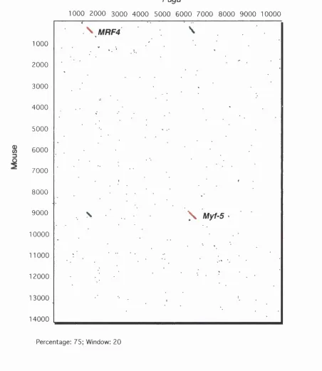
Figure 12A: Dot Plot Analysis of the MRF4fMyf-5 Region... 93
Figure 12B: Dotplot Analysis: Myf-5 Human vs Bovine... 94
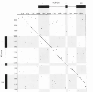
Figure 12C: Dotplot Analysis: Myf-5 Human vs M ouse... 94
Figure 13A: Dotplot Analysis: Mouse vs Human Myf-5 Intron 1 ...97
Figure 13B: Dotplot Analysis: Mouse vs Human Myf-5 Intron 2 ...97
Figure 14A: Size and Location of EMSA Probes... 101
Figure 14B: Subdivision of EMSA Fragment D ... 101
Figure 14C: Origin of the Mouse Tissues for EMSA Extracts... 101
Figure 15A: Electrophoretic Mobility Shift Assays: Fragment A ... 104
Figure 15B: Electrophoretic Mobility Shift Assays: Fragment B ... 104
Figure 15C: Electrophoretic Mobility Shift Assays: Fragment C ... 104
Figure 15D: Electrophoretic Mobility Shift Assays: Fragment D ... 104
Figure 15E: Electrophoretic Mobility Shift Assays: Fragment E ... 105
Figure 15F: Electrophoretic Mobility Shift Assays: Fragment F ... 105
Figure 15G: Electrophoretic Mobility Shift Assays: Fragment G ... 105
Figure 16 A-B : Construct p l 2 ... 114
Figure 16C: Construct H M Z17... 114
Figure 17A-C: Construct p i ...118
Figure 18 A-B: Construct pUTR... 120
Figure 18C-D: Construct p i U TR...120
Figure 19A-J: Construct pl2UTR - Fritz...123
Figure 20A-D: Construct p2UTR...125
Figure 21A-G: Construct pCFUTRR... 128
Figure 22A-B: Construct pCFH... 131
Figure 22C-D: Construct pCFA... 131
Figure 23A: Construct pCFHA...134
Figure 23B-C: Construct pCFAA...134
Figure 24A-B: Construct pAAH... 136
Figure 24C: Construct pACAH... 136
Figure 24D: Construct pAGAH...136
Figure 25 A-B : Construct pACPH... ...140
Figure 25C-D: Construct pAFPH... 140
Figure 26A-B: Construct pAGPH...141
Figure 27A-E: ZMyf-5 Expression Pattern...145
Figure 28A-H: Construct Fugu p l2 U T R ... 151
Figure 29: Myf-5 Homologues: Size Comparison...153
Figure 30: Dotplot: Zebrafish vs Fugu M yf-5... 154
Figure 31A-C: Construct pZ F M ... 156
Figure 32A-C: Construct Fugu pUTR... 156
Figure 33A-D: Construct p Z F F ... 159
List of Tables
Table 1: Exon and Intron Sizes of MRF4 and Myf-5... 84Table 2: Summary of EMSA Results for Myf-5 Intron Fragm ents... 107
Table 3A: Summary of the Mouse Transgenic R esults... I l l Table 3: Mouse Myf-5 Reporter Constructs...112
Table 4A: Summary of the Fugu and Hybrid Transgenic Results...148
Table 4: Fugu and Hybrid Myf-5 Reporter Constructs... 149
Abbreviations
AP anterior-posterior
ATP adenosine 5'-triphosphate
bp base pair
BSA bovine serum albumin
bHLH basic -helix-loop-helix protein structure motif
cDNA complementary DNA
cpm counts per minute
C-terminus carboxy terminus
dH^O distilled water
DNA deoxyribonucleic acid
DNase deoxyribonuclease
dATP deoxyadenosine 5'-triphosphate
dCTP deoxycytidine 5'-triphosphate
dGTP deoxyguanosine 5'-triphosphate
dNTP deoxyribonucleoside 5'-triphosphate
dTTP deoxythymidine 5'-triphosphate
ddNTP dideoxyribonucleoside 5'-triphosphate
dpc days post coitum
DTP dithiothreitol
DV dorsal-ventral
EDTA ethylenediaminetetraacetic acid
EMSA electrophoretic-mobility shift assay
HEPES N-2-hydroxyethylpiperazine-N'-ethanesulphonic acid
hpf hours post fertilisation
kb kilo base (base pairs)
KD kilo dalton
MBq mega bequerel
M molar
Mb mega base (base pairs)
pM micro molar
mM milli molar
l^g micro gram
1^1 micro htre
mg miUigram
MOPS 3-N-morpholinopropanesulphonic acid
MYF MYogenic Factor
ng nano gram
ml milli litre
NP-40 Nonidet P-40
N-terminus amino terminus
OAc acetate
OD optical density
PAGE polyacrylamide gel electrophoresis
PBS phosphate buffered saline
PCR polymerase chain reaction
PMSF phenylmethylsulphonyl fluoride
RNase ribonuclease
rpm revolutions per minute
RT reverse transcriptase
SSC sodium chloride/ sodium citrate
SDS sodium dodecyl sulphate
SPF specific pathogen free
Tris 2-amino-2-(hydroxymethyl)-1,3-propanediol
TBF Tris-borate-FDTA buffer
TF Tris-FDTA buffer
u units
UV ultra violet
W watts
%(w/v) grams per 100ml
%(v/v) ml per 100ml
Chapter 1
In troducti on
An important task in the field of vertebrate embryology is to elucidate the
mechanisms by which mesodermal cells acquire their developmental fates. The
determination of specific cell fates underlies the most fundamental processes of
development. In vertebrates, some of the early steps in determining cell fate take place
during gastrulation when cells become committed to each of the three primary germ layers endoderm, ectoderm and mesoderm. Mesoderm specification begins with the
ingression of epiblast cells through the primitive streak followed by commitment and
allocation to the different mesodermal lineages: midline cells generate axial structures,
notochord and prechordal plate, paraxial mesoderm becomes progressively segmented
into the somites and also gives rise to the head mesoderm, while lateral mesoderm forms
the splanchnopleure and somatopleure. The somites of the paraxial mesoderm give rise
to the axial skeleton, trunk muscle, some head and neck muscle, some bones and muscles of the skull and epidermis of the skin. The work presented here is concerned
with the regulation of the myogenic transcription factor Myf-5 and its role in the paraxial
mesoderm and in the commitment of cells to the myogenic lineages that give rise to
skeletal muscle. Before discussing the process of myogenesis in which mesodermal cells
acquire muscle fate, the establishment of the three germ layers is outlined briefly with
reference to the different model organisms studied as part of project, including the
mouse and teleost fish.
1. Germlayer Formation In the Mouse
Some of the most dramatic stages of differentiation of the embryo take place in the
process of gastrulation by establishing the three germ layers. Various aspects of
gastrulation have been studied in diverse organisms, including insects, birds, amphibia,
fish and mammals. The mouse fate map exhibits topological similarity with those of
chick, amphibia and teleost fish, indicating that even though the physical morphology of
Depending on the particular organism studied, however, the actual process of generating
the germlayers varies considerably.
Initially the undifferentiated cells of the mouse embryo divide to form a cluster of cells
called the morula. Up to the 16 cell stage the morula cells retain their equipotency and
can give rise to complete embryos. After further cell divisions the potency of these cells
becomes gradually restricted. Tight junctions form between the outer cells while a fluid
filled cavity (blastocoel) develops within the embryo. The cells in contact with the
outside become trophectoderm while the cells with no contact to the outside become the
inner cell mass (ICM). Cells of the ICM at the interface with the blastocoel give rise to
the primitive endoderm and cells trapped between the primitive endoderm and the
trophectoderm (black) form the epiblast (red) (see Fig. 1, stage E4.5). Around day 5 of
development, the blastula stage mouse embryo becomes implanted into the uterus and
part of the trophectoderm proliferates and pushes the epiblast ahead of itself (Fig. 1,
stage E5.5). The epiblast adopts epithelial character and becomes the primitive
mesectoderm surrounding the proamniotic cavity (green) (Fig. 1, stage E6). At around
E7 of development the primitive streak is formed from a subset of mesectoderm cells
near the interface of the extraembryonic and embryonic halves of the egg cylinder. The epithelial continuity of mesectoderm is lost in the streak as cells delaminate to form the
three embryonic germlayers: ectoderm, mesoderm and endoderm. Fate mapping studies
of mesectoderm cells using lineage tracers (Gardner 1982, 1983) show that these cells
give rise to mesoderm as they ingress through the streak and some of the cells intercalate
into the visceral endoderm layer to give rise to embryonic endoderm (Fig. 1, see stage
E7) and also to extraembryonic tissues (Gardner and Rossant, 1979). Thus cells from
the mesectoderm (epiblast) are thought to contribute to all three germ layers of the
developing foetus.
At the anterior end of the streak the node is formed by a group of cells that appears to
have organising capacity similar to Hensen's node in the chick embryo and Spemann's
organiser in Xenopus and is likely to be involved in patterning of axial structures. It has
been suggested that the node also contains a proliferative centre from which the
notochord and floor plate cells of the neural tube originate (Selleck and Stem, 1991;
Sulik et a l, 1994). Indeed, transplantation of chick node into zebrafish embryos
generates a secondary axis, suggesting that the underlying signals are evolutionarily
conserved (Hatta and Takahashi, 1996). Formation of the notochord divides the
mesodermal layer into the paraxial mesoderm on either side of the AP axis and marks the
onset of neumlation. Further differentiation of the paraxial mesoderm leads to its
successive segmentation and the formation of the somites, from which most skeletal
muscle is derived. In fish embryos, muscle is also formed from paraxial mesoderm but
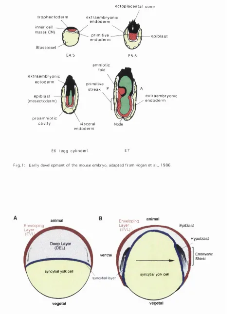
Figure 1: Early Development of the Mouse Embryo. At around E4.5 the
initially equipotent cell mass divides into epithelial trophectoderm (black) and inner cell
mass (red) of the blastula with its fluid filled cavity the blastocoel (yellow).
Around E5 the embryo implants into the uterus, the trophectoderm of the ectoplacental
cone pushes the ICM (epiblast) and the overlaying primitive endoderm (white) inside the
egg cylinder. Within the epiblast cells the proamniotic cavity forms (green) at E6,
followed by the formation of the primitive streak around E7 at the interface with the
extraembryonic ectoderm (striped). Cells delaminating from the mesectoderm (epiblast)
ingress through the streak between the mesectoderm and the visceral endoderm and form
two new layers the embryonic mesoderm and endoderm and also contribute to the
extraembryonic endoderm. At the most anterior end of the streak where the node is
formed there is no visceral endoderm overlaying the streak.
Figure 2: Epiboly in Teleost Fisb.
2A: Schematic representation of the blastuala stage zebrafish embryo. The enveloping
layer (red) is the outermost epidermis of the embryo, enveloping the deep cells (DEL)
shown in gray, which ultimately give rise to all three germ layers. At the margin DEL cells leave the blastoderm and contribute to the syncitial (multinucleated) yolk cell
(arrows).
2B; At 80% epiboly the synctial layer (blue) moves toward the vegetal (yolk rich) pole.
The EVL follows the syncitial layer toward the vegetal pole but remains on the outside.
At the margin of the blastoderm (germ ring), the deep cells involute (arrows) and give
rise to the hypoblast (shaded gradient) from which the presumptive mesoderm and endoderm form. The outermost, noninvoluting DEL cells become epiblast from which
the presumptive ectoderm is formed. Additional cell movement towards the dorsal
midline (black arrow) of the embryo leads to a thickening of the germ ring, the
t r o p h e c t o d e r m
i n n e r cel ma s s ( I CM)
B l a s t o c o e l
e x t r a e m b r y o n i c e n d o d e r m
p r i mi t i v e e n d o d e r m
e p i b l a s t
E 4. 5 E5. 5
e x t r a e m b r y o n i c e c t o d e r m
e p i b l a s t ( m e s e c t o d e r m )
p r o a m n i o t i c c a v i t y
a m n i o t i c fold
p r i m i t i v e s t r e a k ^
v i s c e r a l e n d o d e r m
e x t r a e m b r y o n i c e n d o d e r m
E6 ( e g g c y l i n d e r ) E7
Fi g . l : E a r l y d e v e l o p m e n t of t h e m o u s e e mb r y o , a d a p t e d f r o m Hogan e t al., 1 9 8 6 .
animal Envel opi ng
Layer (EVl
syncytial yolk cell
® Envel opi ng Layer
(EVL)
ventral
syncytial layer
Embryonic Shield
Epiblast
Hypoblast
syncytial yolk cell
vegetal vegetal
2. Germlayer Formation In Teleost Fish
The process of gastrulation in amphibia (e.g. Xenopus) and teleost fish (e.g. pufferfish,
zebrafish) shares many features with higher vertebrates. The most extensively studied
teleost is the zebrafish, Danio rerio. The main stages of gastrulation in these small
translucent embryos can be directly observed.
The fertilised egg at first divides synchronously and these intital cleavages are
incomplete (meroblastic) such that the furrows do not pass through the yolk rich region
of the egg thus giving rise to a giant uncleaved yolk cell. In zebrafish cleavages generate
two populations of blastoderm cells: the enveloping layer (EVL) forming the outer
epidermis (periderm) of the embryo (Bouvet, 1976) and the deep cell layer (DEL)
beneath the EVL that gives rise to the major ectodermal, mesodermal and endodermal
portions of the embryo (Martindale et al, 1987). At the midblastula stage DEL cells
located near the margin leave the blastoderm and contribute their nuclei to the syncitial
multinucleated yolk cell (Fig. 2A). During gastrulation, blastodermal cells gradually
move over the yolk cell towards the vegetal (yolk rich) pole, enveloping the yolk cell
which is destined for digestion in the prospective gut of the embryo. This process, for which there is no counterpart in higher vertebrate development, requires spreading of the
blastoderm layer and is driven by radial intercalation of blastomeres (epiboly)
accompanied by concomitant thinning of the blastoderm. In zebrafish, during this stage
extensive cell mixing within the DVL takes place and descendants from single
blastomeres move to diverse locations and will eventually give rise to diverse cell types
(Kimmel and Warga, 1987). Thus fate maps of pregastrulation zebrafish embryos are
difficult to obtain. Involution can be regarded as a similar event as invagination during
development of higher vertebrates (Trinkaus, 1988). Involution marks the onset of
mesoderm formation and takes place at 50% epiboly, when half of the yolk cell is
covered by the enveloping blastoderm. In teleosts involution does not initiate at the
dorsal side of the embryo (as it does in amphibia) but begins more or less
simultaneously around the circumference of the blastoderm. The involuted internal layer
of blastoderm cells forms the hypoblast and the outer blastoderm DEL layer becomes the
epiblast (see Fig. 2B). Convergent movement of cells from lateral positions to the dorsal
midline of the gastrula leads to formation of the embryonic shield (Fig. 2B), a thickening
of the germ ring with some similarity to the primitive streak or the blastopore of amniote embryos and amphibia, respectively. Fate mapping studies in zebrafish have shown that
cells from the most lateral (outermost) layer of the epiblast do not involute and these cells
give rise to ectodermal derivatives. In contrast, the fate of the involuting cells as
described for the streak in amniotes, dependent on when they enter the hypoblast. Cells
from the region near the blastoderm margin enter early and give rise to endoderm while
1990). The specification of mesodermal fate at the molecular level is a complicated and
as yet not fully understood process, in which temporal and spatial distribution of
inductive signalling molecules play a major role.
3. Mesodermal Patterning
It is not clear if a homogeneous population of mesoderm cells ever exists, or if cells
ingressing through the primitive streak are already predetermined to become one of the
various mesoderm derivatives. Indeed, it appears that the first cells ingressing through
the primitive streak adopt the fate of lateral mesoderm, while the cells that follow go on
to develop into the paraxial mesoderm. Thus, mesodermal fate could be determined by
the timing of ingression through the streak alone, but it is likely that additional signalling
molecules are involved in patterning of the mesodermal cells. Candidate patterning
molecules, some with graded or partially restricted, some with uniform expression along
the AP axis of the primitive streak have been found, including peptide growth factors,
signalling molecules and transcription factors of the Zinc-finger and homeodomain- families. Their complex expression patterns suggest combinatorial effects and indicate a
degree of overlap and redundancy in these molecules (Tam and Trainor, 1994). Since dye labelled cells of the rostral presomitic mesoderm of mouse and chick embryos can
give rise to lateral plate mesoderm and endothelium, they can not be totally committed to
a particular cell fate until after somite segmentation (Bagnall, 1988; Beddington and
Martin, 1989; Selleck and Stem, 1991, 1992; Veini and Bellairs, 1991). Cells may be
prepattemed as they ingress through the streak and receive further reinforcing signals
depending on the distances from the primitive streak in order to adopt the fate of paraxial
or lateral mesoderm. FGF-3, FGF-4 and FGF-5 are known to be expressed in the
primitive streak and combinations of FGFs may act to specify the different mesodermal
derivatives passing through the primitive streak. However, targeted mutations for each
of these genes do not show defects in early gastmlation (Mansour et al, 1993; Hebert et
al, 1994; Feldman et al, 1995), probably because they can partly substitute for each
other's function. In contrast, homozygous mutations of the FGFR-1 receptor result in
severe mesodermal patterning defects (Yamaguchi et al, 1994), suggesting that specific
domains of expression of the FGFR-1 receptor rather than the FGF ligands play an
essential role in determining the ability of various cells in the streak to respond to
overlapping FGF signals. Given the complexity of the patterning mechanisms a
combination of both ligand and receptor distribution seems most likely.
Although the mechanisms underlying the fate of cell populations passing through the
streak is at present poorly understood, it is clear that epiblast cells entering the primitive
gives rise to notochord. Intermediate mesoderm gives rise to the kidney primordium and
lateral mesoderm provides much of the mesenchyme involved in development of the
viscera and the cartilage of the hmb buds enveloped by surface ectoderm. Paraxial
mesoderm along both sides of the axial structures gives rise to the somites from which
the vertebrae and ribs, skeletal muscle and the dermis of the back are derived.
4. Somitogenesis
Amongst the first segmental structures formed in the mouse are the somites of the
paraxial mesoderm. Somites are continuously formed between E8 and E14 at the rostral
edge of the presomitic mesoderm and they are displaced anteriorly as the next somite is
bom. The necessary cell mass is acquired by continuous recmitment of cells to the
caudal end of the presomitic mesoderm from different sources including cells ingressing
through the primitive streak contributing mainly to anterior somites, while tail bud
mesenchyme contributes to posterior somites (Beddington, 1981; Tam and Trainor,
1994). Accordingly, in the absence of primitive streak or caudal tissue, the presomitic
mesoderm can only give rise to a limited number of somites (Packard, 1978; Tam,
1986). At the anterior end of the paraxial mesoderm, in the cranial region and at the posterior end of the presomitic mesoderm, additional segmental structures, termed
somitomers, have been identified by electron microscopy but according to Tam and (
Beddington (see Tam and Trainor, (1994)), lineage tracing experiments indicate that the \
somitomers of the paraxial mesoderm are unhkely to be the direct precursors of the '
somites. In the mouse, about 65 pairs of somites are sequentially generated in the
process of somitogenesis, one pair every 1.5 hours. In zebrafish, roughly 30 somite
pairs are formed, starting at 10 hours postfertilisation (hpf), one pair every 20-30 minutes.
In mammals and birds, the newly formed somites are epithelial spheres of cells
surrounding mesenchymal cells within a central cavity, the somitocoel (Fig. 3A). The
specification of lineages in the somite is linked to the establishment of dorsoventral
polarity shortly after segmentation. Reorienting the dorsoventral polarity of the youngest
three somites has shown that the dorsoventral axis is not determined until about three
hours after segmentation. In contrast, rostrocaudal identity is already determined when '
the somites are formed (Aoyama and Asamoto, 1988). Following segmentation, the
spherical shape of the somites disintegrates into the ventral sclerotome and a dorsal
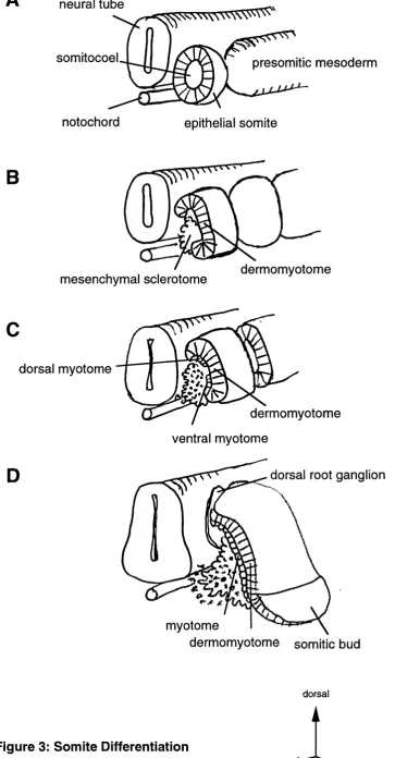
Figure 3: Somite Differentiation.
3A: Early epithelial somite budded off from the presomitic mesoderm, adjacent to the
axial structures, neural tube and notochord.
3B: The epithelial somite becomes divided into the dorsal dermomyotome and ventral
sclerotome compartments: Following axial signals, the somite breaks open and
mesenchymal sclerotome migrates from the ventromedial quadrant of the somite
medially, dorsally the epithehal character is retained in the dermomyotome.
3C: Myotome forms as cells move over the ventral and dorsal margins of the
dermomyotome. Myf-5 is first expressed in spatially separated ventral and dorsal
myotomes.
3D: The somitic bud derived from the lateral dermomyotome migrates laterally to
somitocoel
notochord
presomitic mesoderm
epithelial somite
B
u
mesenchymal sclerotome dermomyotome
dorsal myotome
dermomyotome
ventral myotome
dorsal root ganglion
myotome
dermomyotome somitic bud
dorsal
i
Figure 3: Somite Differentiation
Redrawn and modified from
ventral wall of the somite and the somitocoel, giving rise to the precursor population for
the skeleton and cartilaginous tissues, while the dorsal compartment retains its epithelial
structure as it differentiates into dermomyotome and generates a second layer of
myotome mainly from its dorsal medial margin (Fig. 3C). At the lateral margin of the
dermomyotome, the somitic bud is formed from which hypaxial muscle precursors are
derived (Bober et al, 1994; Daston et al, 1996) (Fig. 3D). The formation of the
myotome has recently been studied by dye labelhng of prospective myotomal precursors
in the dermomyotome of chick embryos (Denetclaw et al, 1997). These studies showed
that as the dermomyotome differentiates, the myotome is formed by the cells at the
dorsal lip migrating over the entire width of the dorsomedial edge. The myotome extends
in the ventrolateral direction, as the myotome fibres progressively lengthen in the
anteroposterior direction. Later the dorsal myotome gives rise to the muscle of the
vertebrae and the deep muscle of the back, while the ventral part of the myotome forms
the body wall muscle and the muscle of the limb. The dermotome differentiates into the
dermis of the trunk and tail. Signalling molecules from the axial structures, the surface
ectoderm and lateral plate mesoderm play an important role in the patterning and
maintenance of these somite compartments as well as in the commitment of cells to the
specific myogenic fates.
5. Somite Patterning
5. 1. Dorsoventral Patterning
The first visible differentiation of cell types in the somite is the formation of the
dermomyotome dorsolateraly, and the formation of sclerotome on the ventromedial
aspect of the somite. In the mouse sclerotome differentiation occurs 6-7 hours after the
somite was bom at which time the next 4-5 somites have already formed. The timing of
somite differentiation is hnked to the number of cell divisions the somite has to undergo
depending on its axial level (Snow, 1981; Tam 1981; Power and Tam 1993).
Dorsoventral rotation of chick somites shows that polarity along the dorsoventral axis is
first observed in somite IV, shortly prior to sclerotome formation (Aoyama and Asamoto
1988; Ordahl and Le Douarin, 1992) whereas rostrocaudal polarity is aquired much
earlier. This has been demonstrated by altering both the dorsoventral and rostrocaudal axis at the same time. Rotated somites i m lost their DV identity but retained their
original rostrocaudal polarity, demonstrating that rostrocaudal patterning occurs
independently and prior to dorsoventral somite patterning.
Induction and maintenance of dorsal dermomyotome and ventral sclerotome identity
requires persistent signalling from axial structures (notochord and neural tube). If the
dorsal half of the somite is missing, while body wall muscle from the ventral half
develops normally (Christ et al, 1992; Rong et al, 1992), indicating that separate
signals are involved in patterning of the dorsal and ventral half of the somite and that the
neural tube is required for dorsal patterning of the somite (Brand-Saberi et al, 1993;
Pourquie et al, 1993; Goulding et al, 1994). Similarly, removal of the notochord prior
to somite formation results in absence of ventral sclerotome and fusion of the
dermomyotome under the neural tube while grafting of ectopic notochord between
presomitic mesoderm and surface ectoderm can inhibit dorsal fate, i.e.. dermomyotome
differentiation (Pourquie et al, 1993; Goulding et al, 1994), demonstrating that the
notochord and the floorplate produce ventralising signals.
These experiments indicate that a morphogenetic gradient might exist along the DV axis,
of ventralising signals emanating from the notochord and floorplate and dorsalising
signals from the dorsal neural tube and surface ectoderm. Different concentrations of
these signals could induce the epithelial somite to give rise to sclerotome ventrally,
dermomyotome dorsally and myotome in the middle where both signals compete.
5. 2. Sonic Hedgehog and Wnt Signalling
The dorsalising effect of the neural tube can be mimicked by molecules of the Wnt
family. Wntl, Wnt3 and Wnt4 genes are expressed in the dorsal neural tube and surface
ectoderm and in culture of chick somites I-IQ induce the myotomal marker MyoD
(Muensterberg et al, 1995) (Fig. 4A, note, that the roles of MyoD and Myf-5 in birds
are reversed compared with the mouse or human and that the diagram in Fig. 4 is a
summary of the situation in the mouse embryo). The ability of Wnt genes to activate
myotomal differentiation appears to be evolutionarily conserved. In Drosophila,
wingless (W ntl) can activate the Drosophila homologue of the myogenic bHLH factors
nautilus (Couso and Martinez Arias, 1994) and activation of XMyoD by XWnt-8 in
Xenopus has also been observed (Hoppler et al, 1996). However, experiments in chick
have also shown that Wnt signals alone are not sufficient to induce myogenesis in the
youngest somites i m and in the presomitic mesoderm. In these tissues a combination of
Wntl, Wnt3 and sonic hedgehog (shh) is required to induce MyoD (Fig. 4)
(Muensterberg et al, 1995). Since sonic hedghog is primarily expressed ventrally, first
in the notochord and subsequently in the floorplate of the ventral neural tube, the
question arises how shh could play a role in dorsal Wnt signalling. Interestingly, sonic
hedgehog is a secreted protein whose N-terminal fragment becomes autoactivated and
has short and long range signalling functions (Lee et al, 1994; Marti et al, 1995;
Roelink et al, 1995). The N-terminal domain can, after nucleophilic attack by the 38-
OH group of cholesterol, become covalently linked to cholesterol which affects its
subcellular distribution (Porter et al, 1996). Shh concentrated in this way in the cell
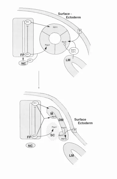
Figure 4: Somite Patterning.
Model of the somite patterning signals and their sources in the mouse embryo. The
medial and lateral somite halves respond to signals from the dorsal neural tube and lateral
plate mesoderm (LM) plus surface ectoderm by expressing Myf-5 or MyoD respectively.
The dorsal neural tube signal is likely to be a combination of Wnt family members, and
the signal from the lateral plate and surface ectoderm can be mimicked by BMP-4.
Members of the FGF family may also play a role. The action of BMP4 and FGF
members is probably to delay terminal differentiation (blunt arrow) in the lateral somite
half to allow proliferation and cell migration of hypaxial muscle precursors. Additional
positive signals (?) may also be required. Short range signalling of Shh secreted from the
notochord (NC) plays a part in floorplate (FP) - induction. Subsequently Shh is also
secreted from the floorplate. The ventromedial part of the somite is exposed to high
levels of sonic hedgehog (Shh) and develops into sclerotome, expressing Pax-1, a
sclerotomal marker. As the somite matures, mesenchymal sclerotome migrates medially
and the remaining dermomyotome (DM) begins to differentiate into myotome (M).
Initially the dorsal myotome is positive for Myf-5 and the ventral myotome for MyoD,
later Myf-5 is also observed ventrally. The signals activating Myf-5 and MyoD ventrally
are not known, but might involve Pax-3. Activation of Pax-3 by surface ectoderm has
Surface -Ectoderm
Wnt
Myf-5
/ MyoD
Paxi
BMP4 FGF's
LM
Shh
NC
B
Wnt
Myf-i
Surface Ectoderm
SC MyoD
(Myf-5)
formation in the ventral neural tube. Subsequent long range signalling from the
floorplate may be a function of the unmodified N-terminal domain. Long range
signalling of Shh is clearly important in sclerotome induction (Fig. 4B). When somites
that are already expressing the sclerotomal marker Pax-1 are cultured in the absence of
Shh, Pax-1 expression is gradually lost, but can be restored after incubation with Shh
demonstrating that Shh can induce sclerotomal markers in somites that are competent to
respond to it (Muensterberg et al, 1995). Remarkably, it appears that another long range
function of Shh is to induce competence in somites I-DI to respond to dorsalising Wnt
signals from the dorsal neural tube. Indeed temporary exposure to Shh is required to
induce competence in presomitic mesoderm to subsequently respond to Wnt signals and
express the myotomal marker MyoD in chick. Conversely, exposure to Wnt signals
alone is not sufficient to induce myogenesis in presomitic mesoderm that had not
previously been exposed to Shh (Muensterberg et al, 1995).
Thus, not only is a combination of diverse signals required for the formation of the
somite compartments, but competence of the somite to respond to such signals is
acquired during somite maturation. The effect of ectopic Shh expression on sclerotome
and myotome fonhation has been examined in chick embryos and mouse explants and
confirms the role of Shh as a ventralising signal in DV patterning (Fan and Tessier-
Lavigne, 1994; Johnson et a l, 1994). Chick embryos injected with a virus encoding
Shh show a dorsal expansion of the Pax-I expression domain on the infected side
(Johnson et al, 1994), while ectopic expression of Shh in the dorsal mesoderm
abolished Pax-3, a dermomyotomal marker (Fan et al, 1995). Remarkably Shh-
induced Pax-1 activation in these experiments was observed over several somite
diameters, and yet in vivo the dorsal somite does not adopt sclerotomal fate. This is
consistent with a model in which competitive signals from the surface ectoderm and the
dorsal neural tube antagonise Shh signals from the floorplate and notochord.
Accordingly, high concentrations of Shh secreted from the notochord and floorplate
maintain the Pax-1 expression domain and promote proliferation of the sclerotome,
whereas myotome differentiation requires the presence of both sonic hedgehog and Wnt
signals (Fig. 4B).
5. 3. Mediolateral Somite Patterning
The medial and lateral halves of the somite probably give rise to separate lineages of
muscle cells. Epaxial muscle of the back is derived from the medial half of the somite
and hypaxial muscle, including limb and body wall musculature, is derived from the
lateral half (Ordahl and Le Douarin, 1992). In culture, cells from the medial half of the
presomitic mesoderm of mouse embryos will first express the myogenic bHLH factor
Myf-5 in response to axial signals whereas cells from the lateral half activate MyoD
that hypaxial and epaxial muscle is derived by separate MyoD and Myf-5 dependent
pathways in the lateral and medial somite halves respectively (Ordahl and Le Douarin,
1992).
Medially muscle is formed rapidly from the myotome. In contrast, the lateral somite
compartment gives rise to the migratory muscle cell populations of the limb and ventral
body wall that are committed to myogenesis but whose differentiation is delayed.
What are the mechanisms activating cell differentiation medially and cell proliferation
laterally? It has been shown that medial but not lateral cells require the presence of axial
structures for their differentiation, indicating that medial and lateral differentiation of the
somites are independently controlled by competing signals (Rong et al, 1992).
Interestingly, lateral mesoderm alone fails to activate MyoD in presomitic mesoderm,
however, surface ectoderm associated with its underlying lateral mesoderm is sufficient
to induce MyoD laterally (Cossu et al, 1996a). In addition to providing a lateralising
signal, lateral plate mesoderm is believed to secrete inhibitory signals that suspend
myogenesis in limb muscle precursors of mouse and chick embryos by two days by
delaying expression of MyoD and Myf-5 (Fig. 4) (Buckingham, 1992; Pownall and
Emerson, 1992; Pourquie et a l, 1995). Thus migratory limb muscle precursors are
normally prevented from expressing any member of the MRF family until they reach their respective target sites in the hmb where they enter myogenesis (Tajbakhsh and
Buckingham, 1994; Pourquie et a/., 1995).
Candidate signalling molecules mediating these effects of the lateral plate and surface ectoderm include members of the TGF(3 and FGF families like bone morphogenetic
protein-4 (BMP-4). BMP-4 is a diffusible growth factor expressed primarily in the
lateral plate for which it can substitute in patterning of the lateral somite compartment. Consistent with this, lateral tissue grafts expressing virally encoded BMP-4 promote an
expansion of lateral somitic cells into more medial domains (Pourquie et al, 1996).
Interestingly, signals from the neural tube, probably involving Shh, can counteract the
effect of BMP-4, suggesting that mediolateral patterning of the somites, like dorso-
ventral patterning, is established by competition between signals emanating from
opposite poles along the mediolateral axis. In chick limb buds laterally high
concentrations of FGFs and FGFR coincide with suspension of terminal differentiation
during migration from the somite to the limb bud (Haub and Goldfarb, 1991; Niswander
and Martin, 1992). Following arrival at the target site FGFR levels are down regulated,
thus reducing FGFR signalling thereby inducing terminal muscle differentiation. The mechanisms underlying the negative effect of growth factors of the FGF and TGF-(3
family on myogenesis have been examined by Gerber et a l, (1997), who showed that
these growth factors interfere with the ability of MyoD to remodel the chromatin
structure at the myogenin locus and block the initiation of myogenin transcription.
myogenin was found to be one order of magnitude less efficient, reflecting the role of
Myf-5 and MyoD in early myoblast determination. Such changes induced in the local
chromatin stmcture may influence gene expression within a lineage by determining the
access of other lineage specific transcription factors. The myogenic factors share a C/H
rich amino acid domain associated with the remodelling function and this domain is also
conserved between mammals, birds, amphibia and fish (this study), reflecting the
functional role of these factors in lineage restriction and suggesting that the functional
mechanism of lineage restriction is also conserved. Other studies indicate that FGF
mediated activation of protein kinase C (PKC) in vivo and in vitro (Davis et al, 1987; Li
et al, 1992) plays a role in inhibiting myogenic bHLH proteins. Phosphorylation of a
conserved site in the DNA binding domain of myogenin is thought to inhibit DNA
binding. Taken together these findings show that the bHLH factors respond to a diverse
range of positive and negative regulatory mechanisms.
6. Somites in Teleost Fish
In teleost fish, during gastrulation cells involute at the blastoderm margin and converge
to the dorsal side to form the embryonic shield, from which the axial mesoderm is generated. As the axial mesoderm extends it becomes separated from the paraxial
mesoderm that gives rise to the somites. In zebrafish about 30 somites are formed
between 10 and 24 hpf (hours post fertilisation), giving rise to a new pair of somites
every 20-30 minutes (Hanneman and Westerfield, 1989). The myotome is the major
component of the somites in zebrafish, while sclerotome is formed by only a small
number of cells in the ventromedial region (see Fig. 5 A) (Morin-Kensicki and Eisen,
1997). In wild type zebrafish embryos, until about 13 hpf, the somites have the shape of
epithelial spheres and subsequently become transformed into chevron-shaped myotomes
(Fig. 5B). Somite development in teleosts is characterised by the establishment of three
specialised stmctures: the adaxial cells, the pioneer cells and the horizontal myoseptum
(HMS) (Fig. 5B). As in other species, the notochord in zebra fish plays an important
role in somite patterning and seems to be required for the formation of these structures.
Sonic hedgehog, secreted from the axial mesoderm, has been shown to be involved in
the recruitment of adaxial cells to the myotomal lineage and subsequently the muscle
pioneers become specified by a different member of the hedgehog family: echidna
hedgehog (Currie and Ingham, 1996).
The adaxial cells are first visible in the presomitic mesoderm as large block shaped cells
organised in three to five rows adjacent to the notochord (Fig. 5C) (Waterman, 1969;
Figure 5: Somites in Teleost Fish.
5A: Schematic transverse section through trunk region of a zebrafish embryo.
Compared with other vertebrates the sclerotome (SC) in teleost fish constitutes a very
small portion of the somite and is located at its ventromedial margin (black). The
myotome makes up most of the somites. Notochord (NC), neural keel (NK). (Redrawn
and modified from Morin-Kensicki and Eisen 1997).
5B: Schematic drawing of a lateral view of zebrafish trunk somites. The chevron shape
somites are devided in the middle of the myotome by a fibrous sheet, the horizontal
myoseptum (HMS) that is formed from the muscle pioneer cells (not shown). The
adaxial cells (AD) migrate radially outwards through the myotome to become the most
superficial muscle cells in the somite (Morin-Kensicki and Eisen, 1997).
5C: Schematic drawing of the segmental plate in zebrafish. The adaxial cells (AD) are
arranged as a sheet between the notochord (NC) and the lateral presomitic cells (EPS).
NK
NC
Sclerotome (Sc) in Teleost Fish
SB
HMS AD
Fish Trunk Somites, Lateral View,
5 0
paraxial mesoderm elongate to span the length of the somite and eventually migrate
radially through the somite. Following migration they form a monolayer of superficial
muscle cells that differentiates into slow muscle fibres (Devoto et al, 1996). A subset
of the adaxial cells, the muscle pioneers, extends from the tip of the V-shaped somites
along the AP axis (see Fig. 5B). Unlike the other adaxial cells, muscle pioneers do not
migrate radially but rather extend from the notochord to the lateral surface of the somite,
where, after 28 hpf, they form the horizontal myoseptum (Hatta et al, 1991). The
horizontal myoseptum is a fibrous sheet that segregates the hypaxial and epaxial muscle
precursors of the somite. Mutations affecting formation of the myoseptum have been
designated you-type mutations, because they invariable cause curved tails resulting from
U-shaped rather than V shaped myotomes (Van Eeden et al, 1996). The paraxial cells
lateral to the adaxial cells belong to the lateral presomitic mesoderm (LPS). Vital dye
labelling experiments have shown that these cells give rise to fast muscle fibres (Fig.
5C) (Devoto e ta l, 1996).
In contrast to higher vertebrates, expression of MyoD in zebrafish is seen prior to somite
formation and is maintained throughout somitogenesis in the adaxial cells along the entire AP axis, including the presomitic mesoderm and two laterally extending bands at
the rostral edge of the segmental plate immediately preceding somite formation
(Weinberg et al, 1996). If this pattern of expression is a general feature amongst teleost
fish and if it is true for other myogenic regulatory factors like Myf-5 is not yet known.
In Xenopus, MyoD and Myf-5 are expressed in the unsegmented paraxial mesoderm, but transcripts are found throughout the entire presomitic mesoderm and are not confined
to the adaxial cells of the segmental plate (Frank and Harland, 1991; Harvey, 1992;
Hopwood et al, 1992). It would be of interest to investigate if in zebrafish Myf-5- and
MyoD-dependent muscle lineages exist in the presomitic mesoderm or in the somites or
if zebrafish MyoD and Myf-5 are expressed in overlapping domains.
7. The Myogenic bHLH Factors
Initially research on the specification of muscle focused on the transition from
undifferentiated myoblasts to myotubes. Genomic DNA from myoblasts was transfected
into fibroblasts and shown to convert the fibroblast cells into myoblasts (Konieczny and
Emerson, 1984; Lassar et al, 1986). Similar results were obtained when fibroblasts
were treated with 5-azacytidine, an inhibitor of méthylation. Based on these data,
subtractive hybridisation of cDNA expression libraries from treated and untreated
fibroblasts identified the first myogenic factor gene encoding MyoD (Davis et al, 1987;
Weintraub e ta l, 1991). Subsequently three further myogenic factors were cloned from
1989; Wright et al, 1989; Salminen et al, 1991) and MRF4 (Rhodes and Konieczny
1989; Miner and Wold, 1990; Hinterberger et al, 1992) A variety of tissue culture cells
transfected with cDNA for each of these proteins can activate the myogenic program,
confirming that the bHLH proteins act as myogenic factors (Davis et a l 1987; Weintraub
et al, 1989; Choi et al, 1990). Myogenic bHLH regulatory genes have also been
identified in birds, frogs, sea urchins (Venuti et al, 1991), insects (Michelson et al,
1990), nematodes (Weintraub et al, 1991) and amphioxus (Araki et al, 1996). Most
invertebrate species only have a single member of the MyoD gene family but it has been
shown that the myogenic factors from sea urchin and nematode (Krause et a l, 1992) can
also activate the myogenic program in mammalian cells, suggesting that the regulatory
mechanisms are extremely ancient.
Surprisingly, evidence from Drosophila and nematode indicates that even in the absence
of the myogenic factors early myogenesis proceeds normally. The most likely
explanation is that alternative pathways for myogenic determination and differentiation
exist in invertebrates.
Recently a MyoD homologue from the ascidian C. intestinalis, CiMDF, has been cloned
(Meedel et al, 1997). Interestingly the CiMDF gene is differentially transcribed to
produce distinct transcripts CiMDFa and CiMDFb that have separate functions in
myogenesis and overlapping temporal expression, that may distinguish between primary
and secondary muscle lineages of ascidians. The presence of E-box motifs in CiMDF
suggests auto and cross-regulation similar to the vertebrate genes (Meedel et a l, 1997).
Interestingly, there is little similarity between invertebrate and vertebrate species apart
from a highly conserved structural motif, the basic-helix-loop-helix domain of about 60
amino acids, which mediates protein dimérisation and DNA binding. The myogenic
factors heterodimerise with ubiquitous E2 proteins (Lassar et al, 1989,1991; Braun et
al, 1990; Brennan and Olson, 1990) in order to bind to E-box motifs (CANNTG) that
were initially found to be important in the immunoglobulin and muscle creatine kinase
enhancers (Buskin and Hauschka, 1989; Gossett et al, 1989; Murre et a l, 1989) and
are also present in other muscle specific genes including those encoding the myogenic
factors themselves. The MRFs can therefore transactivate the expression of muscle
structural genes, and auto- or transactivate their own expression (Braun et a l, 1989a,b;
Edmondson and Olson 1989; Rhodes and Konieczny, 1989; Thayer et al, 1989; Miner
and Wold, 1990; Yee and Rigby, 1993).
8. The Myogenic Cascade
In vitro studies on muscle cell lines have shown that the bHLH factors are expressed in a
myoblasts prior to and after differentiation, while myogenin and MRF4 are expressed
only after differentiation (Hinterberger et al, 1991). These results suggested that MyoD
and Myf-5 might act early in determining myoblast fate, while MRF4 and myogenin are
involved in later differentiation of myoblasts into myotubes. When Myf-5 is expressed
in lOTl/2 cells, myogenesis is associated with expression of MyoD but the converse is
not true: MyoD expression does not lead to activation of Myf-5 (Braun et al, 1989b),
suggesting that Myf-5 acts upstream of MyoD at least in vitro. Studies of the expression
patterns of the myogenic factors in vivo have revealed the temporal sequence of their
expression and are in agreement with a transcriptional hierarchy of the myogenic factors
(Thayer et al, 1989; Braun et a l, 1990; Edmondson et al, 1992; Naidu et a l, 1995). In
skeletal muscle, Myf-5 is the first myogenic regulatory gene to be expressed at E8.0, in
the dermomyotome of the newly formed somites (Ott e ta l, 1991). 12 hours later,
myogenin appears in the myotome of successive somites, (Sassoon et a l, 1989)
followed by MRF4 (Bober et al, 1991; Hinterberger et al, 1991) and MyoD (Sassoon
et al, 1989) (see Fig. 6). MRF4 is reactivated in a second phase of expression from
E14.5 onwards.
The temporal expression of the myogenic factors in the limb differs from that of the trunk musculature. The migratory myoblasts that colonise the limb bud leave the
ventrolateral edge of the somite and do not express MRFs until they have reached their
destination (Sassoon et al, 1989). Therefore, Myf-5 is first expressed at E10.5,
followed by myogenin and MyoD which are coexpressed in the limb myoblasts rather
than being sequentially activated as in the somites. MRF4 mRNA is not detectable in the
limb until late in development at E16 (Bober et al, 1991).
There are also species dependent variations in the order and timing of expression of the
myogenic factors. In birds, the homologue of MyoD instead of Myf-5 appears first,
followed by myogenin and Myf-5 (Pownall and Emerson, 1992). In Xenopus and
zebrafish, MyoD and Myf-5 are activated in the presumptive mesoderm (Hopwood et
a l, 1992; Weinberg et al, 1996; this study) whereas myogenic factors are not expressed
at significant levels prior to somite formation in the mouse. This raises the possibility
that the function of XMyoD and ZMyf-5 may be quite distinct from higher vertebrate
species. However, the differences in the temporal expression are less significant if one
considers MyoD and Myf-5 as a functionally equivalent pair of genes with a common
evolutionary origin (Atchley et al, 1994). MyoD and Myf-5 probably arose from a
common gene, as did myogenin and MRF4, which suggests that the partners in each of
BIRTH
Myf-5
Myogenin
MRF4
MyoD
6 8 1 0 1 2 1 4 1 6 1 8 6 8 10 12
DAYS
limb bud som ite
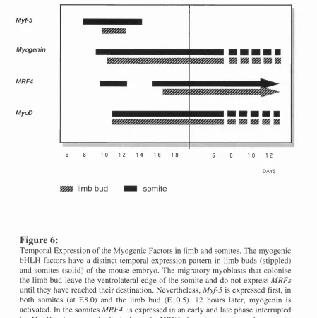
F ig u re 6:
Temporal Expression o f the M yogenic Factors in limb and somites. The m yogenic bHLH factors have a distinct temporal expression pattern in limb buds (stippled) and somites (solid) o f the mouse embryo. The migratory myoblasts that colonise the limb bud leave the ventrolateral edge o f the somite and do not express MRFs
until they have reached their destination. Nevertheless, Myf-5 is expressed first, in both somites (at E8.0) and the limb bud (E10.5). 12 hours later, myogenin is activated. In the somites MRF4 is expressed in an early and late phase interrupted by M yoD whereas in the limb the early MRF4 phase is m issing, and myogenin and M yoD are coexpressed. In the limb MRF4 mRNA is not detectable until late in development at E l 6. Expression o f M yoD and Myf-5 (black) converts premyogenic cells into skeletal myoblasts while the expression o f myogenin and
9. MRF Null Mutations
To examine the individual roles of the myogenic factors in myogenesis, targeted null
mutations have been introduced into each of the four myogenic regulatory genes and in
addition double mutants for MyoD/Myf-5 (Rudnicki et al, 1993), MRF4/Myf-5 (Braun
et ai, 1995), myogenin/MyoD and myogenin/Myf-5 (Rawls et al, 1995) have been
obtained by interbreeding these targeted mouse hues. This strategy has been particularly
successful in studying myogenesis, because the redundancy between the regulatory
factors makes it otherwise impossible to dissect their individual function.
9. 1. MyoD Null Mutants
The introduction of a homozygous null mutation in the MyoD gene does not produce an
embryonic muscle phenotype. Moreover, MyoD mutant mice are viable and fertile and
indistinguishable from wild type litter mates (Rudnicki et al, 1992) suggesting that
MyoD and Myf-5 have overlapping functions. However, preferential expression of
MyoD in fast twitch muscle fibres raises the possibility that subtle fibre type changes
occur in the absence of MyoD (Hughes et al, 1993). Furthermore, recent evidence
shows that satellite cells fail to divide in mdx/MyoD-/- double mutant mice, suggesting
that MyoD is required for satellite cell activation during muscle fibre regeneration
(Megeney et al, 1996). Clearly, the functional overlap of Myf-5 and MyoD is limited
since a single Myf-5 allele can not rescue the MyoD phenotype and results in lethality
due to the apparent reduction in skeletal muscles, whereas a single copy of MyoD is
sufficient to rescue Myf-5 knockout mice. It has been suggested that MyoD may be able
to recruit more cells possibly from different precursor populations to the myogenic
lineage than Myf-5 (Braun and Arnold, 1996). Assuming that competition exists
between cell lineages alternatively determined by MyoD and Myf-5, the upregulation of
Myf-5 in MyoD deficient mice can be explained as an expansion of the Myf-5 myogenic
cell lineage. According to this model, the MyoD cell lineage seems to be enlarged at the
expense of the Myf-5 cell lineage in the wild type situation, suggesting that MyoD
expression is either more stable or more responsive to environmental cues compared
with Myf-5. The different expression domains of MyoD and Myf-5 within the somite
might support such a model (Braun and Arnold, 1996). The most interesting observation
in MyoD mutant mice is that Myf-5 mRNA levels are elevated two-fold, suggesting that
Myf-5 may substitute for the absence of MyoD in the development of skeletal muscle.
This is supported by the finding that the levels of MRF4 and myogenin transcripts are