O R I G I N A L A R T I C L E
Changes in the cellulose crystallinity of moso bamboo cell walls
during the growth process by X-ray diffraction techniques
Keisuke Toba1•Takahisa Nakai2•Tatsuya Shirai3• Hiroyuki Yamamoto3
Received: 26 November 2014 / Accepted: 20 May 2015 / Published online: 7 June 2015
ÓThe Japan Wood Research Society 2015
Abstract We investigated the changes in cellulose
crys-tallinity in the cell walls of moso bamboo (Phyllostachys pubescensMazel) during the growth process. Changes in crystallinity in bamboo cell walls were traced along the longitudinal and radial directions using flat-sawn speci-mens obtained from each internode using X-ray diffraction measurements. Cellulose crystallinity in cell walls in the same culm was higher around the base of the bamboo culms in less than 10 days after the emergence of bamboo shoots, while the upper internodes remained lower in crystallinity. Thereafter, crystallinity in the upper intern-odes of bamboo culms became higher. This suggests that cellulose crystallinity within bamboo cell walls has grad-ually increased from the base to the top with the elongation of the internode during the growth process of bamboo shoots. In addition, cellulose crystallinity in cell walls increased particularly in the outer part of the bamboo culms in all internodes. Moreover, increases of crystallinity occurred after elongation growth of internodes. It is con-sidered that increases in crystallinity in the outer portion of each internode are one of the countermeasures of bamboo culms against increased bending moment due to the elon-gation growth.
Keywords Bamboo shootMaturationCrystallization
Internodal growthMicrofibrils
Introduction
Bamboos are monocotyledonous plants of the family Gramineae found in tropical, subtropical, and temperate regions around the world [1]. Bamboo is a perennial plant of which the culms (main stem) are lignified during elon-gation growth, without secondary growth in thickness [2, 3]. Bamboo has been used for various daily necessities, although many artificial materials become widely used today. For example, it is used as a structural material in the field of architecture, e.g., laminated bamboo lumber [4,5]. The structural features of bamboo are different from wood. Bamboo culms consist of parenchyma and rigid bundle sheaths. The rigid bundle sheath, which aligns nearly parallel to the longitudinal axis of the culm, is found more in the outer section of the culm than in the inner side [2,6,7]. This optimized spatial construction gives bamboo culms better physical and mechanical properties [8–11]. Bamboo is also one of the fastest-growing plants, and can reach 15–30 m in height within 2–4 months [6]. Nomura [2] reported that moso bamboo (Phyllostachys pubescens) and madake bamboo (Phyllostachys bambusoides) can grow more than a meter in a day. This rapid growth rate is due to the sum of the growth amount at each internode.
Some researchers have reported the time-dependent variation in the macroscopic morphology of bamboo dur-ing the growth process [2, 12–14]. In addition, a few studies have focused on the changes in crystalline proper-ties of cellulose in bamboo cell walls during the growth process. Taniguchi [15] analyzed the chemical components of bamboo, and revealed that the cellulose content in
& Keisuke Toba [email protected]
1 Gifu University Composite Materials Center, Gifu
University, Gifu 501-1193, Japan
2 Interdisciplinary Graduate School of Science and
Engineering, Shimane University, Matsue 690-8504, Japan
3 Graduate School of Bioagricultural Sciences, Nagoya
bamboo shoot rapidly increased after stripping off the leaf during the growth process. Nomura and Yamada [16] performed X-ray diffraction (XRD) measurements at ages of 2, 4, and 5 weeks, and obtained similar results as Taniguchi [15]. In addition, they demonstrated that the crystallinity near the base of bamboo shoots (internodal growth) was higher than at the top. Wang et al. [17] measured the radial distribution of crystallinity, crystal size, and microfibril angle in 0.5- to 10.5-year-old bamboo culms using X-ray scattering techniques; however, there have been no detailed studies on the crystalline properties of bamboo shoots during the process of internodal growth at the intervals of several days.
In the present study, we investigated the changes in cellulose crystallinity in the cell walls of moso bamboo during the growth process; thus, we collected detailed information on the growth of bamboo using XRD tech-niques, and assessed how the bamboo culm forms mechanically optimized conformations during the growth process.
Methods
Bamboo shoots
The studied specimens were prepared from culms of moso bamboo (Phyllostachys pubescens Mazel), which are grown in the forests of Matsue (35.26°N, 133.50°E), Japan. Dozens of bamboo shoots, which emerged a few cen-timeters off the ground, were selected on April 23, 2012,
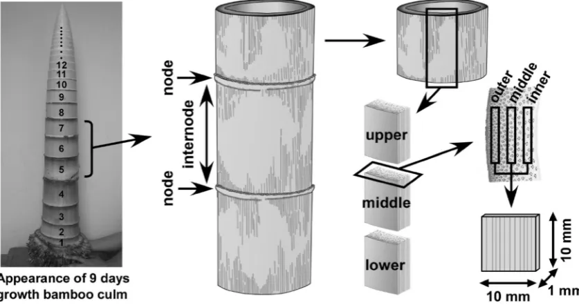
and were sequentially cut down at intervals of several days until early December. We dug up the ground about 20 cm around the each bamboo culm to cut off the bamboo base from the rhizome. All of the specimens were covered with plastic wrap after cutting, and were immediately taken to the laboratory without drying. All of the internodes, except the bamboo base, were numbered from the base to the top as shown in Fig. 1. To avoid decaying and dimensional changes due to drying, specimens were temporarily stored in 50 % (v/v) aqueous ethanol solution. Table1shows the characteristics of the tested bamboos.
Specimen preparation
Flat-sawn specimens of dimensions 1910910 mm (thickness9length9width) were made from each internode using a saw, chisel, and cutter knife. All of the specimens were prepared under wet conditions to prevent shrinkage of cell walls. Figure 1 shows the process of specimen preparation. First, both the upper and lower parts of each bamboo node were removed from each internode. The rest of the tubular internode was divided longitudi-nally, and then it was vertically segmented into three blocks (upper, middle, lower). After removing a few mil-limeters from both surfaces which included the epidermal tissues, the outer and inner sides of specimens in each block were obtained. The middle specimens were prepared from the central region of each remaining block. Then all of the specimens were freeze-dried using a vacuum freeze dryer (VD-400, Taiyo Kagaku Co., Ltd., Japan) to retain the intact condition of the cell wall structure.
XRD measurements
XRD measurements were performed to assess the crys-talline properties of air-dried bamboo cell walls using an X-ray diffractometer (XD-D1w, Shimadzu Co., Japan). All XRD measurements were made with the reflection tech-nique. The incident X-ray radiation was the characteristic Cu KaX-ray passing through a nickel filter with a power of 30 kV and 30 mA. Both the air-scattering prevention slit and the divergence slit were 1°. The width of the detection slit was 0.1 mm, and scanning speed and integration time in the measurements of crystallinity were 2.0°/min and 2.0 s, respectively. Scanning ranges of 2hangle was 5.0°– 40.0°. Flat-sawn specimens obtained from each internode were vertically fixed on the specimen holder of the XRD device to irradiate the tangential section.
The crystallinity of the crystalline cellulose in the cell wall of bamboo was calculated using the following Segal method [18,19]:
Cs¼ I200IAm
I200 100ð%Þ; ð1Þ
where Cs is the crystallinity (%), I200 is the reflection
intensity of (200) plane diffraction, andIAmis the intensity
at the minimum near 18.5°of 2hangle. The Segal method is simple to apply and does not require peak separation between (110) and (110) reflections.
In the present study, the cellulose microfibril angle of the bamboo cell walls was also measured in some speci-mens to confirm its effect on the crystallinity. Cave’s method [20] was adopted to calculate the cellulose microfibril angle in the S2 layer (MFA) by using an X-ray diffractometer (XD-D1w, Shimadzu Co., Japan) with a symmetrical transmission mode. A point-focused X-ray beam (Cu-KaX-ray, beam diameter 2 mm) was applied to the flat-sawn specimens. Each specimen was rotated
around its normal axis with a rotation angle from 180° to 330°at a rotation speed of 6°per minute in a position of 2h=22.4°: the diffraction angle of the (200) plane of celluloseIb. The scattered X-rays were detected by a Na-I scintillation counter behind a receiving slit of 0.6 mm width. Estimation of the mean MFA was based on fol-lowing formula:
MFA (Þ ¼ 0:6T; ð2Þ
where T was ‘‘angle T’’ which was calculated from the diffraction pattern of the (200) plane [20].
Results and discussion
Figures2 and 3 show the relationship between the crys-tallinity of the bamboo cell walls and the internode number for each growing period. Both figures focused on the dif-ferences in spatial position of each internode, as shown in Fig.1. That is, Fig.2shows the changes in crystallinity in the longitudinal direction, whereas Fig.3 shows it in the radial direction. These two figures suggest that the crys-tallinity near the base of the bamboo culms is higher than in the upper internodes of the same culm in less than 10 days after the emergence of bamboo shoots (Figs.2a, b, 3a, b). On the other hand, the crystallinity in the upper internode of bamboo culm was relatively lower even after 29 days of growth (Figs. 2f,3f), although the bamboo culm became higher than 12 m. This suggests that cellulose crystallinity in cell walls in bamboo shoots gradually increased from the base to the top during the elongation process. In addition, development of the bamboo cell wall was more matured around the base of the bamboo culm than in the upper internodes during the growth process. Figure4 shows the typical changes in XRD pattern in the 23 days growth specimen that corresponding to Figs.2e
Table 1 Characteristics of moso bamboo specimens in the present study
Growing period (day) Culm height (cm) Number of internodes Maximum diameter (mm) Number of XRD specimen
2 19.3 60 81.1 51
9 102.0 61 128.1 66
15 143.4 62 124.5 96
18 125.6 60 144.0 81
23 399.0 50 122.6 57
29 1272.4 55 121.8 63
36 1752.3 55 161.7 78
43 1540.0 60 129.9 87
51 1813.9 63 143.4 72
57 1803.6 57 125.7 84
Fig.
2
Cellulose
crystallinity
in
the
vertical
position
in
bamboo
cell
walls
in
each
growth
period.
Black
,g
ray
,
and
white
circles
indicate
the
results
in
the
specimens
prepared
from
upper
,
middle
,
and
lower
positions
in
each
internode,
respectively.
Specimens
in
a
,
b
,
and
d
were
not
divided
into
three
sections
due
to
the
shortness
of
the
longitudinal
direction.
Each
plot
with
an
error
bar
represents
an
averaged
value
of
three
Fig.
3
Cellulose
crystallinity
in
the
radial
position
in
bamboo
cell
walls
in
each
growth
period.
Black
,
gray
,
and
white
circles
indicate
the
results
in
specimens
prepared
from
outer
,
middle
,
and
inner
positions
in
each
internode,
respectively.
Each
plot
with
an
error
bar
represents
an
averaged
value
of
three
and3e. Lower internodes showed sharp (200) diffraction peaks (Fig.4a). The outer part also showed sharp (200) diffraction peaks (Fig.4b).
Figure2 focuses on changes in crystallinity related to the vertical positions within each internode. There was no significant variation in crystallinity in the longitudinal direction within each internode, even after 225 days of growth (Fig.2k) (i.e., the crystallinity of each block showed similar values without regularity, regardless of growing period). Nomura and Yamada [16] performed XRD measurements using the 30th internode of a bamboo culm, the height of which was 865 cm after 5 weeks of growth, and showed that crystallinity was higher in the upper part of the internode. Additional investigations are necessary to clarify the reason for the different results regarding the crystallinity of bamboo cell walls in the longitudinal direction within the internode, such as the relationship between bamboo growth and lignification, and analysis of chemical composition during the growth pro-cess of bamboo shoot.
Figure3focuses on the changes in crystallinity related to the radial position in each internode. The crystallinity showed clear differences in the radial direction within each internode, that is, crystallinity gradually became higher in the outer side as growth progressed. This broad distinction of crystallinity was never observed in young bamboo culms (Fig.3a–d), whose growing period was within 20 days. Differences in crystallinity in the radial direction began to appear near the base of the bamboo culm in early stages of growth, while the bamboo top still showed low values (Fig.3e, f); the differences gradually progressed to the
upper internodes (Fig.3g–j). The crystallinity profiles of bamboo culms that grew more than 50 days (Fig.3i, j) were almost identical to those that grew 225 days (Fig.3k). Therefore, it is considered that the cellulose crystallinity in bamboo cell walls became constant in approximately 50 days, although it is considered that the completion of bamboo maturation, such as distribution of bundle sheath, fiber length and so forth, are much later [2]. These results suggest that increases in crystallinity in the outer portion of each internode are one of the counter-measures of bamboo culms against increased bending moment due to the elongation growth, for example, in the case when the bamboo culm bows by crosswind.
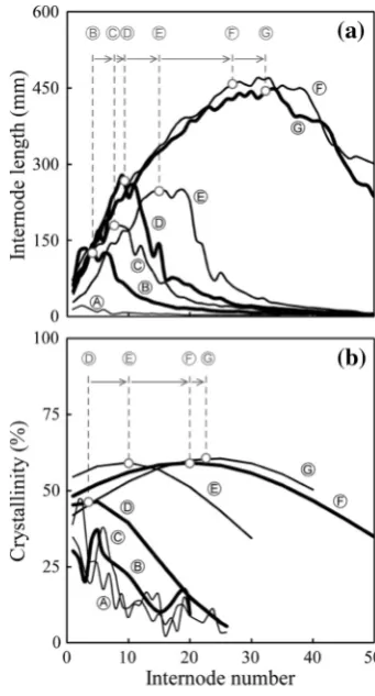
Figure5 shows how the (a) internode length and (b) crystallinity varied with internode position. The seven data plot, which are indicated by circled black letters, are for samples with growth periods of 2 (A), 15 (B), 18 (C), 23 (D), 29 (E), 36 (F) and 43 (G) days. Gaussian curves
Fig. 4 Typical changes in XRD pattern in 23 days growth bamboo specimen.aDifferences induced by vertical position of internodes. Five XRD patterns in this figure correspond to the outer part of specimens in each internode number. Thenumbers on the right sideof each XRD patterns show internode number. On the other hand,
bdifferences in XRD pattern induced by radial position. Three XRD patterns in this figure correspond to the radial position in number 1 for internode number. The lower and outer parts of bamboo specimens had sharp (200) diffraction peak
Fig. 5 Comparisons of changes in a internode lengths andb crys-tallinity at each internode number for samples with different growth periods. The samples with different growth periods of 2 (A), 15 (B), 18 (C), 23 (D), 29 (E), 36 (F) and 43 (G) days are indicated with
were fitted to these data plots, and the maxima are indi-cated by the open circles. Internode lengths were measured by image processing software (QuickGrain Strandard, Inotech Co., Japan). Images of the vertical section in each internode were used for internode length analysis, and our results for internode length coincide with previous studies [2,16]. Thus, the maximum internode length was 457 mm for node 28 for the sample that grew for 36 days. Likewise, the maximum crystallinity was 61 % for node 23 for the sample that grew 43 days. The internode numbers for each maximum are indicated by the circled gray letters. As the growth periods increased, the maximum internode length and maximum crystallinity were found at higher-numbered internodes. This indicates that increasing crystallinity in bamboo cell walls occurs after elongation growth of the internode from the base to the top.
The structure of a bamboo cell wall is different from a wood cell wall. The model of a bamboo cell wall consists of 7–9 layers [21,22]. In this model, the MFA of cellulose in each layer is nearly parallel or perpendicular to the fiber direction. Similar to the cell walls of wood, the layers with small MFAs are dominant in the bamboo cell wall. In
the present study, the MFA of some specimens were measured to confirm its effect on the differences in crystallinity in the radial direction. Figure6 shows the relationship between internode number and the MFA. MFA showed no significant change in the radial direction in each specimen. In particular, the specimens that showed clear differences in crystallinity (Fig.3i, k) had almost identical MFA values, except in the base. Therefore, MFA had little influence on crystallinity in our study, although we also found that the MFA was larger in 23-day-old specimens compared to others (Fig. 6b). Wang et al. [17] reported large variation in the MFA in the radial direction using moso bamboo that grew more than half a year. The degree of maturation of bamboo cell walls may affect the variation in MFA; however, further investigations of the formation of the cell wall structure of bamboo shoots are necessary for a complete understanding of bamboo growth.
References
1. Fujii T (2008) Basic science and advanced technologies for industrial applications of bamboo (in Japanese). CMC, Tokyo 2. Nomura T (1980) Growth of bamboo (in Japanese). Wood Res
Tech Notes 15:6–33
3. Li XB, Shupe TF, Peter GF, Hse CY, Eberharrdt TL (2007) Chemical changes with maturation of the bamboo species phyl-lostachys pubescens. J Trop For Sci 19:6–12
4. Mahdavi M, Clouston PL, Arwade SR (2011) Development of laminated bamboo lumber: review of processing, performance, and economical considerations. J Mater Civ Eng 23:1036–1042 5. Xiao Y, Yang RZ, Shan B (2013) Production, environmental
impact and mechanical properties of glubam. Constr Build Mater 44:765–773
6. Liese W (1987) Research on bamboo. Wood Sci Technol 21:189–209
7. Iguchi Y, Fishitani M, Kubo T, Sato K (2002) Effect of volume fraction of bundle sheath and water extractives on bending creep behavior of bamboo under changing moisture conditions (in Japanese). Mokuzai Gakkaishi 48:413–424
8. Urakami H (1996) Relationships between dynamic viscoelasticity of mosochiku (Phyllostachys pubescens) and internode number, age, and specific gravity (in Japanese). Mokuzai Gakkaishi 42:832–838
9. Obataya E, Kitin P, Yamauchi H (2007) Bending characteristics of bamboo (Phyllostachys pubescens) with respect to its fiber-foam composite structure. Wood Sci Technol 41:385–400 10. Tsubaki T, Nakano T (2010) Creep behavior of bamboo under
various desorption conditions. Holzforschung 64:489–493 11. Wang X, Ren H, Zhang B, Fei B, Burgert I (2012) Cell wall
structure and formation of maturing fibres of moso bamboo (Phyllostachys pubescens) increase buckling resistance. J R Soc Interface 9:988–996
12. Nomura T, Yamada T (1974) X-ray analysis of tyrosine in growing stage of bamboo (Phyllostachs eduisA. & C. Riviere). Wood Res 56:21–27
13. Nomura T, Yamada T (1991) Growth of moso bamboo ( Phyl-lostachys heterocycla) I. Internodeal growth (in Japanese). Mokuzai Gakkashi 37:1115–1122
Fig. 6 Microfibril angle (MFA) in the radial position in bamboo cell walls in each growth period.Black,gray, andwhite circlesindicate the results in the specimens prepared fromouter,middle, andinner
14. Liese W, Weiner G (1996) Aging of bamboo culms. A review. Wood Sci Technol 30:77–89
15. Taniguchi E (1956) Chemical studies on the crystalline region of cellulose materials. XIV. Variation of fine structure in Akamatsu (Pinus densifloraSieb. et Zucc.) and Mosochiku (Phyllostachys edulis Riv.) through growth (in Japanese). Mokuzai Gakkaishi 2:152–157
16. Nomura T, Yamada T (1974) Crystallinity change in the growing stage of bamboo (Phyllostachys mitis). Wood Res 57:23–30 17. Wang Y, Leppa¨nen K, Andersson S, Serimaa R, Ren H, Fei B
(2012) Studies on the nanostructure of the cell wall of bamboo using X-ray scattering. Wood Sci Technol 46:317–332
18. Segal L, Creely JJ, Martin AE, Conrad CM (1959) An empirical method for estimating the degree of crystallinity of native cel-lulose using the X-ray diffractometer. Text Res J 29:786–794
19. Toba K, Yamamoto H, Yoshida M (2013) Crystallization of cellulose microfibrils in wood cell wall by repeated dry-and-wet treatment, using X-ray diffraction technique. Cellulose 20:633–643
20. Cave ID (1966) Theory of X-ray measurement of microfibril angle. For Prod J 16:37–42
21. Tono T, Ono K (1962) Researches on the morphological structure and the physical properties of bamboo fiber for paper making. II. The layered structure and its morphological transformation by acid treatment (in Japanese). Mokuzai Gakkaishi 8:245–249 22. Parameswaran N, Liese W (1976) On the fine structure of