Gout, Hyperuricemia and Crystal-Associated Disease Network (G-CAN) consensus statement regarding labels and definitions of disease states of gout
David Bursill1, William J Taylor2,3, Robert Terkeltaub4, Abhishek Abhishek5, Alexander K
So6, Ana Beatriz Vargas-Santos7, Angelo Gaffo8, Ann K Rosenthal9, Anne-Kathrin Tausche10,
Anthony M Reginato11, Bernhard Manger12, Carlo Alberto Scirè13-14, Carlos Pineda15, Caroline
van Durme16, Ching-Tsai Lin17, Congcong Yin18, Daniel A Albert19, Edyta Biernat-Kaluza20,
Edward Roddy21, Eliseo Pascual22,23, Fabio Becce24, Fernando Perez-Ruiz25-27, Francisca
Sivera28, Frédéric Lioté29, Georg Schett30, George Nuki31, Georgios Filippou13, Geraldine M
McCarthy32,33, Geraldo Da Rocha Castelar-Pinheiro7, Hang-Korng Ea29, Helena De Almeida
Tupinambá34, Hisashi Yamanaka35,36, Hyon K Choi37, James M Mackay38, James R O’Dell39,
Janitzia Vázquez-Mellado40, Jasvinder A Singh41-43, John D Fitzgerald44, Lennart TH
Jacobsson45, Leo AB Joosten46, Leslie R Harrold47,48, Lisa K Stamp49, Mariano Andrés22,23,
Marwin Gutierrez50, Masanari Kuwabara51,52, Mats Dehlin53, Matthijs Janssen54, Michael
Doherty5, Michael S Hershfield55, Michael H Pillinger56, N Lawrence Edwards57, Naomi
Schlesinger58, Nitin Kumar59, Ole Slot60, Sebastien Ottaviani61, Pascal Richette29, Paul
MacMullan62, Peter Chapman63, Peter E Lipsky64, Philip C Robinson65, Puja P Khanna66, Rada
N Gancheva67, Rebecca Grainger2,3, Richard J Johnson68, Ritch Te Kampe16, Robert T
Keenan69, Sara K Tedeschi70, Seoyoung C Kim70, Sungjae Choi71, Theodore R Fields72,
Thomas Bardin29, Tillmann Uhlig73, Tim L Jansen54, Tony R Merriman74, Tristan Pascart75,
Tuhina Neogi76, Viola Klück77, Worawit Louthrenoo78,Nicola Dalbeth79.
1. Adelaide Medical School, University of Adelaide, South Australia, Australia.
3. Wellington Regional Rheumatology Unit, Hutt Valley District Health Board, Lower
Hutt, New Zealand.
4. Veterans Affairs Medical Center, University of California, San Diego, California,
USA.
5. The University of Nottingham, Nottingham, UK.
6. Service de Rhumatologie, CHUV 1011 Lausanne, Switzerland.
7. Internal Medicine Department, Rheumatology Unit, State University of Rio de
Janeiro, Avenida Marechal Rondon, Rio de Janeiro, Brazil.
8. University of Alabama at Birmingham, USA.
9. Medical College of Wisconsin and the Clement J. Zablocki Veterans Affairs Medical
Center, Milwaukee, USA.
10. Department of Rheumatology, University Hospital "Carl Gustav Carus" of the
Technical University Dresden, Dresden, Germany.
11. Warren Alpert School of Medicine at Brown University, Providence, Rhode Island,
USA.
12. University Erlangen-Nuremberg, Germany.
13. Section of Rheumatology, Department of Medical Sciences, University of Ferrara,
Ferrara, Italy.
14. Epidemiology Unit, Italian Society for Rheumatology, Milan, Italy.
15. Instituto Nacional de Rehabilitación Luis Guillermo Ibarra Ibarra. Mexico City.
16. Department of Internal Medicine, Division of Rheumatology, Maastricht University
Medical Centre, Maastricht, the Netherlands.
17. Division of Allergy, Immunology and Rheumatology, Taichung Veterans General
18. Henry Ford Immunology and Dermatology Department, Henry Ford Health System,
Detroit, MI, USA.
19. Dartmouth-Hitchcock Medical Center, New Hampshire, USA
20. ORLIK, Warsaw, Poland.
21. Research Institute for Primary Care and Health Sciences, Keele University, Keele,
Staffordshire, ST5 5BG, UK.
22. Sección de Reumatología, Hospital Universitario de Alicante, Alicante, Spain.
23. Departamento de Medicina Clínica, Universidad Miguel Hernández, Alicante, Spain.
24. Department of Diagnostic and Interventional Radiology, Lausanne University
Hospital and University of Lausanne, Lausanne, Switzerland.
25. University of the Basque Country, Biscay, Spain.
26. Rheumatology Division, Cruces University Hospital, Baracaldo, Spain.
27. Biocruces Health Research Institute, Baracaldo, Spain.
28. Department of Rheumatology, Hospital General Universitario Elda, Elda, Spain.
29. Hôpital Lariboisière, Assistance Publique-Hopitaux de Paris, and INSERM
UMR-1132 and Université de Paris, Paris, France.
30. Department of Internal Medicine III, Friedrich-Alexander University
Erlangen-Nürnberg and Universitatsklinikum Erlangen, Erlangen, Germany.
31. University of Edinburgh, Edinburgh, UK.
32. School of Medicine and Medical Science, University College Dublin, Ireland.
33. Mater Misericordiae University Hospital, Dublin, Ireland.
34. State University of Rio de Janeiro, Brazil.
35. School of Medicine, Tokyo Women’s Medical University, Tokyo, Japan.
36. Institute of Rheumatology, Tokyo Women’s Medical University Hospital, Tokyo,
37. Harvard Medical School and Massachusetts General Hospital, Boston, USA.
38. Aristea Therapeutics, San Diego, California, USA.
39. University of Nebraska Medical Center, Nebraska, USA.
40. Hospital General de México and Universidad Nacional Autónoma de México, Mexico
City, Mexico.
41. Medicine Service, VA Medical Center, Birmingham, Alabama, USA.
42. Department of Medicine at School of Medicine, University of Alabama at
Birmingham, Birmingham, Alabama, USA.
43. Division of Epidemiology at School of Public Health, University of Alabama at
Birmingham, Birmingham, Alabama, USA.
44. University of California, Los Angeles, California, USA.
45. Department of Rheumatology & Inflammation Research, Institute of Medicine, The
Sahlgrenska Academy, University of Gothenburg, Göteborg, Sweden.
46. Department of Internal Medicine, Radboud University Medical Center Nijmegen,
Nijmegen, The Netherlands.
47. University of Massachusetts Medical School, Worcester, Massachusetts, USA.
48. Corrona, LLC, Southborough, Massachusetts, USA.
49. Department of Medicine, University of Otago, Christchurch, New Zealand.
50. Division of Musculoskeletal and Rheumatic Diseases, Instituto Nacional
Rehabilitación, México City, México.
51. Department of Cardiology, Toranomon Hospital, Tokyo, Japan.
52. Division of Renal Diseases and Hypertension, School of Medicine, University of
Colorado Denver, Aurora, Colorado, USA.
53. Sahlgrenska Academy, University of Gothenburg, Gothenburg, Sweden.
55. Duke University Medical Center, Durham, North Carolina.
56. New York University School of Medicine, New York, USA.
57. University of Florida College of Medicine, Gainesville, Florida, USA.
58. Rutgers-Robert Wood Johnson Medical School, Piscataway, New Jersey, USA.
59. Department of Internal Medicine, Division of Cardiovascular Medicine, University of
Michigan, Ann Arbor, MI, USA.
60. Copenhagen Center for Arthritis Research, Center for Rheumatology and Spinal
Disorders. Rigshospitalet Glostrup, DK-2600 Glostrup, Denmark.
61. Rheumatology department, Bichat-Claude Bernard Hospital, University of Sorbonne
Paris Cité, 46 rue Henri Huchard, 75018 Paris, France.
62. University of Calgary, Canada.
63. Department of Rheumatology, Immunology and Allergy, Canterbury District Health
Board, Christchurch, New Zealand.
64. AMPEL BioSolutions, LLC, Charlottesville, Virginia, USA.
65. School of Clinical Medicine, Faculty of Medicine, University of Queensland,
Queensland, Australia.
66. University of Michigan, Michigan, USA.
67. University Hospital ‘St. Ivan Rilski’, Clinic of Rheumatology, Sofia, Bulgaria
68. Division of Renal Diseases and Hypertension, University of Colorado Denver,
Aurora, Colorado, USA.
69. Division of Rheumatology, Duke University School of Medicine, Durham, N.C. USA.
70. Harvard Medical School, and Brigham and Women's Hospital, Boston,
Massachusetts, USA.
71. Division of Rheumatology, Department of Internal Medicine, Korea University
72. Weill Cornell Medical College, Hospital for Special Surgery, New York, USA.
73. Department of Rheumatology, Diakonhjemmet Hospital, Oslo, Norway.
74. Department of Biochemistry, University of Otago, Dunedin, New Zealand.
75. Lille Catholic University, Saint-Philibert Hospital, Department of Rheumatology,
France.
76. Boston University School of Medicine, Section of Rheumatology, Department of
Medicine, Boston, Massachusetts, USA.
77. Department of Internal Medicine, Radboud Institute for Molecular Life Sciences,
Radboud University Medical Center, Nijmegen, The Netherlands.
78. Division of Rheumatology, Department of Internal Medicine, Faculty of Medicine,
Chiang Mai University, Chiang Mai, Thailand.
79. Department of Medicine, Faculty of Medical and Health Sciences, University of
Auckland, Auckland, New Zealand.
Corresponding author: Prof Nicola Dalbeth, Bone and Joint Research Group, Department of Medicine, Faculty of Medical and Health Sciences, University of Auckland, 85 Park Rd,
Grafton, Auckland 1023, New Zealand. Phone +64 9 3737999 x82568, Fax +64 9 3737677
x84377. Email: n.dalbeth@auckland.ac.nz.
Word count: 2951, Tables: 5, Figures: 1, Supplementary materials: 1, Supplementary tables: 2.
Keywords: gout, urate, hyperuricemia, monosodium urate crystals, nomenclature, language,
ABSTRACT
Objective: There is a lack of standardisation in the terminology used to describe gout. The aim of this project was to develop a consensus statement describing the recommended
nomenclature for disease states of gout.
Methods: A content analysis of gout-related articles from rheumatology and general internal medicine journals published over a five year period identified potential disease states and the
labels commonly assigned to them. Based on these findings, experts in gout were invited to
participate in a Delphi exercise and face-to-face consensus meeting to reach agreement on
disease state labels and definitions.
Results: The content analysis identified 13 unique disease states and a total of 63 unique labels. The Delphi exercise (n=76 respondents) and face-to-face meeting (n=35 attendees)
established consensus agreement for eight disease state labels and definitions. The agreed
labels were: ‘asymptomatic hyperuricemia’, ‘asymptomatic monosodium urate crystal
deposition’, ‘asymptomatic hyperuricemia with monosodium urate crystal deposition’, ‘gout’,
‘tophaceous gout’, ‘erosive gout’, ‘first gout flare’ and ‘recurrent gout flares’. There was
consensus agreement that the label ‘gout’ should be restricted to current or prior clinically
evident disease caused by monosodium urate crystal deposition (gout flare, chronic gouty
arthritis, or subcutaneous tophus).
Conclusion: Consensus agreement has been established for the labels and definitions of eight gout disease states, including ‘gout’ itself. The Gout, Hyperuricemia and
Crystal-Associated Disease Network (G-CAN) recommends the use of these labels when describing
KEY MESSAGES
The language used to describe gout is characterised by a lack of consistent terminology
and definitions.
Consensus agreement has been reached about the labels and definitions of disease states
of gout.
The agreed labels are: ‘asymptomatic hyperuricemia’, ‘asymptomatic monosodium urate
crystal deposition’, ‘asymptomatic hyperuricemia with monosodium urate crystal
deposition’, ‘gout’, ‘tophaceous gout’, ‘erosive gout’, ‘first gout flare’ and ‘recurrent gout
flares’.
The label ‘gout’ should be restricted to current or prior clinically evident disease caused
by monosodium urate crystal deposition.
The Gout, Hyperuricemia and Crystal-Associated Disease Network (G-CAN)
recommends the use of these labels when communicating in the scientific literature and in
professional practice.
INTRODUCTION
The language used to describe gout is characterised by a lack of consistent terminology and
definitions.1,2 In particular, many different terms are used interchangeably to describe
different disease states and their constituent features. This lack of agreement and clarity has
implications for how disease related concepts are communicated in both clinical and research
settings.3-5 Notably, there is no universally accepted definition of ‘gout’ itself. 6
The Gout, Hyperuricemia and Crystal-Associated Disease Network (G-CAN) is an
international, multidisciplinary network for collaborative research, committed to advancing
all aspects of the crystal deposition-associated disorders. G-CAN has supported a project to
establish consensus agreement on the nomenclature of hyperuricaemia and gout, its primary
objective being the promotion of accurate, well defined, terms that facilitate understanding of
disease related concepts. The intended audience is health care professionals and
non-physician scientists in clinical and research settings.
In the first stage of the G-CAN gout nomenclature project, consensus agreement was reached
on the labels and definitions of the disease elements of gout. The content analysis of the
literature and subsequent G-CAN-endorsed consensus statement have been published, with
the results of the latter summarised in Table 1.1,7 This initial work provided labels and
definitions for clinical elements including gout flare, chronic gouty arthritis and subcutaneous
tophus, as well as imaging elements such as gouty bone erosion. Using these results as a
framework, the objective of this second stage of the G-CAN gout nomenclature project was
to reach agreement on the nomenclature of disease states of gout. For the purpose of this
absence, of two or more disease elements’. Here, we describe the process and outcomes of
this project addressing the labels and definitions of the disease states of gout.
METHODS
This work consisted of three components: a content analysis of the literature, a Delphi
exercise and a face-to-face consensus meeting. The content analysis of the literature was
performed to identify the language currently used to represent disease states of gout. The
results of this analysis were then used as the basis for two group consensus exercises - a
Delphi exercise and a face-to-face meeting - with the overall objective of reaching agreement
on a nomenclature for disease states of gout. A schematic representation of these project
components is shown in Figure 1.
Content analysis of the literature
This component of the project had two aims: first, to establish the range of disease states
described in the contemporary gout- and hyperuricemia-related literature; and second, to
identify the labels currently used to denote these disease states. Articles were extracted from
the ten highest-ranked general rheumatology journals, and the five highest-ranked general
internal medicine journals (according to Impact Factor, 2016 Thomson-Reuters Journal
Citation Reports) published between 1st January 2013 and 31st January 2018. These journals
are shown in Supplementary Table S1. Relevant articles within each journal were identified
through MEDLINE using the search terms ‘gout’ or ‘urate’ or ‘hyperuricemia’ without
exclusion criteria. This methodology was used to provide a suitably large representation of
contemporary literature for the extraction of disease states and their labels, with the intention
of reflecting the current language of gout and hyperuricaemia, rather than its progression over
For the purpose of this project, a disease state was defined as a ‘clinically meaningful cluster
of the presence, or absence, of two or more disease elements’. The G-CAN-endorsed labels
and definitions for the disease elements of gout are summarised in Table 1. A cluster was
considered ‘meaningful’ if the co-occurrence of these disease elements had the potential to
impact either disease prognosis or management. Articles were manually searched for
passages of text referring to the collective presence, or absence, of two or more disease
elements. Labels for each identified disease state were extracted to determine the range and
frequency of unique labels. Disease state labels were taken verbatim from the examined text,
except where the labels for component disease elements were modified to comply with
existing G-CAN consensus statement for disease elements (as shown in Table 1). Labels
were considered ‘unique’ if they used different words or phrases to describe a disease state.
For each article, the use of a unique label was recorded only once. All articles were analyzed
by a single investigator (DB). To ensure the accuracy of the disease state and label
identification, the first 10 articles examined were jointly reviewed by a second investigator
(ND) with 98% agreement on identified disease element clusters.
Delphi exercise
The Delphi exercise was conducted as a series of three web-based surveys using Survey
MonkeyTM software (SurveyMonkey Inc., San Mateo, CA). Physicians and non-physician
scientists with expertise in gout were identified through their membership of G-CAN and
invited by email to participate in the first round of the survey. Subsequent rounds were only
made available to those who had engaged in the previous surveys. In each survey,
respondents were presented with disease states identified by the content analysis of the
proposed disease state was meaningful for disease prognosis or management. Next,
respondents were asked to select and rank their preferred labels for each disease state from a
list of options derived from the content analysis of the literature; labels were included if
present in at least two of the articles analysed, with the frequency with which they occurred
in the literature also shown. In the first round, respondents were also able to nominate their
own preferred disease states or labels that had not already been presented; these were
included as voting options in the second round of the Delphi if nominated by at least two
respondents. Respondents were given the option to comment on disease states or labels that
they felt either strongly for or against; a thematic summary of these comments was provided
as group feedback in subsequent rounds according to Delphi principles. Disease state label
options were refined as the Delphi rounds progressed. Voting on whether a disease state was
meaningful, and for its preferred label, ceased once consensus agreement was achieved,
defined as at least 80% agreement.
Face-to-face meeting
The face-to-face meeting took place on the 20th of October 2018 in Chicago, IL. All G-CAN
members were invited to attend irrespective of their involvement in the Delphi exercise.
There were two main objectives for this meeting. The first objective was to address those
disease states for which consensus agreement was not met at the conclusion of the Delphi
exercises, either for whether they were meaningful, or for the preferred label. The second
objective was to agree on a definition for each disease state included in the final consensus
statement. Attendees were provided pre-reading that included a summary of the content
analysis of the literature, results of the Delphi exercise, and draft definitions of the disease
states as a starting point for discussion. The meeting was conducted as a facilitated
were summarised, refined by group discussion, and then brought forward for voting by show
of hands. Consensus agreement was defined as at least 80% agreement by those present at
the time of voting.
The group was first asked to consider which of the proposed disease states should be included
in the nomenclature based on the results of the Delphi exercise. It was agreed that only those
disease states that had achieved consensus agreement as being meaningful following the three
rounds of the Delphi exercise would be included. Next, disease state labels for which
consensus agreement had not been reached during the Delphi exercise were discussed and
voted on. Finally, the definitions for each disease state were developed and iteratively
modified until consensus agreement was reached.
G-CAN endorsement
The results of the project and consensus nomenclature statement have been reviewed and
endorsed by the G-CAN Board of Directors.
RESULTS
Content analysis of the literature
A total of 539 articles were extracted using the search criteria. Analysis of these articles
identified 13 disease states that were categorised into preclinical states, clinical states, and
states describing the disease course of gout (Table 2). In total, there were 63 unique labels
identified for these 13 disease states. A detailed description of these results is shown in the
Supplementary Material.
Seventy-six G-CAN members responded to the first round of the survey; of these, 72 (95%)
completed all three rounds. The respondents included 34 members from Europe (45%), 24
from North America (32%), 13 from the Asia-Pacific region (17%), and five from Latin
America (7%). The majority of respondents were rheumatologists (n=67, 88%); other
physician specialists (n=4, 5%) and non-physician scientists (n=5, 7%) also participated.
Of the 13 disease states identified from the content analysis of the literature, nine were
deemed to be meaningful by consensus agreement (Table 3). Of these nine disease states
deemed to be meaningful, seven disease states reached consensus agreement on their
preferred label: ‘asymptomatic hyperuricemia’, ‘asymptomatic monosodium urate crystal
deposition’, ‘severe gout’, ‘tophaceous gout’, ‘erosive gout’, ‘first gout flare’ and ‘recurrent
gout flares’ (Table 4). A detailed description of the Delphi exercise results regarding whether
disease states were meaningful and preferred labels is shown in the Supplementary Material.
Face-to-face meeting
A total of 35 G-CAN members attended the face-to-face meeting, the majority of whom were
rheumatologists (n=33, 94%). Of those attending, 32 (91%) had also participated in all three
rounds of the Delphi exercise. The panel included 18 members from Europe (51%), 11 from
North America (31%), four from the Asia-Pacific region (11%), and two from Latin America
(6%). The number of attendees participating in voting activities during the meeting varied
from 28 to 35.
Agreement about which disease states are meaningful
The first item raised was the proposal that only disease states reaching consensus agreement
state consensus statement. This proposal was unanimously agreed upon (35 of 35 voting in
favour), reducing the total number of disease states for consideration to nine; this was further
reduced to eight when it was unanimously agreed to eliminate the disease state ‘the presence
of monosodium urate crystals with any of the following: frequent recurrent gout flares,
chronic gouty arthritis, subcutaneous tophi or imaging disease elements of gout’. This
disease state, labelled ‘severe gout’ through the Delphi exercise, was thought to be a broad,
non-specific state that would be difficult to define in clinical and research settings. It was
also considered to be potentially misleading for gout treatment; for example, it might imply
that patients not fulfilling this definition have ‘non-severe gout’ and that urate lowering
therapy is not warranted in this case. For the cluster of disease elements: ‘hyperuricemia with
imaging evidence of monosodium urate crystal deposition but without clinical disease
elements of gout’, consensus agreement on this state being meaningful was achieved through
the Delphi exercise. However, a number of respondents commented that this state was
similar to the disease state, ‘asymptomatic monosodium urate crystal deposition’, and
therefore may be redundant. After being put to vote, it was unanimously agreed (35/35 in
favour) that this represented a unique and meaningful disease state, distinct from
‘asymptomatic monosodium urate crystal deposition’ which could represent a state of
asymptomatic crystal deposition irrespective of serum urate concentration. The final eight
disease states deemed meaningful by consensus agreement at the conclusion of both the
Delphi exercise and face-to-face meeting are shown in Table 5.
Disease state labels
Consensus agreement was achieved on two disease state labels that remained unresolved after
monosodium urate crystal deposition’ and ‘gout’ (Table 4). Further details on voting results
are shown in Supplementary Table S2.
For the disease state referring to ‘hyperuricemia with imaging evidence of monosodium urate
crystal deposition but without clinical disease elements of gout’, the label ‘asymptomatic
hyperuricemia with monosodium urate crystal deposition’ was very close to reaching
consensus following the Delphi exercise with 79% agreement; after being put to vote,
consensus agreement was reached with 33 of 35 (94%) in favor of this label.
The second disease state label that remain unresolved following the Delphi exercise
concerned the disease state ‘the presence of monosodium urate crystals with clinical disease
elements of gout’. The two most preferred labels for this disease state following the Delphi
exercise were ‘gout’ (56% agreement) and ‘symptomatic gout’ (43% agreement). This
situation raised the fundamental question of whether ‘gout’ refers to the underlying
pathophysiological process of monosodium urate crystal deposition or the clinically evident
sequelae of crystal deposition. Consensus agreement for the label ‘gout’ to describe the
disease state ‘the presence of monosodium urate crystals with clinical disease elements of
gout’ was achieved with 34 of 34 (100%, one abstention) voting in favour. Thus, consensus
was reached that the label ‘gout’ should be reserved for clinically evident disease.
Disease state definitions
Consensus agreement was achieved for the definitions of all eight disease states of gout
(Table 5). Relevant issues arising from group discussions on the composition of these
definitions are outlined here. Further details on voting results are shown in Supplementary
When considering the definition of the disease state of gout it was considered important to
include reference to ‘a disease caused by monosodium urate crystal deposition’ resulting in
clinical disease elements. Therefore ‘gout’, according to this definition, requires current or
prior clinically evident symptoms or signs resulting from monosodium urate crystal
deposition. The issue was also raised as to whether ‘monosodium urate crystal-proven’
should be used as a modifier for the label ‘gout’. Although use of this descriptor is popular in
clinical practice, it strictly refers to method of diagnosis, which can be achieved through a
number of modalities, including synovial fluid analysis, ultrasound or dual-energy computed
tomography. As this does not represent a separate disease state, it was not included in the
recommended nomenclature.
Disease state labels not specifically addressed by the nomenclature
Throughout discussions it was acknowledged that disease states are not necessarily mutually
exclusive and that the potential for overlap exists. It was also recognised that a consensus
nomenclature cannot formally address all combinations of disease elements of gout. This led
to the suggestion of a hierarchical approach to address those disease states that are not
formally included in the agreed nomenclature. Specifically, the following recommendation
was proposed: ‘Where there is more than one disease state present, these can be combined
(for example, ‘tophaceous and erosive gout’). Where there are additional elements present,
not recognized as disease states, these will be labelled as the recognized disease state with or
without additional disease elements (for example, ‘tophaceous gout with chronic gouty
arthritis’)’. This proposal was unanimously agreed on with 27 of 27 voting in favour (100%,
DISCUSSION
In this project, we have achieved consensus agreement on the labels and definitions for
disease states of gout. This project builds on the G-CAN-endorsed nomenclature for the
disease elements of gout,7 which provided a foundation for both the extraction of disease
element clusters in the content analysis of the literature, and for the formulation of disease
state terminology. The G-CAN endorsed labels for disease elements and for disease states
should be used concurrently where appropriate. These technical language labels and
definitions for disease states which have been endorsed by G-CAN have been developed for
use by health care professionals and non-physician scientists in clinical and research settings.
Our content analysis of the literature demonstrated that the existing terminology of the
disease states of gout is deficient in a number of key areas. Disease states were, in general,
infrequently mentioned, poorly defined or inconsistently labelled in the large body of
contemporary gout-related literature that was analysed. With the exception of ‘asymptomatic
hyperuricemia’, little mention was made of pre-clinical disease states defined by the presence
of monosodium urate crystal deposition on imaging and the absence of clinical disease
elements of gout. Increasing availability of advanced imaging such as ultrasound and
dual-energy computed tomography (DECT) will inevitably lead to increased detection of
monosodium urate crystal deposition prior to the development of clinical disease. While
further research is required regarding the sensitivity and specificity of these imaging
modalities, and the implications of these findings for disease management, there is a need to
consistently label and define these pre-clinical states. This project has provided consensus
labels and definitions for two further pre-clinical disease states: ‘asymptomatic monosodium
urate crystal deposition’ and ‘asymptomatic hyperuricemia with monosodium urate crystal
One of the key outcomes of this project was defining the label ‘gout’. There was much
discussion about what constitutes ‘gout’, whether it is the presence of monosodium urate
crystal deposition, or more specifically, the clinical manifestations resulting from this crystal
deposition. In this consensus statement, we recommend the label ‘gout’ be used only when
there are current or prior clinical symptoms or signs of monosodium urate crystal deposition
(includinggout flare, chronic gouty arthritis, or subcutaneous tophus). The prognostic
significance of asymptomatic monosodium urate crystal deposition is currently uncertain and
we recommend that the label ‘gout’ is not used in the absence of current or prior clinical
symptoms or signs caused by monosodium urate crystal deposition. Another key outcome
was the rejection of non-specific labels of the clinical features of gout, such as ‘severe gout’,
which are, despite their ambiguity, present in a number of international gout management
guidelines.8-11 Where cluster of elements cannot be described using a single label, guidance
has been provided for the use of consistent nomenclature.
In summary, this consensus statement presents recommended labels and definitions for
disease states of gout. The Gout, Hyperuricemia and Crystal-Associated Disease Network
(G-CAN) recommends the use of these labels when communicating in the scientific literature
and in professional practice.
Acknowledgements
We wish to thank Pamela Love (G-CAN Executive Director and Board Secretary), Sharon
Andrews (G-CAN Executive Assistant) and Andrea Love for assisting in the organisation of
Disclosures
AKT has received speaking fees and honoraria for advisory boards from Berlin Chemie
Menarini, Novartis, Grünenthal and AstraZeneca.
JAS has received consultant fees from Crealta/Horizon, Medisys, Fidia, UBM LLC,
Medscape, WebMD, the National Institutes of Health and the American College of
Rheumatology. JAS owns stock options in Amarin pharmaceuticals and Viking therapeutics.
JAS is a member of the executive of OMERACT, an organization that develops outcome
measures in rheumatology and receives arms-length funding from 36 companies. JAS is a
member of the Veterans Affairs Rheumatology Field Advisory Committee. JAS is the editor
and the Director of the UAB Cochrane Musculoskeletal Group Satellite Center on Network
Meta-analysis. JAS previously served as a member of the following committees: member,
the American College of Rheumatology's (ACR) Annual Meeting Planning Committee
(AMPC) and Quality of Care Committees, the Chair of the ACR Meet-the-Professor,
Workshop and Study Group Subcommittee and the co-chair of the ACR Criteria and
Response Criteria subcommittee.
ND has received speaking fees from Pfizer, Horizon, Janssen, and AbbVie, consulting fees
from Horizon, AstraZeneca, Dyve Biosciences, Hengrui, and Kowa, and research funding
from Amgen and AstraZeneca.
Funding
Work by DB was supported by an Australian Rheumatology Association/Arthritis South
Australia Post-Graduate Rheumatology grant.
ND (the guarantor) accepts full responsibility for the work and the conduct of the project, had
access to the data, and controlled the decision to publish. ND, DB, WJT and RT conceived of
the project. DB and ND were responsible for devising the Delphi exercise surveys and the
running of the face-to-face meeting, including the analysis of results. All authors participated
in either or both of the Delphi exercise and face-to-face consensus meeting. DB and ND
drafted the first version of the manuscript. All authors contributed to manuscript revisions
and approved the final manuscript.
Competing Interest: None declared.
REFERENCES
1. Bursill D, Taylor WJ, Terkeltaub R, Dalbeth N. The nomenclature of the basic disease
elements of gout: A content analysis of contemporary medical journals. Semin Arthritis
Rheum 2018; 48(3): 456-61.
2. Edwards NL, Malouf R, Perez-Ruiz F, Richette P, Southam S, DiChiara M.
Computational Lexical Analysis of the Language Commonly Used to Describe Gout.
Arthritis Care Res (Hoboken) 2016; 68(6): 763-8.
3. Liddle J, Roddy E, Mallen CD, et al. Mapping patients' experiences from initial
symptoms to gout diagnosis: a qualitative exploration. BMJ Open 2015; 5(9): e008323. 4. Vaccher S, Kannangara DR, Baysari MT, et al. Barriers to Care in Gout: From
Prescriber to Patient. J Rheumatol 2016; 43(1): 144-9.
5. Walsh CP, Prior JA, Chandratre P, Belcher J, Mallen CD, Roddy E. Illness
perceptions of gout patients and the use of allopurinol in primary care: baseline findings from
a prospective cohort study. BMC Musculoskelet Disord 2016; 17(1): 394.
6. Bardin T, Richette P. Definition of hyperuricemia and gouty conditions. Curr Opin
Rheumatol 2014; 26(2): 186-91.
7. Bursill D, Taylor WJ, Terkeltaub R, et al. Gout, Hyperuricemia, and
Crystal-Associated Disease Network Consensus Statement Regarding Labels and Definitions for
Disease Elements in Gout. Arthritis Care Res (Hoboken) 2019; 71(3): 427-34.
8. Hui M, Carr A, Cameron S, et al. The British Society for Rheumatology Guideline for
the Management of Gout. Rheumatology (Oxford) 2017; 56(7): e1-e20.
9. Khanna D, Fitzgerald JD, Khanna PP, et al. 2012 American College of Rheumatology
guidelines for management of gout. Part 1: systematic nonpharmacologic and pharmacologic
10. Richette P, Doherty M, Pascual E, et al. 2016 updated EULAR evidence-based
recommendations for the management of gout. Ann Rheum Dis 2017; 76(1): 29-42. 11. Sivera F, Andres M, Carmona L, et al. Multinational evidence-based
recommendations for the diagnosis and management of gout: integrating systematic literature
review and expert opinion of a broad panel of rheumatologists in the 3e initiative. Ann Rheum
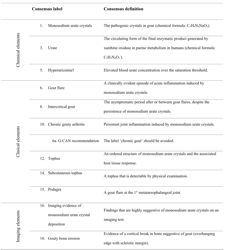
Table 1. G-CAN endorsed labels and definitions of the disease elements of gout 7
Consensus label Consensus definition
C he m ic al e le m en ts
1. Monosodium urate crystals The pathogenic crystals in gout (chemical formula: C5H4N4NaO3).
3. Urate
The circulating form of the final enzymatic product generated by xanthine oxidase in purine metabolism in humans (chemical formula: C5H3N4O3-).
5. Hyperuricemia† Elevated blood urate concentration over the saturation threshold.
C lin ic al e le m en ts
6. Gout flare A clinically evident episode of acute inflammation induced by monosodium urate crystals.
8. Intercritical gout The asymptomatic period after or between gout flares, despite the persistence of monosodium urate crystals.
10. Chronic gouty arthritis Persistent joint inflammation induced by monosodium urate crystals.
6a. G-CAN recommendation The label ‘chronic gout’ should be avoided.
12. Tophus
An ordered structure of monosodium urate crystals and the associated host tissue response.
14. Subcutaneous tophus
A tophus that is detectable by physical examination.
15. Podagra
A gout flare at the 1st metatarsophalangeal joint.
Im ag in g el em en ts
16. Imaging evidence of monosodium urate crystal deposition
Findings that are highly suggestive of monosodium urate crystals on an imaging test.
18. Gouty bone erosion Evidence of a cortical break in bone suggestive of gout (overhanging edge with sclerotic margin).
Table 2. Results of the content analysis of 539 gout- and hyperuricemia-related articles: disease element clusters identified as potentially meaningful disease states of gout and characteristics of their labels.
Disease states represented by disease element clusters
Number of articles
labelling disease state (%
of total articles)
Number of unique
labels identified
Most frequently used labels
(% of articles referencing disease
state) P re cl in ic al s ta te s
Hyperuricemia† without clinical disease elements2 of gout 79 (14.5%) 1 Asymptomatic hyperuricemia† (100%)
Imaging evidence of MSU1 crystal deposition without clinical disease
elements2 of gout
32 (5.9%) 8 Asymptomatic MSU
1 crystal deposition
(43.8%)
Hyperuricemia† with imaging evidence of MSU1 crystal deposition and
without clinical disease elements2 of gout
32 (5.9%) 4 Asymptomatic hyperuricemia† with MSU
1
crystal deposition (90.6%)
C li ni ca l s ta te s
Presence of MSU1 crystals with
Clinical disease elements2 of gout 61 (11.3%) 14 Symptomatic gout (50.8%)
Presence of MSU1 crystals with any of the following: frequent recurrent
gout flares, chronic gouty arthritis, subcutaneous tophi or imaging disease elements3 of gout
72 (13.4%) 6 Severe gout (81.9%)
Presence of MSU1 crystals with at least one subcutaneous tophus 106 (19.7%) 3 Tophaceous gout (81.1%)
Chronic gouty arthritis with at least one subcutaneous tophus
(40.0%) Presence of MSU1 crystals with any of the following: gout flare, chronic
gouty arthritis and without subcutaneous tophi 10 (1.9%) 3 Non-tophaceous gout (80%) Presence of MSU1 crystals with clinical disease elements2 of gout and with
at least one gouty bone erosion 6 (1.1%) 1 Erosive gout (100%)
D
is
ea
se
c
ou
rs
e
st
at
es
The first episode of gout flare without preceding intercritical gout 73 (13.5%) 5 Incident gout (75.3%)
More than one episode of gout flare with intercritical gout 79 (14.7%) 8 Recurrent gout flares (94.9%)
Presence of MSU1 crystals with clinical disease elements2 of gout and early
in the course of disease natural history
19 (3.5%) 4 Early gout (68.4%)
Presence of MSU1 crystals with clinical disease elements2 of gout and late
in the course of disease natural history
9 (1.7%) 2 Longstanding gout (66.7%)
†In British English, hyperuricaemia.
1. Monosodium urate.
2. Clinical disease elements: gout flare, intercritical gout, chronic gouty arthritis, subcutaneous tophus.
Table 3. Results of the Delphi exercise for agreement about whether the proposed gout disease states are meaningful1.
Disease states represented by disease element clusters
Delphi exercise
Consensus achieved2
(round)
Agreement (%)
P
re
cl
in
ic
al
s
ta
te
s Hyperuricemia† without clinical disease elements3 of gout Yes (1) 84%
Imaging evidence of MSU4 crystal deposition without clinical disease elements3 of gout Yes (1) 89%
Hyperuricemia† with imaging evidence of MSU4 crystal deposition without clinical disease elements3 of gout Yes (1) 86%
C
li
ni
ca
l s
ta
te
s
Presence of MSU4 crystals with clinical disease elements3 of gout Yes (1) 97%
Presence of MSU4 crystals with any of the following: frequent recurrent gout flares, chronic gouty arthritis, subcutaneous tophi
or imaging disease elements5 of gout
Yes (1) 93%
Presence of MSU4 crystals with subcutaneous tophi Yes (1) 89%
Chronic gouty arthritis with at least one subcutaneous tophus No 74% after round 3
Presence of MSU4 crystals with any of the following: gout flare, chronic gouty arthritis; without subcutaneous tophi No 74% after round 3
D
is
ea
se
c
ou
rs
e
st
at
es
The first episode of gout flare without preceding intercritical gout Yes (1) 92%
More than one episode of gout flare with intercritical gout Yes (1) 88%
Presence of MSU4 crystals with clinical disease elements3 of gout and early in the course of disease natural history No 67% after round 3
Presence of MSU4 crystals with clinical disease elements3 of gout and late in the course of disease natural history No 69% after round 3
†In British English, hyperuricaemia.
1. ‘Meaningful’ defined as ‘having important implications for disease management and/or prognosis’.
2. Consensus defined as ≥80% agreement on preferred label.
3. Clinical disease elements: gout flare, intercritical gout, chronic gouty arthritis, subcutaneous tophus.
4. Monosodium urate.
Table 4. Results of the Delphi exercise and face-to-face consensus meeting for agreement on the labels for the disease states of gout.
Disease states represented by disease element clusters
Delphi exercise Face-to-face meeting
Agreed label Consensus achieved1 (round) Agreement (%) Consensus achieved2 Agreement (%) P re cl in ic al s ta te s
Hyperuricemia† without clinical disease elements2 of gout Yes (3) 85% - - Asymptomatic hyperuricemia†
Imaging evidence of MSU3 crystal deposition without clinical
disease elements3 of gout
Yes (3) 86% - - Asymptomatic monosodium urate
crystal deposition
Hyperuricemia† with imaging evidence of MSU3 crystal
deposition without clinical disease elements2 of gout No - Yes 100%
Asymptomatic hyperuricemia† with monosodium urate crystal
deposition C li ni ca l s ta te s
Presence of MSU3 crystals with clinical disease elements2 of
gout
No - Yes 97% Gout
Presence of MSU3 crystals with any of the following: frequent
recurrent gout flares, chronic gouty arthritis, subcutaneous tophi or imaging disease elements4 of gout
Presence of MSU3 crystals with subcutaneous tophi Yes (1) 89% - - Tophaceous gout
Presence of MSU3 crystals with clinical disease elements2 of
gout and with at least one gouty bone erosion
Yes (1) 82% - - Erosive gout
D
is
ea
se
c
ou
rs
e
st
at
es The first episode of gout flare without preceding intercritical
gout Yes (3) 83% - - First gout flare
More than one episode of gout flare with intercritical gout Yes (3) 89% - - Recurrent gout flares
†In British English, hyperuricaemia.
1. Consensus defined as ≥80% agreement on preferred label.
2. Clinical disease elements: gout flare, intercritical gout, chronic gouty arthritis, subcutaneous tophus.
3. Monosodium urate.
4. Imaging disease elements: imaging evidence of monosodium urate crystal deposition, gouty bone erosion.
Table 5. G-CAN endorsed labels and definitions for the disease states of gout.
Consensus label Consensus definition
P re cl in ic al s ta te s
1. Asymptomatic hyperuricemia† Hyperuricemia† in the absence of gout.
2. Asymptomatic monosodium urate crystal deposition
Evidence of monosodium urate crystal deposition in the absence of gout. Monosodium urate crystal deposition may be demonstrated by imaging or microscopic analysis.
3. Asymptomatic hyperuricemia† with monosodium urate crystal deposition
Hyperuricemia† with evidence of monosodium urate crystal deposition in the absence of gout. Monosodium urate crystal deposition may be demonstrated by imaging or microscopic analysis.
C lin ic al s ta te s
4. Gout A disease caused by monosodium urate crystal deposition with any of the following clinical presentations (current or prior): gout flare, chronic gouty arthritis, or subcutaneous tophus.
5. Tophaceous gout Gout with at least one subcutaneous tophus.
6. Erosive gout Gout with at least one gouty bone erosion.
D is ea se co u rs e st at es
7. First gout flare The first episode of gout flare.
8. Recurrent gout flares More than one gout flare.
Additional recommendation on disease states not
addressed by the nomenclature
Where there is more than one disease state present, these can be combined (for example: tophaceous and erosive gout). Where there are additional elements present, not recognized as disease states, these will be labelled as the recognized disease state with or without additional disease elements (for example: tophaceous gout with chronic gouty arthritis).