RESEARCH
Identifying local structural states
in atomic imaging by computer vision
Nouamane Laanait
1,2*, Maxim Ziatdinov
1,2, Qian He
3and Albina Borisevich
1,3Abstract
The availability of atomically resolved imaging modalities enables an unprecedented view into the local structural states of materials, which manifest themselves by deviations from the fundamental assumptions of periodicity and symmetry. Consequently, approaches that aim to extract these local structural states from atomic imaging data with minimal assumptions regarding the average crystallographic configuration of a material are indispensable to advances in structural and chemical investigations of materials. Here, we present an approach to identify and clas-sify local structural states that is rooted in computer vision. This approach introduces a definition of a structural state that is composed of both local and nonlocal information extracted from atomically resolved images, and is wholly untethered from the familiar concepts of symmetry and periodicity. Instead, this approach relies on computer vision techniques such as feature detection, and concepts such as scale invariance. We present the fundamental aspects of local structural state extraction and classification by application to simulated scanning transmission electron micros-copy images, and analyze the robustness of this approach in the presence of common instrumental factors such as noise, limited spatial resolution, and weak contrast. Finally, we apply this computer vision-based approach for the unsupervised detection and classification of local structural states in an experimental electron micrograph of a com-plex oxides interface, and a scanning tunneling micrograph of a defect-engineered multilayer graphene surface. Keywords: Scanning transmission electron microscopy, Scanning tunneling microscopy, Computer vision, Unsupervised machine learning
© The Author(s) 2016. This article is distributed under the terms of the Creative Commons Attribution 4.0 International License (http://creativecommons.org/licenses/by/4.0/), which permits unrestricted use, distribution, and reproduction in any medium, provided you give appropriate credit to the original author(s) and the source, provide a link to the Creative Commons license, and indicate if changes were made.
Background
A multitude of imaging probes such as scanning trans-mission electron microscopy (STEM) have reached the requisite spatial resolution, at least in two dimensions, to directly distinguish the individual structural microstate of a material, namely an atom and its local neighbors [1]. In addition, the prevalence of auxiliary information channels such as electron energy-loss spectra acquired at similar spatial resolutions allows one to append to these structural microstates additional chemical/electronic state information [2–4]. The data that emanate from such modalities reveal a wealth of information regarding the static modulation of material properties by local struc-tural deviations [5], competing structural ground states [6], and even dynamic phase transformations or ensuing
structural reordering during in situ atomic resolution imaging of materials growth [7]. These imaging modali-ties are crucial to fundamental investigations of modern materials, which often display a range of structural con-figurations and order parameter phases. In many cases, some structural phases are not directly discernible by the diffraction-based methods of X-rays and neutron scatter-ing [8, 9] due to either their small volume fraction and/or their lack of long-range periodicity, and therefore require an imaging approach [10, 11] for identification.
To identify and classify local structural states and their correlations as resolved by atomic resolution imag-ing, the traditional language of crystallography with its restrictive assumptions of symmetry and periodicity leaves much to be desired [12]. Nevertheless, many suc-cessful approaches that extract structural information from atomically resolved data [13, 14] still adopt many of the underlying assumptions of traditional crystal-lography, through the use of integral transforms such as
Open Access
*Correspondence: [email protected]
1 Institute for Functional Imaging of Materials, Oak Ridge 37831, TN, USA
Fourier transforms (e.g., in geometric phase analysis [15]) and other techniques from harmonic analysis. Such tech-niques explicitly transform the local spatial information into a space that presupposes the presence of a coher-ent superposition of componcoher-ents to classify the struc-tural states present in an image. Recent work has taken a different route to identify local structural states by analyzing the intrinsic intensity signatures in atomically resolved images through multivariate statistics [16]. The feature identification method used is strictly local, how-ever, and does not incorporate the information present in neighboring intensity distributions around an atom or defect site. Here, we explore an alternative method to identify and classify local structural states in atomically resolved images that is rooted in a multi-scale extraction and classification of structural states present in an image. The presented approach, in essence, provides a middle ground between structure identification that relies on “single-point” intensities and those that analyze informa-tion obtained from an extended region through integral transforms.
The underlying assumptions of the presented approach are contextual information and scale invariance. The for-mer implies that the local intensity distribution in the neighborhood of a particular structural state, e.g., atomic coordination surrounding a defect site, is the key measure by which we perform detection of local structural states. Furthermore, to not assume a priori, the spatial extent of these local states our approach should be scale invariant, whereby we would like to detect not only atoms but also clusters of atoms whose intensity distribution becomes more localized at larger length scales in the image (i.e., obtained through progressive down-sampling).
Our methodology borrows heavily from techniques developed in the field of computer vision to perform tasks such as pattern recognition, through the use of a scale-invariant feature detectors and descriptors [17]. Following detection, we classify the structural states by a hierarchical clustering strategy [18, 19] using the scale-invariant descriptor associated with each state. We tested the fundamental assumptions of our approach, namely scale invariance and contextual information, by applying it to simulated scanning transmission electron microscopy images of ideal crystals and atomically sharp interfaces between crystals. To explore the utility of this analysis in practice, we performed an extensive quantita-tive study of the accuracy in detection of local structural states in the presence of instrumental factors such as noise- and material-dependent factors such as low con-trast, finding that this approach is robust under common experimental conditions. Finally, we conclude by demon-strating automated extraction and classification of local
structural states in STEM images of strained interfaces of SrTiO3/LaCoO3 and local modulations in the electron
density of states near defects on graphite surfaces imaged by scanning tunneling microscopy.
Methods
In what follows, we restrict our attention to 2-dimen-sional atomically resolved images with gray scale value, where the image I is defined as a mapping from a 2-dimensional spatial domain x (i.e., pixels) to a strictly positive real number (i.e., intensity): I:x→R+.
Fea-ture detection proceeds by locating keypoint feaFea-tures, denoted by Kp(x), in an image I(x), that are extrema of a detector function F(ζ, x), where ζ is a parameter or set of parameters that specify the feature detector. The detector function is an operator that transforms the image locally, and often involves spatial derivatives of the image. A key-point can be then generally expressed as
Numerous feature detection methods have been devel-oped in the field of computer vision that achieve scale invariance [20, 21]. Here, we restricted our attention to the Laplacian of Gaussian operator (LoG). The latter is one of the most widely used feature detectors and defined as
where G(.) is a multivariate Gaussian distribution with variance σ and ∇2 is the Laplacian operator, evaluated in
the spatial domain of the image.
The LoG operator is efficient in detecting local inten-sity curvatures in images. Given that atomically resolved images show pronounced local intensity curvatures, we use the LoG throughout as a detector to extract features. As first pointed out by Lindeberg [22, 23], the Laplacian of Gaussian kernel provides a natural way to extract key-point features that are stable in both the image spatial domain and the scale space of the image. The latter is constructed by consecutive blurring (convolution with a Gaussian filter) and down-sampling of the original image I(x) [23]. With additional approximations in regard to the detector function, the construction of a scale space, and search strategies for the extrema in the spatial and scale domains, Lowe constructed a feature extraction and description framework known as the scale-invariant feature transform (SIFT) [24]. SIFT is widely regarded as one of the most effective detector-based feature extrac-tion techniques with wide-range applicaextrac-tions from pat-tern recognition [25] to image registration [20], and was used throughout this work as descriptor for a local struc-tural state.
(1)
Kp=argmaxζ,x(F◦I)(x) or argminζ,x(F◦I)(x).
Results
Scale‑invariant detection and description of structural states
We used simulated electron microscopy images of bulk SrTiO3 and SrTiO3/BaTiO3 interface projected on the
[100] direction. The images were generated using an implementation of the standard multislice code using standard imaging conditions for Nion UltraSTEM200 for 200 kV operation and an aberration-free probe [26].
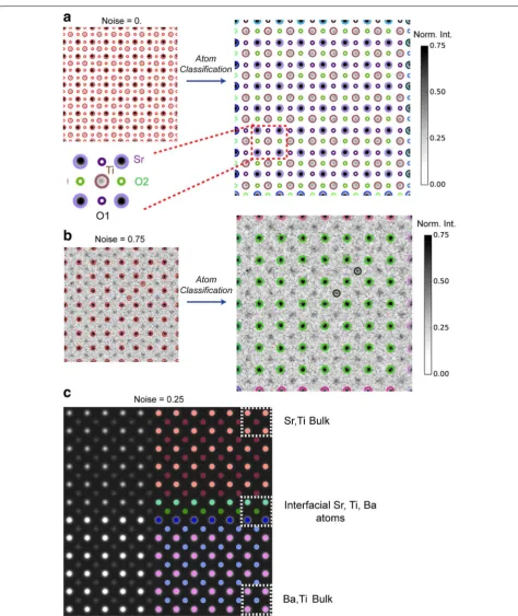
The raw simulated images were convoluted with a Gaussian probe size with a full-width-half-max of 0.7 Å to account for the finite source size of the electron beam. No other preprocessing of the images was performed. A global scaling of the intensity was applied. This intensity scaling has no effect on the Laplacian of Gaussian detec-tor, since the detector is only sensitive to the local image contrast gradient (Fig. 1a).
The atoms detected in each image by the LoG are indi-cated by circles. The size of each circle is proportional to the scale (i.e., σ) at which the feature was found to be an extrema of the LoG operator (Fig. 1b). Note that while the oxygen columns in the bulk SrTiO3 are not clearly
evident in Fig. 1a due to their low intensity relative to Sr and Ti, they are readily detected by LoG albeit at a smaller scale than either Sr or Ti columns. The detected feature is commonly referred as a keypoint, Kp, in com-puter vision. Associated with each keypoint are the coor-dinates of the feature (x,y) and its scale (Fig. 1c), as well as other properties that we do not make use of in this work.
Given a particular Kp, we use the scale-invariant feature transform to compute a descriptor, Ds. The descriptor is centered around Kp(x,y) and encodes the intensity distributions around that feature (Fig. 1c). Both the spatial extent of Ds and the intensities it contains are sampled from the spatial domain of the image but at the appropriate scale. Consequently, the image patch from which Ds is extracted (16 × 16 pixels centered on
Kp(x,y)) varies in size with respect to the spatial domain in the original image. The SIFT descriptor is composed of intensity gradient magnitudes and orientations that are appropriately weighted to decrease their contribution to the descriptor as a function of their distance from Kp(x,y) [24]. Furthermore, the intensity values in Ds are trans-formed to a local frame of reference, i.e., with respect to
Kp(x,y) The latter provides a description of the feature that is rotation invariant and reduced sensitivity to global changes in imaging conditions such as illumination [17,
27]. The resultant SIFT descriptor is a 128-dimensional unit vector and is shown in Fig. 1c in a vector format for the different detected columns in Fig. 1b. In this work, we modified the SIFT descriptor, by intentionally break-ing its rotational invariance through a choice of a pre-ferred orientation angle of the Ds image patch (0° defined
with respect to the x-axis of the image) (see Fig. 1c). This modification leads to a minimalistic descriptor that is only translation invariant and does not incorporate other symmetry assumptions. Consequently, Ds provides a dis-tinct description of intensity gradients that are dissimi-lar for atomic columns such as O1 and O2 despite them having identical local intensities, since their neighboring columns (Sr, Ti) are in a different orientation order. Given
Kp and Ds, we then define a structural state,
as a pair composed of a keypoint, which gives a local description of the image intensity, and Ds which provides a nonlocal description of neighboring intensity gradients. This description of a structural state, such as an atomic column, is both scale invariant and context dependent.
Noise and contrast behavior of structural state detection
We assumed that the imaging is free from all geomet-ric distortions due to scanning of the electron probe, and focused on testing the robustness of the above formulation at different noise levels and local con-trast values. Each simulated STEM image (Fig. 1a) is altered with noise that is sampled from a Poisson dis-tribution and added in a linear convex fashion to the ideal image, with the noise level given by λ. The accu-racy of the atomic column detection as a function of
λ is calculated by direct comparison to the ideal case (i.e., λ = 0, accuracy = 1). Furthermore, in the case of bulk SrTiO3, we split the accuracy into two classes
depending on the local contrast of the detect atoms. We found that a detection accuracy of Sr and Ti atoms fluctuates about 0.85 (±0.06) for λ ≤ 0.4, and falls off precipitously for λ > 0.4. As expected, local intensity fluctuations affect the detection of Ti atoms first, as shown in Fig. 2a. The detection accuracy of O atoms, on the other hand, becomes unreliable for noise levels that even exceed 0.05 due to their low contrast values (<0.05). Such behavior is well known in experimental
Z-contrast STEM images [28], where oxygen columns, while in principle resolvable, are often not detectable due their weak Rutherford cross-sections relative to heavier atoms and the finite dynamic range of the detec-tor. The detection accuracy of Sr, Ti, and Ba columns in the simulated image of SrTiO3/BaTiO3 as a function of
noise level behaves in an analogous manner to simu-lated bulk SrTiO3. Robust image de-noising strategies
can, of course, be employed in practice to increase the accuracy of atomic column detection by the LoG detec-tor, but this was not performed here as the de-noising constitutes a separate problem from the focus of this paper, and is well covered in both electron microscopy and image recognition literature.
The primary reason for the reduced accuracy in detected atomic columns is the delocalization of their response to the LoG kernel in scale space [29]. Note,
however, that the LoG detector has a strong response to features near edges (of an image), which, in practice, can lead to an overestimation of the detection accuracy.
Fig. 1 Structural states as scale-invariant features. a Simulated STEM images of bulk SrTiO3 and SrTiO3/BaTiO3 interface with the electron beam
propagating along the [100] crystallographic direction. Images are convoluted with a Gaussian function with FWHM of 0.7 Å to account for the finite source size of the electron beam. b Features extracted by the Laplacian of Gaussian detector are shown as an overlay of circles on the images in a. The intensity scale was inverted to improve the visibility. The size of the circle indicates the scale at which the feature was detected.
For simplicity in the ensuing analyses, the contrast threshold of the LoG is tuned so that oxygen columns in the right image in b are not detected
(see Additional file 1 for all atomic columns). c Close-up of the left image in b indicating both the keypoint, Kp, which describes the atom locally
and the descriptor vectors, Ds, which encode the intensity distribution of neighboring columns to provide a nonlocal description of the column.
Descriptors for the different atomic columns are shown as 1-dimensional vectors, indicating that columns with the same intensity can have differ-ent descriptors due to the differdiffer-ent angular configuration of their neighboring atoms. The structural state, in this case an atomic column, is then
defined by the pair composed of (Kp, Ds). The implementations of the LoG detector in the Python scikit-image library [41] and SIFT in OpenCV [42]
From the above analysis, we conclude that for noise levels <0.4Cmax, where Cmax is the maximum image
contrast of the structural feature of interest, the pre-sented approach can produce a meaningful and robust detection. An additional aspect of the LoG worth men-tioning is that the presence of other instrumental fac-tors, such as blurring, only affects the scale at which the feature is detected, but not the accuracy of the LoG detector. Finally, we emphasize that the LoG searches for both maxima and minima in the local imaging con-trast as a function of scale and therefore can be used to detect missing atoms or used in imaging modes such as
bright-field imaging where atomic columns can also be represented by the image minima. In such an instance, its detection robustness will be affected by the presence of noise in a manner similar to the above analysis.
Structural state classification
The definition of a structural state in Eq. 3 allows us to classify the different detected atomic columns to find the main structural classes present in a particular image. Numerous methods exist to perform these classification tasks. Here, we focus on unsupervised machine learning to explore the effectiveness of the presented approach
Fig. 2 Atomic column detection in the presence of noise and low contrast. The accuracy of atom detection is analyzed as a function of noise level,
λ. The noise, sampled from a Poisson distribution, is added to the STEM simulated images of bulk SrTiO3a and SrTiO3/BaTiO3 (b). The accuracy is
computed by comparison of detected features Kp(x,y) at some �=0 to the ideal images (λ = 0). To demonstrate the dependency of atom
to “learn” the overall structural configuration in a mate-rial. To that effect, we use hierarchical agglomerative clustering.
In agglomerative clustering, each structural state S is initially considered to belong to a distinct class Ci.
Follow-ing this initial assignment, different classes Ci and Cj are
merged into a new class Ck if their respective members
(i.e., structural states) are similar, given some notion of similarity, g. In our case, the similarity (or affinity) meas-ure between two structural states, Si and Sj, is naturally
defined by the (Euclidean) distance between their respec-tive descriptors, Dsi and Dsj,
and is used to merge the different structural classes. Different methods, known as linkage, apply the similar-ity measure to the classes in a specific way. We use the average linkage method which uses the average similarity between classes:
where NC (ND) are the number of structural states
belonging to each class C(D). With g¯ as similarity
meas-ure, agglomerative clustering results in a classification that groups structural states into relatively compact classes that are well separated [30]. The only remaining parameter that must be specified to perform the hierar-chical clustering of structural states is the level at which we must truncate the merging procedure. This was deter-mined by a statistical measure that optimizes the similar-ity between structural states that belong to the same class (see Additional file 1 for additional details and illustration of this analysis for the classification used here, Additional file 1: Fig. S2).
The results of the classification of atomic columns in the SrTiO3 and SrTiO3/BaTiO3 images (shown in Fig. 1b)
using agglomerative clustering at various noise levels are shown in Fig. 3, where each structural class is represented by a different color coding. For bulk SrTiO3, we find that
the classification clearly distinguishes between the differ-ent atomic columns in the unit cell. Note that although O1 and O2 oxygen columns have identical imaging inten-sities and are equivalent under the rotational symmetry of SrTiO3 (P2 mm), they are grouped into different
clus-ters, since their descriptors are not rotationally invariant as discussed above.
We found that even in the presence of large noise levels (λ = 0.75), columns of different types (Sr, Ti) are still classified separately, giving good evidence of the robustness of Eq. 3 in the presence of noise. In the case of SrTiO3/BaTiO3, a complete classification of the unit (4) g
Si,Sj
=Dsi−Ds2 j,
(5) ¯
g(C,D)= 1
NCND
i∈C
j∈D
g
i,j
,
cell configurations is achieved, with Ti columns in bulk STO, at the interface, and in bulk BTO grouped as dis-tinct states. Similar results are also obtained for Sr and Ba atomic columns. These observations are crucial evidence that the definition of an atomic column given in Eq. 3
does encapsulate the local coordination environment necessary to discriminate between different structural states and further reinforce the utility of formulating a structural state as a combination of local and nonlocal image intensities.
Strained interfaces and defects
We illustrate the utility of the structural state extraction and classification in experimental images by two case studies from some of the most widely used atomic imag-ing modalities, namely scannimag-ing transmission electron microscopy data of interfaces in heteroepitaxial systems and scanning tunneling microscopy (STM) data of defect states found on the surface of graphite.
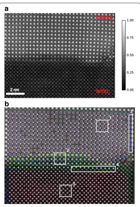
In studies of solid/solid interfaces, in particular those interfaces that originate through epitaxial growth, char-acterizing the structural nature of the interface is crucial to tailoring the materials properties. For instance, solid/ solid interfaces are often the starting point of extended defects such as misfit dislocations that arise to compen-sate epitaxial strain, and lead to elastic fields propagating in both directions from the interface, substantially modi-fying its crystal structure and potentially its properties. It has also been demonstrated that, even for a case of coherent epitaxy, the different symmetry of the film and substrate can result in a progression of distinct structural states localized in the vicinity of the interface [31]. In all these instances, it is crucial to precisely extract and iden-tify the local structural states present at interfaces. We applied the presented approach to a Z-contrast STEM image of SrTiO3(STO)/LaCoO3 (LCO) interface. This
image was acquired using Nion UltraSTEM 100 operated at 100 kV (Fig. 4a).
The classification of structural states leads to a succinct representation of the evolution of structural states at the interface, as represented by classes of LaCoO3 unit cells
Fig. 3 Classification of Local Structural States. Detected atomic columns at different noise levels in simulated STEM images are classified by hierar-chical clustering, with different structural classes represented by circles with different colors. The different atomic columns in the [100] projection of the SrTiO3 unit cell are all classified as distinct structural states by the presented approach (a). In the presence of noise, the distinction between
Sr and Ti atomic columns is still maintained (b). Note that Sr atoms at the edge of the image belong to separate classes since their coordination is different than that of Sr atoms in the “bulk”. c Classification of atoms in the image of a SrTiO3/BaTiO3 interface distinguishes the interfacial atoms (Sr,
classification of the atomic columns in this system, addi-tional properties (e.g., displacements of Co with respect to the center of LCO unit cell) can be then readily computed for a structural class and compared to others to fully char-acterize the nature of the interface in this system.
Next, we extract and classify structural states that arise due to point-like defects on a graphite surface. Point defects such as monovacancies, adsorbed atoms, intersti-tials, and Stone–Wales defects are known to affect strongly the electronic and magnetic properties of graphene layers [32]. Recently, it was realized that the electronic structure of atomic vacancy is highly sensitive to the details of the passivation of its dangling σ bonds with foreign chemical species, such as hydrogen and oxygen [33]. Here, we focus on the so-called V111 type of the
monovacancy–hydro-gen complexes [33, 34]. The V111 complex, in which each
σ dangling bond is passivated with one hydrogen atom, is characterized by the formation of a localized nonbonding
π electronic state at the Fermi level [34] whose decay into the “clean” area of the lattice can be described by r−2 law
[35]. To date, the studies of monovacancy–hydrogen com-plexes (as well as other types of point defects) in graphene-like materials have been limited to either the single-layer structure or AB (Bernal)-stacked structure. On the other hand, a rotation of graphene layers with respect to each other, particularly in the case of low twist angles (below 10°), may result in an alternation of the system’s low-energy electronic structure, such as a reduction of the Fermi velocity and associated localization of charge car-riers [36, 37], which may in turn alter the electronic and magnetic properties of the vacancy. Below, we analyze the scanning tunneling microscopy (STM) data on hydrogen-passivated single atomic vacancies of the V111 type in the
topmost graphene layer of graphite that is rotated relative to the underlying layer(s).
Figure 5a shows the STM image of the topmost gra-phene layer of graphite that features a well-defined Moiré pattern and is peppered with monovacancy–hydrogen complexes of the V111 type. The V111 complexes were
prepared by sputtering the surface of a graphite sample with low-energy Ar+ ions and its subsequent exposure to
atomic hydrogen environment and annealing. The choice of experimental parameters was the same as reported in the study of V111 complexes in Ref. [34]. The extracted
and classified structural states by our methodology are shown in Fig. 5b. First, note that the “edge” atoms around the vacancy produce a strongly nonequivalent response in terms of the corresponding local intensity of the STM signal (see inset to Fig. 5a). Given that the STM signal is a convolution between topographic and electronic features, this in-equivalency may reflect the out-of-the-plane struc-tural distortions at the vacancy site. Our analysis allows the extraction of detailed information on the distribution of the vacancy’s nonbonding state for each V111 complex
(associated with magenta, green, and orange circles, e.g., region 1 in Fig. 5b). In particular, we found that the dis-tribution of the STM signal associated with the vacancy’s nonbonding state (i) does not follow the threefold symme-try of underlying atomic lattice, which can be related either to the aforementioned structural distortions or to the rota-tional direction of the topmost graphene layer (nonzero twist angle), and (ii) the details of its propagation appear to be sensitive to the relative position of vacancy with respect to Moiré spots on the surface. To confirm the lat-ter, our analysis must be carried out on a larger set of STM images and sample conditions, and is beyond the scope of this article. Nonetheless, the efficient extraction and clas-sification of structural states associated with the monova-cancy–hydrogen complexes represents a crucial first step in a more systematic study of modulating the electronic configurations of graphene through point defects.
Fig. 4 a HAADF-STEM images of LaCoO3/SrTiO3 interface. The color
scale in normalized intensity. b Classified structural states clearly highlight the diffuse nature of the interface, with each boxed region outlining a particular structural configuration: 1 bulk LaCoO3, 2
Inter-facial LaCoO3, 3 distorted column of Co atoms, 4 interfacial SrTiO3, 5
Discussion
A key ingredient to the success of the Computer Vision-based analysis of local structural states resides in the defi-nition of a structural state that combines both local and nonlocal image intensity distributions, in contrast with previous methods that rely on single-point intensities [14,
16]. For instance, a single-point intensity method would not differentiate between the Ti columns present in bulk
BaTiO3, Ti columns in bulk SrTiO3, and those at the
interface of STO/BTO, since they all have indistinguish-able intensity values, and spatial separations and angles with respect to their neighboring atoms, yet, differ only in the type of atoms that constitutes their coordination (Fig. 3c). The latter is a direct consequence of the defini-tion of a structural state given in Eq. 3, whereby intensity gradients in a neighborhood around Kp are encoded in
Ds, with the size of this neighborhood directly given by the appropriate scale at which the keypoint was found to be an extremum of the Laplacian of Gaussian detector. Another illustrative example of the advantages of the pre-sent approach is in detecting a range of distinct classes in the local configuration of (La, Co) columns at the inter-face of LCO/STO that clearly reflect the strained nature of the latter. Given the success of our approach in detect-ing these subtle variations in the structure of materials, it would be interesting to explore in future work if one can reconstruct the fundamental ingredients of the lattice and unit cell directly from the more primitive definition of a structural state in Eq. 3, which relies solely on real space image information and the concept of scale invari-ance, without relying on the priori knowledge of the aver-age crystallographic symmetry.
The classification procedure used here, namely hier-archical clustering, enabled a physically meaningful cat-egorization of structural states in a number of cases, both for simulated and experimental data. This unsupervised learning approach, however, lacks a clear connection to the physics of the problem. In many contexts, one often seeks the identification/classification of local structural states subject to well-defined physical principles such as spatial connectivity, or localization due to the presence of interfaces, defects, etc. Under these conditions, one can supplement hierarchical clustering with connectivity constraints to generate structural classes that obey a set of physical assumptions. In essence, it allows one to test different physical hypotheses regarding the local struc-ture present in the system at hand.
We have shown that representing a structural state with computer vision-based descriptors that are effi-cient at encoding image information leads to an analysis approach that can discriminate between the myriad of local states in the presented data across vastly different imaging modalities. The preponderance of atomically resolved images both in the literature and open databases provides an opportunity to begin data exploration of local structural states that are shared by a variety of materials and their evolution during varying experimental condi-tions. The SIFT descriptor with its scale invariance could provide one of the promising methods by which one can fingerprint local structural states of interest to per-form structural recognition against the above databases.
Fig. 5 Scanning tunneling microscopy of defects on graphite. The
image was acquired with a sample bias voltage of 100 mV and tunneling current setpoint of 0.7 nA. a The defects (box outline) are
monovacancy–hydrogen complexes generated through Ar+ ion
Furthermore, the structural identification we presented could also be used to identify recurring artifacts in atom-ically resolved imaging such as dynamic scattering and electron beam channeling [38], by comparing local state descriptors obtained from a library of simulated images, for instance, as a function of thickness, to those local descriptors extracted from experimental data.
Modern imaging modalities such as STEM are hyper-spectral in nature, where in addition to atomic resolu-tion images (by Z-contrast), a full electron energy-loss spectrum can be acquired. In the case of STM, tunneling spectroscopy can be performed to measure the full elec-tronic density of states. As such, incorporating this addi-tional information into the feature detection/description method is an important task that should be explored in future work [39], to construct descriptors that are more physics based, thereby taking full advantage of all the information present in modern imaging modalities. This would benefit, in particular, atomic imaging modalities, such as atom probe tomography, that provide a full three-dimensional view of a material’s structure [40].
Conclusion
In summary, we have explored a novel approach by which one can detect, identify, and classify local structural states in spatially resolved atomic images. We showed that the principles of scale invariance and contextual structural state identification, defined based on neighbor-ing intensity distributions, give an efficient and discrimi-native approach by which one can extract and identify local states without the assumptions of symmetry, and illustrated the application of this method to simulated and experimental images from electron microscopy and scanning tunneling microscopy. Moreover, we showed that the more primitive concept of a structural state is sufficient to extract the salient structural configura-tions present in atomic imaging of materials. We foresee that our approach may provide a natural and powerful method by which one can express more complex struc-tural correlations such as those present in frustrated and disordered systems, correlations that may lie obscured by the rigid assumptions of classical crystallography in two dimensions.
Additional file
Additional file 1: Figure S1. Detected Keypoints from all atomic columns present in the simulated. STEM of a SrTiO3/BaTiO3. In the main
text, only Sr, Ti, and Ba columns are included to simplify the analysis of noise dependency and classification. The addition of detected oxygen col-umns shown above does not modify the results in the main text. Figure S2. Silhouette Coefficient Analysis of the Classification is shown here for the simulated STEM image of a bulk SrTiO3 lattice. Top is a plot of the
silhouette coefficient with different number of clusters. Bottom are the 4 structural classes (Sr, Ti, O1, O2).
Abbreviations
STEM: scanning transmission electron microscopy; STM: scanning tunneling microscopy; SIFT: scale-invariant feature transform; LoG: Laplacian of Gaussian; STO: SrTiO3; LCO: LaCoO3; BTO: BaTiO3.
Authors’ contributions
NL conceived and designed the research, and performed the analysis. QH and AB performed the simulations and collected the STEM data. MZ collected the STM data. NL and AB wrote the manuscript with contributions from MZ. All authors read and approved the final manuscript.
Author details
1 Institute for Functional Imaging of Materials, Oak Ridge 37831, TN, USA. 2 Center for Nanophase Materials Sciences, Oak Ridge 37831, TN, USA. 3
Materi-als Sciences and Technology Division, Oak Ridge National Laboratory, Oak Ridge 37831, TN, USA.
Acknowledgements
NL thanks Sergei V. Kalinin for insightful discussions and for bringing his atten-tion to this research topic. This work was supported by the Eugene P. Wigner Fellowship (NL) at Oak Ridge National Laboratory (ORNL), a US Department of Energy (DOE) facility managed by UT-Battelle, LLC for US DOE Office of Science under Contract No. DE-AC05-00OR22725. Data analysis was performed at the Center for Nanophase Materials Sciences, a DOE Office of Science User Facility at ORNL. Electron microscopy imaging and simulations (AB, QH) were sup-ported by Materials Science and Engineering Division of the US DOE Office of Science. MZ acknowledges the support from Materials Science and Engineer-ing Division of the US DOE Office of Science.
Competing interests
The authors declare that they have no competing interests.
Received: 23 August 2016 Accepted: 26 October 2016
References
1. Pennycook, S.J., Kalinin, S.V.: Microscopy: hasten high resolution. Nature
515, 487–488 (2014)
2. Zhou, W., et al.: Direct determination of the chemical bonding of indi-vidual impurities in graphene. Phys. Rev. Lett. 109(20), 206803 (2012) 3. Krivanek, O.L., et al.: Atom-by-atom structural and chemical analysis
by annular dark-field electron microscopy. Nature 464(7288), 571–574 (2010)
4. Erni, R., et al.: Atomic-resolution imaging with a sub-50-pm electron probe. Phys. Rev. Lett. 102(9), 096101 (2009)
5. Kim, Y.M., et al.: Probing oxygen vacancy concentration and homogeneity in solid-oxide fuel-cell cathode materials on the subunit-cell level. Nat. Mater. 11(10), 888–894 (2012)
6. Catalan, G., et al.: Flexoelectric rotation of polarization in ferroelectric thin films. Nat. Mater. 10(12), 963–967 (2011)
7. Nagao, K., et al.: Experimental observation of quasicrystal growth. Phys. Rev. Lett. 115(7), 075501 (2015)
8. Als-Nielsen, J., McMorrow, D.: Elements of Modern X-ray Physics, 2nd edn. Wiley, Hoboken (2011)
9. Cross, J.O., et al.: Materials characterization and the evolution of materials. MRS. Bull. 40(12), 1019–1033 (2015)
10. Laanait, N., et al.: Full-field X-ray reflection microscopy of epitaxial thin-films. J. Synchrotron Radiat 21(6), 1252–1261 (2014)
11. Holt, M., et al.: Nanoscale hard X-ray microscopy methods for materials studies. Ann. Rev. Mater. Res. 43(1), 183–211 (2013)
12. Keen, D.A., Goodwin, A.L.: The crystallography of correlated disorder. Nature 521(7552), 303–309 (2015)
13. Borisevich, A.Y., et al.: Suppression of octahedral tilts and associated changes in electronic properties at epitaxial oxide heterostructure inter-faces. Phys. Rev. Lett. 105(8), 087204 (2010)
15. Hytch, M.J., Snoeck, E., Kilaas, R.: Quantitative measurement of displace-ment and strain fields from HREM micrographs. Ultramicroscopy 74(3), 131–146 (1998)
16. Belianinov, A., et al.: Identification of phases, symmetries and defects through local crystallography. Nat. Commun 6, 7801 (2015) 17. Szeliski, R.: Computer vision—algorithms and applications. Springer
London, London (2011)
18. Bishop, C.: Pattern recognition and machine learning. Springer, Heidel-berg (2006)
19. Ward, J.H.: Hierarchical Grouping to Optimize an Objective Function. J. Am. Stat. Assoc 58(301), 236–244 (1963)
20. Mikolajczyk, K., Schmid, C.: A performance evaluation of local descriptors. IEEE Trans. Pattern Anal. Mach. Intell. 27(10), 1615–1630 (2005)
21. Triggs, B. Detecting keypoints with stable position, orientation, and scale under illumination changes. In: Eighth European conference on computer vision. Prague (2004)
22. Lindeberg, T.: Scale-space theory: a basic tool for analysing structures at different scales. J. Appl. Stat 21(2), 224–270 (1994)
23. Burt, P.J., Adelson, E.H.: The Laplacian pyramid as a compact image code. IEEE Trans. Commun. 31(4), 532–540 (1983)
24. Lowe, D.G.: Distinctive image features from scale-invariant keypoints. Int. J. Comput. Vision 60(2), 91–110 (2004)
25. Obdrzˇa ́lek, S., Matas, J. Object recognition using local affine frames on maximally stable extremal regions. In: Ponce, J. (ed) Toward Category-Level Object Recognition, New York: Springer (2006)
26. Kirkland, E.J.: Advanced computing in electron microscopy. Plenum Press, New York (1998)
27. McLachlan, G., Peel, D.: Finite mixture models: wiley series in probability and mathematical statistics. Wiley, Hoboken (2000)
28. Pennycook, S.J.: Z-contrast transmission electron-microscopy—direct atomic imaging of materials. Ann. Rev. Mater. Sci. 22, 171–195 (1992)
29. Rublee, E., et al. ORB: An efficient alternative to SIFT or SURF. In: Computer Vision (ICCV), 2011 IEEE international conference on. 2011
30. Hastie, T., Tobshirani, R., Friedman, J.: The elements of statistical learn-ing: data mining, inference, and prediction. Springer series in statistics. Springer Science+ Business Media, New York (2009)
31. He, Q., et al.: Towards 3D mapping of BO6 octahedron rotations at per-ovskite heterointerfaces, unit cell by unit cell. ACS. Nano 9(8), 8412–8419 (2015)
32. Humberto, T., et al.: The role of defects and doping in 2D graphene sheets and 1D nanoribbons. Rep. Prog. Phys. 75(6), 062501 (2012)
33. Fujii, S., et al.: Role of edge geometry and chemistry in the electronic properties of graphene nanostructures. Faraday Discuss. 173, 173–199 (2014)
34. Ziatdinov, M., et al.: Direct imaging of monovacancy-hydrogen com-plexes in a single graphitic layer. Phys. Rev. B 89(15), 155405 (2014) 35. Ugeda, M.M., et al.: Missing atom as a source of carbon magnetism. Phys.
Rev. Lett. 104(9), 096804 (2010)
36. Bistritzer, R., MacDonald, A.H.: Moiré bands in twisted double-layer gra-phene. Proc. Natl. Acad. Sci. 108(30), 12233–12237 (2011)
37. de Trambly Laissardière, G., Mayou, D., Magaud, L.: Localization of Dirac Electrons in Rotated Graphene Bilayers. Nano. Lett 10(3), 804–808 (2010) 38. Loane, R.F., Xu, P., Silcox, J.: Incoherent imaging of zone axis crystals with
ADF stem. Ultramicroscopy 40(2), 121–138 (1992)
39. Brown, M., Hua, G., Winder, S.: Discriminative learning of local image descriptors. IEEE Trans. Pattern Anal. Mach. Intell. 33(1), 43–57 (2011) 40. Amouyal, Y., Schmitz, G.: Atom probe tomography—a cornerstone in
materials characterization. MRS. Bull. 41, 13 (2016)
41. van der Walt, S., et al.: Scikit-image: image processing in Python. PeerJ 2, e453 (2014)
![Fig. 1 Structural states as scale-invariant features. a Simulated STEM images of bulk SrTiO3 and SrTiO3/BaTiO3 interface with the electron beam propagating along the [100] crystallographic direction](https://thumb-us.123doks.com/thumbv2/123dok_us/911539.1588950/4.595.59.538.85.554/structural-invariant-features-simulated-interface-propagating-crystallographic-direction.webp)