regulatory T cells (Tregs)
in vivo
:
lessons from TCR-transgenic Tregs
Authors’ addresses
Kesley Attridge1, Lucy S. K. Walker2
1Kennedy Institute of Rheumatology, University of Oxford, Oxford, UK.
2
Institute for Immunity and Transplantation, University College London Medical School, London, UK.
Correspondence to: Lucy Walker
Institute for Immunity and Transplantation University College London Medical School Royal Free Campus
Rowland Hill Street London NW3 2PF, UK Tel.: +44 020 7794 0500 Fax: +44 020 7433 1943 e-mail: lucy.walker@ucl.ac.uk
Acknowledgements
We are grateful to Rupert Kenefeck and Chun Jing Wang for Treg Ki67 staining (Fig. 2), and to Gemma Ryan for artwork (Fig. 3). We thank David Sansom for critical reading of the manuscript. L. S. K. W. is funded by an MRC Senior Fellowship. The authors declare no conflicts of interest.
This is an open access article under the terms of the Creative Commons Attribution License, which permits use, distribution and reproduction in any medium, provided the original work is properly cited.
This article is part of a series of reviews covering Regulatory Cells in Health and Disease appearing in Volume 259 of Immunological Reviews.
Summary: The identification of CD25 and subsequently Forkhead box protein 3 (Foxp3) as markers for regulatory T cells (Tregs) has revolu-tionized our ability to explore this population experimentally. In a sim-ilar vein, our understanding of antigen-specific Treg responses in vivo
owes much to the fortuitous generation of T-cell receptor (TCR)-transgenic Tregs. This has permitted tracking of Tregs with a defined specificity in vivo, facilitating analysis of how encounter with cognate antigen shapes Treg homeostasis and function. Here, we review the key lessons learned from a decade of analysis of TCR-transgenic Tregs and set this in the broader context of general progress in the field. Use of TCR-transgenic Tregs has led to an appreciation that Tregs are a highly dynamic proliferative population in vivo, rather than an anergic population as they were initially portrayed. It is now clear that Treg homeostasis is positively regulated by encounter with self-antigen expressed on peripheral tissues, which is likely to be relevant to the phenomenon of peripheral repertoire reshaping that has been described for Tregs and the observation that the Treg TCR specificities vary by anatomical location. Substantial evidence has also accumulated to sup-port the role of CD28 costimulation and interleukin-2 in Treg homeo-stasis. The availability of TCR-transgenic Tregs has enabled analysis of Treg populations that are sufficient or deficient in particular genes, without the comparison being confounded by repertoire alterations. This approach has yielded insights into genes required for Treg func-tionin vivo, with particular progress being made on the role ofctla-4in this context. As the prospect of manipulating Treg populations in the clinic becomes reality, a full appreciation of the rules governing their homeostasis will prove increasingly important.
Keywords: Tregs, immune regulation, tolerance, TCR-transgenic, Treg proliferation, Treg function
Introduction
Despite the theoretical capacity to form >1015 unique ab T-cell receptors (TCRs) (1), humans contain only around 1012 T cells (2), indicating that only a small fraction of potential TCR specificities is deployed at any one time. Protective immunity against a wealth of unpredictable infec-tions therefore relies on a high level of TCR cross-reactivity (3, 4) that is beginning to be documented experimentally (5). In light of this understanding, the simplistic notion that T cells are purged of self-reactive specificities during thymic Immunological Reviews 2014
Vol. 259: 23–39
Printed in Singapore. All rights reserved
©2014 The Authors. Immunological Reviews Published by John Wiley & Sons Ltd.
development becomes untenable; removing all cells with the potential to cross-react with self-antigens would leave us with a dangerously narrow protective repertoire. In parallel with this realization, support has grown for the existence of additional tolerance mechanisms that might compensate for the inherent limitations of negative selection. Indeed, the concept of dominant tolerance, whereby T cells with regula-tory function actively control the self-reactive T cells that enter the peripheral repertoire, is now well recognized (6– 10). Considerable research effort has focused on unraveling the identity and characteristics of the regulatory T-cell popu-lation(s) and a large body of knowledge has now been gath-ered in this area. One approach that has proved informative is the use of regulatory T cells (Tregs) expressing transgenic TCRs, permitting analysis of the antigen-specific activation of this population in vivo. Here, we review some of the insights that have been made using this approach and how it has shaped our understanding of Treg biology.
TCR-transgenic Tregs: a window on Treg thymic selection
While the focus of this review is the homeostasis and func-tion of Tregs in the periphery, it is important to first briefly consider Treg thymic selection. Study of TCR-transgenic Tregs has been formative in shaping many of our current ideas about how Tregs are generated intrathymically. In fact, the important observation that thymocytes could be directed to differentiate into Treg in response to thymic self-antigen expression was made using a TCR-transgenic system with specificity for influenza hemagglutinin (HA) (11). This par-adigm was subsequently shown to hold true for other trans-genic TCR/cognate antigen double-transtrans-genic systems (12, 13). For example, DO11.10 TCR-transgenic mice on a recombinase activating gene-deficient (RAG / ) background fail to develop Tregs (14); however, transgenic provision of ovalbumin (OVA) in the thymus permits Treg generation in these mice (13). In the latter experiments, OVA was expressed under the control of the rat insulin promoter (rip-mOVA), which is known to drive expression of functionally significant levels of OVA intrathymically (15), like the endogenous insulin promoter (16). Interestingly, RAG-sufficient DO11.10 TCR-transgenic mice were found to select a small number of Tregs in the absence of thymic OVA expression (13, 14, 17), but these Tregs were enriched for endogenous TCRa chains (13, 17) consistent with the use of an alternative TCR to support their differentiation. Thus, T cells with TCRs that cannot
recognize intrathymic antigen (DO11.10/RAG / ) were precluded from differentiating into Treg, but provision of relevant antigen in the thymus (DO11.10+ rip-mOVA+ RAG / ), or the potential to recognize intrathymic antigen using non-transgenic TCR chains (DO11.10/RAG+) permitted Treg generation.
Leading on from the use of TCRs specific for model anti-gens, a subsequent wave of experiments employed trans-genic TCRs that were derived from naturally occurring Tregs (18, 19). These experiments reinforced the role of TCR specificity in Treg thymic selection and suggested a model in which intraclonal competition limits the number of T cells of a given specificity that can develop into Tregs (18, 19). Whether such niche constraints restrict Treg differentia-tion in the context of a diverse repertoire of precursors remains less clear (20).
Tregs as a highly proliferative population
Lack of Treg anergy in vivo
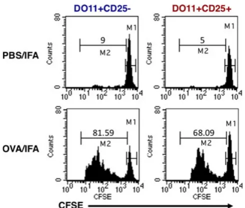
Early analysis of mature Tregs using in vitro assays identified a striking lack of proliferation following TCR engagement, leading to the characterization of this population as naturally anergic (14, 21, 22). In fact, anergy came to be known as one of the defining features of Treg populations. The gener-ation of mice bearing TCR-transgenic Tregs provided the first opportunity to monitor Treg behaviorin vivo, using clo-notypic antibodies to track a discrete cohort of adoptively transferred Tregs responding to a specific antigen. Surpris-ingly, this approach revealed a remarkable capacity for Tregs to proliferate when responding to cognate antigen in vivo. Accordingly, OVA-specific Tregs mounted a robust prolifera-tive response to OVA protein in incomplete Freund’s adju-vant (IFA) (13) (Fig. 1), while HA-specific TCR-transgenic Tregs proliferated and accumulated in the draining lymph node (LN) following subcutaneous immunization with HA peptide in IFA (23).
development of Forkhead box protein 3 (Foxp3) staining protocols, but several lines of evidence argued against the CD25+ cells being activated conventional T cells rather than Tregs. Notably, the extensive proliferative response of this population was uncoupled from production of cytokines such as IL-2, interferon-c (IFNc), and IL-4 (13, 23), and instead IL-10 production was observed in one of the studies (23). In addition, while TCR-transgenic conventional T cells upregulated CD40L following antigen encounter, this response was completely lacking in the TCR-transgenic CD25+ fraction (13). Perhaps, the most compel-ling demonstration that the TCR-transgenic CD25+ cells were in fact Tregs was that despite their capacity to prolifer-ate in vivo, they were strictly anergic when assessed in vitro and elicited robust suppression in standard co-culture assays (13, 23). Thus, TCR-transgenic Tregs recapitulated the in vitro behavior previously ascribed to this subset, yet permit-ted new insights into the antigen-responsiveness of this populationin vivo.
In addition to responding to protein antigen emulsified in adjuvant, TCR-transgenic Tregs were also shown to proliferate following provision of peptide-pulsed bone marrow-derived dendritic cells (DCs) (24). In the latter study, subcutaneous injection of OVA-pulsed DCs triggered the proliferation of OVA-specific Tregs in the draining LN, leading to an eight- to 10-fold expansion over the course of 3 days. Elegant studies
in which mice were engineered to transgenically express TCR chains from a scurfy mouse-derived CD4+ T cell also revealed robust proliferation within the Foxp3+compartment (25), demonstrating that the phenomenon extended beyond the extensively utilized TCRs specific for model antigens.
Validation that TCR-transgenic Tregs were accurately mim-icking the proliferative capacity of normal polyclonal Tregs came from experiments in which CD4+ CD25hi CD62Lhi cells were carboxyfluorescein succinimidyl ester (CFSE)-labeled and adoptively transferred into congenically distinct recipient mice (26). Thirty-five days later, approximately 70% of the donor Tregs had divided, most of them more than six times. Interestingly, the CD25hi phenotype of the injected Treg population was remarkably stable, with approximately 90% of the cells retaining high CD25 expres-sion even 70 days following transfer. This is consistent with the subsequent demonstration that Treg Foxp3 expression is also very stable under physiologic conditions (27). Bromo-deoxyuridine (BrdU) labeling experiments further supported the notion that Tregs represent a highly proliferative popula-tion in normal mice, with Tregs consistently incorporating higher levels of this thymidine analog than conventional T cells (26, 28). In line with this, the proportion of CD4+ Foxp3+ Tregs staining positive for the proliferation marker Ki67 is consistently higher than that of the conven-tional T-cell fraction (Fig. 2), and is still higher if Tregs are rendered deficient in the lipid phosphatase PTEN (29). Further insight into the biochemical control of Treg prolifer-ation was recently gleaned by analysis of animals in which raptor, an obligatory component of the mammalian target of rapamycin complex 1 (mTORC1) complex, was selectively deleted in Tregs (30). Raptor-deficient Tregs showed a striking lack of proliferation, compared with control Tregs, when cell trace labeled and adoptively transferred into nor-mal recipients. These experiments established a critical role for raptor/mTORC1 signaling in Treg proliferation, with the mevalonate pathway proving particularly important (30).
Experiments examining the proliferative capacity of human Tregs have largely recapitulated findings in mice (Fig. 2). Despite in vitro anergy (31, 32), when analyzed ex vivo, on average 22% of blood CD4+ CD25+ CD127loFoxp3+ T cells stained positive for the proliferation marker Ki67 compared with 5% of the CD4+ CD25intcells and 2% of the CD4+ CD25 cells (33). This high proliferative capacity was maintained over a period of 3–4 months in longitudinally tracked individuals (33). Detailed characterization of human
Treg populations by the Sakaguchi group has clarified that the proliferative fraction resides within the CD45RA Foxp3hi‘activated Treg’ population (34). Interest-ingly, recent analysis indicates that Tregs isolated from human secondary lymphoid organs show evidence of an even greater proliferative capacity than those found in blood, with approximately 60% staining positive for Ki67 compared with around 20% in blood-derived Tregs (35). Collectively, these studies have radically changed our view of Tregs from an anergic and unresponsive population to a highly dynamic one, equipped to respond rapidly to homeostatic and anti-genic cues (36).
Treg proliferation in infection, autoimmunity, and cancer
In addition to proliferating in the steady state, it is now well established that a variety of stimuli can enhance Treg proliferation. These include a wide range of immunological perturbations including exposure to infectious agents. For example, it has been demonstrated that Tregs proliferate and accumulate at sites of Leishmania major infection, with up to 80% of Tregs from such sites showing the capacity to respond specifically to Leishmania-infected DCs ex vivo (37). Similarly, infection of mice with Mycobacterium tuberculosis (Mtb) was shown to trigger proliferation of pathogen-specific TCR-transgenic Tregs (38). Subsequent studies by the same group used tetramers to show that endogenous Mtb-specific Tregs also proliferated and expanded in the pulmonary lymph node of infected mice. Importantly, Treg accumulation was short-lived due to selective loss of these
cells via an IL-12 and Tbet-dependent mechanism, thereby lifting regulation during the latter stages of infection (39). Viruses can also trigger the expansion of Tregs (40–42), and in one case, this was shown to occur by a superanti-gen-dependent pathway (43). Similarly, Tregs are known to proliferate in response to helminths with the proportion of Foxp3+ Tregs incorporating BrdU in the pleural cavity increasing from approximately 20% to over 50% following Litomosoides sigmondontis infection (44). In a separate study, Tregs expanded in response to Heligmosomoides polygyrus infec-tion in an inducible costimulatory (ICOS)-dependent man-ner, and this was shown to reflect a role for ICOS in supporting the survival of divided Tregs, rather than an obligate role in proliferation itself (45).
In the context of autoimmune disease, Tregs have been shown to exhibit heightened proliferation in the peripheral blood of individuals with systemic autoimmunity (46) and at the site of inflammation in tissue-specific autoimmunity. For example, Tregs from the synovial fluid of arthritis patients showed substantially higher proliferation than those in peripheral blood (47), and increased Treg proliferation has been documented in the inflamed CNS of mice with EAE (48) and the pancreas of BDC2.5 NOD mice (49). In the latter model, mice express a transgenic TCR comprising rearranged TCRa and b genes from a diabetogenic T-cell clone isolated from a non-obese diabetic (NOD) mouse (50). Both Treg and effector T cells infiltrate the islets in BDC2.5 NOD mice, and disease incidence is low unless Treg development is precluded by introduction of the
Foxp3 scurfy mutation (51). Tregs infiltrating the pancreas of BDC2.5 NOD mice were shown to incorporate higher levels of BrdU than conventional T cells present at this site (49). Consistent with this observation, Tregs infiltrating the islets in non-TCR-transgenic NOD mice showed increased staining for Ki67 compared with conventional T cells; intriguingly, this was the case in new onset disease but not in prediabetic animals (52). The loss of pancreas-resident Tregs following administration of the chemothera-peutic alkylating agent cyclophosphamide (53) may in part reflect its ability to efficiently inhibit Treg proliferation (54).
Tregs have long been recognized to be overrepresented in tumors (55), prompting interest in their proliferation at such sites. In the context of B16F10 and 4T1 tumors, analy-sis of tumor-draining LNs showed that Tregs proliferated substantially more than effector CD4+ or CD8+ T cells (56). Tregs infiltrating brain tumors in a mouse model of glio-blastoma showed markedly increased proliferation compared with their Foxp3-negative counterparts (57). Furthermore, analysis of carcinogen-induced sarcomas in mice revealed that over 60% of the tumor-infiltrating Tregs incorporated BrdU following a 3-day pulse, clearly illustrating the high proliferative potential of the tumor-resident Treg population (58). Expansion of intratumoral Tregs is thought to reflect the marked proliferation of a few dominant clones (59) and could be driven in part by the high levels of TGFb that typi-fies the tumor microenvironment (60) or by factors associ-ated with angiogenesis such as vascular endothelial growth factor (61).
The discovery that Tregs can proliferate in the steady state has been rapidly followed by an appreciation that this pro-liferative response can be significantly augmented in a vari-ety of medically relevant settings including infection, autoimmunity, and cancer. Such expansion could potentially serve to bolster the Treg population during immunological insults, such that they are poised to efficiently terminate immune responses (62).
Factors controlling peripheral Treg homeostasis
Role of TCR signaling in shaping Treg homeostasis
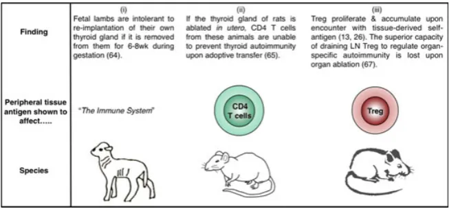
A major force that shapes the homeostasis of Treg populations in the periphery is signaling through the TCR. The seeds of this idea were sown by Peter McCullagh (63), who hypothe-sized that within any given T-cell clone, both ‘high’ and ‘low’ pathogenicity cells are selected in the thymus, with the latter able to regulate the pathogenicity of the former. Importantly, the low pathogenicity (regulatory) clones were postulated to have a short duration of survival following release from the thymus unless exposed to their specific ligand (63). The experimental basis for McCullagh’s hypothesis was the obser-vation that temporary removal of the thyroid gland in gestat-ing lambs triggered its destruction upon re-implantation, but rejection did not occur if part of the gland was allowed to remain in the animal (64). Further support for the notion that Treg homeostasis was controlled by access to peripheral self-antigen (Fig. 3) emerged from studies in rats; ablation of thyroidsin uterorendered peripheral CD4+T cells incapable of preventing thyroid autoimmunity, while preserving their
capacity to control other autoimmune diseases (65). The implication of these data is that CD4+T cells with regulatory activity fail to expand to a sufficient frequency or fail to sur-vive, if denied the opportunity to encounter relevant autoanti-gen in the periphery.
The advent of TCR-transgenic Tregs permitted the direct assessment of the role of peripheral self-antigen in control-ling Treg homeostasis. CFSE-labeled OVA-specific (13) or HA-specific (26) Tregs were adoptively transferred into mice expressing the relevant antigen as a tissue-specific self-protein, and the proliferative response in the draining LN was assessed a few days later. In both cases, antigen expres-sion was directed to the pancreatic islets through use of the rat insulin promoter, and the antigen-specific Tregs were demonstrated to proliferate specifically in the pancreatic LNs but not in non-draining LNs (13, 26) (Fig. 3). Importantly, the absolute number of OVA-specific Tregs in the draining LN was higher after adoptive transfer into mice expressing OVA as a self-antigen compared with antigen-negative litter-mates (13), consistent with a role for peripheral antigen in positively regulating Treg homeostasis. Similarly, Tregs endowed with the capacity to regulate autoimmune ovarian disease were found to be concentrated in the ovary-draining LN (66). In fact, it was shown for a range of organ-specific autoimmune diseases that Treg activity was enriched in the autoantigen-draining LN in a manner that could be abol-ished by selective autoantigen ablation (67). These data add a CD4+ CD25+ Foxp3+ dimension to the thyroid ablation studies performed a decade earlier (65) that examined the regulatory capacity of the CD4+ population as a whole (Fig. 3). As LNs draining the site of autoantigen expression have been shown to be key sites for the priming of autore-active pathogenic T cells (68, 69), it appears that regional LNs are actively involved in both the generation and sup-pression of tissue-specific autoimmune responses.
The considerable proliferative capacity of Tregsin vivobegs the question of which cell type supports such a response. In this regard, most attention to date has focused on DCs. DCs have been shown to be capable of driving Treg proliferation both in vitro and in vivo (24), particularly those bearing a CD8a-negative phenotype (70). DCs are also required for the expansion of Tregs in the setting of chronic LCMV infection (43). Ablation of DCs in mice expressing diphtheria toxin receptor under the control of the CD11c promoter signifi-cantly reduced Treg proliferation (71), although it should be noted that mice constitutively lacking DCs from birth showed only a marginal reduction in Treg numbers (72). Neverthe-less, augmenting the DC population by exogenous Flt3 ligand
administration significantly increases the Treg population, even in thymectomized mice, arguing for effects on periph-eral homeostasis rather than thymic generation (73). Further support for an influential role for DCs in Treg homeostasis derives from the observation that Treg numbers correlate with the number of major histocompatibility complex (MHC) class II-expressing DCs (74). The requirement for DCs to express MHC class II to influence Treg homeostasis (74) is consistent with a role for Treg TCR signaling and fits with the observa-tion that Tregs rendered p56(Lck)-deficient are impaired in their proliferative responsein vivo(75).
The above observations suggest a model in which the size of the peripheral Treg niche is set by the availability of MHC class II-expressing DCs bearing relevant peptides. This model is supported by the demonstration that wildtype Tregs, pre-sumably selected on a diverse array of peptides in the thymus, failed to divide in the periphery if transferred to mice in the which the MHC class II molecules were loaded with a single peptide, CLIP (76). In contrast, Tregs selected within a thymic environment expressing only CLIP proliferated to a greater extent in the periphery of these animals, consistent with hav-ing more Tregs-bearhav-ing TCRs that recognize this peptide (76). Artificially supplementing TCR engagement by adminis-tration of anti-CD3 permits enlargement of the Treg popula-tion by driving addipopula-tional Tregs into cycle, offering further support for the involvement of TCR signaling in setting the size of the peripheral Treg niche (77).
The proliferation (13, 26) and selective accumulation (66, 67, 78) of Tregs in LN draining sites of self-antigen expres-sion implies that Treg with a given TCR specificity is not uni-formly distributed throughout the body but rather are subject to local homeostatic control. In line with this idea, by sequencing TCRachains in mice with a fixed TCRbchain, the Hsieh laboratory (79) showed that the TCR repertoire of Tregs varies substantially according to anatomical location. This variation differs from similar analysis of the naive T-cell population, which revealed little repertoire skewing between anatomical sites (79), consistent with the free recirculation of naive T cells between secondary lymphoid organs.
Role of IL-2 in peripheral Treg homeostasis
It is well accepted that the cytokine most influential in Treg homeostasis is IL-2. The immunoregulatory role of this cytokine first came to light with the observation that mice deficient in IL-2 signaling exhibit polyclonal expansion of lymphocytes with tissue infiltration, autoantibody produc-tion, anemia, and in some cases, inflammatory bowel dis-ease (83–85). Available evidence, albeit limited, suggests that IL-2Ra deficiency also causes a similar phenotype in humans, with lymphadenopathy, anemia, and multi-organ lymphocytic infiltration (86). The prospect that this might reflect impairment of regulatory cells was raised by mixing experiments in which the presence of IL-2-sufficient T cells curbed the uncontrolled expansion of those rendered IL-2 deficient (87). The advent of CD25 as a marker of the Treg population permitted a more refined version of this experi-ment in which CD4+ CD25+ cells were shown to prevent lethal autoimmunity following adoptive transfer into IL-2Rb-deficient mice (88). Along similar lines, CD4+
CD25+ cells prevented disease caused by the introduction of IL-2Ra /
bone marrow into rag-deficient hosts (89). Thus, the dysregulated immunity in mice lacking IL-2 signaling could be attributed to a failure to maintain CD4+ CD25+ Tregs.
The central role for IL-2 in Treg homeostasis and meta-bolic fitness has since been clearly demonstrated (90, 91). IL-2 has been shown to reinforce qualitative aspects of the Treg program in addition to simply conferring a survival advantage, as Tregs that lack IL-2 signaling express low lev-els of numerous markers associated with the Treg phenotype (CTLA-4, CD39, CD73) (92). Interestingly, the role of the microRNA miR155 in promoting Treg homeostasis is thought to reflect its ability to inhibit SOCS1, thereby sensi-tizing Treg to IL-2 signaling (93). Along similar lines, Tregs are characterized by low SOCS3 expression, compared with conventional T cells, further increasing their IL-2 respon-siveness (94).
Elegant experiments using bone marrow chimeric mice revealed that the size of the peripheral CD4+ CD25+ Treg compartment is directly related to the number of conven-tional T cells capable of producing IL-2 (95). Consistent with this scenario, neutralizing IL-2 with antibodies reduces the steady state in vivo proliferation of Treg in normal mice (71, 96). Conversely, acute Treg ablation triggers a dramatic increase in the proliferation of those Tregs remaining (from approximately 20% to 70% Ki67+), and this increase corre-lates with increased plasma levels of IL-2 (97). The pivotal
role of IL-2 in setting the size of the peripheral Treg com-partment has led to therapeutic IL-2 administration being tested in mouse models of autoimmunity (52, 98, 99) and subsequently in several human clinical trials (100–103).
The coupling of Treg numbers to conventional T-cell IL-2 production (95) laid the foundation for the notion that Treg homeostasis and function can be ‘boosted’ by the conven-tional T-cell compartment (104). Accordingly, experiments with TCR-transgenic T cells and Tregs sharing the same specificity revealed that T-cell activation triggers a feedback loop whereby suppressive capacity is augmented hand in hand with the conventional T-cell response (104). One manifestation of this is the striking observation that when conventional T cells are activated by antigen in vivo, the early wave of IL-2 produced serves primarily to activate Treg in the locality (105). However, IL-2 cannot explain the whole of the boost effect, and a role for TNFa in augment-ing Treg proliferation was also identified (104). Intriguingly, recent data suggest that in certain settings, Treg function may also be boosted by engagement of their surface Nrp1 by the ligand Sema4a (106). The feedback loops operating to ensure appropriate suppression during immune responses are only now beginning to emerge and alterations in these may well underlie the immune imbal-ance observed in the context of autoimmunity.
Role of CD28 in Treg homeostasis
Further evidence for the importance of costimulatory signals in Treg homeostasis arose from analysis of CTLA-4-deficient mice that have a lymphoproliferative phenotype (113, 114) driven by excessive CD28 signaling (115). These animals exhibit increased Treg proliferation and an augmented Treg population (116, 117) consistent with the role of CD28 in promoting Treg proliferation. Likewise, short-term blockade of CTLA-4 in wildtype mice increases Treg proliferation (116, 118, 119), an effect that is recapitulated by administration of anti-CTLA-4 antibody to humans (120). Given the impor-tance of CD28 in driving Treg proliferation and the role of CTLA-4 in limiting CD28 engagement (121–123), the Treg proliferation induced in CTLA-4 / mice and by anti-CTLA-4 antibody likely results from enhanced CD28 signaling due to increased availability of costimulatory ligands.
The recent generation of mice in which CD28 can be in-ducibly deleted has allowed further exploration of the role of this receptor in Treg homeostasis (110). Accordingly, tamoxifen-induced CD28 deletion was shown to trigger a dramatic reduction in Treg frequencies as early as 1 week after treatment (110). The decrease in Tregs was also observed in thymectomized mice and was associated with reduced Ki67 staining, implying a crucial role for CD28 in peripheral Treg maintenance. To home in on the role of CD28 specifically in Tregs, the Turka group (124) generated an animal model in which CD28 was selectively deleted from Foxp3-expressing cells. Mice with a floxed CD28 gene were crossed with Foxp3-YFP-Cre mice, such that Foxp3 promoter activity was linked both to yellow fluorescence protein (YFP) expression and excision of the floxed CD28 gene. A decrease in the proportion of Foxp3+ Treg was observed within the CD4 single-positive population in the thymus (124), in line with the known role of CD28 in pro-moting thymic selection of Tregs (108, 109, 125). How-ever, the peripheral Treg compartment was replete in these animals, consistent with the notion that peripheral Treg homeostasis can be uncoupled from thymic output (89). Intriguingly, the animals lacking CD28 in Tregs developed autoimmune symptoms over time, primarily focused on the liver and skin, supporting a requirement for CD28 in Treg function (124). In female mice heterozygous for Foxp3-Cre in which approximately half the Tregs delete CD28 (due to random inactivation of the X-chromosome), CD28-depen-dent effects on Treg homeostasis were revealed. Accord-ingly, the CD28-sufficient Tregs in these animals incorporated significantly more BrdU than those lacking CD28 in the same animal (124). The competitive advantage conferred by Treg CD28 expression was further revealed by
mixed bone marrow transfers into irradiated recipients: when analyzed 6 months later, the entire Treg compartment appeared to derive from sufficient rather than CD28-deficient cells, while the rest of the hematopoietic compart-ment remained of mixed origin (124).
The early studies showing that DCs could drive Treg proliferation in vivo highlighted the importance of CD86/ CD80 expression for this function; OT-II TCR-transgenic Treg proliferated robustly in the presence of OVA peptide-pulsed lipopolysaccharide-matured DCs, but the response was reduced by around 70% if the DCs derived from CD80/CD86 knockout mice (24). The key roles for DCs (71, 73, 74) and the CD80/CD86 axis (107, 109) in main-taining Treg homeostasis begged the question of whether DCs represent the critical source of costimulatory ligands for Treg in vivo. This issue was elegantly explored by experi-ments in which DC-deficient (CD11c:DTA) bone marrow and CD80/CD86-deficient bone marrow were mixed and used to reconstitute lethally irradiated wildtype mice (126). The only DCs able to develop in the resulting chimeras lacked expression of CD80 and CD86, but other cell types (such as B cells) expressed these ligands at normal levels. In the absence of DC-expressed CD80/CD86, Tregs developed normally in the thymus, but their numbers were markedly reduced in the periphery (126). This suggests that DCs are a crucial source of CD28 ligands for maintaining peripheral Treg homeostasis.
The role of CD28 in positively regulating Treg homeosta-sis also appears to hold true in humans (127). Accordingly, the proliferative response of human Tregs cultured for 6 days with allogeneic mature DCs is largely inhibited by blocking anti-CD86 antibody (128). In addition, IDO activa-tion was shown to modulate proliferaactiva-tion of human Tregs in the context of mixed lymphocyte reaction responses in a manner dependent on CD80/CD86 (129). Thus, CD28 costimulation represents a primary pathway for promotion of Treg homeostasis in both mouse and human.
Apoptosis of peripheral Tregs
common c-chain cytokines in countering this process was subsequently demonstrated (130). Intriguingly, it has recently been shown that Foxp3 itself can exhibit pro-apop-totic activity, increasing the activity of Puma and Bim and repressing Bcl2 expression in thymic Treg precursors, such that these cells must compete for cytokine signals to ensure their survival (131). IL-2 is normally critical for generating a replete peripheral Treg compartment, but this requirement can be circumvented if Treg apoptosis is prevented by Bim deficiency (132).
The importance of apoptosis in setting the size of the peripheral Treg niche has recently been demonstrated by the generation of mice that selectively lack the pro-apopto-tic molecules Bak1 and Bax in Tregs; these mice show increased peripheral accumulation of Tregs, despite normal thymic development (97). A surprise finding from this study was that the anti-apoptotic protein responsible for promoting survival in the Treg compartment is Mcl-1 (97) rather than Bcl-2 or Bcl-XL, as has been tacitly assumed for years. Thus, the decreased level of Bcl2 observed in apop-tosis-prone Tregs may actually be correlative rather than causal.
Experiments in mice suggest that the propensity of Tregs to apoptose may decline with age in a manner that is associated with decreased Bim expression. The latter conclusion was based on the observation that Bim levels were markedly lower in Tregs from 20-month-old mice compared with 1.5-month-old animals (133); how this relates to Treg homeostasis in aging humans remains to be established.
Use of TCR-transgenic Tregs to probe suppressive
mechanismsin vivo
It is increasingly clear that Tregs can call upon a vast array of mechanisms to maintain dominant tolerance (134, 135). These are suggested to encompass use of granzyme B (136), CD73/CD39 (137), CTLA-4 (138, 139), TGFb (140, 141), IL-10 (142), and IL-35 (143). Tregs have also been demon-strated to act as an ‘IL-2 sink’ (144–146) and to induce the expression of enzymes that consume essential amino acids (147), in both cases depriving conventional T cells of essential growth factors. Moreover, Tregs are thought to interact with multiple target populations to effect suppression, including conventional T cells and antigen-presenting cells.
The role of CTLA-4 in Treg-mediated suppression has been a matter of contention for more than a decade (108, 148). The issue first came to light after the demonstration that unlike conventional CD4+ T cells, Tregs constitutively
express CTLA-4 (138, 139, 149). Many approaches have since been adopted in an effort to clearly define the function of Treg-expressed CTLA-4, an endeavor that has been complicated by a lack of clarity on the basic nature of CTLA-4-mediated inhibition. In addition, the lethal lympho-proliferative syndrome observed in CTLA-4 / mice (113, 114) has rendered data obtained from these animals difficult to interpret. However, recent studies using antigen-specific Tregs have proved insightful. Indeed, our own laboratory has overcome some of the issues outlined above by using CTLA-4 / mice expressing the DO11.10 transgenic TCR on a RAG / background. As the only TCR expressed in these mice is directed against a non-self-antigen, T cells retain a naive phenotype and the severe lymphoproliferative disease characteristic of CTLA-4 / mice is notably absent (150). Because Treg development is known to require cog-nate antigen expression in the thymus, crossing these mice with those expressing OVA intrathymically due to the rip-mOVA transgene permits the induction of Tregs with a sin-gle specificity that can be studied over the long term (116). An added benefit of this system is that the CD28 pathway is unaffected, unlike in systems where CTLA-4 immunoglobu-lin fusion protein (CTLA-4-Ig) or blocking antibodies against CD80 or CD86 is used to prevent lymphoprolifera-tive disease.
We utilized Tregs from these mice in an adoptive transfer model of type-1 diabetes and were able to show that while CTLA-4-sufficient Tregs were effective at suppressing disease onset, lack of CTLA-4 expression specifically within the Tregs compartment prevented suppressive function, with all recipients rapidly becoming hyperglycemic (116). This was the first time that Treg populations with an identical speci-ficity and affinity for antigen (conferred by their DO11.10/ RAG / status), either expressing or lacking CTLA-4, had been directly compared. The importance of CTLA-4 expres-sion for Treg suppresexpres-sion of diabetes was consistent with the immune dysregulation triggered by Treg-specific dele-tion of the ctla-4 gene (117) and has since been confirmed in another diabetes system where disease is driven by T cells responding to an endogenous pancreatic self-antigen (151).
2,3-dioxygenase) in APCs, with the ensuing local trypto-phan depletion leading to inhibition of T-cell proliferation. However, others were unable to replicate the induction of IDO activity in APCs by CTLA-4-Ig (154) and abatacept, a CTLA-4-Ig fusion protein used clinically, was shown to sup-press T-cell responses without upregulating IDO (155). Moreover, when the response of DCs to abatacept was assessed by Affymetrix microarray, minimal gene changes were observed and none at all if belatacept, a CTLA-4-Ig molecule with higher affinity for ligands, was used (156). Strikingly, a commercially available CTLA-4-Ig protein that retains full Fc-dependent effector functions induced robust gene changes in DCs (156). Among the genes upregulated was IFNc, which had been shown to mediate CTLA-4-Ig-dependent induction of IDO (152). These findings raise the possibility that some of the data generated with CTLA-4-Ig fusion proteins might reflect Fc-dependent signaling rather than reverse signaling through CD80/CD86.
An alternative model of cell-extrinsic CTLA-4 function has emerged involving the downregulation of CD80/CD86 on APCs. In common with the reverse signaling model, this involves the APCs being rendered immunosuppressive, but this time as a result of impaired costimulatory ligand expression. The downregulation of CD80/CD86 expression on APCs by incubation with CTLA-4-expressing cells has been noted in numerous studies (116, 117, 157–163) and in many cases is blocked by anti-CTLA-4 antibody (116, 159, 162, 163) or CTLA-4 deficiency (117). In collabora-tion with the Sansom group (164), we identified a novel molecular mechanism by which CTLA-4 can downregu-late CD80/CD86 that involves removal of these ligands from APCs by a process of trans-endocytosis. Using TCR-transgenic Tregs, we demonstrated that this process could occur in vivo, establishing that adoptively transferred Tregs could acquire GFP-tagged CD86 molecules from host APCs (164). Using CTLA-4 / TCR-transgenic Tregs that, we had shown, lack the capacity to regulate diabetes (116), we demonstrated that ligand trans-endocytosis in vivo was strictly dependent on CTLA-4 expression (164). Interest-ingly, we found that activated CTLA-4-expressing conven-tional T cells were also capable of trans-endocytosis, consistent with the recent demonstration that conventional T cells can utilize CTLA-4 in a cell-extrinsic manner in vivo (165, 166).
In addition to exploring the role of CTLA-4, TCR-trans-genic Tregs have also been used to study the involvement of the TGFb pathway in Treg suppression. Although the importance of TGFbfor maintaining immune homeostasis is
unquestioned, whether Tregs need TGFbfor their suppressive function and whether conventional T cells need to perceive TGFb to be suppressed has been unclear. Using the BDC2.5 TCR, Ishigame et al. (157) investigated the necessity of TGFbR-mediated signals for Treg function. In this study, BDC2.5 T cells were adoptively transferred into SCID/NOD mice, with diabetes effectively prevented by co-transfer of Foxp3-RFP+BDC2.5 Tregs. Repeating these co-transfers using the same Treg lacking TGFbRII revealed that TGFbsignaling via this receptor is not required for Treg function, as all recip-ients remained disease-free (167). Likewise Tregs lacking TGFbRII expression retained the capacity to regulate colitis, resulting in colon histopathology indistinguishable from that of mice treated with TGFbRII-sufficient Tregs (168). This dif-fers from results seen when effector T cells were rendered insensitive to TGFb using a dominant negative form of the TGFbRII; in mouse models of diabetes (169) and colitis (170), effector cells that were unable to respond to TGFb failed to be appropriately controlled by Tregs. Curiously, in the colitis model, when TCR-transgenic Tregs (DO11.10) that were either sufficient or deficient for TGFb1 were compared, no difference was observed in their ability to suppress pathol-ogy (170) and yet suppression was abrogated by anti-TGFb antibody in both cases. This observation suggests that success-ful suppression of a destructive immune response by Tregs can require TGFb even when the Tregs themselves are not producing this cytokine. Using T cells lacking TGFbRII expression, rather than those expressing dominant negative TGFbRII, it was recently shown that conventional T cells do not require the capacity to respond to TGFbsignaling for sup-pression to occur in vivo (168), mirroring results obtained earlier using in vitroassays (171). While at face value, these data are hard to reconcile, one possible interpretation is that TGFb is not a direct mediator of Treg function per se but is involved in generating an environment permissive of sup-pression. Thus, TGFb availability would support Treg func-tion, without being an absolute requirement for it. Since strong TCR signaling can release T cells from Treg suppres-sion (172, 173), the observation that TGFb decreases T-cell sensitivity to TCR stimulation (168) could potentially be important in this regard.
Insights into peripheral Treg induction
with a mono-specific conventional T-cell population free from Tregs is a major advantage of such systems. In elegant experiments that utilized the known specificity of the 5C.C7 TCR, the Allison laboratory (175) demonstrated that optimal Foxp3 induction in adoptively transferred CD4+ T cells occurred in response to low doses of high-affinity ligand. Furthermore, the small population of cells that did express Foxp3 in response to low affinity peptide was found to have contracted by day 5 post immunization and did not persist over the long term (175). These data mirror findings in the thymus suggesting a requirement for relatively strong TCR stimulation for efficient Treg generation (11).
To determine whether Tregs arose in the periphery in response to a pancreatic self-antigen, Wong et al. (176) uti-lized transgenic mice expressing the diabetogenic BDC2.5 TCR. Using TCR-based lineage tracing, the authors showed that conversion from the conventional to the regulatory T-cell compartment occurred only at very low frequencies both within the pancreas and its draining lymph node (176). In addressing the same question, the Ziegler labora-tory (177) adoptively transferred naive CD4+ Foxp3 GFP T cells from DO11.10 TCR-transgenic RAG / bone mar-row chimeric mice into RAG / recipients expressing OVA under the control of the rat insulin promoter. In this sys-tem, any Treg occurring in recipient mice must have arisen from Foxp3 donor cells. At 18 days post cell transfer, Tregs were observed in both the spleen and pancreatic lymph node, affording long-term protection against diabetes onset (177). The contrasting results observed between these reports are likely due to differences in the experimental sys-tems utilized, including the presence of existing Treg popu-lations in the BDC2.5 TCR study and the use of lymphopenic recipients in the DO11.10 TCR study. In this regard, it has recently been shown that thymus-derived and peripherally induced Tregs share a common peripheral niche, and that the former can outcompete the latter when both are present together (178).
TCR-transgenic systems have also been used to probe the differential characteristics of thymic-derived tTregs versus peripherally induced pTregs. By adoptively transferring OT-II TCR-transgenic CD4+ Foxp3 cells into congenic hosts, the Shevach laboratory (179) demonstrated that unlike their thymic-derived counterparts, pTregs did not express the transcription factor Helios when induced with low-dose OVA. However, the validity of Helios as a marker of the tTreg population is now disputed (180–182). It has recently been proposed that tTregs and pTregs can be effectively distinguished based on their expression of the receptor
Neuropilin-1 (Nrp-1). To examine this possibility, two groups used TCR-transgenic mice on a RAG-deficient back-ground, which generate progeny lacking tTregs. Yadav et al. (183) found that despite the lack of Tregs in the thymus, 1B3 TCR-transgenic RAG / mice demonstrated the emer-gence of a peripheral population of CD4+ Foxp3+ cells within roughly 3 weeks of birth. Transcriptional analyses comparing these Tregs with those from 1B3 TCR-transgenic animals, which generate tTregs normally, showed lower expression of Nrp-1 by pTregs (183). Along these same lines, Weiss et al. (184) used DO11.10 TCR-transgenic RAG / mice, which similarly lack tTregs, to show that pTregs generated in the gut mucosa in response to OVA antigen express lower levels of Nrp-1 mRNA than the total Tregs pool in wildtype BALB/c mice.
TCR-transgenic systems have also facilitated functional comparison of thymus-derived Tregs and peripherally induced Tregs. In one study, IL-10 differentiated DCs (DC10) were used to generate induced Tregs either in vitro orin vivo and these were directly compared with tTregs in a model of airway inflammation (185). The presence of the OT-II TCR transgene controlled for antigen specificity between the different Treg populations. Following adoptive transfer of pTregs or tTregs into recipients at day 46 post sensitization with OVA, pTregs were found to be superior at ameliorating airway hyper-reactivity and controlling IgE lev-els (185). More work is required to delineate the extent to which thymic-derived and peripherally induced Treg popu-lations play distinct versus overlapping roles in immune regulation.
Bypassing Treg suppressionin vivo
3 days following birth (188); pTregs can be detected in these animals (189) and have the potential to elicit regula-tory function (190), yet they fail to prevent the emergence of autoimmune pathology. It is now known that Treg reper-toires undergo ‘reshaping’ in the periphery (191, 192), likely reflecting encounter with self or environmental anti-gens. While Treg cohorts with a lower TCR diversity are also subject to peripheral ‘reshaping’, their lack of breadth ulti-mately results in a heightened risk of autoimmunity (193). Thus, conventional T-cell responses can escape appropriate control in cases where the TCR repertoire of the Treg popu-lation is not sufficiently diverse.
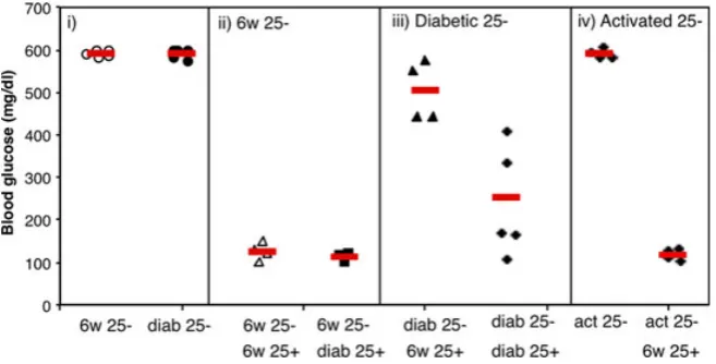
In the setting of diabetes, despite no obvious deficit of Treg numbers (reviewed in 194), we were keen to explore whether Tregs might be functionally impaired in diabetic animals. Using the DO11 x rip-OVA model of type-1 diabe-tes, we found that Tregs in diabetic animals appear to be fully functional, even showing somewhat enhanced suppres-sive capacity (78) (Fig. 4). We observed that conventional CD4+ T cells (Tconv) taken from either prediabetic or dis-eased animals induced diabetes with equivalent efficacy when transferred into Rip-mOVA RAG / recipients but showed differing sensitivity to regulation. Co-transfer of Tregs with Tconv cells from prediabetic mice was highly successful at inhibiting diabetes but was ineffective at sup-pressing disease induced by Tconv cells from diabetic ani-mals (78). These data suggested that during progression to overt diabetes, conventional T cells in this model develop resistance to Treg-mediated suppression. Similar results from
the Mathis laboratory have shown that Tregs from NOD mice show no defect in suppressive capacity compared to those from wildtype animals; rather, NOD Tconv are less prone to suppression than wildtype Tconv (195) consistent with earlier observations in this model (196, 197). Importantly, analysis of lymphocyte populations from Type 1 diabetes patients has confirmed that Tconv cells can exhibit resistance to Treg suppression in this setting (198, 199).
In the DO119rip-mOVA mouse model, levels of IL-21 mRNA increased in Tconv isolated from the draining pan-creatic lymph node as this population acquired resistance to Treg suppression (78). IL-21 was shown to interfere with Treg suppressionin vitro, and this was associated with inhibi-tion of Tconv IL-2 producinhibi-tion, thereby starving Tregs of a critical survival factor (200). Interestingly, IL-21 could sub-stitute for the lack of IL-2 in the Tconv population, permit-ting their responses to proceed unimpaired (200). Other reported examples of cytokines capable of counteracting Treg suppression include IL-2 (201), IL-6 (202), IL-7 and IL-15 (203), IL-1 and IL-18 (204), and TNFa (205). Inter-estingly, although these cytokines utilize a variety of path-ways to counteract suppression, IL-27 has also been shown to impair Treg homeostasis in a similar fashion to IL-21, by inhibiting IL-2 productionin vivo(206). In other cases, cyto-kine signaling may simply substitute for CD28 costimula-tion, thus bypassing the need for Tconv to compete with constitutively CTLA-4-equipped Tregs for costimulatory ligands.
Concluding remarks
The tools of modern immunology, most notably TCR transgenesis and fate-mapping reporters (e.g. Foxp3-GFP), have revolutionized our capacity to study Treg biology. The key feature of both approaches is that they facilitate study of Tregs in a physiologic setting in vivo. In particular, the serendipitous generation of animal models in which Tregs express transgenic TCRs has permitted analysis of cohorts of antigen-specific Tregs responding to immunized-antigen or tissue-expressed self-antigen in a whole animal. The lessons that we have learned from such analyses serve to underline the fundamental similarity between conventional and regulatory T cells, with both populations feeding off TCR
stimulation, CD28 costimulation, and cytokine signaling. A key difference appears to be the constitutively active state of the Treg population, epitomized by higher levels of prolifer-ation that presumably reflect encounter with self-antigen. Given the remarkable proliferative capacity, site-specific accumulation, and dynamic repertoire reshaping that charac-terizes the Treg population in the periphery, it is somewhat incongruous that they were initially defined as anergic. A better understanding of the parameters that determine the size of the peripheral Treg niche and the division of labor between thymus-derived and peripherally derived popula-tions should provide guidance on the best strategies to manipulate these cells clinically.
References
1. Davis MM, Bjorkman PJ. T-cell antigen receptor genes and T-cell recognition. Nature 1988;334:395–402.
2. Arstila TP, Casrouge A, Baron V, Even J, Kanellopoulos J, Kourilsky P. A direct estimate of the human alphabeta T cell receptor diversity. Science 1999;286:958–961.
3. Mason D. A very high level of crossreactivity is an essential feature of the T-cell receptor. Immunol Today 1998;19:395–404. 4. Sewell AK. Why must T cells be cross-reactive?
Nat Rev Immunol 2012;12:669–677. 5. Su LF, Kidd BA, Han A, Kotzin JJ, Davis MM.
Virus-specific CD4(+) memory-phenotype T cells are abundant in unexposed adults. Immunity 2013;38:373–383.
6. Sakaguchi S, et al. Immunologic tolerance maintained by CD25+CD4+regulatory T cells: their common role in controlling autoimmunity, tumor immunity, and transplantation tolerance. Immunol Rev 2001;182:18–32.
7. Walker LS, Abbas AK. The enemy within: keeping self-reactive T cells at bay in the periphery. Nat Rev Immunol 2002;2:11–19. 8. Graca L, Le Moine A, Cobbold SP, Waldmann H.
Dominant transplantation tolerance. Opinion. Curr Opin Immunol 2003;15:499–506. 9. Fontenot JD, Rudensky AY. A well adapted
regulatory contrivance: regulatory T cell development and the forkhead family transcription factor Foxp3. Nat Immunol 2005;6:331–337.
10. Schwartz RH. Natural regulatory T cells and self-tolerance. Nat Immunol 2005;6:327–330. 11. Jordan MS, et al. Thymic selection of
CD4+CD25+regulatory T cells induced by an agonist self-peptide. Nat Immunol 2001;2:301– 306.
12. Apostolou I, Sarukhan A, Klein L, von Boehmer H. Origin of regulatory T cells with known specificity for antigen. Nat Immunol 2002;3:756–763.
13. Walker LS, Chodos A, Eggena M, Dooms H, Abbas AK. Antigen-dependent proliferation of
CD4+CD25+regulatory T cells in vivo. J Exp Med 2003;198:249–258.
14. Itoh M, et al. Thymus and autoimmunity: production of CD25+CD4+naturally anergic and suppressive T cells as a key function of the thymus in maintaining immunologic self-tolerance. J Immunol 1999;162:5317–5326. 15. Kurts C, Heath WR, Carbone FR, Allison J, Miller
JF, Kosaka H. Constitutive class I-restricted exogenous presentation of self antigens in vivo. J Exp Med 1996;184:923–930.
16. Jolicoeur C, Hanahan D, Smith KM. T-cell tolerance toward a transgenic beta-cell antigen and transcription of endogenous pancreatic genes in thymus. Proc Natl Acad Sci USA
1994;91:6707–6711.
17. Kawahata K, et al. Generation of CD4(+)CD25 (+) regulatory T cells from autoreactive T cells simultaneously with their negative selection in the thymus and from nonautoreactive T cells by endogenous TCR expression. J Immunol 2002;168:4399–4405.
18. Leung MW, Shen S, Lafaille JJ. TCR-dependent differentiation of thymic Foxp3+cells is limited to small clonal sizes. J Exp Med 2009;206:2121– 2130.
19. Bautista JL, et al. Intraclonal competition limits the fate determination of regulatory T cells in the thymus. Nat Immunol 2009;10:610–617. 20. Romagnoli P, et al. The thymic niche does not
limit development of the naturally diverse population of mouse regulatory T lymphocytes. J Immunol 2012;189:3831–3837.
21. Takahashi T, et al. Immunologic self-tolerance maintained by CD25+CD4+naturally anergic and suppressive T cells: induction of autoimmune disease by breaking their anergic/suppressive state. Int Immunol 1998;10:1969–1980. 22. Kuniyasu Y, Takahashi T, Itoh M, Shimizu J,
Toda G, Sakaguchi S. Naturally anergic and suppressive CD25(+)CD4(+) T cells as a functionally and phenotypically distinct immunoregulatory T cell subpopulation. Int Immunol 2000;12:1145–1155.
23. Klein L, Khazaie K, von Boehmer H. In vivo dynamics of antigen-specific regulatory T cells not predicted from behavior in vitro. Proc Natl Acad Sci USA 2003;100:8886–8891. 24. Yamazaki S, et al. Direct expansion of functional
CD25+CD4+regulatory T cells by antigen-processing dendritic cells. J Exp Med 2003;198:235–247.
25. Killebrew JR, Perdue N, Kwan A, Thornton AM, Shevach EM, Campbell DJ. A self-reactive TCR drives the development of Foxp3+regulatory T cells that prevent autoimmune disease. J Immunol 2011;187:861–869. 26. Fisson S, et al. Continuous activation of
autoreactive CD4+CD25+regulatory T cells in the steady state. J Exp Med 2003;198:737–746. 27. Rubtsov YP, et al. Stability of the regulatory T
cell lineage in vivo. Science 2010;329:1667– 1671.
28. Hori S, Haury M, Lafaille JJ, Demengeot J, Coutinho A. Peripheral expansion of thymus-derived regulatory cells in anti-myelin basic protein T cell receptor transgenic mice. Eur J Immunol 2002;32:3729–3735.
29. Walsh PT, et al. PTEN inhibits IL-2 receptor-mediated expansion of CD4+CD25+ Tregs. J Clin Invest 2006;116:2521–2531. 30. Zeng H, Yang K, Cloer C, Neale G, Vogel P, Chi
H. mTORC1 couples immune signals and metabolic programming to establish T(reg)-cell function. Nature 2013;499:485–490. 31. Ng WF, et al. Human CD4(+)CD25(+) cells: a
naturally occurring population of regulatory T cells. Blood 2001;98:2736–2744.
32. Taams LS, Smith J, Rustin MH, Salmon M, Poulter LW, Akbar AN. Human anergic/ suppressive CD4(+)CD25(+) T cells: a highly differentiated and apoptosis-prone population. Eur J Immunol 2001;31:1122–1131. 33. Vukmanovic-Stejic M, et al. The kinetics of
34. Miyara M, et al. Functional delineation and differentiation dynamics of human CD4+T cells expressing the FoxP3 transcription factor. Immunity 2009;30:899–911.
35. Peters JH, et al. Human secondary lymphoid organs typically contain polyclonally-activated proliferating regulatory T cells. Blood 2013;122:2213–2223.
36. Walker LS. CD4+CD25+Treg: divide and rule? Immunology 2004;111:129–137.
37. Suffia IJ, Reckling SK, Piccirillo CA, Goldszmid RS, Belkaid Y. Infected site-restricted Foxp3+ natural regulatory T cells are specific for microbial antigens. J Exp Med 2006;203:777–788.
38. Shafiani S, Tucker-Heard G, Kariyone A, Takatsu K, Urdahl KB. Pathogen-specific regulatory T cells delay the arrival of effector T cells in the lung during early tuberculosis. J Exp Med 2010;207:1409–1420.
39. Shafiani S, et al. Pathogen-specific Treg cells expand early during mycobacterium tuberculosis infection but are later eliminated in response to Interleukin-12. Immunity 2013;38:1261–1270. 40. Betts RJ, et al. Influenza A virus infection results
in a robust, antigen-responsive, and widely disseminated Foxp3+regulatory T cell response. J Virol 2012;86:2817–2825.
41. Schmitz I, et al. IL-21 restricts virus-driven Treg cell expansion in chronic LCMV infection. PLoS Pathog 2013;9:e1003362.
42. Veiga-Parga T, Sehrawat S, Rouse BT. Role of regulatory T cells during virus infection. Immunol Rev 2013;255:182–196.
43. Punkosdy GA, et al. Regulatory T-cell expansion during chronic viral infection is dependent on endogenous retroviral superantigens. Proc Natl Acad Sci USA 2011;108:3677–3682. 44. Taylor MD, et al. Early recruitment of natural
CD4+Foxp3+Treg cells by infective larvae determines the outcome of filarial infection. Eur J Immunol 2009;39:192–206.
45. Redpath SA, et al. ICOS controls Foxp3(+) regulatory T-cell expansion, maintenance and IL-10 production during helminth infection. Eur J Immunol 2013;43:705–715.
46. Alexander T, et al. Foxp3+Helios+regulatory T cells are expanded in active systemic lupus erythematosus. Ann Rheum Dis 2013;72:1549–1558.
47. Herrath J, et al. The inflammatory milieu in the rheumatic joint reduces regulatory T-cell function. Eur J Immunol 2011;41:2279–2290. 48. O’Connor RA, Malpass KH, Anderton SM. The
inflamed central nervous system drives the activation and rapid proliferation of Foxp3+ regulatory T cells. J Immunol
2007;179:958–966.
49. Herman AE, Freeman GJ, Mathis D, Benoist C. CD4+CD25+T regulatory cells dependent on ICOS promote regulation of effector cells in the prediabetic lesion. J Exp Med
2004;199:1479–1489.
50. Haskins K, Portas M, Bergman B, Lafferty K, Bradley B. Pancreatic islet-specific T-cell clones from nonobese diabetic mice. Proc Natl Acad Sci USA 1989;86:8000–8004.
51. Chen Z, Herman AE, Matos M, Mathis D, Benoist C. Where CD4+CD25+T reg cells impinge on autoimmune diabetes. J Exp Med
2005;202:1387–1397.
52. Tang Q, et al. Central role of defective interleukin-2 production in the triggering of islet autoimmune destruction. Immunity
2008;28:687–697.
53. Brode S, Raine T, Zaccone P, Cooke A. Cyclophosphamide-induced type-1 diabetes in the NOD mouse is associated with a reduction of CD4+CD25+Foxp3+regulatory T cells. J Immunol 2006;177:6603–6612.
54. Lutsiak ME, Semnani RT, De Pascalis R, Kashmiri SV, Schlom J, Sabzevari H. Inhibition of CD4(+) 25+T regulatory cell function implicated in enhanced immune response by low-dose cyclophosphamide. Blood 2005;105:2862–2868. 55. Nishikawa H, Sakaguchi S. Regulatory T cells in
tumor immunity. Int J Cancer 2010;127:759–767.
56. Darrasse-Jeze G, et al. Tumor emergence is sensed by self-specific CD44hi memory Tregs that create a dominant tolerogenic environment for tumors in mice. J Clin Invest
2009;119:2648–2662.
57. Wainwright DA, Sengupta S, Han Y, Lesniak MS. Thymus-derived rather than tumor-induced regulatory T cells predominate in brain tumors. Neuro Oncol 2011;13:1308–1323.
58. Bui JD, Uppaluri R, Hsieh CS, Schreiber RD. Comparative analysis of regulatory and effector T cells in progressively growing versus rejecting tumors of similar origins. Cancer Res 2006;66:7301–7309.
59. Sainz-Perez A, Lim A, Lemercier B, Leclerc C. The T-cell receptor repertoire of
tumor-infiltrating regulatory T lymphocytes is skewed toward public sequences. Cancer Res 2012;72:3557–3569.
60. Ghiringhelli F, et al. CD4+CD25+regulatory T cells inhibit natural killer cell functions in a transforming growth factor-beta-dependent manner. J Exp Med 2005;202:1075–1085. 61. Terme M, et al. VEGFA-VEGFR pathway blockade
inhibits tumor-induced regulatory T-cell proliferation in colorectal cancer. Cancer Res 2013;73:539–549.
62. Walker LS. Regulatory T cells overturned: the effectors fight back. Immunology
2009;126:466–474.
63. McCullagh P. The significance of immune suppression in normal self tolerance. Immunol Rev 1996;149:127–153.
64. McCullagh P. Interception of the development of self tolerance in fetal lambs. Eur J Immunol 1989;19:1387–1392.
65. Seddon B, Mason D. Peripheral autoantigen induces regulatory T cells that prevent autoimmunity. J Exp Med 1999;189:877–882. 66. Samy ET, Parker LA, Sharp CP, Tung KS.
Continuous control of autoimmune disease by antigen-dependent polyclonal CD4+CD25+ regulatory T cells in the regional lymph node. J Exp Med 2005;202:771–781.
67. Wheeler KM, Samy ET, Tung KS. Cutting edge: normal regional lymph node enrichment of
antigen-specific regulatory T cells with autoimmune disease-suppressive capacity. J Immunol 2009;183:7635–7638. 68. Gagnerault MC, Luan JJ, Lotton C, Lepault F.
Pancreatic lymph nodes are required for priming of beta cell reactive T cells in NOD mice. J Exp Med 2002;196:369–377.
69. Scheinecker C, McHugh R, Shevach EM, Germain RN. Constitutive presentation of a natural tissue autoantigen exclusively by dendritic cells in the draining lymph node. J Exp Med
2002;196:1079–1090.
70. Zou T, Caton AJ, Koretzky GA, Kambayashi T. Dendritic cells induce regulatory T cell proliferation through antigen-dependent and -independent interactions. J Immunol 2010;185:2790–2799.
71. Suffner J, et al. Dendritic cells support homeostatic expansion of Foxp3+regulatory T cells in Foxp3.LuciDTR mice. J Immunol 2010;184:1810–1820.
72. Ohnmacht C, et al. Constitutive ablation of dendritic cells breaks self-tolerance of CD4 T cells and results in spontaneous fatal autoimmunity. J Exp Med 2009;206:549–559.
73. Swee LK, Bosco N, Malissen B, Ceredig R, Rolink A. Expansion of peripheral naturally occurring T regulatory cells by Fms-like tyrosine kinase 3 ligand treatment. Blood 2009;113:6277–6287. 74. Darrasse-Jeze G, et al. Feedback control of
regulatory T cell homeostasis by dendritic cells in vivo. J Exp Med 2009;206:1853–1862. 75. Kim JK, et al. Impact of the TCR signal on
regulatory T cell homeostasis, function, and trafficking. PLoS ONE 2009;4:e6580.
76. Zou T, et al. Cutting edge: IL-2 signals determine the degree of TCR signaling necessary to support regulatory T cell proliferation in vivo. J Immunol 2012;189:28–32.
77. Nishio J, Feuerer M, Wong J, Mathis D, Benoist C. Anti-CD3 therapy permits regulatory T cells to surmount T cell receptor-specified peripheral niche constraints. J Exp Med
2010;207:1879–1889.
78. Clough LE, et al. Release from regulatory T cell-mediated suppression during the onset of tissue-specific autoimmunity is associated with elevated IL-21. J Immunol 2008;180:5393–5401. 79. Lathrop SK, Santacruz NA, Pham D, Luo J, Hsieh
CS. Antigen-specific peripheral shaping of the natural regulatory T cell population. J Exp Med 2008;205:3105–3117.
80. Burzyn D, Benoist C, Mathis D. Regulatory T cells in nonlymphoid tissues. Nat Immunol 2013;14:1007–1013.
81. Feuerer M, et al. Lean, but not obese, fat is enriched for a unique population of regulatory T cells that affect metabolic parameters. Nat Med 2009;15:930–939.
82. Sathaliyawala T, et al. Distribution and compartmentalization of human circulating and tissue-resident memory T cell subsets. Immunity 2013;38:187–197.
84. Willerford DM, Chen J, Ferry JA, Davidson L, Ma A, Alt FW. Interleukin-2 receptor alpha chain regulates the size and content of the peripheral lymphoid compartment. Immunity 1995;3:521– 530.
85. Suzuki H, et al. Deregulated T cell activation and autoimmunity in mice lacking interleukin-2 receptor beta. Science 1995;268:1472–1476. 86. Sharfe N, Dadi HK, Shahar M, Roifman CM.
Human immune disorder arising from mutation of the alpha chain of the interleukin-2 receptor. Proc Natl Acad Sci USA 1997;94:3168–3171. 87. Kramer S, Schimpl A, Hunig T.
Immunopathology of interleukin (IL) 2-deficient mice: thymus dependence and suppression by thymus-dependent cells with an intact IL-2 gene. J Exp Med 1995;182:1769–1776.
88. Malek TR, Yu A, Vincek V, Scibelli P, Kong L. CD4 regulatory T cells prevent lethal autoimmunity in IL-2Rbeta-deficient mice. Implications for the nonredundant function of IL-2. Immunity 2002;17:167–178.
89. Almeida AR, Legrand N, Papiernik M, Freitas AA. Homeostasis of peripheral CD4+T cells: IL-2R alpha and IL-2 shape a population of regulatory cells that controls CD4+T cell numbers. J Immunol 2002;169:4850–4860.
90. Fontenot JD, Rasmussen JP, Gavin MA, Rudensky AY. A function for interleukin 2 in
Foxp3-expressing regulatory T cells. Nat Immunol 2005;6:1142–1151.
91. D’Cruz LM, Klein L. Development and function of agonist-induced CD25+Foxp3+regulatory T cells in the absence of interleukin 2 signaling. Nat Immunol 2005;6:1152–1159. 92. Cheng G, Yu A, Dee MJ, Malek TR. IL-2R
signaling is essential for functional maturation of regulatory T cells during thymic development. J Immunol 2013;190:1567–1575.
93. Lu LF, et al. Foxp3-dependent microRNA155 confers competitive fitness to regulatory T cells by targeting SOCS1 protein. Immunity 2009;30:80–91.
94. Pillemer BB, Xu H, Oriss TB, Qi Z, Ray A. Deficient SOCS3 expression in
CD4+CD25+FoxP3+regulatory T cells and SOCS3-mediated suppression of Treg function. Eur J Immunol 2007;37:2082–2089. 95. Almeida AR, Zaragoza B, Freitas AA. Indexation
as a novel mechanism of lymphocyte homeostasis: the number of CD4+CD25+ regulatory T cells is indexed to the number of IL-2-producing cells. J Immunol 2006;177:192– 200.
96. Setoguchi R, Hori S, Takahashi T, Sakaguchi S. Homeostatic maintenance of natural Foxp3(+) CD25(+) CD4(+) regulatory T cells by interleukin (IL)-2 and induction of autoimmune disease by IL-2 neutralization. J Exp Med 2005;201:723–735.
97. Pierson W, et al. Antiapoptotic Mcl-1 is critical for the survival and niche-filling capacity of Foxp3(+) regulatory T cells. Nat Immunol 2013;14:959–965.
98. Rabinovitch A, Suarez-Pinzon WL, Shapiro AM, Rajotte RV, Power R. Combination therapy with sirolimus and interleukin-2 prevents spontaneous
and recurrent autoimmune diabetes in NOD mice. Diabetes 2002;51:638–645.
99. Grinberg-Bleyer Y, et al. IL-2 reverses established type 1 diabetes in NOD mice by a local effect on pancreatic regulatory T cells. J Exp Med 2010;207:1871–1878.
100. Saadoun D, et al. Regulatory T-cell responses to low-dose interleukin-2 in HCV-induced vasculitis. N Engl J Med 2011;365:2067–2077. 101. Koreth J, et al. Interleukin-2 and regulatory T
cells in graft-versus-host disease. N Engl J Med 2011;365:2055–2066.
102. Bluestone JA. The yin and yang of
interleukin-2-mediated immunotherapy. N Engl J Med 2011;365:2129–2131.
103. Matsuoka K, et al. Low-dose interleukin-2 therapy restores regulatory T cell homeostasis in patients with chronic graft-versus-host disease. Sci Transl Med 2013;5:179ra143.
104. Grinberg-Bleyer Y, et al. Pathogenic T cells have a paradoxical protective effect in murine autoimmune diabetes by boosting Tregs. J Clin Invest 2010;120:4558–4568.
105. O’Gorman WE, et al. The initial phase of an immune response functions to activate regulatory T cells. J Immunol 2009;183:332–339. 106. Delgoffe GM, et al. Stability and function of
regulatory T cells is maintained by a neuropilin-1-semaphorin-4a axis. Nature 2013;501:252–256.
107. Salomon B, et al. B7/CD28 costimulation is essential for the homeostasis of the CD4+CD25+ immunoregulatory T cells that control autoimmune diabetes. Immunity 2000;12:431–440.
108. Sansom DM, Walker LS. The role of CD28 and cytotoxic T-lymphocyte antigen-4 (CTLA-4) in regulatory T-cell biology. Immunol Rev 2006;212:131–148.
109. Tang Q, et al. Cutting edge: CD28 controls peripheral homeostasis of CD4+CD25+ regulatory T cells. J Immunol 2003;171:3348–3352.
110. Gogishvili T, Luhder F, Goebbels S,
Beer-Hammer S, Pfeffer K, Hunig T. Cell-intrinsic and -extrinsic control of Treg-cell homeostasis and function revealed by induced CD28 deletion. Eur J Immunol 2013;43:188–193.
111. Sanchez-Valdepenas C, Martin AG, Ramakrishnan P, Wallach D, Fresno M. NF-kappaB-inducing kinase is involved in the activation of the CD28 responsive element through phosphorylation of c-Rel and regulation of its transactivating activity. J Immunol 2006;176:4666–4674.
112. Murray SE. Cell-intrinsic role for NF-kappa B-inducing kinase in peripheral maintenance but not thymic development of Foxp3+regulatory T cells in mice. PLoS ONE 2013;8:e76216. 113. Tivol EA, Borriello F, Schweitzer AN, Lynch WP,
Bluestone JA, Sharpe AH. Loss of CTLA-4 leads to massive lymphoproliferation and fatal multiorgan tissue destruction, revealing a critical negative regulatory role of CTLA-4. Immunity 1995;3:541–547.
114. Waterhouse P, et al. Lymphoproliferative disorders with early lethality in mice deficient in Ctla-4. Science 1995;270:985–988.
115. Tai X, Van Laethem F, Sharpe AH, Singer A. Induction of autoimmune disease in CTLA-4-/-mice depends on a specific CD28 motif that is required for in vivo costimulation. Proc Natl Acad Sci USA 2007;104:13756–13761. 116. Schmidt EM, et al. CTLA-4 controls regulatory T
cell peripheral homeostasis and is required for suppression of pancreatic islet autoimmunity. J Immunol 2009;182:274–282.
117. Wing K, et al. CTLA-4 control over Foxp3+ regulatory T cell function. Science 2008;322:271–275.
118. Quezada SA, Peggs KS, Curran MA, Allison JP. CTLA4 blockade and GM-CSF combination immunotherapy alters the intratumor balance of effector and regulatory T cells. J Clin Invest 2006;116:1935–1945.
119. Tang AL, et al. CTLA4 expression is an indicator and regulator of steady-state CD4+ FoxP3+T cell homeostasis. J Immunol 2008;181:1806–1813.
120. Kavanagh B, et al. CTLA4 blockade expands FoxP3+regulatory and activated effector CD4+T cells in a dose-dependant fashion. Blood 2008;112:1175–1183.
121. Thompson CB, Allison JP. The emerging role of CTLA-4 as an immune attenuator. Immunity 1997;7:445–450.
122. Walker LS, Sansom DM. The emerging role of CTLA4 as a cell-extrinsic regulator of T cell responses. Nat Rev Immunol 2011;11:852–863. 123. Bour-Jordan H, Esensten JH, Martinez-Llordella
M, Penaranda C, Stumpf M, Bluestone JA. Intrinsic and extrinsic control of peripheral T-cell tolerance by costimulatory molecules of the CD28/B7 family. Immunol Rev 2011;241:180–205. 124. Zhang R, Huynh A, Whitcher G, Chang J,
Maltzman JS, Turka LA. An obligate cell-intrinsic function for CD28 in Tregs. J Clin Invest 2013;123:580–593.
125. Tai X, Cowan M, Feigenbaum L, Singer A. CD28 costimulation of developing thymocytes induces Foxp3 expression and regulatory T cell differentiation independently of interleukin 2. Nat Immunol 2005;6:152–162.
126. Bar-On L, Birnberg T, Kim KW, Jung S. Dendritic cell-restricted CD80/86 deficiency results in peripheral regulatory T-cell reduction but is not associated with lymphocyte hyperactivation. Eur J Immunol 2011;41:291–298.
127. Golovina TN, et al. CD28 costimulation is essential for human T regulatory expansion and function. J Immunol 2008;181:2855–2868. 128. Zheng Y, Manzotti CN, Liu M, Burke F, Mead KI,
Sansom DM. CD86 and CD80 differentially modulate the suppressive function of human regulatory T cells. J Immunol
2004;172:2778–2784. 129. Chung DJ, et al. Indoleamine
2,3-dioxygenase-expressing mature human monocyte-derived dendritic cells expand potent autologous regulatory T cells. Blood 2009;114:555–563.