Can Changes in Clinical Practice Decrease the Incidence of Severe
Retinopathy of Prematurity in Very Low Birth Weight Infants?
Lily C. Chow, MD*; Kenneth W. Wright, MD*; Augusto Sola, MD‡; and the CSMC Oxygen Administration
Study Group
ABSTRACT. Objective. A wide variability in the inci-dence of severe retinopathy of prematurity (ROP) is re-ported by different centers. The altered regulation of vascular endothelial growth factor from repeated epi-sodes of hyperoxia and hypoxia is 1 important factor in the pathogenesis of ROP. Strict management of O2
de-livery and monitoring to minimize these episodes may be associated with decreased rates of ROP. The objective of this study was to compare the incidence of and need for surgery for severe ROP (stages>3) in infants of 500 to 1500 g birth weight before and after the implementation of a new clinical practice of O2 management in a large
level 3 neonatal intensive care unit (NICU).
Methods. An oxygen management policy that in-cluded strict guidelines in the practices of increasing and weaning of fraction of inspired oxygen (FIO2) and the
monitoring of O2 saturation parameters in the delivery
room, during in-house transport of infants to the NICU, and throughout hospitalization was implemented in April 1998. The main objectives were to monitor oxygen-ation levels more precisely and to avoid hyperoxia and repeated episodes of hypoxia-hyperoxia in very low birth weight infants. Included in the policy were equipment for monitoring, initiation of monitoring at birth, avoid-ance of repeated increases and decreases of the FIO2,
minimization of “titration” of FIO2, modification of
pre-viously used alarm limits, and others. After an educa-tional process, each staff member signed an agreement stating understanding of and future compliance with the guidelines. Examinations were performed by experi-enced ophthalmologists following international classifi-cation and American Academy of Pediatrics recommen-dations. ROP data from January 1997 to December 2002 for infants of 500 to 1500 g were analyzed as usual and also have been reported to Vermont Oxford Network since 1998.
Results. The incidence of ROP 3 to 4 at this center decreased consistently in a 5-year period from 12.5% in 1997 to 2.5% in 2001. The need for ROP laser treatment decreased from 4.5% in 1997 to 0% in the last 3 years.
Conclusion. We observed a significant decrease in the rate of severe ROP in very low birth weight infants in association with an educational program provided to all
NICU staff and the implementation and enforcement of clinical practices of O2management and monitoring.
Al-though several confounders cannot be excluded, it is likely that differences in these clinical practices may be, at least in part, responsible for the documented inter-center variability in rates of ROP. Pediatrics 2003;111: 339 –345;retinopathy of prematurity, oxygen, pulse oxim-etry, very low birth weight.
ABBREVIATIONS. ROP, retinopathy of prematurity; VEGF, vas-cular endothelial growth factor; VLBW, very low birth weight; NICU, neonatal intensive care unit; VON, Vermont Oxford Net-work; Fio2, fraction of inspired oxygen; Spo2, oxygen saturation
levels as measured by pulse oximetry; AAP, American Academy of Pediatrics; RN, registered nurse; RT, respiratory therapist.
K
nown risk factors for retinopathy of
prematu-rity (ROP) include prematuprematu-rity and low birth
weight. In addition, worsening ROP has been
linked to severity of illness and extent of
complica-tions.
1,2Much has been described in the literature
regarding the role of supplemental oxygen in the
development of ROP.
3–7The altered regulation of vascular endothelial
growth factor (VEGF) has been suggested as 1 of the
factors in the pathogenesis of ROP.
8,9In premature
infants, the retina is incompletely vascularized. In
utero, the arterial oxygen pressure of the fetus is 22
to 24 mm Hg. After premature birth, relative
hyper-oxia may downregulate VEGF production.
Adminis-tration of supplemental O
2may lead to sustained
hyperoxia, setting the stage for vaso-obliteration of
existing vessels and arrest of the vascularization.
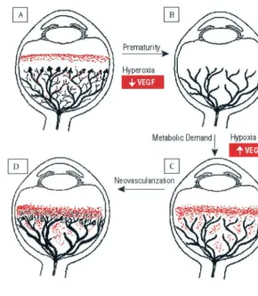
Over time, as the metabolic demands of the
devel-oping eye increases, the immature nonperfused area
of the retina becomes hypoxic and may overproduce
VEGF pathologically. High levels of VEGF stimulate
neovascularization of the retina, which in severe
cases may result in retinal fibrosis and retinal
detach-ment (Fig 1). It thus is possible that repeated cycles of
hyperoxia and hypoxia favor the progression of
ROP.
10,11With the improved survival of very low birth
weight (VLBW) infants during the past decade, ROP
continues to be a source of significant morbidity.
Wide intercenter variability exists in the incidence of
severe ROP (stage
ⱖ
3), with interquartile ranges
be-tween 4% and 18% in different centers.
1,2,12,13These
differences could be attributed to the combination of
many known and unknown factors; 1 explanation
From the *Division of Neonatology, Department of Pediatrics, Cedars-SinaiMedical Center and UCLA School of Medicine, Los Angeles, California; and ‡Division of Neonatal-Perinatal Medicine, Emory University, Atlanta, Geor-gia.
Presented in part at Society for Pediatric Research Annual Meeting; May 1–5, 2000; Baltimore, MD.
Received for publication Apr 15, 2002; accepted Sep 23, 2002.
Reprint requests to (A.S.) Division of Neonatal-Perinatal Medicine, Emory University, 2040 Ridgewood Dr, Atlanta, GA 30322. E-mail: asola2@ emory.edu
might be that differences in clinical practices affect
the rates of ROP.
In our level 3 neonatal intensive care unit (NICU),
data from 1997 and before showed elevated rates of
severe ROP and the need for surgical treatment in
comparison with data reported by the Vermont
Ox-ford Network (VON; quartile ranges), publications
that show lower rates in similar groups, and centers
where authors had previously worked, particularly
for infants with birth weight
⬍
1250 g. Therefore, a
continuous quality improvement process, including
an educational program and a new policy for O
2delivery and monitoring, was implemented at our
center in 1998 to address the strict management of O
2in all VLBW infants (Appendix). The main goal was
to minimize repeated episodes of alternating
hypox-ia/hyperoxia by modifying the practice of wide
frac-tion of inspired oxygen (F
io
2) adjustments made in
response to transient (and/or artifactual) readings
on the oxygen saturation monitors and to avoid
un-desired episodes of high oxygen saturation levels as
measured by pulse oximetry (Sp
o
2). The policy
ad-dressed equipment to be used for monitoring,
ac-ceptable Sp
o
2parameters; alarm settings and clinical
responses to alarms; bedside care after increasing
F
io
2; and the careful regulation of F
io
2in the
deliv-ery room, during transport to the NICU from the
delivery room, and throughout the entire course in
the NICU (Appendix). The purpose of this report is
to describe the incidence of severe ROP (stage
ⱖ
3)
and laser therapy for ROP in VLBW infants (birth
weight 500-1500 g) before and after the
implementa-tion of these practice changes at our level 3 NICU.
METHODS
This is a clinical descriptive study in which data were collected during a 5-year period (January 1997–December 2001) in a single tertiary neonatal center. Data were collected prospectively in real time and stored in the local database; for 1998 to 2001, they were also submitted to the VON, using their usual data collection forms. The VON is a nonprofit voluntary collaboration of⬎400 NICUs in North America, Europe, the Middle East, Asia, and the Pacific Rim that, among other efforts, includes a low birth weight database. The database is an independent and neutral source of information regarding clinical practices and outcomes for high-risk infants and is used for providing unique, comprehensive, individualized and confidential reports to participating neonatal units. The database includes⬎25 000 infants, and this number changes year by year. A large number of these infants are screened for ROP; we used the same methods to calculate the rates of severe ROP and therapy as used by the VON (see later).
The infants were examined by the same pediatric ophthalmol-ogy service, following ROP international classification14 and
American Academy of Pediatrics (AAP) guidelines.15,16Data are
reported for VLBW inborn infants, as the number of outborn infants was minimal for this birth weight group and did not contribute to the number of cases of severe ROP or of cases of ROP requiring surgery. The data were collected and analyzed in dif-ferent birth weight categories (500 –749 g, 750 –999 g, 1000 –1249 g, and 1250 –1500 g). The rates of severe ROP (stages ⱖ3) were calculated using the number of cases of severe ROP diagnosed as the numerator and the total number of infants who received retinal examinations as the denominator. For calculating the rates of laser therapy, the number of infants who underwent therapy is used as the numerator. Furthermore, the percentage of infants screened (nscreened/neligible for screening⫻100) was moni-tored through the years. For presentation in the figures, we elected to use the rates calculated as described, as opposed to using actual numbers. The absolute numbers in the VON database are very discrepant from the ones at an individual center; we therefore chose to present the rates defined and calculated in the same way to make visual comparison easier. Data collected also included gender, race, infection, prenatal steroid administration, transfu-sion practices, and use of postnatal steroids, among others.
nally, data regarding survival rates were also collected and ana-lyzed for this same period. The mortality rate includes all deaths at any age that occurred before discharge from the NICU.
Eye examinations were performed by 3 experienced pediatric ophthalmologists. Between January 1998 and December 2001, the same 3 ophthalmologists screened the infants and a dedicated registered nurse (RN) was also hired to ensure that all eligible infants had eye examinations at the appropriate time according to AAP guidelines.15Eye drops to dilate the pupils (Cyclomydril and
Mydriacyl 0.5%; Alcon, Dallas, TX, 1 drop to both eyes ⫻ 2 instilled 5 minutes apart) were administered by the bedside RN 1 hour before examination. The examination was done by indirect ophthalmoscopy, with an eyelid speculum in place and with gentle scleral depression. After discharge, developmental assess-ment was performed in our Infant Progress Clinic, and ophthal-mological follow-up was performed by 2 of the ophthalmologists. The policy for O2monitoring and administration for VLBW
infants at birth and during their first few weeks of life was imple-mented in April 1998 (Appendix). The main objectives were to monitor oxygenation levels more precisely to attempt to decrease the numbers of “false alarms” and to avoid hyperoxia and re-peated episodes of hypoxia-hyperoxia in VLBW infants. Included in the policy was new equipment for monitoring Spo2to measure
more accurately arterial blood oxygen saturation levels and pulse rates in the presence of the infant’s movement and low perfusion17
(Masimo Radical Signal Extraction Technology Pulse Oximeter, Irvine, CA). Other aspects of the policy included initiation of monitoring at birth, during in-hospital transport, and during off-unit procedure and avoidance of repeated increases and decreases of the Fio2, minimization of “titration” of Fio2, modification of
previously used alarm limits, and others (Appendix). The process also included the development and establishment of the “CRADLE Club” (Caring, Responsible Approach to Development in the Lives of Extremely low birth weight infants), which in-cluded a specialized designated care group of neonatal nurses and neonatal respiratory therapists. These individuals were specifi-cally assigned as leaders of care teams and/or as direct care providers, participating actively in the continuous and timely care of these tiny newborn infants. An in-depth staff educational pro-cess for doctors, respiratory therapists (RTs), RNs, and housestaff. The goal of the policy was to curtail unnecessary O2 use and
administration, to minimize abrupt changes in Fio2, avoid periods
with Spo2⬎93% to 95%, and prevent large swings in O2
satura-tion. The saturation goal limits chosen for treatment of VLBW infants at our center were based on physiologic principles and the known relationship between Pao2and oxygen saturation in
new-born infants18,19 and were used in the delivery room, from the
moment of birth, until 2 to 8 weeks of age, depending on gesta-tional age at birth. Accepted values in Spo2were from 85% to 95%
for infants⬎32 weeks’ gestation and 85% to 93% for thoseⱕ32 weeks’ gestation. In addition, saturation limits of 83% to 93% were adapted with the discretion of the attending neonatologist for the smallest, highest risk infants. In accordance with this policy, alarms are not to be turned off or changed after increasing Fio2.
When and if Fio2needs to be adjusted, both the infant and the
monitor are to be evaluated. Weaning of Fio2is required when the
Spo2 is on the “high” side or above the desired range. This
weaning could be done as quickly as necessary but decreasing Fio2by not⬎2% to 5% at a time to avoid sustained hyperoxia
while at the same time decreasing the likelihood of episodes of subsequent “unexpected” hypoxia. When there is a need to in-crease the Fio2, the nurse or RT who increases the Fio2remains at
the bedside until the infant is adequately assessed, the Spo2is
within the desired range, and the new Fio2requirement is fully
documented, with the objective to avoid subsequent hyperoxic periods. If to maintain the infant’s saturation within the
preestab-lished saturation range it becomes necessary to increase the Fio2
by⬎5% from the previous “stable” Fio2, a doctor needs to be
notified. The policy also addressed responses to spontaneous ox-ygen desaturations and to apneic episodes. All personnel were required to sign the policy stating understanding of and compli-ance with the guidelines. The new policy of oxygen management is used until 4 to 8 weeks or longer after birth, depending on duration of oxygen therapy, gestational age at birth, and retinal vascular maturity. If the retina does not show complete vascular-ization to the periphery at the time of discharge, then strict in-structions were given for follow-up outpatient ophthalmology examination and reinforced with the infant’s parents.
We discussed and consulted about performing and reporting statistical analysis of this data. The large numbers and “sample size” of the VON database compared with a small sample at 1 center may show statistically significant differences that could lead to overinterpretation of this descriptive clinical data. Further-more, we did not do a priori any sample size calculations of event rates and expected reduced rates (% reduction) to perform mean-ingfully a valid statistical analysis to compare our own population from 1 year to subsequent years. Therefore, we report only de-scriptive statistics.
RESULTS
The number of inborn infants admitted to the
NICU with birth weights 500 to 1500 g was fairly
stable through the years, varying from 86 to 92 per
year (Table 1). Rates of survival until the time of
discharge by birth weight and by year are also
sum-marized in Table 1. Survival rates showed a trend
toward improvement for the whole group, especially
for infants with birth weight of 500 to 749 g. In
addition, the percentage of infants screened (data not
shown) improved in 1998 and has been 100% for
1999 to 2001.
After the O
2policy implementation in 1998, the
incidence of ROP stages 3 to 4 for all infants
⬍
1500 g
decreased from 12.5% to 2.5% between 1997 and 2001
(Fig 2). This figure also shows the global incidence
reported by the VON for the same period, which has
not changed significantly in the same time period.
Figure 3 depicts the percentage of infants who
sur-vived without severe ROP at our center (76% in 1997
and 88% in 2001.)
In Fig 4, the incidence of severe ROP (stages 3– 4)
at this center is shown by birth weight category. The
rate for ROP stages 3 to 4 in infants of 500 to 749 g
was 38% before the implementation of the policy and
has decreased to 10% to 12% in 2000 to 2001. In
addition, the rates of severe ROP decreased
signifi-cantly in infants with birth weight of 750 to 999 g
(12%–15% in 1997 and 1998 to no severe cases in the
past 3 years), similar to what occurred for infants of
1000 to 1249 g between 1997 and later. For infants
with birth weight of 1250 to 1500 g, the rates were
very low before implementing the practice change
and there have been no severe cases of ROP since
1998.
TABLE 1. Survival Rates
Birth Weight (g) 1997 1998 1999 2000 2001 Total
n % Survive n % Survive n % Survive n % Survive n % Survive n % Survive
500–749 14 48 15 40 18 73 15 87 12 75 74 58
750–999 25 74 27 78 18 78 21 82 17 81 108 83
1000–1249 24 88 20 100 26 96 28 100 21 94 119 95
1250–1500 29 97 27 100 26 100 28 100 36 97 146 99
The incidence of ROP laser therapy for the whole
group (Fig 5) was 4.4% in 1997 and decreased to 1.3%
in 1998. Since December 31, 1998, 0% of infants have
required laser therapy for ROP. Figure 5 also shows
the operative rates as reported by the VON.
DISCUSSION
As part of an organized process of improvement in
quality of care, the implementation of a clinical
prac-tice change of curtailed O
2was associated with an
important and clinically significant decrease in the
incidence of both severe ROP and the need for ROP
therapy. The greatest change was in infants with
birth weight between 500 and 999 g, with virtual
elimination of severe ROP in the 750- to 999-g birth
weight category. In these most immature infants, the
area of avascular retina is the largest and is subject to
periods of relative hyperoxia even while on room air.
In addition, these infants have longer, more
compli-cated hospitalization with longer periods of assisted
ventilation and oxygen supplementation, widely
known comorbid conditions associated with
worsen-ing ROP. However, the decrease in the incidence of
severe ROP was gradual, not abrupt. We speculate
that this was attributable to several factors, which
included resistance to change and difficulties in
achieving consistent implementation of the
manage-ment of monitors and oxygen in all shifts at all times.
This was attributable to “nonuniform” acceptance of
staff. This made us provide training, retraining,
ed-ucation, and sharing of evidence as an ongoing
pro-cess. Even then, witnessing lack of compliance by
some staff, we requested a signed statement by all
personnel in the NICU, acknowledging
understand-ing of the policy and the mandate to comply. Only
then was the gap between the policy and the practice
minimized or eliminated. We can speculate that the
decrease in incidence of ROP was “gradual” because
the change in practice was also gradual, as a result of
the time that it took for the “buy in” of all bedside
nurses and RTs to deliver the practice at all times for
all infants. In addition, many care providers reported
greater ease in following the policy with the use of
new Sp
o
2monitors (Masimo Signal Extraction
Tech-nology) with less artifact and fewer false low
alarms.
20 –22The visual prognosis for children who have ROP
and reach threshold disease is poor, despite available
current medical interventions. The multicenter
cryo-therapy study
23showed that approximately 30% of
the infants who had threshold disease and received
cryotherapy still had unfavorable vision at 3-month
follow-up, and similar results were seen at 1-year,
5.5-year, and 10-year follow-ups.
24 –26Although laser
therapy is an accepted therapy for threshold
ROP,
27–30reducing threshold disease is the main goal
to avoid unfavorable visual outcomes. In our
popu-lation, we observed that, since January 1999, no
in-fant among 238 surviving inin-fants with birth weight
⬍
1500 g and 148 surviving infants with birth weight
500 to 1250 g reached threshold disease or required
laser therapy.
In the recent multicenter STOP-ROP trial,
31Sp
o
2target was higher (96%–99%), but patients who were
entered into this trial had already reached
prethresh-old ROP. The study findings concluded that the use
of supplemental O
2did not cause the progression of
Fig 2. Incidence of ROP stages 3 to 4 for infants with birth weight of 500 to 1500 g at CSMC (f) and VON (p) for the years 1997 to
2001. (Rates are calculated as described in “Methods.”)
Fig 3. Percentage of infants who were born at CSMC between 1997 and 2001 and had birth weight of 500 to 1500 g and survived without ROP stages 3 to 4 (number of infants is shown in paren-theses on top of each bar).
Fig 4. Incidence of ROP stages 3 to 4 (ninfants with ROP 3– 4/n infants screened) by birth weight–specific groups for infants
⬍1500 g born at CSMC (f, 500 –749 g;p, 750 –999 g;`, 1000 –1249 g;z, 1250 –1500 g).
Fig 5. Incidence of ROP laser therapy for infants with birth weight of 500 to 1500 g and born at CSMC (f) and in the VON (p)
prethreshold ROP but did increase the risk of other
systemic complications. As discussed by Hay and
Bell,
32it would be a misconception to assume from
the STOP-ROP study that it could be safe or
accept-able for preterm infants to undergo liberal oxygen
administration during the early phases of their
neo-natal course, in the early periods of retinal
vascular-ization (ie, before reaching prethreshold disease).
The guidelines for saturation goal limits for
treat-ment of VLBW infants at our center (Sp
o
2from 85%
to 95% for infants
⬎
32 weeks’ gestation, from 85% to
93% for those
ⱕ
32 weeks’ gestation, and from 83% to
93% in some cases based on the discretion of the
neonatologist) were based on the lack of evidence to
support the need of Sp
o
2⬎
95% to 98% and on
ref-erences cited in “Methods.”
17,18,33However, we
ac-knowledge that there are no well-established data for
such recommendations and that different Sp
o
2mon-itors do not measure exactly the same values of
saturation under the same conditions in the same
infant, particularly during motion or low perfusion
states.
20 –22Recently, Tin et al
34compared whether differing
policies on the control of oxygen saturation had any
impact on the number of infants who develop ROP
with or without signs of cerebral palsy. The authors
found that infants who were given enough
supple-mental oxygen to maintain a saturation of 88% to
98% developed ROP that required cryotherapy 4
times as often as infants who were given enough O
2to maintain oxygen saturation of 70% to 90%,
whereas there was no change in the incidence of
cerebral palsy between the groups. In our own
cen-ter, we are still evaluating the long-term outcomes of
infants who were treated aiming for lower oxygen
saturation after the implementation of the protocol.
Although none of the infants whom we treated were
“truly hypoxic” (assumed as Sp
o
2⬍
75%) for any
period of time using the new O
2saturation limits of
our policy, it is important to ensure that the
long-term neurodevelopmental outcome is not adversely
affected by using these new alarm limits. Although
detailed developmental assessment to answer this
question is in progress at our center, our preliminary
information of infants born until mid-2000 does not
show unfavorable effects. The developmental
assess-ment is still being investigated, because infants who
were born late in 2000 and in 2001 are not yet 18
months of postconceptional age. The rate of
devel-opmental disabilities (Bayley scale score Mental
De-velopment Index
⬍
70 or Psychomotor Development
Index
⬍
70)
35and of cerebral palsy for the infants
born in 1997 to June 2000 has not changed
signifi-cantly during the study years (being approximately
17%), but there is a trend toward diminishing
unfa-vorable outcomes (currently at 10%). On the basis of
the neurodevelopmental findings in Tin’s report and
in our preliminary findings, it is possible to speculate
that maintaining even lower values of oxygen
satu-ration than the ones we used could have been of
more benefit for the infants with birth weight of 500
to 749 g in this study, in whom the rate of severe ROP
improved to approximately 10% but has not
de-creased further since the year 2000.
The findings at our center do not provide sufficient
evidence to support a cause and effect relationship
between the described educational process, the
change in clinical practice, and the decreased ROP
rates. This is not a controlled, randomized study, and
the duration of Sp
o
2readings above or below
pre-defined ranges was not accurately quantified for
each infant in a concurrent manner in this study. In
addition, the role of several confounders cannot be
excluded in this descriptive study. However, some of
the known factors involved in lower ROP rates can
be excluded. For example, increased mortality rates
in VLBW infants could cause an apparent decrease in
ROP rates. The opposite occurred during the time
period of this study, with overall improved survival
rates (Table 1). In addition, lower ROP rates can be
observed if the number of the infants at highest risk
varies or if the survival rate for infants
⬍
750 g
de-creases. Again, the reverse was true in this study. In
addition, a change in patient demographics,
includ-ing gender, race, rate of infection, or blood
transfu-sion practices, may be associated with varying rates
of severe ROP. We carefully analyzed these factors in
our own database and in the VON reports and found
that no significant changes had occurred over time
(data not shown). Finally, “falsely low” rates of
se-vere ROP in a NICU can also be related to screening
deficits or inaccuracies of the examination. A
proto-col for ROP screening examinations was
imple-mented as of 1998 with dedicated pediatric
ophthal-mologists and RNs. All infants who met criteria
based on AAP guidelines
15(
ⱕ
1500 g, oxygen
expo-sure, or difficult postnatal course as determined by
neonatologist) were examined while in the NICU,
and outpatient follow-up was ensured when needed.
We are confident that no infant with severe ROP
and/or blindness has been missed since 1998.
Other changes in clinical practice that could also be
associated with a change in ROP rates include the
utilization rate of prenatal steroids; this has been and
has remained at approximately 89% in our
institu-tion. Other potential confounders could be related to
different ventilation strategies and to the use of
post-natal steroids. At our institution, there was a
de-crease in the use of high-frequency oscillatory
venti-lation during these years (from 38% to 18%), but the
partial arterial pressure of carbon dioxide and pH
values were not examined specifically, so we cannot
comment on their potential effect on the decreased
rates of severe ROP observed. Between 1998 and
2000, the use of postnatal steroids in VLBW infants at
our NICU decreased from 24% to 6%. This
corre-sponds to the 42nd percentile in the VON in 1998 and
the 9th percentile in 2000. Whether this change is
partly involved in the decreased ROP rates remains
to be understood. Some randomized studies have not
shown an association between postnatal steroids and
ROP,
36,37but a recent multiple regression analysis
has shown an increase in the rate of ROP associated
with the use of postnatal steroids.
38Ex-traction Technology), the staff education and
in-creased awareness related to O
2administration and
monitoring, and the objective to avoid Sp
o
2⬎
93% to
95% played a positive role in the decreasing rates of
severe ROP observed. The steps included a detailed
and lengthy educational process and the buy in of
care providers, ensuring universal implementation.
We attribute this to many factors, including learning
behavior and reinforcement of practices over time,
clinical competence of caregivers, and the
develop-ment and impledevelop-mentation of a specialized
desig-nated care group of neonatal nurses and neonatal
RTs specifically for tiny infants. We consider that the
clinical significance of these changes in 1 center is
high, but we acknowledge that the association of
other factors may also have played a significant role
in the improved outcomes in ROP.
CONCLUSION
The rate of severe ROP and the need for laser
therapy for severe ROP can decrease significantly in
association with the use of adequate pulse oximetry
monitoring equipment and the implementation of a
strict clinical practice of oxygen administration and
management in infants with birth weight of 500 to
1500 g. The findings of this report lend support to the
assumption that some of the intercenter variability
described for ROP rates is related to differences in
clinical practices and could be related, at least in part,
to the differences in the minute-to-minute handling
of O
2administration and monitoring.
APPENDIX: MANAGEMENT OF FIO2AND OXYGEN
SATURATION MONITORS IN VLBW INFANTS
(Policy originally prepared by Augusto Sola, MD, on March 12, 1998)
Objective of this policy: To avoid hyperoxia and repeated epi-sodes of hypoxia-hyperoxia in VLBW infants (birth weight⬍1500 g).
The issues covered in this policy are to start at the time of birth and to be maintained at all times (ie, during “transit,” for diag-nostic procedures, and in NICU). The pulse oximeter equipment (oxygen saturation monitor) to be used for these infants is Masimo Signal Extraction Technology.
1. No VLBW infant will be subject at any time to repeated increases and decreases of the Fio2in response only to the
read-ings in the oxygen saturation monitors.
2. The Fio2will not be “titrated” (ie, changed frequently “up
and down and up again”) to try to maintain the oxygen saturation monitor reading between “acceptable” levels.
The following issues related to oxygen, oxygen saturation mon-itors, and changes in Fio2are to be understood and implemented
in daily practice.
1. Oxygen is a drug: It could be a very dangerous medicine with potentially significant side effects in VLBW preterm infants. Avoiding hypoxia is important, but prolonged hyperoxia can lead to oxidative stress and injury. There is no evidence that VLBW infants need to be managed with an Fio2that leads to surface
oxygen saturation levels (Spo2) of 95% to 100%. Actually, these
levels are potentially dangerous. In addition, repeated episodes of alternating hyperoxia/hypoxia can promote significant alterations in vascular tone in immature infants. By avoiding these episodes, risks to the developing vascular bed in various organ systems could be minimized.
2. Low oxygen saturation alarms: When an infant shows a low alarm for oxygen saturation (ie, Spo2⬍85%) both the infant and
the monitor should be evaluated before any changes are made in Fio2.
a. Is the pulse wave appropriate? b. Is their motion artifact?
c. How is the heart rate and respiratory effort?
d. How low is the saturation and for what period of time has it been below acceptable values?
3. Alarm settings of oxygen saturation monitor: The monitor will be used immediately after birth. The usual alarm setting for low Spo2is 85% and for high Spo2is 93% (or up to 95% in larger
VLBW infants). Settings should not be changed because the mon-itor alarms frequently. The settings will never be changed after an increase in Fio2. The alarms will not be turned off at any time.
4. Weaning Fio2and oxygen saturation levels:
• Fio2 can be weaned by 2% to 5% at a time if the oxygen
saturation (Spo2) is on the “high” side. In the delivery room,
wean Fio2rapidly to avoid Spo2⬎93%. Maintain same plan
during transport to NICU.
• This “high” side of Spo2 during the NICU stay has to be
decided for each individual VLBW infant per MD/NNP. (“High” is usually an Spo2⬎92% for infants who are⬍1000 –
1100 g or⬍32 weeks of gestation and⬎94% for infants⬎1200 g or⬎32 weeks of gestation).
• The weaning can be done as fast as necessary, to avoid periods of hyperoxia, but by no more than 2% to 5% at a time. • Exercise caution to avoid an exaggerated decrease in Fio2that
can produce an undesired decrease in Spo2that could
subse-quently lead to hypoxia (and then a subsequent abrupt increase in Fio2with risk for hyperoxia.)
• Weaning can usually be done as long as the oxygen saturation is stable and⬎92 to 93%. However, the oxygen saturation at which weaning will occur will be determined for every infant every morning at around 8:30am, jointly between MD/NNP and RT. Subsequently, a written order will be placed in the chart. If needed, this will be revised during the evening shift.
5. Charting: When the infant and the oxygen saturation have been stable at a certain weaned Fio2(for at least an hour), this is
the Fio2that will be charted in the records, as the last stable Fio2.
This will then represent the Fio2that the infant requires to
main-tain normoxia (without exposure to frequent hyperoxia and/or hypoxia episodes).
6. Increases in Fio2:
• In every case that a VLBW infant requires an increase in Fio2,
the nurse or RT who made this change will not leave the bedside until the infant is adequately assessed and stable and appropriate documentation has been performed. This includes a stable Spo2within the desired ranges at a certain increased
Fio2. This will decrease the likelihood of a high O2saturation
for any period of time after increases in Fio2.
• The nurse and RT will together make the decision as to whether an MD/NNP should also be notified when Fio2needs to be
increased.
• An MD/NNP must always be notified when it is necessary to keep the Fio2⬎5% from the previous “stable” Fio2to maintain
the infant’s oxygen saturation within the preestablished satura-tion range.
7. Oxygen desaturation after handling or a procedure (ie, air-way suctioning): In these cases, instead of “simply” increasing the Fio2, it may be more appropriate to transiently increase positive
end-expiratory pressure or to use faster respiratory rates. (In some cases, it may be necessary to increase the peak pressure by 2 cm H2O and to evaluate “lung volumes” clinically or with bedside
monitoring.) NEVER increase Fio2⬎5% to 10% as the only action.
It is Important to avoid hypoxia, but it is also important to avoid subsequent hyperoxia. (After suctioning, observe infant for at least 10 minutes, because adjustment of respiratory settings may be needed.)
8. Spontaneous oxygen desaturations: If the Fio2needs to be
increased to avoid persisting and/or recurring oxygen desatura-tion in a short period of time, then the nurse and RT need to assess the infant appropriately. The RT will also assess ventilator func-tion and/or oxygen delivery to the infant. Judgments should be made jointly by nurse and RT as to whether MD/NNP has to be notified to evaluate the need of changing ventilator settings and not only the Fio2. A new order will then be written.
9. “Apneic” spells and oxygen desaturation: The adequate re-sponse is to increase respiratory rate, increase respiratory param-eters, or use tactile stimulation and/or, in severe cases, manual ventilation. In general, with these steps, the same Fio2that the
immedi-ately. A new Fio2order may be required after the significant
episode.
In summary, for VLBW infants (birth weight⬍1500 g): • Masimo Signal Extraction Technology oxygen saturation
mon-itor
• No “titration” of Fio2(can produce dangerous and risky “ups
and downs” in infant’s oxygenation levels).
• Important to wean actively (according to individual assess-ment).
• No increase in Fio2without assessing infant (and monitor).
• Every increase in Fio2requires careful assessment and
docu-mentation.
• Do not keep increased Fio2without additional assessment by
MD/NNP. Changes in respiratory parameters may be neces-sary.
• The Fio2 requirement of the infant should be documented
clearly.
• Do not leave bedside if any changes in Fio2have been made.
• No infant should be left as stable if the condition had required an increase in Fio2⬎3% to 5%.
ACKNOWLEDGMENTS
This study was supported in part by the Ruth and Harry Roman Chair in Neonatology and the Discovery Fund for Eye Research, The Henry L. Guenther Foundation.
The CSMC oxygen administration study group: Sherry Fillafer, RN; Sandy Forbis, MSW; Ellen Mack, MN, RNC; Jorge Raber, RCP; and Marta Rogido, MD (currently at Emory University School of Medicine, Division of Neonatal Perinatal Medicine).
We specially thank the CSMC NICU nurses and respiratory therapists who actively participated in the continuous and timely care of these infants. In particular, we thank the staff members of the CRADLE Club, a team created to improve overall delivery of care for tiny newborn infants. In addition, we thank Lynn Ep-pinger, MHA, for making all of this possible, and the reviewers of Pediatricsfor insightful comments and suggestions that markedly improved this article.
REFERENCES
1. Gunn TR, Easdown J, Outerbridge EW, Aranda JV. Risk factors in retrolental fibroplasia.Pediatrics.1980;65:1096 –1100
2. Palmer EA, Flynn JT, Hardy RJ, et al. Incidence and early course of retinopathy of prematurity. The Cryotherapy for Retinopathy of Pre-maturity Cooperative Group.Ophthalmology.1991;98:1628 –1640 3. Bedrossian RH, Carmichael P, Ritter A. Retinopathy of prematurity
(retrolental fibroplasia) and oxygen: part I. Clinical study: part II. Fur-ther observations on the disease.Am J Ophthalmol.1954;37:78 – 86 4. Gaynon MW, Stevenson DK, Sunshine P, Fleisher BE, Landers MB.
Supplemental oxygen may decrease progression of prethreshold dis-ease to threshold retinopathy of prematurity. J Perinatol. 1997;17: 434 – 438
5. Kinsey VE, Arnold HJ, Kalina RE, et al. PaO2levels and retrolental fibroplasia: a report of the cooperative study.Pediatrics.1977;60:655– 668 6. Phelps DL. Reduced severity of oxygen-induced retinopathy in kittens
recovered in 28% oxygen.Pediatr Res.1988;24:106 –109
7. Stuart MJ, Phelps DL, Setty BN. Changes in oxygen tension and effects on cyclooxygenase metabolites: III. Decrease of retinal prostacyclin in kittens exposed to hyperoxia.Pediatrics.1988;82:367–372
8. Pierce EA, Foley ED, Smith LE. Regulation of vascular endothelial growth factor by oxygen in a model of retinopathy of prematurity.Arch Ophthalmol.1996;114:1219 –1228
9. Robbins SG, Rajaratnam VS, Penn JS. Evidence for upregulation and redistribution of vascular endothelial growth factor (VEGF) receptors flt-1 and flk-1 in the oxygen-injured rat retina.Growth Factors.1998;16:1–9 10. Saito Y, Omoto T, Cho Y, Hatsukawa Y, Fujimura M, Takeuchi T. The progression of retinopathy of prematurity and fluctuation in blood gas tension.Graefes Arch Clin Exp Ophthalmol.1993;231:151–156
11. Penn JS, Henry MM, Wall PT, Tolman BL. The range of PaO2variation determines the severity of oxygen-induced retinopathy in newborn rats. Invest Ophthalmol Vis Sci.1995;36:2063–2070
12. Gibson DL, Sheps SB, Uh SH, Schechter MT, McCormick AQ. Retinop-athy of prematurity-induced blindness: birth weight-specific survival and the new epidemic.Pediatrics. 1991;87:953–954
13. Hussain N, Clive J, Bhandari V. Current incidence of retinopathy of pre-maturity, 1989–1997.Pediatrics. 1999;104(3). Available at: www.pediatrics. org/cgi/content/full/104/3/e26
14. International Committee for the Classification of Retinopathy of
Prema-turity. An international classification of retinopathy of premaPrema-turity. Arch Ophthalmol. 1984;102:1130 –1134
15. American Academy of Pediatrics, American Academy of Ophthalmol-ogy, American Association for Pediatric Ophthalmology and Strabis-mus. Screening examination of premature infants for retinopathy of prematurity.Pediatrics. 1997;100:273
16. Screening examination of premature infants for retinopathy of prema-turity. A joint statement of the American Academy of Pediatrics, the American Association for Pediatric Ophthalmology and Strabismus, and the American Academy of Ophthalmology.Ophthalmology. 1997; 104:888 – 889
17. Wischniewski E, Erler T, Avenarius S. Multicenter trial of neonatal pulse oximeter sensor usage: a difference between manufacturers. Anesth Analg. 2002;94:S110
18. Wilkinson AR, Phibbs RH, Gregory GA. Continuous measurement of oxygen saturation in sick newborn infants.J Pediatr.1978;93:1016 –1019 19. Boyle KM, Baker VL, Cassaday CJ. Neonatal pulmonary disorders. In: Barnhart SL, Czervinske MP, eds.Perinatal and Pediatric Respiratory Care. Philadelphia, PA: WB Saunders; 1995:445– 479
20. Liberman R, Holmes M, Taschuk R, Snelling L. Accuracy of pulse oximeters during neonatal motion.Respir Care.1999;44:1499
21. Poets CF, Urschitz MS, Bohnhorst B, Peter CS. Pulse oximetry in the neonatal intensive care unit (NICU): detection of hyperoxemia and false alarm rates.Anesth Analg.2002;94:S37–S40
22. Goldstein MR, Barnum PT, Vogt J, Gangitano ES, Stephenson CG, Liberman RL. Conventional pulse oximetry can give spurious data in a neonatal population at risk for retinopathy of prematurity (ROP) [ab-stract].Pediatr Res.1998;43:216A
23. Cryotherapy for Retinopathy of Prematurity Cooperative Group. Mul-ticenter trial of cryotherapy for retinopathy of prematurity: three-month outcome.Arch Ophthalmol. 1990;108:195–204
24. Cryotherapy for Retinopathy of Prematurity Cooperative Group. Mul-ticenter trial of cryotherapy for retinopathy of prematurity. One-year outcome-structure and function.Arch Ophthalmol. 1990;108:1408 –1416 25. Cryotherapy for Retinopathy of Prematurity Cooperative Group.
Mul-ticenter trial of cryotherapy for retinopathy of prematurity: Snellen visual acuity and structural outcome at 5 1/2 years after randomization. Arch Ophthalmol. 1996;114:417– 424
26. Cryotherapy for Retinopathy of Prematurity Cooperative Group. Mul-ticenter trial of cryotherapy for retinopathy of prematurity. Ophthal-mological outcomes at 10 years.Arch Ophthalmol. 2001;119:1110 –1118 27. McNamara JA, Tasman W, Brown GC, Federman JL. Laser
photocoag-ulation for stage 3⫹retinopathy of prematurity.Ophthalmology.1991; 98:576 –580
28. Hunter DG, Repka MX. Diode laser photocoagulation for threshold retinopathy of prematurity. A randomized study.Ophthalmology.1993; 100:238 –244
29. Laser ROP Study Group. Laser therapy for retinopathy of prematurity. Arch Ophthalmol. 1994;112:154 –156
30. Connolly BP, Ng EY, McNamara JA, Regillo CD, Vander JF, Tasman W. A comparison of laser photocoagulation with cryotherapy for threshold retinopathy of prematurity at 10 years: part 2. Refractive outcome. Ophthalmology.2002;109:936 –941
31. The STOP-ROP Multicenter Study Group. Supplemental therapeutic oxygen for prethreshold retinopathy of prematurity (STOP-ROP), a randomized, controlled trial. I. Primary outcomes.Pediatrics. 2000;105: 295–310
32. Hay WW Jr, Bell EF. Oxygen therapy, oxygen toxicity, and the STOP-ROP trial.Pediatrics.2000;105:424 – 425
33. Tin W, Milligan DWA, Pennefather P, Hey E. Pulse oximetry, severe retinopathy, and outcome at one year in babies of less than 28 weeks gestation.Arch Dis Child Fetal Neonatal Ed. 2001;84:F106 –F110 34. Gramlicht T. Oxygen therapy. In: Barnhart SL, Czervinske MP, eds.
Perinatal and Pediatric Respiratory Care. Philadelphia, PA: WB Saunders; 1995:156 –179
35. Bayley N.Bayley Scales of Infant Development.2nd ed. San Antonio, TX: Psychological Corporation; 1993
36. Sobel DB, Philip AG. Prolonged dexamethasone therapy reduces the incidence of cryotherapy for retinopathy of prematurity in infants of less than 1 kilogram birth weight with bronchopulmonary dysplasia. Pediatrics.1992;90:529 –533
37. Cuculich PS, DeLozier KA, Mellen BG, Shenai JP. Postnatal dexameth-asone treatment and retinopathy of prematurity in very-low-birth-weight neonates.Biol Neonate2001;79:9 –14