S H O R T R E P O R T
Open Access
Cone beam computed tomography for the
assessment of linear scleroderma of the
face
C. Di Giovanni
1, S. Puggina
2, A. Meneghel
1, F. Vittadello
1, G. Martini
1and F. Zulian
1*Abstract
Background:To date, standardized methods for assessing the disease progression of linear scleroderma of the face (LSF) are lacking.
Objectives:We investigated whether Cone Beam Computed Tomography (CBCT) may represent a reliable tool for assessing linear scleroderma of the face (LSF).
Methods:Ten patients with LSF and five age-matched controls underwent CBCT assessment. The transverse sections at three anatomic levels of the maxillofacial bones were analyzed. Measurements of soft tissue and total thickness of both affected and unaffected side of the face were made by a standardized methodology. Six raters evaluated CBCTs twice and blindly one from the other. The intra- and inter-rater reliability was assessed by the Intraclass Correlation Coefficient (ICC).
Results:CBCT was fast and well tolerated by the patients. The inter-rater concordance for the total thickness was excellent, mean ICC 0.75 for patients, 0.89 for controls. The mean ICC for soft tissue thickness was 0.49 for patients, 0.66 for controls. 58.3% of the measurements for patients and 91.2% of those for controls showed excellent ICC results (≥0.75). The intra-rater concordance resulted optimal (ICC 0.77–0.99).
Conclusions:CBCT is a reliable technique to assess skin and bony changes of LSF.
Keywords:Localized scleroderma, Morphea, Scleroderma en coup de sabre, Linear scleroderma of the face, Cone beam computed tomography, Outcome measure
Juvenile localized scleroderma (JLS) is often difficult to assess, both at disease onset and during the disease course [1–3]. When the disease involves the face, as in linear scleroderma of the face (LSF) subtype, clinical scores, such as the LoScat [4, 5] and other tools such as infrared thermography [6], laser doppler flowmetry [7], doppler ultrasound [8, 9], computerized skin scoring [10] and MRI [11] have significant limitations in moni-toring the disease course.
The Cone Beam Computed Tomography (CBCT) scanner uses a 2D detector and a cone-shaped x-ray beam which allows a scan of the region of interest repro-ducing a digital volume, thus providing both 2D and 3D
images (12). CBCT is less costly and the radiation dose is considerably less than a traditional computed tomog-raphy (CT) and has a better spatial resolution [12]. For this reason it has a wide range of applications in pediatric dentistry and maxillofacial surgery and has also been applied for the evaluation of the odontostomatolo-gic involvement in LSF [13].
We investigated whether CBCT may represent a po-tential tool for the assessment of LSF.
Patients and methods
Patients with the diagnosis of LSF [1], and followed at our Pediatric Rheumatology Centre entered the study. The control group consisted of healthy age-matched subjects with no evident facial asymmetry who needed CBCT for dental or orthodontic purposes.
* Correspondence:[email protected]
1Department of Woman and Child Health, University of Padua, Via Giustiniani
3, 35128 Padova, Italy
Full list of author information is available at the end of the article
Maxillofacial CBCT was performed with a New Tom equipment according with the following parameters: num-ber of slices: 606, axial thickness: 0.250 mm, air kerma 2.77 mGy, DAP 507.56 mGy/cm2, CTDIw 2.41 mGy, CTDIvol 2.41 mGy.
Information about the two half-face images were ob-tained in order to measure the total thickness (TT) and the soft tissue thickness (STT) of standardized segments and planes of the maxillofacial bones.
Before starting the measurements, each CBCT image was oriented in multi planar reconstruction (MPR) mode with the aim of refining the precision of the mea-surements. We ensured that the frontal plane was per-pendicular to the temporal bones and the sagittal plane was perpendicular to the hard palate.
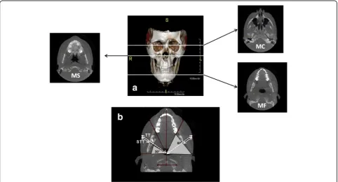
In order to include the entire face volume, three main CBCT transverse sections were identified and named by anatomical references as the upper margin of the man-dibular condyle (MC), the floor of the maxillary sinus (MS) and the mandibular foramen (MF) (Fig. 1).
Measurements of both affected and unaffected side of the face were made on standardized CBCT sections ac-cording to defined anatomical references. At first, a front-to-back and lateral-lateral axis of symmetry were identified. From the intersection axes, an origin point was generated. From this point, 30° and 60° lines, cross-ing bony and soft structures, were drawn. For each given degree, the soft tissue thickness (STT) and the total
thickness (TT), including both bony and soft tissue, of the right and left side were calculated by using the soft-ware Onis 2.4 free edition and expressed in millimeters and decimals (Fig. 1). Twenty-four measures for each subject were therefore obtained.
Six raters, all physicians, evaluated 10 LSF patients’ and 5 controls’CBCTs blindly one from the other. Three raters were experienced with the use of the measure-ment method while the three not experienced received a two hour training session before starting the measure-ments. Patients’CBCTs have been analyzed twice after a time interval of one week in order to evaluate the intra-rater variability which was assessed by the repeatability coefficient with 95% limits of confidence [14].
To assess the inter-rater reliability, we used the intra-class correlation coefficient (ICC) two-way random effect model [15]. The ICC was analysed by calculating and pooling the standard deviations (SD) of the measurements recorded on each subject. The mean SD within patient and variance of all measurements on a single patient were calculated, pro-viding a quantitative assessment of inter-observer reliability. As relative measure of dispersion, the coefficient of vari-ation was calculated for each patient and for each factor. ICC values ranging 0.75–1 were considered signs of excel-lent reliability, ICC ranging 0.4–0.74 signs of good reliability [16]. The 95% confidence interval (CI) defined the degree of significance and the accuracy of the estimate. All analyses were performed by using IBM SPSS (Version 18.0).
Results
Ten patients with LSF (7 Parry Romberg Syndrome-PRS, 3 En Coupe De Sabre-PRS), aged 3–21 years, mean dis-ease duration 1.8 years (range 0.5–8 years) and five age-matched healthy controls underwent CBCT assessment. As each patient was independently evaluated twice by each examiner, a set of 48 measures (24 in the first round and 24 in the second one) were obtained. Each examiner also performed 240 measures in 5 controls.
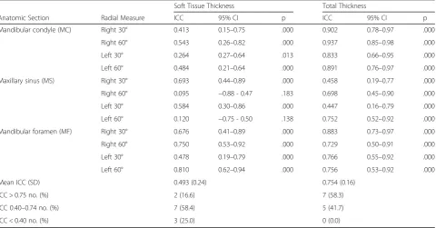
The inter-rater concordance for the total thickness was excellent with mean ICC 0.75 (SD 0.16) for patients (Table 1) and 0.89 (SD 0.09) for controls. The mean ICC for the soft tissue thickness was 0.49 (SD 0.24) for pa-tients and 0.66 (SD 0.28) for controls, respectively. 58.3% of the measurements for patients and 91.2% of those for controls showed excellent ICC results.
As for the soft tissue thickness, the mean ICC was 0.49 (SD 0.24) with 75% of the measurements being good or excellent (Table 1). As shown, the best perform-ance was obtained at the level of the MF and MC sec-tions. The controls showed an even better concordance for the soft tissue measurements with 41.7% with ICC > 0.75 and 41.7% with ICC 0.40–0.74.
The group of three non-experienced physicians reached significant results in 83.3% of the cases, with ICC ranging 0.62–0.98. The group of experts reached significant results in 73% of cases, with an ICC ranging 0.47–0.94.
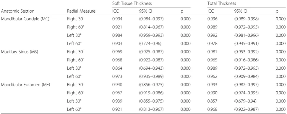
The intra-rater repeatability coefficient for the total thickness ranged between 0.857 and 0.996, for the soft tis-sue thickness ranged between 0.864 and 0.973 (Table 2). All the measurements reached significance and no
differences were reported between non-experienced and experienced examiners, as well.
Maxillofacial CBCT was fast (mean duration 3.6 s) and well tolerated by the patients, even the youngest. The average time needed to conduct the measurements of CBCTs’imaging ranged from 15 to 25 min.
Discussion
The assessment of LSF is still a challenging issue as the tools usually applied for the other subtypes of localized scleroderma have shown significant limitations when ap-plied to LSF [3]. Indeed, there are no validated methods to establish when LSF is in full remission in order to get the surgical reconstruction started.
To date, the most used clinical score, the localized scleroderma assessment tool (LoSCAT) [4, 5], aimed to quantify the skin involvement by clinical signs of activity and damage, is not able to detect changes in the deeper layers of the skin nor bone deformities.
More sensitive assessment tools have both advantages and limitations [3]. Infrared thermography and laser dop-pler flowmetry present with high sensitivity but low speci-ficity in detecting active LS lesions as an increased number of false positive results are reported. Indeed, it has been demonstrated that they are not appropriate to evaluate deep lesions of the face [6, 7]. Doppler ultrasound has also limitations when applied to LSF due to the lack of standardization and the operator/equipment dependence [8, 9]. Magnetic Resonance Imaging (MRI), currently used to detect musculoskeletal involvement and central ner-vous system abnormalities in LSF [10, 11], is a long-lasting
Table 1Inter-rater variability of CBCT scan measures
Soft Tissue Thickness Total Thickness
Anatomic Section Radial Measure ICC 95% CI p ICC 95% CI p Mandibular condyle (MC) Right 30° 0.413 0.15–0.75 .000 0.902 0.78–0.97 .000
Right 60° 0.543 0.26–0.82 .000 0.937 0.85–0.98 .000 Left 30° 0.264 0.27–0.64 .013 0.833 0.66–0.95 .000 Left 60° 0.484 0.21–0.64 .000 0.891 0.76–0.97 .000 Maxillary sinus (MS) Right 30° 0.693 0.44–0.89 .000 0.458 0.19–0.77 .000 Right 60° 0.095 −0.88 - 0.47 .183 0.698 0.45–0.90 .000 Left 30° 0.584 0.30–0.86 .000 0.447 0.16–0.79 .000 Left 60° 0.120 −0.75 - 0.50 .138 0.752 0.52–0.92 .000 Mandibular foramen (MF) Right 30° 0.676 0.41–0.89 .000 0.883 0.73–0.97 .000 Right 60° 0.750 0.53–0.92 .000 0.729 0.50–0.91 .000 Left 30° 0.478 0.19–0.79 .000 0.766 0.55–0.92 .000 Left 60° 0.810 0.62–0.94 .000 0.756 0.53–0.92 .000 Mean ICC (SD) 0.493 (0.24) 0.754 (0.16)
procedure therefore needs sedation for patients aged less than seven years. As evident, none of these tools are either feasible or sensitive enough to detect disease changes of LSF over time.
For these reasons, given the young age of the majority of the patients with LSF and the need of a fast technique for a concomitant bony and soft tissue evaluation, we in-vestigated the potential role of CBCT for the assessment of LSF, in view of a possible future application of this technique for monitoring the disease over time.
CBCT is currently used in dentistry and maxillofacial sur-gery, with good sensitivity for both soft and bony tissues [12, 14]. It is fast to perform and minimally invasive since it does not require sedation and results in a radiation expos-ure 20–50 times lower than a conventional CT [17, 18].
In the present study, we found very good inter-rater concordance for the total thickness, among the asses-sors, with mean ICC value of 0.75 for patients (Table 1) and 0.89 for controls. Almost 60% of the measurements for patients and more than 90% for controls showed ex-cellent ICC results. The mean ICC for the soft tissue thickness at the MS level was a bit lower than at MF and MC levels, probably because of the objective diffi-culty in measuring the distance between the superficial skin and the bone surface at that level. However, the intra-rater ICC at the same level was excellent (Table 2), confirming the validity of this technique, particularly when performed by the same operator.
CBCT scan evaluation was feasible also for non-experienced physicians as measurements were not diver-gent from the experienced ones, confirming the face validity of the method and the efficacy of preliminary training sessions.
CBCT was fast and well tolerated even by the youn-gest patients. Once obtained the digital images, the
average time needed to conduct the standardized ana-lysis of CBCTs’imaging ranged from 15 to 25 min.
This is the first application of CBCT for the standardized assessment of LSF. Formerly, we applied this technique for the detection of odontostomatologic abnormalities in pa-tients with LSF [13].
As for the possible limitations of CBCT, besides the radiation exposure, this technique does not allow a reli-able evaluation of the forehead region, limiting its appli-cation for patients with isolated frontal lesions. In fact, due to its conical X-ray emission, the definition and re-producibility of the scan images result to be low for the forehead area, making it inaccurate for comparing mea-sures overtime. We should underline, however, that in a large international study involving 750 patients with ju-venile localized scleroderma, 23% presented with LSF but only very few of them had isolated forehead involve-ment with no maxillary involveinvolve-ment [2]. Therefore, we can evaluate with CBCT the vast majority of patients with LSF. For the few with isolated forehead lesions, as in the initial phase of ECDS, sequential clinical photog-raphy and computerized skin scoring can be used with quite good results [10].
In conclusion, we showed that CBCT represents a reli-able and relatively safe technique to assess and quantify the disease involvement in LSF. It is fast to be performed and covers an important clinical gap in the clinical prac-tice, not only for the disease assessment but also for is-sues related to facial reconstructive procedures. In fact, to date, no technique has been able to define the remis-sion status of LSF and the timing for reconstructive plastic surgery procedures. A prospective study on sensitivity to change and disease progression definition by using se-quential CBCT checks is ongoing and the preliminary re-sults obtained so far are promising.
Table 2Intra-rater repeatability and limits of agreement of CBCT scan measures
Soft Tissue Thickness Total Thickness
Anatomic Section Radial Measure ICC 95% CI p ICC 95% CI p Mandibular Condyle (MC) Right 30° 0.994 (0.984–0.997) 0.000 0.996 (0.989–0.998) 0.000
Abbreviations
CBCT:Cone Beam Computed Tomography; ICC: Intraclass Correlation Coefficient; LoSCAT: Localized scleroderma assessment tool; LSF: Linear scleroderma of the face; MC: Mandibular condyle; MF: Mandibular foramen; MPR: Multi planar reconstruction; MRI: Magnetic Resonance Imaging; MS: Maxillary sinus; SD: Standard deviations; STT: Soft tissue thickness; TT: Total thickness
Acknowledgements
We thank S. Trainito, V. Cecchin, R. Culpo, L. Nai Fovino and F. Sperotto for their contribution in assessing the CBCT images and IL VOLO, Association for childhood rheumatic diseases for having partially supported this project.
Funding
No funding resources for this work.
Availability of data and materials
The dataset used and analysed during the current study is available from the corresponding author on reasonable request.
Authors’contributions
CDG, FZ wrote the draft; CDG, SP, FV, FZ designed the study; AM, GM collected the patient’s data; CDG, SP, AM, GM, FZ revised the test; FV made the statistical analysis. All authors read and approved the final manuscript.
Ethics approval and consent to participate
Ethics approval and informed consent was not needed since CBCT is already in use for maxillofacial assessment in pediatric patients.
Consent for publication
Not applicable.
Competing interests
The Authors declare that they have no competing interests.
Publisher’s Note
Springer Nature remains neutral with regard to jurisdictional claims in published maps and institutional affiliations.
Author details
1Department of Woman and Child Health, University of Padua, Via Giustiniani
3, 35128 Padova, Italy.2Affidea Group, Unix Radiology Service, Piove di Sacco,
Italy.
Received: 5 October 2017 Accepted: 21 December 2017
References
1. Laxer RM, Zulian F. Localized scleroderma. Curr Opin Rheumatol. 2006;18: 606–13.
2. Zulian F, Athreya BH, Laxer R, Nelson AM, Feitosa de Oliveira SK, Punaro MG, et al. Juvenile localized scleroderma: clinical and epidemiological features in 750 children. An international study. Rheumatology (Oxford). 2006;45:614–20. 3. Zulian F. New developments in localized scleroderma. Curr Opin Rheumatol.
2008;20:601–7.
4. Arkachaisri T, Pino S. Localized scleroderma severity index and global assessments: a pilot study of outcome instruments. J Rheumatol. 2008;35: 650–7.
5. Arkachaisri T, Vilaiyuk S, Torok KS, Medsger TA Jr. Development and initial validation of the localized scleroderma skin damage index and physician global assessment of disease damage: a proof-of-concept study. Rheumatology (Oxford). 2010;49:373–81.
6. Martini G, Murray KJ, Howell KJ, Harper J, Atherton D, Zulian F, et al. Juvenile-onset localized scleroderma activity detection by infrared thermography. Rheumatology (Oxford). 2002;41:1178–82.15.
7. Weibel L, Howell KJ, Visentin MT, Rudiger A, Denton CP, Zulian F, et al. Laser Doppler flowmetry for assessing localized scleroderma in children. Arthritis Rheum. 2007;56:3489–95.
8. Li SC, Liebling MS, Haines KA, Weiss JE, Prann A. Initial evaluation of an ultrasound measure for assessing the activity of skin lesions in juvenile localized scleroderma. Arthritis Care Res. 2011;63:735–42.
9. Wortsman X, Wortsman J, Sazunic I, Carreño L. Activity assessment in morphea using color Doppler ultrasound. J Am Acad Dermatol. 2011;65:942. 10. Zulian F, Meneghesso D, Grisan E, Vittadello F, Belloni Fortina A, Pigozzi B,
et al. A new computerized method for the assessment of skin lesions in localized scleroderma. Rheumatology (Oxford). 2007;46:856–60.
11. Schanz S, Fierlbeck G, Ulmer A, Schmalzing M, Kümmerle-Deschner J, Claussen CD, et al. Localized scleroderma: MR findings and clinical features. Radiology. 2011;260:817–24.
12. Carter JB, Stone JD, Clark RS, Mercer JE. Applications of cone-beam computed tomography in oral and maxillofacial surgery: an overview of published indications and clinical usage in United States academic centers and oral and maxillofacial surgery practices. J Oral Maxillofac Surg. 2016;74: 668–79.
13. Trainito S, Favero L, Martini G, Pedersen TK, Herlin T, Zulian F. Odontostomatologic involvement in juvenile localised scleroderma of the face. J Paediatr Child Health. 2012;48:572–6.
14. Bland MJ, Altman DG. Measurement agreement in method comparison studies. Statistical methods in Med Res. 1999;8:135–60.
15. McGraw KO, Wong SP. Forming inferences about some intraclass correlation coefficients. Psychol Methods. 1996;1:30–46.
16. Fleiss JL, Nee CM, Landis JR. Large sample variance of kappa in the case of different sets of raters. Psychol Bull. 1979;86:974–7.
17. Federal Guidance Report 14: Radiation Protection Guidance for Diagnostic and Interventional X-ray Procedures; 2014. Washington, DC, Interagency Working Group on Medical Radiation, US Environmental Protection Agency (EPA). Available at: https://www.epa.gov/sites/production/files/2015-05/ documents/fgr14-2014.pdf
18. Scarfe WC, Farman AG, Sukovic P. Clinical applications of cone beam computed tomography in dental practice. J Can Dent Assoc. 2006;72:75.
• We accept pre-submission inquiries
• Our selector tool helps you to find the most relevant journal
• We provide round the clock customer support
• Convenient online submission
• Thorough peer review
• Inclusion in PubMed and all major indexing services
• Maximum visibility for your research
Submit your manuscript at www.biomedcentral.com/submit