FORMULATION AND EVALUATION OF ETHOSOMAL TRANSDERMAL DELIVERY SYSTEM OF KETOCONAZOLE
7
0
0
Full text
(2) Shaik.Harun Rasheed. et al., Int. J. Pharm. & H. Care Res., Vol.–01 (01) 2013 [36 - 42]. outermost layer of the skin - the stratum corneum (SC).1, 2 To overcome the stratum corneum barrier, many techniques have been assessed to disrupt and weaken the highly organized intercellular lipids in an attempt to enhance the percutaneous drug penetration.3-5 It was previously discovered that significant enhanced delivery of drugs through the skin could be obtained by using a novel-permeation enhanced carrier, the ethosome.6-9 So, Novel drug delivery system (NDDS) has revolutionized the method of medication. The NDDS should ideally fulfill two prerequisites. Firstly, it should deliver the drug at a rate directed by the needs of the body, over the period of treatment. Secondly, it should channel the active entity to the site of action. Sincere attempts have been made to achieve them through various novel approaches in drug delivery. Several technological advances have been made over many years. One such technique is vesicular systems which shows a great promise and opens up new market for pharmaceutical and cosmetic industry for drug delivery through skin. Hence, significant enhanced delivery of Ketoconazole through transdermal route could be obtained by using vesicular drug carrier systems - Ethosomes. The current study was aimed to investigate the potential of ethosomes in enhancement of Ketoconazole transport across the skin, characteristics of ethosomes and their in-vitro skin permeation behavior.. 37. Equipment, Mumbai, India) in a house built closed container. Mixing was continued for additional 5 min. The system was maintained at 30 ± 10C during the preparation and then left to cool at room temperature for 30 min. Entrapment efficiency Ethosomal preparations containing Ketoconazole were estimated by ultra centrifugation technique for measuring its entrapment efficiency, for that the total volume of the ethosomal preparation was measured and 2 ml of the formulation was transferred to 10 ml centrifuge tube. The preparation was diluted with distilled water up to 5 ml and centrifuged at 2000 rpm for 20 minute to separate out undissolved drug in the formulation. Ethosomes were separated by ultra centrifugation at 20,000 rpm for 20 minute. Supernatant and sediment were recovered and their volume was measured. Sediment was dilute with the distilled water up to 5 ml. The unentrapped and entrapped drug content was analyzed by estimating the drug in supernatant and ethosomes (Sediment) by spectroscopic method.11-12 The method was repeated at least three times.. Ketoconazole was obtained as a gift sample from Medreich pharmaceutical company, Bangalore. Lipoid S phosphatidyl choline-3, containing not less than 98% phosphatidyl choline was a kind gift from Lipoid GmbH (Ludwigshafen, Germany). Ethanol and methanol was purchased from Sigma Lab, New Delhi. All other chemicals used in our work were of analytical grade.. Vesicular shape and surface morphology Vesicular shape of the ethosomal preparations were assessed by using transmission electron microscope (TEM). 10 µl of samples were dried on carboncoated grid and negatively stained with aqueous solution of phosphotungstic acid. After drying, the specimen was viewed under the microscope at 10– 100 k-fold enlargements at an accelerating voltage of 100 kV. Scanning electron microscopy (SEM) was also conducted to characterize the surface morphology of the ethosomal vesicle. One drop of ethosomal system was placed on clear glass stub and spread on the glass stub homogenously, then it coated with polaron E 5100 sputter coater (Polaron, UK) and visualized under scanning electron microscope (Leo-435 VP, Cambridge, UK).. Preparation of ketoconazole loaded ethosomal vesicles The ethanolic vesicular system investigated here was composed of 2.0% w/w of Lipoid S PC-3 (PC), 35% w/w of ethanol, drug (ketoconazole, 0.77% w/w). Lipoid S PC-3 (PC) was dissolved along with drug in ethanol. Triple distilled water was added slowly in a fine stream with constant mixing at 700 rpm with a mechanical stirrer (Remi. Vesicular size of the ethosomal formulations The size distribution of ethosomal preparations was measured in two sets of triplicates, in a multimodal mode, by dynamic light scattering (DLS) technique using a computerized Malvern autosizer 5002 inspection system (Malvern, UK). For vesicle size measurement, ethosomal preparation were mixed with the appropriate medium (PBS, pH 6.5) and the measurements were taken in triplicate.13. Materials and methods. www.ijphr.com.
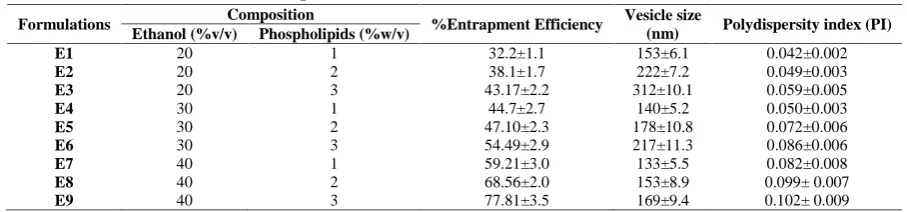
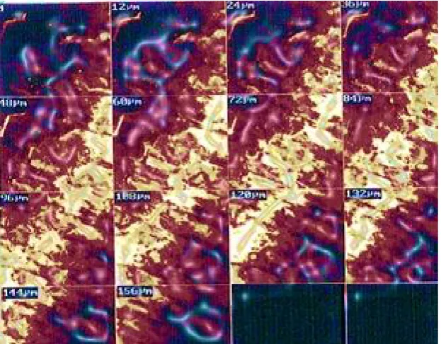
(3) 38. Shaik.Harun Rasheed. et al., Int. J. Pharm. & H. Care Res., Vol.–01 (01) 2013 [36 - 42]. Confocal laser scanning microscopy (CLSM) Depth and mechanism of skin penetration of RR loaded ethosomes was investigated using CLSM, as reported previously.14 Briefly, unentrapped probe was removed from probe-loaded vesicles by minicolumn ultracentrifugation thereafter formulation was applied non-occlusively for 8 h to the dorsal skin of 5–6week old nude albino rat (Sprague Dawley strain). The rat was then sacrificed by heart puncture; dorsal skin was excised, washed, placed on aluminium foil and adhering fat and/or subcutaneous tissue was removed. The skin was then sectioned into the pieces of 1 mm 2 size and evaluated for depth of probe penetration for various formulations. The full skin thickness was optically scanned at different increments through the z-axis of a CLS microscope (LSM 510 with an attached universal Zeiss epifluorescence Microscope). All investigations were performed as per the protocol approved by the Institutional Animals Ethical Committee (Reg. No. 1283/c/09/CPCSEA).15 In vitro permeation study through rat skin Franz diffusion cell was used for the diffusion studies. The study was carried out for 48 h at 37 ± 10C. Rat skin was obtained from the abdominal of albino rats (aged 17–22 weeks, weight 250– 300 g). After depilation and washing, abdominal skin was excised, thoroughly washed with pH 7.4 buffer solution, dried and carefully cleaned and then preserved at 250C. Before using, the skin was thawed, pre-hydrated for 1 h with the pH 7.4 buffer solution and then mounted in the diffusion chamber. of the Franz cell with the horny layer facing the donor compartment and the dermal side toward the receptor fluid, which was stirred with a magnetic bar at 50 rpm. Sample of 1 ml each was withdrawn at the regular time interval for 48 hours (1, 2, 4, 6, 12, 24, 48 hr), Refilling with fresh medium was carried out at the same time. Samples were suitably diluted and analyzed for drug content by a using UV spectrophotometer and mean cumulative of the drug released across the rat skin was calculated. Experiments were performed in triplicate.16 Vesicle skin interaction studies of ethosomal fromulation To observe the ultra structural changes in the skin upon exposure to various ethosomal formulations the vesicles were applied on the skin of rats (Male Sprague Dawley, 5–6 week old, 80–100 g). Preparations were applied topically to the skin for 6 h, animals were sacrificed, skin were excised and stored in formalin solution (10%) in phosphate buffer saline (pH 7.4) followed by dehydration with alcohol. It was then treated with anti-media and embedded in paraffin for fixing. Controls skin section was prepared by similar procedure without application of any preparation. Sections of 5 μm thickness were cut from each piece and stained with heamotoxyline and eosin and histological changes in stratum corneum, epidermis and dermis were examined under optical microscope (Leica, DMLB, Heerbrugg, Switzerland).14. Table No. 01: Composition and evaluation of ethosomal formulations (E1-E9) Formulations E1 E2 E3 E4 E5 E6 E7 E8 E9. Composition Ethanol (%v/v) Phospholipids (%w/v) 20 1 20 2 20 3 30 1 30 2 30 3 40 1 40 2 40 3. %Entrapment Efficiency 32.2±1.1 38.1±1.7 43.17±2.2 44.7±2.7 47.10±2.3 54.49±2.9 59.21±3.0 68.56±2.0 77.81±3.5. Vesicle size (nm) 153±6.1 222±7.2 312±10.1 140±5.2 178±10.8 217±11.3 133±5.5 153±8.9 169±9.4. Polydispersity index (PI). Table No. 02: In vitro drug diffusion study of ethosomes (E1-E9) Time (hr) 1 2 4 12 24 48. % of Drug Diffused Ethosomes 8.9±0.81 13.1±0.80 32.78±2.1 45.81±3.0 69.44±4.1 81.22±5.4. www.ijphr.com. 0.042±0.002 0.049±0.003 0.059±0.005 0.050±0.003 0.072±0.006 0.086±0.006 0.082±0.008 0.099± 0.007 0.102± 0.009.
(4) Shaik.Harun Rasheed. et al., Int. J. Pharm. & H. Care Res., Vol.–01 (01) 2013 [36 - 42]. Fig. No. 01(a): Visualization of ethosomal vesicles TEM (magnification 315 000). Fig. No. 01(b): Visualization of ethosomal vesicles SEM (magnification 10 0000). Fig. No. 02: In vitro drug diffusion study through rat skin. Fig. No. 03: Confocal laser scanning photomicrograph of penetration of Ketoconazole from ethosomes applied non-occlusively onto nude rat skin. www.ijphr.com. 39.
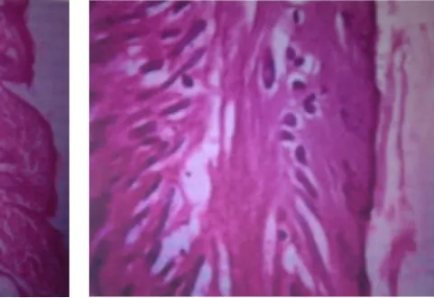
(5) 40. Shaik.Harun Rasheed. et al., Int. J. Pharm. & H. Care Res., Vol.–01 (01) 2013 [36 - 42]. Fig. No. 04 (a): Histopathology of the Normal rat skin. Results and discusions Ketoconazole loaded ethosomes were prepared using varying concentration of Lipoid S PC-3 (PC) and ethanol, when examined by Transmission Electron Microscope (TEM) appeared as unilamellar vesicles with a predominant spherical shape (Fig. 1a). Surface morphology and threedimensional nature of ethosomes were performed by further analysis of the preparation by Scanning Electron Microscopy (SEM), which confirmed the vesicular characteristics possessed by this novel carrier (Fig. 1b). The two basic parameters on the basis of which the formulations were optimized are vesicular size and entrapment efficiency on increasing the phospholipids concentration. It was observed that the vesicular size was increased, though with increase in ethanol concentration the vesicular size decreased (Table 1). This indicates that at higher ethanol the membrane thickness is reduced considerably probably due to the formation of phase with interpenetrating hydrocarbon chain. In terms of entrapment efficiency, among all the Ketoconazole loaded ethosomal formulations, E9 (40% v/v ethanol, 3.0% w/v Lipoid S PC-3 and drug) showed the greatest entrapment efficiency, thus justifying itself as the optimized formulation with greatest entrapment efficiency (77.81±3.5%) and optimum size (169±9.4 nm)thus showing the greatest opportunity to the Ketoconazole loaded ethosomal preparation to attain a better skin penetration, by providing a safe homing to the Ketoconazole and optimized vesicular size which has been reported to affect the skin permeation parameters. An optimum polydispersity index. Fig. No. 06 (b): Histopathology of the rat after application of the ethosome (0.102±0.009) of ketoconazole loaded ethosomal formulation could better justify the homogeneous nature of the prepared ethosomal formulation (Table 1). The rate of the drug release from the ethosomal carrier is an important parameter in the evaluation of the drug delivery for ethosomes. This study was performed by using Franz diffusion cell for the better formulation E9 for 48 hr. and % drug diffused was calculated by using UV spectrophotometer and results are shown in the Table 2 for ethosomes of which Fig. 2 was plotted between time and % of drug diffused to represent cumulative amount of drug permeated across the rat skin. To measure the extent of penetration and transdermal potency of the stored system, confocal laser scanning microscopic studies should be performed, from the study it showed that an increase in the depth penetration of the ketoconazoleloaded ethosomal formulation (up to 156 μm) shown in fig. 3 This prominently efficient delivery of ketoconazoleloaded ethosomal vesicle suggests their enhanced penetration and consequent fusion with the lipid membrane in the depths of the skin supporting the hypothesis.18 The above mentioned parameters were similar for ketoconazoleloaded ethosomal formulation which stored for 120 days (depth of penetration=168 μm) which suggesting a stable nature of ethosomal preparation along with no change in transdermal potency of the stored system. Vesicle skin interaction studies was performed by skin histopathology on application of ethosomal system, it showed that there was no specific changes in the skin histopathology, though skin. www.ijphr.com.
(6) Shaik.Harun Rasheed. et al., Int. J. Pharm. & H. Care Res., Vol.–01 (01) 2013 [36 - 42]. lipid fluidization could be observed in the form of some penetration pathway, which could be followed by these ethanolic vesicles, justifying their proposed mechanism of penetration into the skin and further fusing with the skin lipids in the epidermis and dermis. Further, mild swelling of corneocytes could also be observed, which suggesting the retention of fluids and thus providing an insight on sustained drug delivery mechanism of ketoconazoleloaded ethosomal preparations. Before the proposal of a ketoconazole as a potential carrier for transdermal drug delivery system, an important characteristic to be evaluated is its in vivo skin tolerability/irritancy studies. As, skin non-irritancy of ketoconazoleloaded ethosomal formulation is well justified after applying the preparation.17 It was observed for the erythema scores upon exposure of hairless rabbit skin to various formulations of ethosomes, it was revealed that ketoconazoleloaded ethosomes showed no significant erythema, demonstrating that ethanol present in the ethosomal formulation is not able to act as a skin erythema inducing agent, even though present in high concentration. Vesicle skin interaction study can be performed by observing ultrastructural changes in the skin upon exposure to ethosomal formulations which can be compare with the normal rat skin. This study can be performed by histopathology and the results of the histopathology for the normal rat skin (control skin), ethosomes applied rat skin are compared in the Fig. 4 (a) and (b). Ketoconazole loaded ethosomal formulation showed greater skin penetration, thus justifying its use as a carrier for choice in dermal and transdermal delivery of this anti-fungal drug. Also, the ethosomal formulation is reported to be non-irritant with the skin, which establishing the potential transdermal drug delivery. The enhanced transdermal efficacy obtained from the ethosomal system could be justified on the basis of dual function performed by ethanol present in the ethosomal formulations i.e. fluidizing both the vesicular lipid bilayers and Lipoid S PC-3, thus providing a greater malleability to the vesicles and enhancing permeability of the skin.18. Conclusion Ethosomes have been studied as a possible vehicle for transdermal delivery of ciclopirox olamine, an antifungal agent, from the study it was confirmed. 41. that ethosomal formulation of ketoconazoleshowed a higher entrapment efficiency and better stability profile. The enhanced accumulation of ketoconazolevia ethosomal carrier within the skin might help to optimize targeting of this drug to the epidermal and dermal sites. Thus it concluded that ethosomes is a very promising carrier for transdermal delivery and creating a new opportunities for topical application of ketoconazole in the fungal infections.. References 1.. Blank IH, Scheuplein RJ. Transport into and within the skin. Br J Dermatol., 1969, 81, 4-10. 2. Scheuplein RJ, Blank IH. Permeability of the skin. Physiol Rev., 1971; 51, 702-747. 3. Barry BW. Novel mechanisms and devices to enable successful transdermal drug delivery. Eur. J Pharm Sci., 2001, 14, 101-114. 4. Schreier H, Bouwstra JA. Liposomes and niosomes as topical drug carriers. dermal and transdermal drug delivery. J Control Release., 1994, 30, 1–15. 5. Prausnitz MR et al. Current status and future potential of transdermal drug delivery. Nat. Rev., 2004, 3, 115-124. 6. Touitou E. Compositions for applying active substances to or through the skin. US patent., 5540934, 1996. 7. Touitou E Compositions for applying active substances to or through the skin. US patent., 5716638, 1998. 8. Touitou E, Alkabes M, Dayan M, Eliaz M. Ethosomes novel vesicular carriers for enhanced skin delivery. Pharm Res., 1997, 14, S-305. 9. Touitou E, Dayan M, Bergelson L, Godin B, Eliaz M. Ethosomes- Novel vesicular carriers for enhanced skin delivery: Characterization and skin penetration properties. J Control Rel., 2000, 65, 403-18. 10. Lopez-Pinto JM, Gonzalez-Rodriguez ML, Rabasco AM. Effect of cholesterol and ethanol on dermal delivery from DPPC Liposomes, Int. J. Pharm., 2005,, 298, 60-66. 11. Fry DW, White JC, Goldman ID. Rapid separation of low molecular weight solutes from liposomes without dilution, J Anal Biochem., 1978, 90, 809–815. 12. Sorensen EN, Weisman G, Vidaver GA. A Sephadex column for measuring uptake and loss of low molecular weight solutes from. www.ijphr.com.
(7) 42. Shaik.Harun Rasheed. et al., Int. J. Pharm. & H. Care Res., Vol.–01 (01) 2013 [36 - 42]. small vesicles, Anal Biochem., 1977, 82, 376– 384. 13. New RCC. Preparation of liposomes and size determination. In: New RCC, editor. Liposmes- a practical approach. Oxford: Oxford University Press., 1990, P. 36-39. 14. Dubey Vaibhav, Mishra Dinesh, Dutta Tathagata, Nahar Manoj, Saraf DK, Jain NK. Dermal and transdermal delivery of an antipsoriatic agent via ethanolic liposomes. Journal of Controlled Release., 2007, 123, 148-154. 15. Dubey Vaibhav, Mishra Dinesh, Jain NK. Melatonin loaded ethanolic liposomes: Physicochemical characterization and enhanced transdermal delivery, European J Pharm Biopharm., 2007, 67, 398–405.. 16. Paola Mura, Francesca Maestrelli, Maria Luisa Gonzalez-Rodrıguez, Ilaria Michelacci, Carla Ghelardini, Antonio M Rabasco. Development, characterization and in vivo evaluation of benzocaine-loaded liposomes, European J Pharma and Biopharma., 2007, 67, 86–95. 17. N. Kanninkkanan, T. Jackson, M.S. Shaik, M. Singh, Evaluation of skin sensitization potential of melatonin and nimesulide by murine local lymph node assay, Eur. J. Pharm.Sci., 2001, 14, 217–220. 18. Godin B, Touitou E. Erythromycin ethosomal systems, physicochemical characterization and enhanced antibacterial activity, Curr Drug Deliv., 2005, 2, 269–275.. www.ijphr.com.
(8)
Figure
Related documents