Pharmacophore 2015, Vol. 6 (6), 281-298 USA CODEN: PHARM7 ISSN 2229-5402
Pharmacophore
(An International Research Journal)
Available online at http://www.pharmacophorejournal.com/
Original Research Paper
AN IN SILICO EXPLORATION FOR NOVEL INHIBITORS OF α-AMYLASE AND
α-GLUCOSIDASE EXTRACTED FROM TINOSPORA CORDIFOLIA
Varsha Vasantrao Sonkamble, Nilesh Shirish Wagh and
Laxmikant Haribhau Kamble
*
School of Life Sciences, Swami Ramanand Teerth Marathwada University,
Nanded (MS)-431606, India
ABSTRACT
Present study reports the potential of α-amylase and α-glucosidase inhibitory activities of phytoconstituents of the plant, Tinospora cordifolia using an in silico structure-based molecular docking approach. In silico
screening of seventeen molecules from T. cordifolia was performed and compared with the activity of a known inhibitor acarbose. For this study AutoDock 1.5.6, PyRx and Discovery Studio 4.1 Visualizer softwares were used. Out of the seventeen molecules screened, five: Verbascoside, Hesperetin 7-rhamnoglucoside, 3-(1-Naphthoyl) benzoate, (4-Cinnamoyl-3,5-dihydroxyphenoxy) acetate and 4-(2,4-dimethoxy-3,6-dimethylbenzoyl) oxy-2-hydroxy-3,6-dimethyl benzoate showed lowest binding energies for α-amylase and α-glucosidase. Further, comparative analysis of PDB structures of both enzymes using 3DLigandSite server and Discovery Studio 4.1 Visualizer analysis of docked enzyme structures revealed involvement of almost similar amino acids in ligand binding sites of both the enzymes. This in silico
investigation may felicitate the development of α-amylase and α-glucosidase inhibitors from T. cordifolia
for treating diabetes.
Keywords:
Diabetes Mellitus, Phytoconstituents, Tinospora cordifolia, In silico, Molecular docking, Virtual screening, α-Amylase and α-Glucosidase inhibitors.INTRODUCTION
Diabetes Mellitus is a common endocrine disorder based on deficiency of insulin which leads to chronic hyperglycemia with disturbance of carbohydrate, fat and protein metabolism.1 One of the therapeutic approaches for treating diabetes is to decrease postprandial hyperglycemia which can be mediated by two enzymes, α-amylase and α-glucosidase.2 This can be done by partially inhibiting or blocking the activity of these enzymes with the help of inhibitors which are also called as starch blockers because they contain substances that prevent absorption of dietary starch in the body.3 Acarbose, voglibose, and miglitol are clinically used antidiabetic agents which however confer some adverse effects like
some inhibitors from natural products which could be used as alternatives for prevention and treatment of type 2 diabetes mellitus without any side effects.3
Tinospora cordifolia is one of the well known ayurvedic herbs with tonic, periodic, spasmodic, inflammatory, arthritic, anti-allergic and anti-diabetic properties.5,6 Various concentrations of T. cordifolia extracts as well as powder forms are frequently prescribed for the diabetic patients. So we initially focused on its phytoconstituents analysis. Our previous studies on phytochemical constituents of T. cordifolia
Hesperetin-7-compounds like (2S)-2-Pyrrolidinecarboximidate; (4-Cinnamoyl-3,5-dihydroxyphenoxy) acetate; (15Z)-12-Oxophyto-10,15-dienoate; 1-cyclohexenylmethanone; 2-[(3-Amino-4-
propoxybenzoyl)-oxy]-N,N-diethylethanaminium; 2-Hydroxy-3-oxoicosanoate; 2-Hydroxy-5,10-dioxo-4-phenyl- 3,4,5,10-tetrahydro-2H-benzo[g]chromene-2-carboxylate; 3-(1-Naphthoyl) benzoate; 3,3-Bis-(3,4-dimethoxyphenyl) propanoate; 3,5-dichloro-4-morpholin-4-ylpyridine-2-carboxylate; 3-
Carbamoyl-1-(5-O-phosphono-β-D-ribofuranosyl) pyridinium; 6,9,12,15-Octadecatetraenoatato; 4-(2,4-dimethoxy-3,6-dimethyl-benzoyl)-oxy-2-hydroxy-3,6-dimethyl benzoate; Petunidin-3-O-coumaroylrutinoside-5-O-glucoside and Imidazole-Pyrazole are identified by us.6 These phytochemicals are not reported for anti-diabetic activity. So, in continuation of our research work, we focussed on virtual screening of these molecules from T. cordifolia for their potential amylase and α-glucosidase inhibitory activity. This was achieved using the molecular docking approach of virtually screening and predicting the binding of the small molecules to known target structures.9,10
MATERIALS AND METHODS
Preparation of Macromolecules for Docking The structure of α-amylase (PDB Code: 3BC9) and α-glucosidase (PDB Code: 2QMJ) macromolecules were obtained from protein data bank (http://www.rcsb.org). 3D structures in PDB format were prepared for molecular docking with the help of AutoDock Tools-1.5.611,12 using the steps prescribed in the AutoDock Tutorial.13 From each of the individual macromolecule water molecules were deleted, polar hydrogen were added and then subjected for GRID preparation which helps to define the binding sites where compounds/ligands are to be docked. Later, the structures were saved in PDBQT file format, the format that is accepted by AutoDock tool which contains a protein structure with hydrogen in all polar residues.Preparation of Ligands for Docking
The ligand structures (Compounds from our
earlier studies on T. cordifolia) were obtained
from PubChem database
(http://pubchem.ncbi.nlm.nih.gov) and ChemSpider14 in Mol/SD format. The obtained Mol/SD formats were then converted to PDB formats using Open Babel software.15,16 Ligand molecules in PDB format were further energy minimized by computing gasteiger changes using AutoDock tool and the structures were saved in PDBQT file format.16,17
Molecular Docking of Macromolecules and Ligand
Interaction of the enzymes, amylase and α-glucosidase with individual phytoconstituents of
T. cordifolia was performed using AutoDock Vina11 via PyRx software.13 This was done to obtain a number of possible conformations and orientations for the ligand at binding site of macromolecule. For this, the macromolecules and ligands were loaded in PyRx software, in a PDBQT file obtained from AutoDock and docking was executed by clicking on Run Vina option in PyRx. After completion of docking, the binding energies (kcal/mol) of each ligand with both the individual enzymes were obtained. The best conformation of each ligand with respective enzyme having lowest binding energy was chosen. In case of more than one pose of ligand and enzyme, the pose with lowest binding energy was selected.18 These docked structures i.e. enzyme-ligand complexes were saved in PDB format for further analysis.
Molecular Docking Analysis
Acarbose, a standard amylase and α-glucosidase inhibitor was also docked with both the enzymes as a reference inhibitor and the binding energies with each of the enzymes were predicted. The binding energy of each ligand with respective enzyme was compared with that of the acarbose and ligands with binding energy less than that of acarbose were marked as best inhibitor. Such ligands with low binding energy than acarbose for both the enzymes simultaneously were preferred.
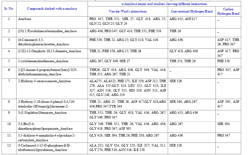
After the docking process, docked structures saved in the PDB format were analyzed for the amino acids involved in the ligand binding sites of the enzyme along with the type of interactions like Vander Waals, hydrogen bonding etc involved in the docking. This was performed using the Discovery Studio 4.1 Visualizer (http://accelrys.com/products/discovery-studio).19 Docked structures showing the amino acid residues involved in the docking as well as the interactions between the residues and ligand were image captured using image save option in Discovery Studio 4.1 Visualizer.
3DLigandSite Analysis of Amylase and α-Glucosidase Enzymes
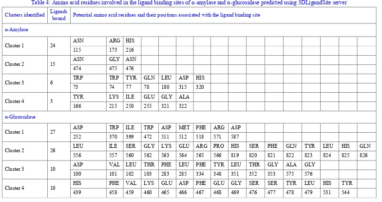
Enzymes α-amylase and α-glucosidase were subjected independently to the 3DLigandSite server (http://www.sbg.bio.ic.ac.uk/3dligandsite) for interpreting the amino acid residues involved in the ligand binding site of the enzyme.20 3DLigandSite is an online server which accepts protein query in FASTA format or PDB structure of the enzyme and predicts the amino acid residues involved in ligand binding site of enzyme through a binding site library comprising of protein-ligand complexes.
RESULTS
Seventeen phytoconstituents of T. cordifolia6 and acarbose, a standard inhibitor, as ligands in PDBQT format as shown in figure 1 were docked with α-amylase and α-glucosidase in PDBQT format shown in figure 2a and 2c. All the seventeen compounds were docked in the active sites of both the enzymes and showed different binding energies in kcal/mol as depicted in table 1. Out of the seventeen, only five compounds showed lower binding energies with both the enzymes as compared to binding energies shown by acarbose (0.8, -8.7 kcal/mol, for α-amylase and α-glucosidase respectively). Verbascoside showed lowest binding energy followed by Hesperetin-7-rhamnoglucoside, 3-(1-Naphthoyl)-benzoate, (4-Cinnamoyl-3,5-dihydroxyphenoxy)-acetate and
4(2,4dimethoxy3,6kcal/mol for αamylase and 11.3, 10.4, 10.9, -9.7 and -9.3 kcal/mol for α-glucosidase respectively. All these five compounds and the standard acarbose docked with amylase and α-glucosidase have been shown in figure 3 and figure 4 respectively. All the compounds were docked with the enzymes in different poses having varying binding energies. However pose with the lowest binding energy indicate highest affinity to the enzyme, thus was considered significant and selected for further docking analysis.
clusters for both the enzymes have been shown in table 4. The probable ligand binding pocket for α-amylase and α-glucosidase, identified in cluster 1 of 3DLigandSite analysis has been shown in figure 2b and 2d respectively.
DISCUSSION
Phytochemical profiling and identification of T. cordifolia plant extracts from our previous reports revealed a diverse range of bioactive phenolics.6 Few of them were reported for antidiabetic activities but we found no details of molecular docking analyses. So, in the present study we tried to put forth the in silico studies of phytochemicals obtained via T. cordifolia plant extracts.
Enzyme and ligand complex models generated after successful docking were obtained based on the parameters such as hydrogen bond interactions, п – п interactions, binding energy, RMSD of active site residues and orientation of the docked compound within the active site.17 In the present study, Verbascoside, Hesperetin-7-rhamnoglucoside, 3-(1-Naphthoyl)-benzoate, (4-Cinnamoyl-3,5-dihydroxyphenoxy)-acetate and 4-(2,4-dimethoxy-3,6-dimethylbenzoyl)oxy-2-hydroxy-3,6-dimethyl-benzoate showed lowest binding energies thus they could be considered as potential inhibitors of amylase as well as α-glucosidase. Earlier reports has revealed the antidiabetic properties of Hesperetin-7-rhamnoglucoside7 and Verbascoside8 which confirms their potency to consider them for further studies on their involvement in antidiabetic drugs. Our study suggest that 3-(1-Naphthoyl)-benzoate, (4-Cinnamoyl-3,5-dihydroxyphenoxy)-acetate, 4-(2,4-dimethoxy-
3,6-dimethyl-benzoyl)-oxy-2-hydroxy-3,6-dimethyl-benzoate could be considered as potential antidiabetic agents. Further, comparing the analysis performed using Discovery Studio 4.1 Visualizer and 3DLigandSite server; it could be revealed that the amino acid residues predicted in the ligand binding site of both the enzymes on 3DLigandSite server were involved in either of the interactions predicted by Discovery Studio 4.1 Visualizer. This was an added advantage of performing two independent analyses of
α-amylase and α-glucosidase enzymes using Discovery Studio 4.1 Visualizer and 3DLigandSite online server.
So, our studies put forward this as a first report on potential antidiabetic property of these compounds based on in silico studies as well as through TLC Autography and α-amylase inhibition assay.6 However, the individual molecules need to be tested through wet lab experiments.
CONCLUSION
In silico studies can provide valuable insights into target drug interactions. Furthermore, this approach can reduce time and cost of clinical trials. So our focus was to screen inhibitors of α-amylase and α-glucosidase especially the phytoconstituents via in silico approach. Owing to the side effects of chemical or synthetic inhibitors already available in the market, screening and selection of phytoconstituents as inhibitors with less or no side effects is the basic purpose of this study. Our results showed the ability to inhibit amylase as well as α-glucosidase by all the compounds. When compared with a standard inhibitor, five compounds: Hesperetin-7-rhamnoglucoside (-10 and 10.4 kcal/mol), Verbascoside (10.5 and -11.3 kcal/mol), 3-(1-Naphthoyl)-benzoate (-9 and -10.9 kcal/mol), (4-Cinnamoyl-3,5-dihydroxyphenoxy)-acetate (-7.5 and -9.7 kcal/mol), and 4-(2,4-dimethoxy-3,6-dimethyl-benzoyl)-oxy-2-hydroxy-3,6-dimethyl-benzoate (-7 and -9.3 kcal/mol) were better than that of the standard drug acarbose (0.8 and -8.7 kcal/mol) with respect to α-amylase and α-glucosidase. This molecular docking analysis could lead to discovery of naturally occurring potent α-amylase and α-glucosidase inhibitors from T. cordifolia
Table 1: Binding affinity (kcal/mol) of compounds with α-amylase and α-glucosidase predicted through virtual docking
Sr. No. Compounds α-Amylase
no. of poses
Binding affinity with α-amylase (kcal/mol)
α-Glucosidase no. of poses
Binding affinity with α-glucosidase (kcal/mol)
1 Acarbose 1 0.8 3 -8.7
2 (2S) 2 Pyrrolidinecarboximidate 9 -4.4 9 -5.7
3 (4-Cinnamoyl-3,5-dihydroxyphenoxy)acetate 3 -7.5 7 -9.7
4 (15Z)-12-Oxophyto-10,15-dienoate 8 -6 9 -7.8
5 1-cyclohexenylmethanone 9 -4.4 9 -5.9
6 2-[(3-Amino-4-propoxybenzoyl)oxy]-N,N-diethylethanaminium 9 -5.6 9 -7.7
7 2-Hydroxy-3-oxoicosanoate 1 -0.7 4 -7.3
8 2-Hydroxy-5,10-dioxo-4-phenyl-3,4,510-tetrahydro-2H-benzo[g]chromene-2-carboxylate
1 -5.8 4 -7.5
9 3-(1-Naphthoyl)benzoate 9 -9 9 -10.9
10 3,3-Bis(3-4-dimethoxyphenyl)propanoate 9 -7.6 6 -8.3
11 3,5-dichloro-4-morpholin-4-ylpyridine-2-carboxylate 9 -7.8 9 -8.3
12 3-Carbamoyl-1-(5-O-phosphono-β-D-ribofuranosyl)pyridinium 3 -6.7 7 -8.5
13 6,9,12,15-Octadecatetraenoatato 9 -4.7 9 -7
14 4-(2,4-dimethoxy-3,6-dimethylbenzoyl)oxy-2-hydroxy-3,6-dimethylbenzoate
7 -7 9 -9.3
15 Petunidin-3-O-coumaroylrutinoside-5-O-glucoside 1 37.6 2 -1
16 Imidazole-Pyrazole 9 -4.6 9 -5.4
17 Hesperetin 7-rhamnoglucoside 10 -10 10 -10.4
Table 2: α-Amylase amino acid residues showing different interactions with ligands predicted using Discovery Studio Visualizer 4.1
Sr. No. Compounds docked with α-amylase
α-Amylase amino acid residues showing different interactions
Van der Waal's interactions Conventional Hydrogen Bond Carbon Hydrogen Bond
1 Acarbose PRO 347; THR 351; SER 27; GLY 418; ARG 25;
GLN 22; GLN 23; GLY 24
ARG 413; ASP 417 _
2 (2S) 2 Pyrrolidinecarboximidate_Amylase ARG 436; PRO 347; GLY 418; THR 351; PHE 350 THR 26 _
3
(4-Cinnamoyl-3,5-dihydroxyphenoxy)acetate_Amylase
PHE 350; THR 21; ARG 25; GLY 418; VAL 416 ARG 436 ASP 417; THR 26; PRO 347
4 (15Z)-12-Oxophyto-10,15-dienoate_Amylase THR 21; PHE 350; ARG 25; THR 26 GLY 418; ARG 436 ASP 417; PRO 347
5 1-cyclohexenylmethanone_Amylase ARG 267; GLY 349; SER 27 THR 351; THR 26 PHE 350
6 2-[(3-Amino-4-propoxybenzoyl)oxy]-N,N-diethylethanaminium_Amylase
THR26; GLY 418; ARG 436; GLY 349, VAL 416; THR 351; ARG 267; THR 21
_ PRO 347; ASP
417
7 2-Hydroxy-3-oxoicosanoate_Amylase ALA275; ALA322; PHE 271; ILE 250; ASP 212; THR 279; ALA 325;GLY 324; LEU 252; GLY 323; ILE 327; ASN 316; GLY 321; HIS 320; ASN 313, ASP 315, GLU 248; ARG 210
SER 326 _
8 2-Hydroxy-5,10-dioxo-4-phenyl-3,4,510- tetrahydro-2H-benzo[g]chromene-2-carboxylate_Amylase
THR 21; ARG 25; THR 26; ASP 417;GLY 418;ARG 436;PRO 347;TYR 348
SER 304; ARG 267 ASP 305; ASP 417
9 3-(1-Naphthoyl)benzoate_Amylase THR 351; THR 26; GLY 418; VAL 416; ARG 267; GLY 349; PRO 347
ARG 413; ARG 436 _
10
3,3-Bis(3-4-dimethoxyphenyl)propanoate_Amylase
GLY 349; THR 351; THR 26; VAL 416; ARG 436; GLY 418; PRO 347; ASP 305
ARG 267 SER 304
11 3,5-dichloro-4-morpholin-4-ylpyridine-2-carboxylate_Amylase
GLY 418; SER 304; THR 26; PHE 350; ARG 267 ARG 436 PRO 347
12 3-Carbamoyl-1-(5-O-phosphono-β-D-ribofuranosyl)pyridinium_Amylase
ALA 325; GLY 324; GLY 323; ILE 327; VAL 311; GLY 274; PHE 310; ASN 316; ILE 250
13 6,9,12,15-Octadecatetraenoatato_Amylase GLY 418; HIS 346; ASP 417; THR 26; PHE 350; PRO 19; ARG 267
ARG 436 PRO 347
14 4-(2,4-dimethoxy-3,6-dimethylbenzoyl)oxy-2-hydroxy-3,6-dimethylbenzoate_Amylase
HIS 346; VAL 416; THR 26; PHE 350; GLY 349; GLY 418; SER 304; ASP 305
ARG 436 PRO 347; ASP
417
15 Petunidin-3-O-coumaroylrutinoside-5-O-glucoside_Amylase
TRP 73; HIS 320; TYR 273; GLY 274; ASP 312, ASP 212; GLU 248; ASN 313; SER 326; GLY 324; GLY 323; ALA 325
ASN 316; ALA 322 PHE 310; GLU 270; GLY 321; ASN 316
16 Imidazole-Pyrazole_Amylase THR 26;GLY 418; ARG 436; PRO 347; GLY 349 _ _
17 Hesperetin 7-rhamnoglucoside_Amylase LEU 180; TYR 77; HIS 116; ALA 213; ARG 210; LEU 177; ILE 250; ALA 322; GLN 78
GLU 248; GLY 321 _
18 Verbascoside_Amylase VAL 249; LYS 215; GLU 164; ASP 162; TYR 166; THR 178; LEU 180; TRP 73
ILE 163; ASP 315; HIS 314; ARG 210; GLU 248
Table 3: α-glucosidase amino acid residues showing different interactions with ligands predicted using Discovery Studio Visualizer 4.1
Sr. No. Compounds docked with α-glucosidase
α-Glucosidase amino acid residues showing different interactions
Van der Waal's interactions Conventional Hydrogen Bond
Carbon Hydrogen Bond
1 Acarbose HIS 688; GLN 355; PHE 821; ARG 565; PRO
566; LEU 556; ILE 557
THR 354 LYS 579
2 (2S) 2 Pyrrolidinecarboximidate_glucosidase PHE 567; VAL 568; TYR 580; VAL 583; PHE 612; ILE 610; SER 611; GLN 578; GLN 355
_ _
3 (4-Cinnamoyl-3,5-dihydroxyphenoxy)acetate_glucosidase TYR 580; PHE 821; PRO 359; MET 357; PHE 567; PRO 358; GLN 355; PHE 684; GLY 609; ILE 610
VAL 568; PHE 612; SER 611
ALA 584; GLY 581; GLN 578
4 (15Z)-12-Oxophyto-10,15-dienoate_glucosidase GLY 581; SER 611; ILE 610; GLN 355; GLN 578; PHE 821; ALA 553; LEU556; ARG 565; VAL 568; PHE 567; SER 362
PRO 566 _
5 1-cyclohexenylmethanone_glucosidase PRO 359; PHE 567; VAL 568; TYR 580; GLN 355; PHE 612
GLN 578 GLY 581
6 2-[(3-Amino-4-propoxybenzoyl)oxy]-N,N-diethylethanaminium_glucosidase
ALA 553; LEU556; PRO 566; PHE 567; SER 362; SER 611; GLN 355; PRO 359; PHE 821; ARG 565; MET 357
VAL 568 TYR 580; GLN
578
7 2-Hydroxy-3-oxoicosanoate_glucosidase HIS 350; GLY 353; THR 354; HIS 688; PHE 821; ALA 553; LEU 556; PRO 359; PHE567; GLY 581; ALA 582; TYR 580; ALA 356; MET 357; LYS 579
ARG 565; PRO 566; VAL 568
PRO 358
8 2-Hydroxy-5,10-dioxo-4-phenyl-3,4,510-tetrahydro-2H-benzo[g]chromene-2-carboxylate_glucosidase
PRO 566; ARG 565; THR 354; HIS 688; LYS 579; MET 357; GLN 355
9 3-(1-Naphthoyl)benzoate_glucosidase SER 362; PHE 612; GLN 578; TYR 580; LEU 556; PHE 821; LYS 579; PRO 566; MET 357; PHE 567
VAL 568 _
10 3,3-Bis(3-4-dimethoxyphenyl)propanoate_glucosidase ILE 557; PRO 566; PHE 567; PRO 358; VAL 568; PRO 359; MET 357; LYS 579; TYR 580; PHE 821
_ _
11 3,5-dichloro-4-morpholin-4-ylpyridine-2-carboxylate_glucosidase
ALA 553; LEU556; TYR 580; PRO 566; GLY 581; PHE 567; PRO 358;PHE 821; ARG 565; ALA 356
VAL 568 PRO 359; MET
357
12 3-Carbamoyl-1-(5-O-phosphono-β-D-ribofuranosyl)pyridinium_glucosidase
PRO 358; PHE567; VAL 568; ALA 582; GLY 581; PHE 612; TYR 580; PRO 566; LEU 556
LYS 579; MET 357 PRO 359; ARG 565
13 6,9,12,15-Octadecatetraenoatato_glucosidase GLY 581; PRO 358; PHE 567; PRO 359; LYS 579; VAL 568; ARG 565; PRO 566; ILE 610; GLY 609; GLN 578; GLN 355; ALA 356
SER 611; PHE 612 ALA 582
14 4-(2,4-dimethoxy-3,6-dimethylbenzoyl)oxy-2-hydroxy-3,6-dimethylbenzoate_glucosidase
TYR 580; PRO 566; GLY 581; SER 362; MET 357; PHE 821
VAL 568 GLN 578
15 Petunidin-3-O-coumaroylrutinoside-5-O-glucoside_glucosidase LEU 102; PHE 821; MET 357; PRO 566; PRO 358; GLY 581; VAL 568; PHE 567; GLN 355
THR 354; ALA 582 GLY 353; THR 354; HIS 688; ALA 356; LYS 579
16 Imidazole-Pyrazole_glucosidase TYR 580; VAL 568; PHE 567; PHE 612 _ _
17 Hesperetin 7-rhamnoglucoside_glucosidase PRO 566; LEU 556; ARG 565; ALA 553; ALA 356; GLN 822
GLU 564; TYR 580; LYS 579
LYS 579; TYR 580
18 Verbascoside_glucosidase GLY 520; VAL 517; PRO 515; SER 516; TRP
472; TRP 584; TRP 509; ASP 398; ILE 435
GLU 522; PHE 518; ARG 571; ASP 587; ASP 511
Table 4: Amino acid residues involved in the ligand binding sites of α-amylase and α-glucosidase predicted using 3DLigandSite server
Clusters identified Ligands
bound Potential amino acid residues and their positions associated with the ligand binding site
α-Amylase
Cluster 1 24 ASN ARG HIS
115 173 216
Cluster 2 15 ASN GLY ASN
474 475 476
Cluster 3 6 TRP TRP TYR GLN LEU ASP HIS
73 74 77 78 180 315 320
Cluster 4 3 TYR LYS ILE GLU GLY ALA
166 215 250 255 321 322
α-Glucosidase
Cluster 1 27 ASP TRP ILE TRP ASP MET PHE ARG ASP
252 370 399 472 511 512 518 571 587
Cluster 2 26 LEU ILE SER GLY LYS GLU ARG PRO HIS SER PHE GLN TYR LEU HIS GLN
556 557 560 562 563 564 565 566 819 820 821 822 823 824 825 826
Cluster 3 10 ASP VAL LEU THR PHE LEU PHE TYR LEU THR GLY ALA GLY
100 101 102 103 283 285 334 348 351 352 353 575 576
Cluster 4 10 HIS PHE VAL LYS GLU ASP PHE GLU GLY SER SER TYR LEU HIS TYR
Figure 3: 3D Docking images of α-amylase-ligand complex generated using Discovery Studio 4.1 Visualizer. a
Figure 4: 3D Docking images of α-glucosidase-ligand complex generated using Discovery Studio 4.1 Visualizer. a
4-(2,4-Figure 6: 2D docking images generated using Discovery Studio 4.1 Visualizer showing amino acid residues involved in interactions between α- glucosidase and ligands. a (4_Cinnamoyl_3_5_dihydroxyphenoxy)acetate_ α-glucosidase complex. b 3-(1-Naphthoyl)benzoate_ α-glucosidase complex. c 4-(2,4-dimethoxy-3,6-dimethylbenzoyl)oxy-2-hydroxy-3,6-dimethylbenzoate_ α-glucosidase complex. d Hesperetin 7_rhamnoglucoside_ α-glucosidase complex. e Verbascoside_ α-glucosidase complex. f Acarbose_ α- glucosidase complex.
REFERENCES
1. Rathinavelusamy, P; Mazumder, PM; Sasmal, D and Jayaprakash V (2014), "Evaluation of in silico, in vitro α-amylase inhibition potential and antidiabetic activity of Pterospermum acerifolium
bark", Pharm Biol., Vol 52 (2), 199-207.
2. Sonkamble, VV; Zore, GB and Kamble LH (2014), "A simple method to screen amylase inhibitors using thin layer chromatography", Sci Res Report., Vol 4 (1), 85-88.
3. Akkarachiyasit, S; Yibchok-Anun, S; Wacharasindhu, S and Adisakwattana, S (2011), "In vitro inhibitory effects of cyandin-3-rutinoside on pancreatic α-amylase and its combined effect with acarbose", Molecules, Vol 16 (3), 2075-2083.
4. Liu, M; Zhang, W; Wei, J and Lin, X (2011), "Synthesis and α-glucosidase inhibitory mechanisms of bis(2,3-dibromo-4,5-dihydroxybenzyl) ether, a potential marine bromophenol α-glucosidase inhibitor", Mar Drugs, Vol 9 (9), 1554-1565.
5. Singh, SS; Pandey, SC; Srivastava, S and Gupta, VS et al. (2003), "Chemistry and medicinal properties of Tinospora cordifolia
(Guduchi)", Indian J Pharmacol., Vol 35, 83-91.
6. Sonkamble, VV and Kamble, LH (2015), "Antidiabetic Potential and Identification of Phytochemicals from Tinospora cordifolia",
Am J Phytomedicine Clin Ther., Vol 3 (1), 097-110.
7. Akiyama, S; Katsumata, S; Suzuki, K and Nakaya, Y et al. (2009), "Hypoglycemic and Hypolipidemic Effects of Hesperidin and Cyclodextrin-Clathrated Hesperetin in Goto-Kakizaki Rats with Type 2 Diabetes", Biosci Biotechnol Biochem., Vol 73 (12), 2779-2782.
8. Boudjelal, A; Henchiri, C; Siracusa, L and Sari, M et al. (2012), "Compositional analysis and in vivo anti-diabetic activity of wild
9. Madeswaran, A; Asokkumar, K; Umamaheswari, M and Sivashanmugam, T et al. (2014), "Computational drug design of potential α-amylase inhibitors using some commercially available flavonoids", Bangladesh J Pharmacol., Vol 9 (1), 72-76.
10.Utomo, DH and Rifa, M (2012), "Identifications small molecules inhibitor of p53- mortalin complex for cancer drug using virtual screening", Bioinformation, Vol 8 (9), 426-429.
11.Trott, O and Olson, AJ (2009), "AutoDock Vina: Improving the speed and accuracy of docking with a new scoring function, efficient optimization, and multithreading", J Comput Chem., Vol 31 (2), 455-461.
12.Morris, GM; Green, LG; Radic, Z and Taylor, P et al. (2014), "Automated Docking with Protein Flexibility in the Design of Femtomolar “Click Chemistry” Inhibitors of Acetylcholinesterase", J Chem Inf Model., Vol 53 (4), 898-906. 13.Dallakyan, S and Olson, A (2015), "Small-Molecule Library
Screening by Docking with PyRx", In: Hempel JE, Williams CH, Hong CC, eds. Chemical Biology SE - 19, Vol 1263. Methods in Molecular Biology. Springer New York, 243-250.
14.Hettne, KM; Williams, AJ; Van Mulligen, EM and Kleinjans, J et al. (2010), "ChemSpider: An Online Chemical Information Resource", J Cheminform., Vol 2 (1), 3.
15.O’Boyle, NM; Banck, M; James, CA and Morley, C et al. (2011), "Open Babel: An open chemical toolbox", J Cheminform., Vol 3 (1), 33.
16.Hyun, TK; Eom, SH and Kim, J (2014), "Molecular docking studies for discovery of plant- derived α-glucosidase inhibitors",
α-Amylase inhibitory activity of Butein and Tricetin", J Comput
Methods Mol Des., Vol 4 (2), 51-56.
18.Herowati, R and Pamudji, G (2014), "Molecular Docking Studies of Chemical Constituents o f Tinospora cordifolia on Glycogen Phosphorylase", Procedia Chem., Vol 13, 63-68.
19.Mamgain, S; Sharma, P; Pathak, RK and Baunthiyal, M (2015), "Computer aided screening of natural compounds targeting the E6
protein of HPV using molecular docking", Bioinformation, Vol 11 (5), 236-242.
20.Wass, MN; Kelley, LA and Sternberg, MJE (2010), "3DLigandSite: predicting ligand-binding sites using similar structures", Nucleic Acids Res., Vol 38 (Web Server issue),W469-W473.
Correspondence Author:
Laxmikant Haribhau Kamble*
School of Life Sciences, Swami Ramanand Teerth Marathwada University, Nanded (MS)-431606, India