Ayushi et al. World Journal of Pharmaceutical and Medical Research
CORRELATION OF FINE NEEDLE ASPIRATION CYTOLOGY AND ITS
HISTOPATHOLOGY IN DIAGNOSIS OF BREAST LUMPS
Dr. Ayushi Narain*1 and Dr. S. Marylilly2
1
Post Graduate, Department of Pathology, Sree Balaji Medical College, Chennai- 44.
2
Professor, Department of Pathology, Sree Balaji Medical College, Chennai- 44.
Article Received on 31/07/2017 Article Revised on 20/08/2017 Article Accepted on 10/08/2017
INTRODUCTION
Breast cancer is the commonest malignancy in women worldwide and the preoperative evaluation of breast lumps is an essential part of the management of breast lesions.[5,6] A palpable breast lump is a common diagnostic problem to both general practitioners and surgeons. Excisional biopsy was accepted practice in the past, but presently needle biopsy makes it possible to reduce surgical excision of benign breast lesions to a minimum. The main purpose of fine needle aspiration cytology (FNAC) of breast lumps is to confirm cancer pre-operatively and to avoid surgery in specific benign conditions.
AIM
To correlate the cytological findings with histopathological examinations of breast lesions.
MATERIALS AND METHODS
The study was conducted in the Department of Pathology of our institution from February 2016 to February 2017.
During this period, 109 fine needle aspiration (FNA) were performed for various breast masses, among which cyto-histopathological correlations were obtained in 72 cases. Both benign and malignant tumors were followed-up. Aspirations were carried out using a 23 gauge needle and 5ml disposable syringe. Cytological smears were fixed in 95% alcohol and stained with H and E. The surgical specimens were fixed in 10% formalin. The gross and cut section findings were noted. Several bits were taken from appropriate sites for processing and paraffin embedding. From each block, sections were cut at 4-5 microns thickness and stained by H and E.
OBSERVATIONS AND RESULTS
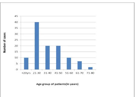
The age of the patients in the present study varied from 16 to 80 years.
Out of 109 cases, female & male patients were 106 and 3 respectively.
This study documented the fact that benign breast lesions were the most common lesions in young females, among
ISSN 2455-3301
WJPMR
AND MEDICAL RESEARCH
www.wjpmr.com
*Corresponding Author: Dr. Ayushi Narain
Post Graduate, Department of Pathology, Sree Balaji Medical College, Chennai- 44.
ABSTRACT
Background: Fine Needle Aspiration Cytology (FNAC) is a simple, minimally invasive, cost effective, outpatient based and a rapid diagnostic method for breast lesions. The aim of the present study was to correlate cytological findings with histopathological findings and to determine the accuracy of FNAC in the diagnosis of breast lesions. Material and Methods: A total of 109 breast aspirates were studied. Histo-cytopathological correlations were obtained in 62 cases. All the aspirates were stained with Haematoxylin and Eosin (H andE) stain. Results: Among 109 patients, 106 were females and 3 were males. Benign breast lesions were found in 74 cases (67.9%); among which fibroadenoma (29.3%) was the commonest lesion which was observed. Malignancy was observed in 27 cases (24.7%); among them, ductal carcinoma was the predominant lesion (23%) which was seen. Histopathological confirmations were obtained in 71 cases out of 72 cases. All 35 malignant aspirates were confirmed by histopathology. Benign reports were confirmed in 35 out of 36 cases by doing histological examinations; except one case which was diagnosed as malignant by studying its histopathology. Sensitivity and specificity of FNAC in breast lesions were reported to be 97.2% and 100% respectively. Conclusion: It is important to remember that a negative FNAC of a breast lesion does not preclude the diagnosis of a carcinoma, particularly in presence of a clinical suspicion of malignancy and/or an abnormal mammogram.
Fig 1: Age distribution of patients with breast lesions.
Table 1: Cytological diagnosis of breast lesions by FNAC(n=109).
Category Cytological Diagnosis No. of Cases Percentage (%)
Inflammatory Lesions(21 cases-19.2%) Duct ectasia 12 11%
Granulomatous Mastitis 4 3.7%
Abscess 3 2.8%
Fat necrosis 2 1.9%
Benign breast lesions(53 cases-48.62%) Fibroadenoma 32 29.3%
Fibrocystic disease 14 12.8%
Galactocoele 3 2.8%
Lactational changes 1 0.9%
Gynacomastia 3 2.8%
Atypical/indeterminate- probably benign
(1 case-0.9%) Atypical epithelial hyperplasia 1 0.9% Lesion not recognized as benign or
malignant(4 cases-3.7%) Phyllodes tumor 4 3.6%
Suspicious for malignancy(2 cases-1.9%) Atypical cells suspicious for malignancy 2 1.9% Positive for malignancy with cystic
change(1 case-0.9%) 1 0.9%
Malignancy(27 cases-24.7%) Ductal carcinoma 25 23%
Medullary carcinoma 2 1.9%
TOTAL 109 CASES 100%
*Five cases in which lymph nodes were palpable revealed evidence of metastasis.
The two cases which were categorized as “suspicious for malignancy” by cytology turned out to be malignant lesions on histopathology and they were diagnosed as infiltrating ductal carcinoma.
The statistical analysis showed high sensitivity (97.2%) and specificity (100%) of FNAC in breast lesions. The diagnostic accuracy was found to be 98.90%. (Table 2).
Table 2: Cyto-Histopathological Correlation (n=72).
HISTOPATHOLOGICAL DIAGNOSIS
FNAC Inflammatory
lesion
Fibroadenoma Fibrocystic disease
Phyllodes tumour
Lactational Hyperplasia
Breast carcinoma Inflammatory lesions 9( 12.5%)
Fibroadenoma 15(20.9%)
Fibrocystic disease 7(9.7%)
Phyllodes Tumor 3(4.2%)
Benign cystic changes 1(1.4%)
Lactational changes 1(1.4%)
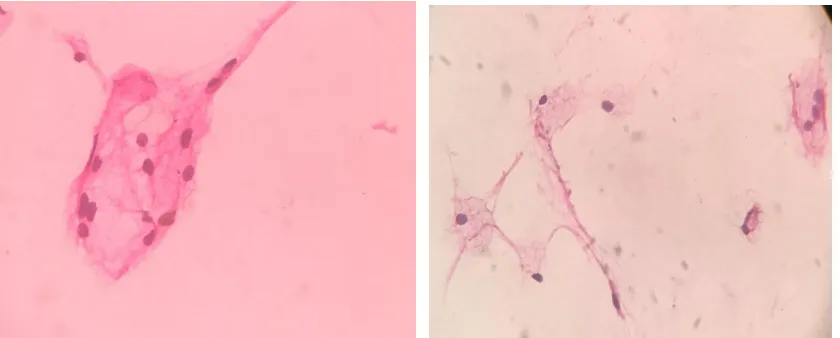
Fig 2: FNAC Suggestive of Gynacomastia,40x: Cohesive clusters of duct epithelial cells and fibrofatty tissue in a clear background.
Fig 3: FNAC Suggestive of Carcinoma breast,40x:Smear showing non cohesive clusters of ductal epithelial cells with pleomorphic nuclei in serofibrinous background.
Fig 4: FNAC suggestive of Fibroadenoma, 40x: Clusters of ductal epithelial cells in tightly cohesive clusters with few benign bare nuclei.
Fig 6: FNAC Suggestive of Galactocele, 40x: Smear shows cyst macrophages, inflammatory cells and occasional duct epithelial cells in a proteinaceous background.
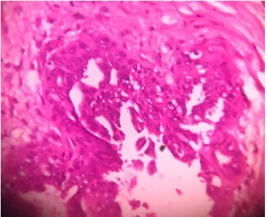
Fig 7: Fibroadenoma: 40x, Section shows a benign neoplasm composed of proliferating mammary ducts & glands in a background of fibromyxoid stroma.
Fig 8: Fibrocystic disease:40x,Section shows breast parenchyma enclosing a cystic lesion surrounded by proliferating mammary glands & cystically dilated ducts with some of the ducts showing epithelial hyperplasia and apocrine metaplasia.
Fig 9: Infiltrating ductal carcinoma: 40x, Section shows fragments of breast parenchyma enclosing a cellular neoplasm composed of round to oval cells with hyperchromatic and pleomorphic nuclei arranged in nests, islands and cords in filtrating surrounding stroma.
DISCUSSION
Many countries have breast cancer screening programs aimed at detecting early disease in asymptomatic women. FNAC occupied a major role in the "Triple test."[1] FNAC of breast lumps is an accepted and established method for determining the natures of breast lumps with a high degree of accuracy[2,3] Application of Fine Needle Aspiration (FNA) for the diagnosis of palpable breast masses was first introduced by Martin and Ellis in 1930 and since then, it has been established as an important tool in the evaluation of breast lesions.
Most of the patients with breast lumps are in a state of anxiety. So, in reducing anxiety and unnecessary surgical procedures as well as in minimization of delay in the diagnosis, FNAC proves very fruitful. FNA procedure is a safe method with only a few reported complications.
In our study, out of 36 cytologically diagnosed benign cases, 35 cases were confirmed histopathologically as benign breast lesions. However, one case which was misinterpreted as a benign cystic lesion by FNAC, was later on diagnosed as a malignant phyllodes tumour on doing a histopathological examination .This might be
due to inadequate sampling, because of the cystic nature of lesion. So, in case of cystic lesions, it is better to re-aspirate the lesion from the solid area after evacuation of cyst or image guided FNA should be performed to locate solid area. It is always necessary to correlate the FNAC findings with clinical diagnoses and mammograms and to go for core biopsies whenever they are needed, to avoid misdiagnoses. The false negative rate varies from 1-8% in different studies[7,8,10,11]
In the present study, all the 35 cytologically diagnosed malignant cases were confirmed as malignant on subsequent histopathological examinations. So, in our study, a 100% cyto-histopathological correlation was observed for malignant lesions. Zhang Qin et al.,[7] AZ Mohammed et al.,[8] Tiwari M.[11] had also observed the same results in their studies.
histopathology holds high significance in diagnosis of breast cancer.
However, cytologically it is difficult to subcategorize the lesions without clinical and mammographical details. Adhering to the principle of Triple test, with the acquisition of technical, observational and interpretative skills will further enhance the diagnostic accuracy of lesions of the breast.[4]
REFERENCES
1. Ahmed I, Nazir R, Chaudhary MY, Kundi S. Triple assessment of breast lump. J Coll Physicians Surg Pak, 2007; 17: 535-8.
2. Purasiri P, Abdalla M, Heys SD,
Ah-See AK, McKean ME, Gilbert FJ, A novel diagnostic index for use in the breast clinic J R Coll Surg Edinb, 1996; 41: 30-34.
3. Kaufman Z, Shpitz B, Shapiro M, Rona R, Lew S, D inbar A, Triple approach in diagnosis of dominant breast masses: combined physical examination, mammography and fine-needle aspiration J Surg Oncol, 1994; 56: 254-57.
4. Venegas R, Rutgers JL, Cameron BL, Vargas H, Butler JA. Fine needle aspiration cytology of breast ductal carcinoma insitu. Acta Cytol, 1994; 38: 136-43.
5. International Agency for Research on Cancer. GLOBOCAN 2012: Estimated Cancer Incidence, Mortality and Prevalence Worldwide in 2012. 2015,http://globocan.iarc.fr/Pages/fact_sheets_popul ation.aspx.
6. Ellis I. O., Humphreys S., Michell M., Pinder S. E., Wells C. A., Zakhour H. D.Guidelines for Non-Operative Diagnostic Procedures and Reporting in Breast Cancer Screening. Sheffield, UK: NHS Cancer Screening Programme, 2001.
7. Zhang Qin, Nie Shigui, Chen Yuhua, Zhou Limei, Fi ne Needle Aspiration Cytology of Breast Lesions: Analysis of 323 Cases The Chinese-German Journal of Clinical Oncology, 2004; 3(3): 172-74.